Embed Size (px)
Citation preview
J. Embryol exp. Morph. Vol. 66, pp. 91-108, 1981Printed in Great Britain © Company of Biologists Limited 1981
Tetraploidy and earlydevelopment: effects on developmental timing and
embryonic metabolism
By MARTIN A. EGLITIS1 AND LYNN M. WILEY2
From the Department of Anatomy, University of Virginia
SUMMARY
The effect of balanced gene dosage changes on the timing of cavitation and on the timingof appearance of a stage-specific embryonic cell surface antigen was studied in preimplantationmouse embryos. Gene dosage was increased by creating tetraploid embryos at the 4-cell stage,either by blastomere fusion with polyethylene glycol (PEG) or by incubation in cytochalasin B(cytB) to block cell division. Removal of the zona pellucida with Pronase from diploidembryos caused a 7 h delay in cavitation. Further manipulations, either with PEG or cytB toinduce tetraploidy, did not produce a statistically significant additional delay in cavitationtiming. Likewise, PEG-induced tetraploidy did not affect the timing of appearance or dis-appearance of the embryonic cell surface antigen as compared with diploid control embryos.In analysing the metabolic effects of tetraploidy, we found that in tetraploid embryos with cellnumber equivalent to intact diploid embryos, MDH activity did not double with the doublingof the genome, being only 50 % greater than diploid levels in cytB-induced tetraploid embryosand only 20 % greater than diploid levels in PEG-induced tetraploid embryos. However, intetraploid embryos with one-half normal cell number, enzyme activity was equal to that inwhole diploid embryos, suggesting that in such embryos, MDH activity increased in parallelwith increases in gene dosage. Further studies showed that levels of RNA synthesis in PEG-induced tetraploid embryos also did not increase in parallel with the doubling of the genome.Rather, these results suggested that in tetraploid embryos, compensation was made for atleast part of the excess genetic material.
INTRODUCTION
Although preimplantation development in the mouse is defined by numerousmorphological, biochemical, and physiological changes, there still remains theproblem of how their timing and coordination are controlled. This study wasinitiated to investigate the control of the timing of formation of the blastocoele(i.e. cavitation).
Recent hypotheses implicate the nucleus in the timing of preimplantationdevelopment. The maternal uterine environment cannot be providing anycritical cues, since development in vitro proceeds normally and at very nearly the
1 Author's address: Department of Cell Biology, Roche Institute of Molecular Biology,Nutley, New Jersey 07110, U.S.A.
2 Department of Human Anatomy, University of California, Davis School of Medicine,Davis, California 95616, U.S.A.
92 M. A. EGLITIS AND L. M. WILEY
rate observed in vivo. Likewise, cytokinesis per se must not be critical in thecontrol of timing, since the number of cells and cell divisions can be alteredwithout affecting the timing of blastocyst formation (Smith & McLaren, 1977).However, neither the number of nuclear divisions (i.e. DNA replications) norchanges in the ratio of nuclear-to-cytoplasmic volume have been excluded fromconsideration as possible repositories of the putative cue for cavitation (Smith &McLaren, 1977; Alexandre, 1979; Braude, 1979; Surani, Barton & Burling,1980).
The embryonic genome is known to be active during preimplantation develop-ment, as shown by the detection of mRNA synthesis (Knowland & Graham,1972) and the expression of paternal isozymes (Chapman, Whitten & Ruddle,1971; Wudl & Chapman, 1976). Also, the inhibitor of mRNA synthesis, a-amanitin, has been used to show that blastocyst formation is dependent on newmRNA synthesis (Braude, 1979). Since the genome is clearly functioning inearly development, it seems reasonable to study the nature of the 'developmentalclock' by determining whether nuclear changes will affect the timing of cavita-tion.
In this report, balanced increases in gene dosage to a tetraploid level wereused to determine whether they would alter the timing of cavitation. We pro-duced tetraploid embryos with a recently developed method to fuse embryonicblastomeres using polyethylene glycol (PEG; Eglitis, 1980), as well as by thestandard procedure using cytochalasin B (cytB; Snow, 1973). The interaction oftetraploidy with cell number changes and its effect on developmental timing wereanalysed by monitoring cavitation rates and the expression of a stage-dependentembryonic cell surface antigen. Additional biochemical studies were done tomonitor the effect of tetraploidy on embryonic metabolism.
MATERIALS AND METHODS
Embryos at the 2-cell stage (47 ~ 49h after hCG injection) were flushed fromthe oviducts of superovulated, random-bred DUB: (1CR) female mice withbicarbonate-free modified Hank's balanced salts solution (BSS; Spindle &Goldstein, 1975). Embryos were grown in modified (Spindle & Goldstein,1975) standard egg culture medium (Biggers, Whitten & Whittingham 1971)under paraffin oil (Fisher) and incubated at 37°C in a humidified atmosphereof 5%C02inair.
Depending upon the particular experiment, zonae pellucidae were removedeither from diploid 2-cell embryos before experimental manipulations (forblastomere fusion) or from tetraploid 2- cell embryos after experimental manipu-lations (for cytB). Zonae pellucidae were removed by a 5 min incubation at37 °C in 0-5 % protease (Pronase, Sigma; Mintz, 1962).
Tetraploid embryos were obtained by two methods: blastomere fusion(Eglitis, 1980), or, inhibition of cell division with cytB (Snow, 1973). Briefly,
Development oftetraploid mouse embryos 93
(1) 48 hpost-hCG
(2) Pronase XjjJ^ 8 h incubation \^M^ (4) Ca2+-free (5)_PHAdisaggregation
(6) 45% PEG 1000V3iii7 (7) PHA
2 min
Fig. 1. A diagram illustrating the general features of the blastomere fusion protocol.For a full description, see Eglitis (1980).
fused blastomeres were produced by disaggregating 4-cell-stage embryos,aggregating individual blastomeres in pairs with phytohaemagglutinin (PHA,Mintz, Gearhart & Guyment, 1973), and treating aggregated pairs for 2 minwith 45 % (w/v) PEG (Sigma, MW 1000; see Eglitis, 1980; Fig. 1). Alternatively,tetraploid embryos were produced by incubating 2-cell embryos for 13 h (from49 - 62 h post-hCG) in standard egg-culture medium containing 10 /*g cytB(CalBiochem) per ml of medium.
To investigate the affect on developmental timing of the interaction of tetra-ploidy and altered cell number, both fusion and cytB-induced tetraploidblastomeres were aggregated with PHA in various cell number combinations.Fused blastomere tetraploid embryos (PEG-4N) were followed after aggregationto restore cell number to that of equivalent (i.e. 4-cell) intact diploid embryos.Alternatively, development of such tetraploid embryos with only half (\ PEG-4N) or one-quarter (£ PEG-4N) the cell number of intact embryos was alsomonitored. CytB-induced tetraploid embryos were followed either as 'half-embryos with ( | w/Z-cytB-4N) or without {\ ZF-cytB-4N) zonae pellucidae, or,after zona removal, as aggregated to restore cell number to that of equivalentintact diploid embryos (cytB-4N).
Controls consisted of intact diploid embryos with (w/Z-2N) or without(ZF-2N) zonae pellucidae, as well as diploid embryos disaggregated at the 4-cellstage to \ {\ ZF-2N) or \ (£ ZF-2N) diploid embryos. Additional controlsincluded blastomeres which did not fuse after PEG treatment, either reaggre-gated to restore cell number to that equivalent to intact embryos (PEG-2N)or grown without reaggregation (\ PEG-2N).
Two parameters were used to time development of the embryos: (1) timing ofblastocoele formation, and (2) timing of expression of a stage-specific embryoniccell surface antigen.
4 EMB 66
94 M. A. EGLITIS AND L. M. WILEY
To time progress towards cavitation, embryos were checked at intervalsthrough an inverted phase-contrast microscope. Embryos were scored as cavitat-ing when an intraembryonic cavity became visible. The timing of cavitation wascompared between different classes of embryos by comparing their t$ of cavita-tion (i.e. the time at which 50 % of the final maximum proportion of cavitationembryos was reached). To determine the t$, the linear regression was taken overthe times at which the proportion of cavitated embryos was uniformly increas-ing. Then, the t$ was calculated by finding the time at which one-half the finalmaximum number of embryos had cavitated.
The timing of expression of the embryonic cell surface antigen was monitoredby indirect immunofluorescence (IIF). The particular antigen assayed was onedetected by an antiserum raised to 8-cell-stage embryos in a male New Zealandwhite rabbit. The cell surface expression of this antigen (or antigens) is very'stage-specific' in that it is restricted to the period immediately preceding andfollowing the onset of cavitation (around 94-106 h post-hCG). If necessary,zonae pellucidae were mechanically removed immediately before the embryoswere observed through a Zeiss phase-contrast microscope equipped withepifluorescence. Negative controls consisted of embryos incubated in the anti-embryo antiserum at times when the antigen was not expressed, as well asembryos incubated in pre-immune (normal) rabbit antiserum.
Two parameters were measured to assess the metabolic levels of tetraploidembryos. The first was the determination of the level of activity of the constitu-tive enzyme, malate dehydrogenase (MDH). The level of MDH activity wasdetermined in diploid and tetraploid morulae (90 h post-hCG) and blastocysts(120 h post-hCG) spectrophotometrically from the rate of reduction of NAD(Brinster, 1966). Enzyme activity in moles NAD reduced/h/embryo wascalculated using the formula:
. . . _ changes in absorbance/min x volume (0-1 ml) = 60 minextinction coefficient (6-22) x 106 M x number of embryos.
In the absence of substrate, embryos reduced only an insignificant amount ofNAD.
The second measure of embryonic metabolism was the determination of thelevel of RNA synthesis per blastomere. This was accomplished autoradio-graphically by quantitating incorporation of [3H]uridine into nuclear RNA.Late morulae, (92-93 h post-hCG) were incubated for 3 h in standard eggculture medium containing 0-5 or 0-05 /iCi/ml of [6-3H]uridine (specific activity22-4Ci/mM, New England Nuclear; concentration 2-23 x 10"5 or 2-33 x 10~6
mM, respectively). Labelled embryos were rinsed thoroughly in BSS containing2-33 x 10~2 mM cold uridine. After labelling, embryos were fixed onto glass slidesby a modification (Epstein, Smith, Travis & Tucker, 1978) of Tarkowski's(1966) air-drying method. The slides were dried overnight in a dessicator at 4 °Cand then dipped in NTB-2 emulsion (Kodak); coated slides were allowed to dry
Development of tetraploid mouse embryos 95
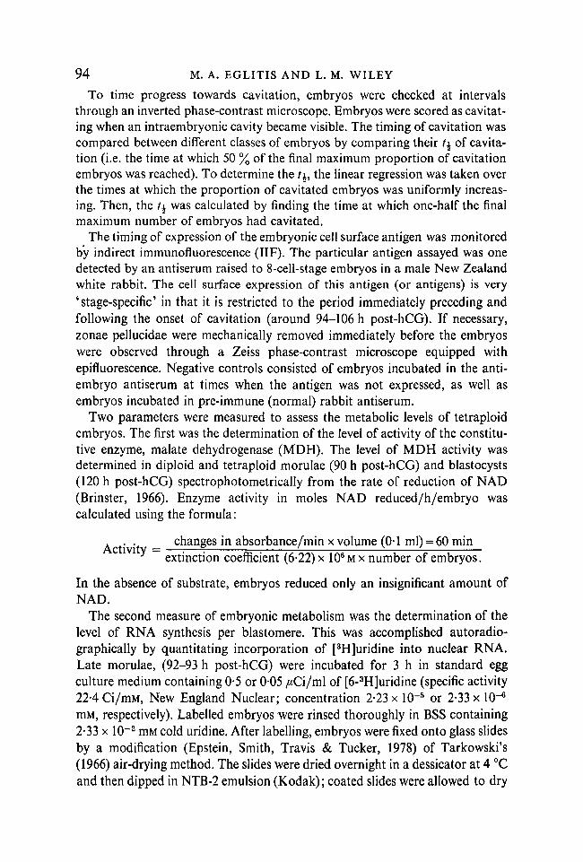
Table 1. Timing of cavitation, t± = h post-hCG
Cell number class
Embryo type Whole Half Quarter
Fused blastomere Tetraploids
Cytochalasin B Tetraploids
Diploid controls
PEG-4N103-2
r = 0-9836
i PEG-4N99-8
r = 0-8952CytB-4N \ CytB-4N
105-9 1020r = 0-9877 r = 0-9979
w/Z-2N96-9
r = 0-9766
ZF-2N103-7
r = 0-9922
i ZF-2N104-2
r = 0-9633
i PEG-4N100-9
r = 0-9374
i ZF-2N104-9
r = 0-9795
r = Correlation coefficient calculated from Fig. 2.
in safe boxes. Embryos labelled in 0-5 ju,Ci [3H]uridine/ml were developed 18 hafter dipping, while embryos labelled in 0-05 JLLC'I [3H]uridine/ml were developed104 h after dipping. Developed slides were counter-stained with methyl green/pyronin and coverslipped. Grains per nucleus were determined by viewing underoil immersion. Only embryos with cell numbers between 8 and 16 were used toquantitate RNA synthesis so as to minimize cleavage stage-dependent vari-ability in RNA synthesis, and so as to maximize the comparability of experi-mental embryos with control embryos. Correction for background levels ofradioactivity were made by counting the number of grains per nucleus forembryos that were fixed without being incubated in [3H]uridine.
RESULTS
Timing of blastocyst formation
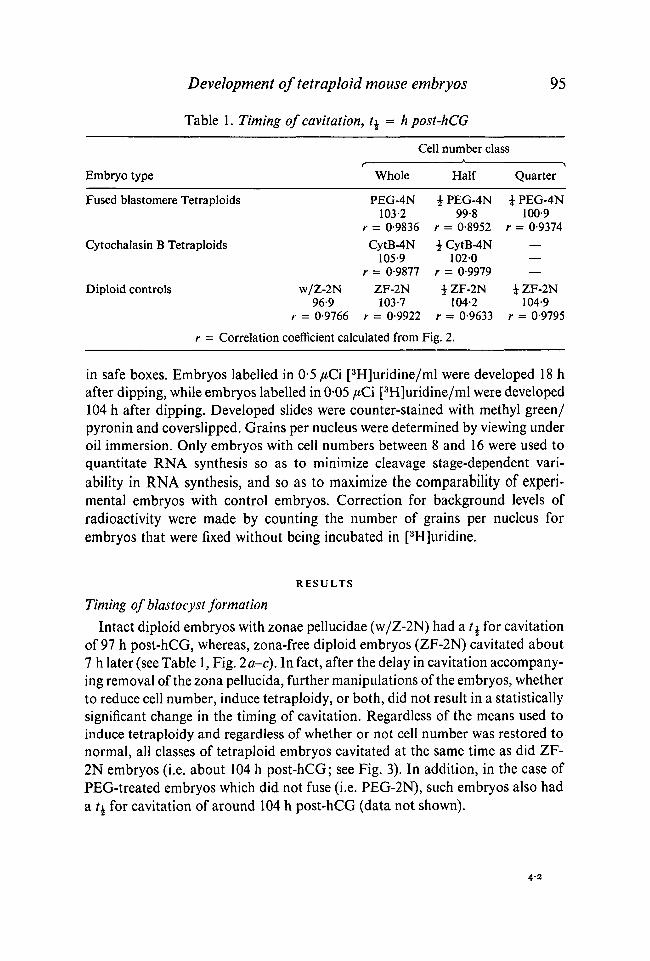
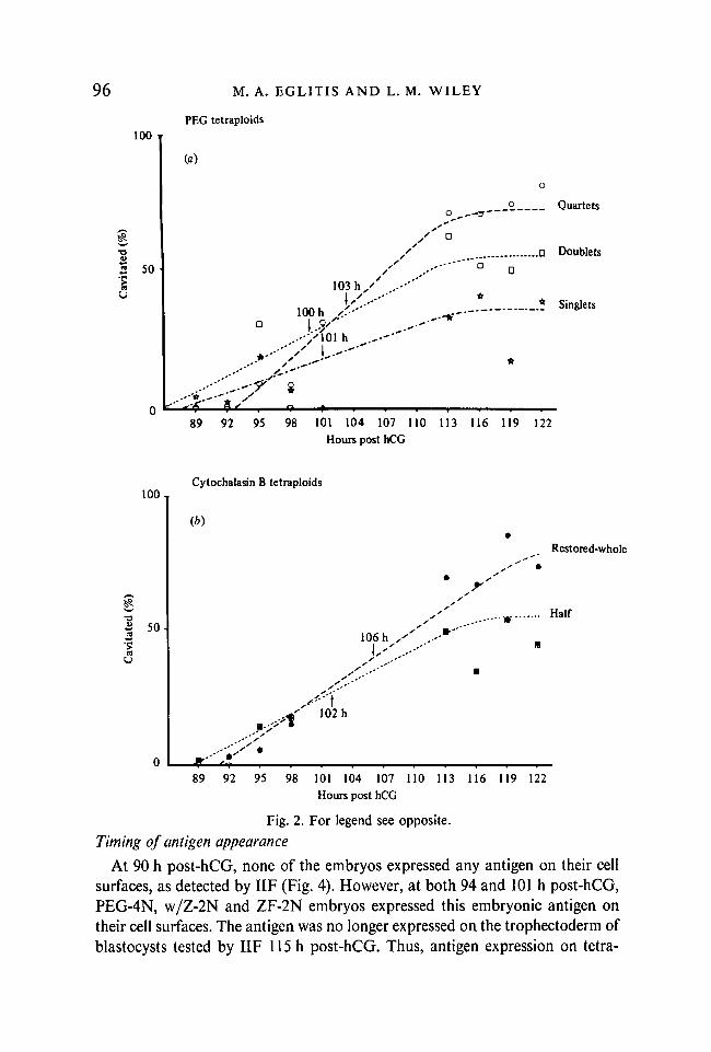
Intact diploid embryos with zonae pellucidae (w/Z-2N) had a t% for cavitationof 97 h post-hCG, whereas, zona-free diploid embryos (ZF-2N) cavitated about7 h later (see Table 1, Fig. 2a-c). In fact, after the delay in cavitation accompany-ing removal of the zona pellucida, further manipulations of the embryos, whetherto reduce cell number, induce tetraploidy, or both, did not result in a statisticallysignificant change in the timing of cavitation. Regardless of the means used toinduce tetraploidy and regardless of whether or not cell number was restored tonormal, all classes of tetraploid embryos cavitated at the same time as did ZF-2N embryos (i.e. about 104 h post-hCG; see Fig. 3). In addition, in the case ofPEG-treated embryos which did not fuse (i.e. PEG-2N), such embryos also hada t± for cavitation of around 104 h post-hCG (data not shown).
4-2
96
100 T
3 50
'S
M. A. EGLITIS AND L. M. WILEY
PEG tetraploids
(a)
103 h ' ..-•'"
lOOh /.-'
..-y\oi h .~--
8
._ Quartets
.D Doublets
.* Singlets.--'*"
89 92 95 98 101 104 107 110 113 116 119 122Hours post hCG
1 0 0 ,
73
| 5 < H
Cytochalasin B tetraploids
Restored-whole
Half
106 h ,' ..--
t1 0 2 h
8 9 9 2 9 5 9 8 101 1 0 4 1 0 7 1 1 0 1 1 3 1 1 6 1 1 9 1 2 2H o u r s p o s t hCG
Fig. 2. For legend see opposite.
Timing of antigen appearance
At 90 h post-hCG, none of the embryos expressed any antigen on their cellsurfaces, as detected by IIF (Fig. 4). However, at both 94 and 101 h post-hCG,PEG-4N, w/Z-2N and ZF-2N embryos expressed this embryonic antigen ontheir cell surfaces. The antigen was no longer expressed on the trophectoderm ofblastocysts tested by IIF 115 h post-hCG. Thus, antigen expression on tetra-
Development of tetraphid mouse embryos 97
1 0 0 ,
3 so -Ito
o
Contro l d iplo ids
(c)
' • .-•"
_^ With zonae
. — Zona-free
... Half
^ Quarter
8 9 9 2 9 5 9 8 101 1 0 4 107 110 113 1 1 6 119 122Hours pos t hCG
Fig. 2. Timing of cavitation. Embryos were monitored by phase contrast microscopyfor the presence or absence of a blastocoele.
number of embryos with a blastocoele.% cavitated = total number of embryos observed
/£ (time at which \ the final maximum number of embryos had cavitated) wasdetermined by calculating the linear regression for the times at which the number ofcavitated embryos was increasing, then calculating t using the value of \ ' % cavitated'that was reached at the end of the culture period. Arrow on graphs point to the t\.(a) PEG Tetraploids: O—O, with cell number restored to whole control levels(quartets); D---D, with cell number equivalent to i controls (doublets); * — , *with cell number equivalent to i controls (singlets), (b) Cytochalasin B Tetraploids:© - - © , with cell number restored to whole control levels (restored-whole); ®---B,with cell number equivalent to i controls (half), (c) Control Diploids: *—*,whole diploid embryos with intact zonae pellucidae (with zonae); # — # , whilediploid embryos with zonae pellucidae removed at the 2-cell stage with 0-5 %Pronase (zona-free); • - - - • , zona-free diploid embryos with % normal cell number(half); • *, zona-free diploid embryos with i normal cell number (quarter).
ploid embryos was neither accelerated delayed, nor prolonged when comparedwith the controls.
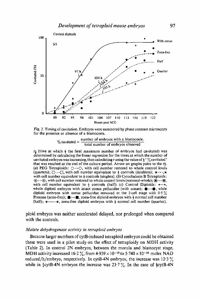
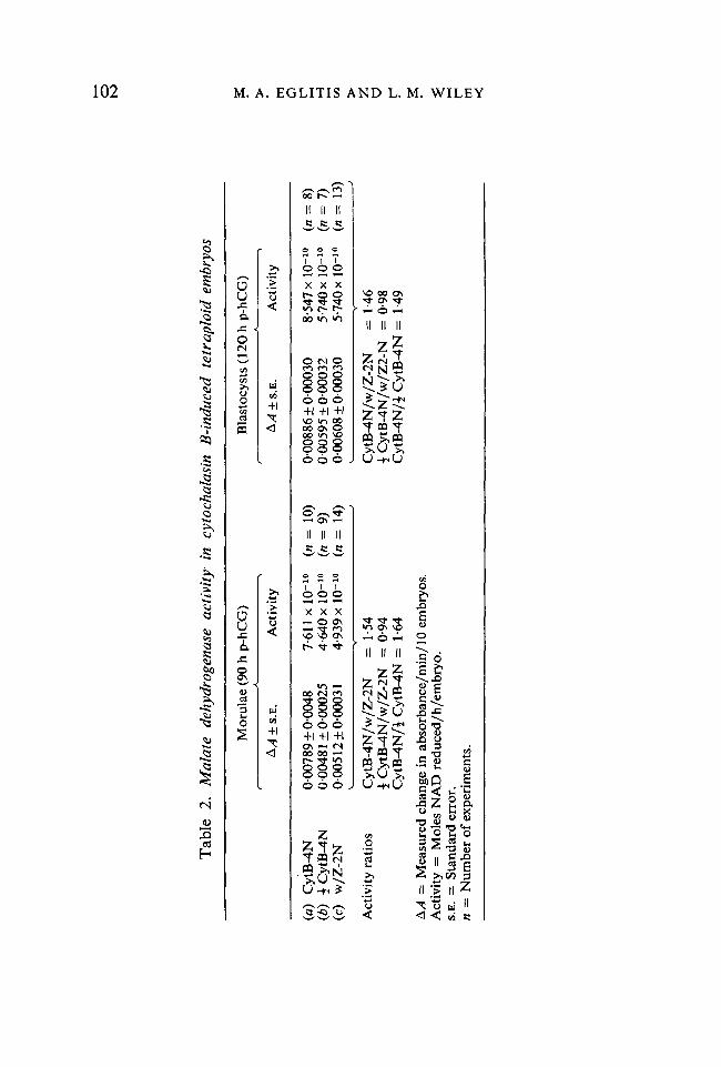
Malate dehydrogenase activity in tetraploid embryos
Because larger numbers of cytB-induced tetraploid embryos could be obtainedthese were used in a pilot study on the effect of tetraploidy on MDH activity(Table 2). In control 2N embryos, between the morula and blastocyst stage,MDH activity increased 16-2 %, from 4-939 x 10"10 to 5-740 x 10"10 moles NADreduced/h/embryo, respectively. In cytB-4N embryos, the increase was 12-3 %while in |cytB-4N embryos the increase was 23-7 %. In the case of £cytB-4N
98 M. A. EGLITIS AND L. M. WILEY
","*&
o
Development of tetraploid mouse embryos 99embryos, the percent increase in MDH activity between the morula and blasto-cyst stage was the same whether such embryos were grown with or withoutzonae. In addition, the actual MDH activity of |cytB-4N embryos at each stagewas also approximately the same regardless of whether or not the zona pellucidawas present. Therefore, in the ratios reported below, MDH activities of |cytB-4N embryos were determined by pooling values of |w/Z- and |ZF-cytB-4Nembryos.
The MDH activity of cytB-4N morulae (90 h post-hCG) was 1-64 times thatof |cytB-4N morulae. When compared to w/Z-2N embryos, cytB-4N embryoshad 1-54 times the 2N level of enzyme activity. Although the ratio of cytB-4N:£cytB-4N differs from 2-0 with only borderline significance (Z = 1-544,0-2 > P> 0-1), the ratio of cytB-4N: w/Z-2N is significantly different from 2-0 (Z =
2-995, P < 0-05). The MDH activity of £cytB-4N morulae was 0-94 times that ofw/Z-2N morulae, not differing significantly from a hypothetical value of 1-0.
At the blastocyst stage (120 h p-hCG), the MDH activity of cytB-4N embryoswas 1-49 times that of £cytB-4N embryos. When compared to w/Z-2N blasto-cysts, cytB-4N blastocysts had 1-46 times the 2N level of enzyme activity. Bothof these values differ significantly from the expected ratio of 2-0:1-0 (Z = 4-094,P < 0-05; Z = 5-996, P < 0-01, respectively). The MDH activity of £cytB-4Nblastocysts was 0-98 times that of w/Z-2N blastocysts, not differing significantlyfrom the hypothetical value of 1-0.
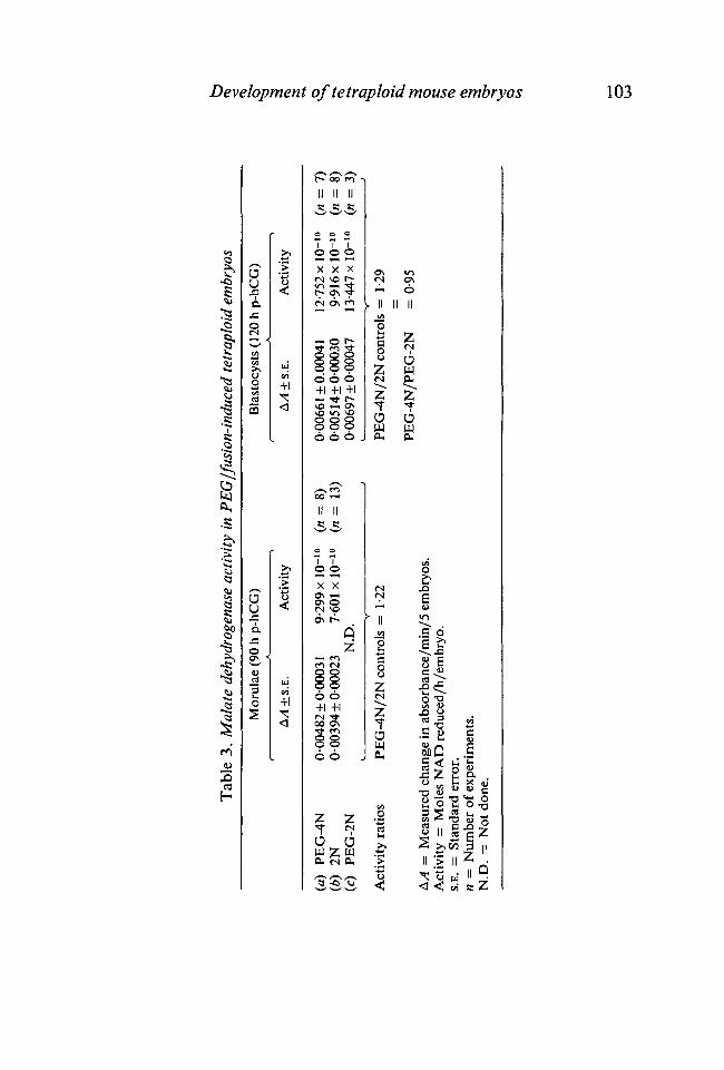
Tetraploid embryos obtained by blastomere fusion were then analysed todetermine if PEG-4N embryos had a depression in MDH activity (relative to theexpected doubling compared to 2N controls) similar to the depression observedin cytB-4N embryos (Table 3). In these experiments, between the morula andblastocyst stage, MDH activity in control 2N embryo increased 30-5 %, from7-601 x 10-10 to 9-916 x 10"10 moles NAD reduced/h/embryo, respectively.Enzyme activities of diploid embryos grown with or without zonae were pooledbecause no differences in activity were detected between the two populations. In
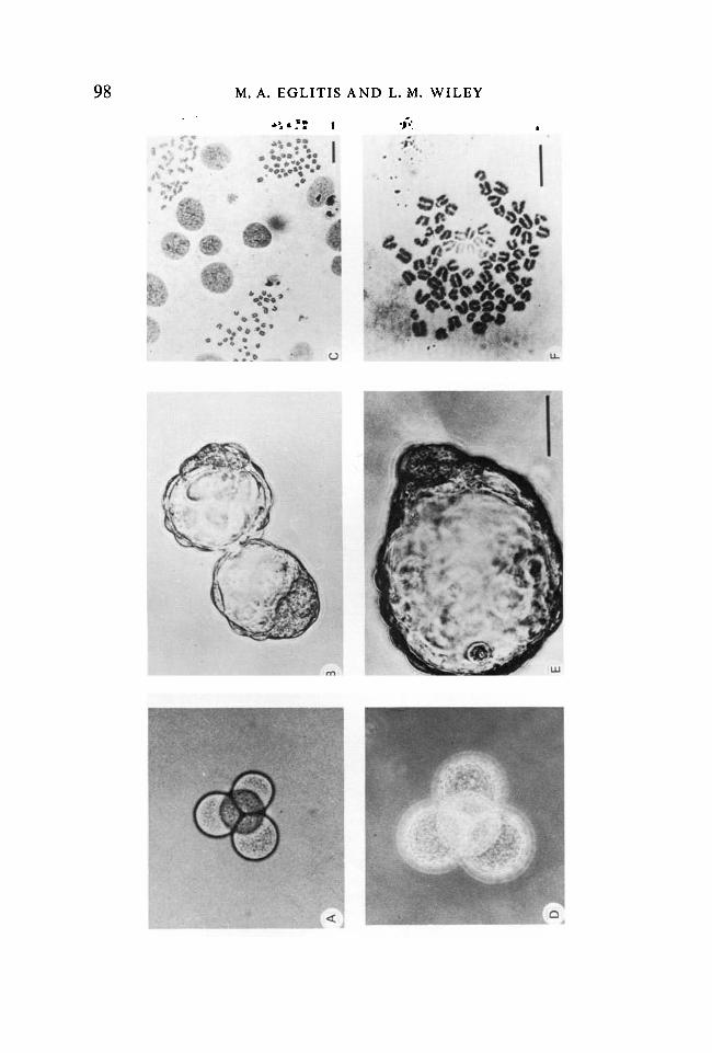
Fig. 3. The appearance, development and ploidy of fused-blastomere tetraploidembryos (a-c) The appearance of control, ZF-2N embryos, including a represent-ative chromosome spread.
(d-f) The appearance of experimental, PEG-4N embryos.(a, d) The appearance of diploid and tetraploid embryos at the end of the exper-
mental manipulations (about 60 hpost-hCG injection). The tetraploid embryo (d)is composed of four fused blastomeres combined by PHA aggregation.
(b, e) Diploid and tetraploid blastocysts about 140 h post-hCG. Note the promi-nent inner cell mass in both diploid and tetraploid embryos. The tetraploid blasto-cyst developed from a restored four cell form as illustrated in (d) and was photo-graphed 83 h after PEG-induced fusion.
(c,/) Metaphase plates obtained from diploid and tetraploid-embryos. Blastocystswere fixed 139 h post-hCG, after a 3 h preincubation in 0-2 /*g/ml colcemid tomaximize cells in metaphase, according to a modification (Epstein et al. 1978) ofTarkowski's (1966) method. Magnification - bar in e applies to a, b, d and e andrepresents 50 /im. Bars in c and/represent 1 /̂ m.
100 M. A. EGLITIS AND L. M. WILEY
I
u
CO
m<D
Development of tetraploid mouse embryos 101PEG-4N embryos, MDH activity increased 37-1 % between the morula andblastocyst stage. Due to a paucity of material, no PEG-2N embryos wereanalyzed at the morula stage, although MDH activities of such embryos weredetermined at the blastocyst stage.
The enzyme activity of PEG-4N morulae (90 h post-hCG) was 1-22 times thatof control 2N embryos, which significantly differs from the expected ratio of2-0 (Z = 12-978, P < 0-01). Thirty hours later, at the blastocyst stage, PEG-4Nembryos had 1-29 times the enzyme activity of 2N embryos, and 0-95 times theenzyme activity of PEG-2N embryos. Both of these values differ significantlyfrom the expected ratio (Z = 2-729, P < 0-1; Z = 16-454, P < 0-05, respec-tively).
Although it might have been expected that the doubled genome of tetraploidembryos would have been accompanied by a doubling in MDH activity relativeto diploid control embryos, these data show that this correlation was notdetected. This result was observed regardless of the means used to inducetetraploidy. Rather than a 100 % elevation in enzyme activity, in cytB-4Nembryos the MDH activity was elevated by only 50 %, while in PEG-4Nembryos the MDH activity was elevated by only about 20 %. In fact, in compar-ing PEG-4N with PEG-2N embryos, their enzyme activities at the blastocyststage were essentially the same.
If the doubled genome had been accompanied by doubled enzyme activity,£PEG-4N or £cytB-4N embryos would be expected to have an enzyme activityequal to that of w/Z-2N embryos. In the case of |cytB-4N embryos, this is whatwas observed. If one assumes that £cytB-4N embryos did indeed have half thecell number of W/Z-2N embryos (see below), then each $cytB-4N embryoblastomere had about 190 % the enzyme activity of a w/Z-2N embryo blastomere.Similarly, the MDH activity of cytB-4N embryos was only 1-5 times the activityof £cytB-4N embryos. Again, if cytB-4N embryos did, indeed, have twice thecell number of £cytB-4N embryos, then each blastomere of a -|cytB-4N embryohad about 130 % the enzyme activity of a blastomere of a cytB-4N embryo.
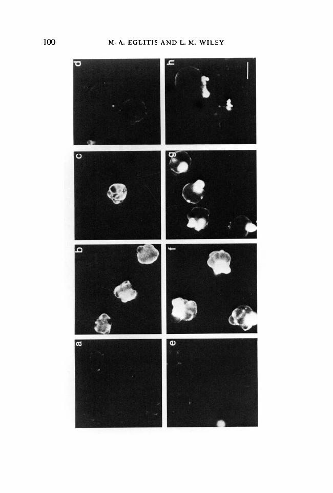
Fig. 4. The expression of a stage-specific embryonic cell surface antigen on diploidand tetraploid morulae.
ia-d) Control diploid embryos, grown in culture from the 2-cell stage with intactzonae pellucidae. Zonas mechanically removed for IIF.
(e-h) PEG-4N embryos.(a, e) 90 h post-hCG, PEG-4N embryos 31 h after PEG-induced fusion.(b,f) 94 h post-hCG, PEG-4N embryos 35 h after PEG-induced fusion.(c, g) 101 h post-hCG, PEG-4N embryos 42 h after PEG-induced fusion.(d, h) 115 h post-hCG, PEG-4N embryos 56 h after PEG-induced fusion. All
photographs taken with 1 -5 min exposures of Kodak Tri-X film. Prints all exposed for35 sec. In h, bright patches of fluorescence are caused by blastomeres killed during11F manipulations which filled with fluorescent antibody. Magnification -bar in// represents 50 fim.
Tab
le
2.
Mal
ate
dehy
drog
enas
e ac
tivi
ty
in
cyto
chal
asin
B
-ind
uced
te
trap
loid
em
bryo
s
o to
id)
Cyt
B-4
N(b
) ±
Cyt
B-4
N(c
) W
/Z-2
N
Act
ivity
rat
ios
Mor
ulae
(90
h p
-hC
G)
Bla
stoc
ysts
(12
0 h
p-hC
G)
± S.
E.
Act
ivity
± S.
E.
Act
ivity
0007
89 ±
0 00
4800
0481
±00
0025
0005
12 ±
0000
31
7 61
1 x
lO-1
0 («
= 1
0)4-
640
xlO
"10
(n =
9)
4-93
9 x
10-10
(n
= 1
4)
Cyt
B-4
N/w
/Z-2
N
=1
-54
i C
ytB
-4N
/w/Z
-2N
=
0-9
4C
ytB
-4N
/i C
ytB
-4N
=
1-64
A A
= M
easu
red
chan
ge i
n ab
sorb
ance
/min
/10
embr
yos.
Act
ivity
= M
oles
NA
D r
educ
ed/h
/em
bryo
.S.
E. =
Sta
ndar
d er
ror.
n =
Num
ber
of e
xper
imen
ts.
0008
86 ±
0000
3000
0595
±00
0032
0-00
608
±00
0030
8-54
7 x
lO"10
(n
= 8
)5-
740
xlO
-10
in =
7)
5-74
0 xl
O"1
0 in
= 1
3)
Cyt
B-4
N/w
/Z-2
N
=
1-46
i C
ytB
-4N
/w/Z
2-N
=
0-9
8C
ytB
-4N
/i C
ytB
-4N
=
1-49
a a r M r m
Tab
le 3
. Mal
ate
dehy
drog
enas
e ac
tivi
ty i
n P
EG
/fus
ion-
indu
ced
tetr
aplo
id e
mbr
yos
Mor
ulae
(90
h p
-hC
G)
Bla
stoc
ysts
(12
0 h
p-hC
G)
+ S
.E.
Act
ivity
A/1
+ S
.E.
Act
ivity
(a)
PE
G-4
N
0-00
482±
0000
31
9-29
9 xl
O~10
(n
= 8
)(b
) 2N
0-
0039
4±0-
0002
3 7-
601
x 10
"10
(n =
13)
(c)
PE
G-2
N
N.D
.«
y '
Act
ivity
rat
ios
PE
G-4
N/2
N c
ontr
ols
=
1-22
AA
— M
easu
red
chan
ge in
abs
orba
nce/
min
/5 e
mbr
yos.
Act
ivity
= M
oles
NA
E> r
educ
ed/h
/em
bryo
.S.
E. =
Sta
ndar
d er
ror.
n =
Num
ber
of e
xper
imen
ts.
N.D
. =
Not
don
e.
0006
61 ±
0.00
041
0-00
514±
0-00
030
0006
97±0
-000
47
12-7
52x
lO"10
(«
= 7
)9-
916x
lO-10
(n
= 8
)13
-447
x 10
"10
in =
3)
PE
G-4
N/2
N c
ontr
ols
=
1-29
PE
G-4
N/P
EG
-2N
=
0-9
5
• O
104 M. A. EGLITIS AND L. M. WILEY
Table 4. Cell numbers per morula*
Diploid controls (1) With Zonae(2) Zonae-free(3) Total Mean
PEG-4N —
* Cell no. determined at 94a standard deviation.n = number of experiments
17-73 ±6-36a
14-47 ±4-021614±5-49
12-73 ±305
h post-hCG.
inin
in
= 30)= 34)= 64)
= 26)
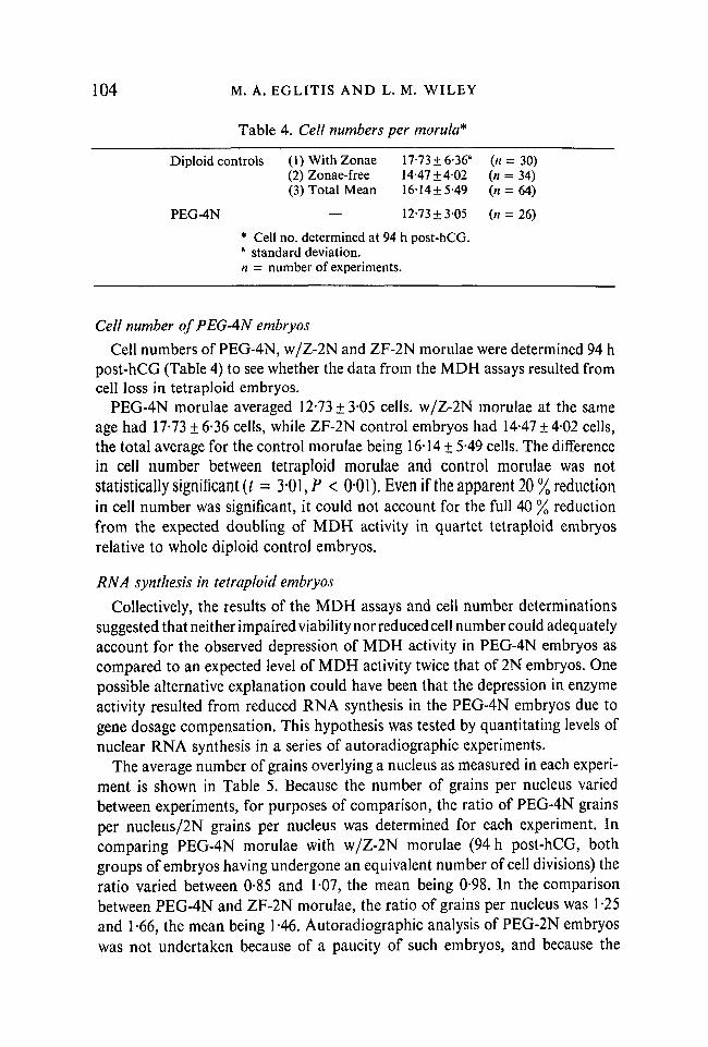
Cell number of PEG-AN embryos
Cell numbers of PEG-4N, w/Z-2N and ZF-2N morulae were determined 94 hpost-hCG (Table 4) to see whether the data from the MDH assays resulted fromcell loss in tetraploid embryos.
PEG-4N morulae averaged 12-73 ±3-05 cells. w/Z-2N morulae at the sameage had 17-73 ± 6-36 cells, while ZF-2N control embryos had 14-47 ± 4-02 cells,the total average for the control morulae being 16-14 ± 5-49 cells. The differencein cell number between tetraploid morulae and control morulae was notstatistically significant (t = 3*01, P < 0*01). Even if the apparent 20% reductionin cell number was significant, it could not account for the full 40 % reductionfrom the expected doubling of MDH activity in quartet tetraploid embryosrelative to whole diploid control embryos.
RNA synthesis in tetraploid embryos
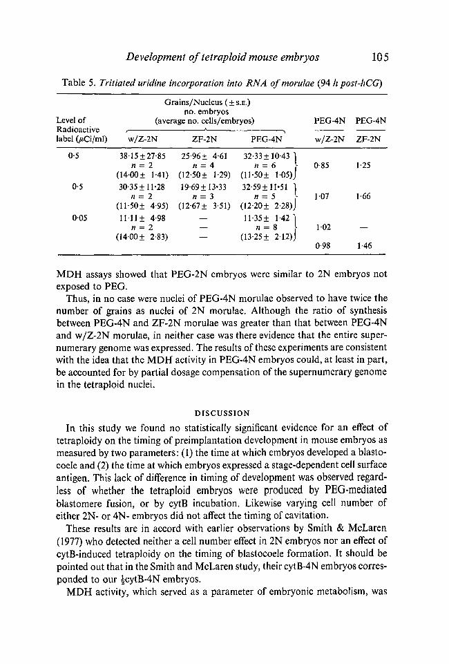
Collectively, the results of the MDH assays and cell number determinationssuggested that neither impaired viability nor reduced cell number could adequatelyaccount for the observed depression of MDH activity in PEG-4N embryos ascompared to an expected level of MDH activity twice that of 2N embryos. Onepossible alternative explanation could have been that the depression in enzymeactivity resulted from reduced RNA synthesis in the PEG-4N embryos due togene dosage compensation. This hypothesis was tested by quantitating levels ofnuclear RNA synthesis in a series of autoradiographic experiments.
The average number of grains overlying a nucleus as measured in each experi-ment is shown in Table 5. Because the number of grains per nucleus variedbetween experiments, for purposes of comparison, the ratio of PEG-4N grainsper nucleus/2N grains per nucleus was determined for each experiment. Incomparing PEG-4N morulae with w/Z-2N morulae (94 h post-hCG, bothgroups of embryos having undergone an equivalent number of cell divisions) theratio varied between 0-85 and 1-07, the mean being 0-98. In the comparisonbetween PEG-4N and ZF-2N morulae, the ratio of grains per nucleus was 1-25and 1-66, the mean being 1-46. Autoradiographic analysis of PEG-2N embryoswas not undertaken because of a paucity of such embryos, and because the
Development of tetraploid mouse embryos 105
Table 5. Tritiated uridine incorporation into RNA ofmorulae (94 h post-hCG)
Grains/Nucleus (±S.E.)no. embryos
Level of (average no. cells/embryos) PEG-4N PEG-4NRadioactive , * *label (yMCi/ml) w/Z-2N ZF-2N PEG-4N w/Z-2N ZF-2N
0-5 38-15±27-85 25-96±4-61 32-33± 10-43]n = 2 n = 4 n = 6 } 0-85 1-25
(1400± 1-41) (12-50± 1-29) (ll-50± 105)J0-5 3O-35± 11-28 19-69± 13*33 32-59± 11-51 ]
n = 2 n = 3 n= 5 \ 107 1 66(ll-50± 4-95) (12-67± 3-51) (12-20± 2-28)J
005 11-11 ± 4-98 — ll-35± 1-42 1n = 2 — n = 8 [ 102 —
(1400± 2-83) — (13-25± 212) J0-98 1-46
MDH assays showed that PEG-2N embryos were similar to 2N embryos notexposed to PEG.
Thus, in no case were nuclei of PEG-4N morulae observed to have twice thenumber of grains as nuclei of 2N morulae. Although the ratio of synthesisbetween PEG-4N and ZF-2N morulae was greater than that between PEG-4Nand w/Z-2N morulae, in neither case was there evidence that the entire super-numerary genome was expressed. The results of these experiments are consistentwith the idea that the MDH activity in PEG-4N embryos could, at least in part,be accounted for by partial dosage compensation of the supernumerary genomein the tetraploid nuclei.
DISCUSSION
In this study we found no statistically significant evidence for an effect oftetraploidy on the timing of preimplantation development in mouse embryos asmeasured by two parameters: (1) the time at which embryos developed a blasto-coele and (2) the time at which embryos expressed a stage-dependent cell surfaceantigen. This lack of difference in timing of development was observed regard-less of whether the tetraploid embryos were produced by PEG-mediatedblastomere fusion, or by cytB incubation. Likewise varying cell number ofeither 2N- or 4N- embryos did not affect the timing of cavitation.
These results are in accord with earlier observations by Smith & McLaren(1977) who detected neither a cell number effect in 2N embryos nor an effect ofcytB-induced tetraploidy on the timing of blastocoele formation. It should bepointed out that in the Smith and McLaren study, their cytB-4N embryos corres-ponded to our JcytB-4N embryos.
MDH activity, which served as a parameter of embryonic metabolism, was
106 M. A. EGLITIS AND L. M. WILEY
monitored to determine whether the response of embryonic metabolism to tetra-ploidy might provide an explanation for the timing data. We found that cytB-4N embryos had only 1-5 times the MDH activity of 2N embryos, while PEG-4N embryos had only 1-2 times the MDH activity of 2N embryos. Thus, MDHactivity, and presumably, overall embryonic metabolism, did not increase by afactor of two in response to a 2-fold increase in the embryonic genome.
These data on MDH activity might simply be a result of depressed embryoviability of 4N embryos, with perhaps, PEG reducing embryo viability morethan cytB. However, although there is some difference in MDH activities betweencytB-4N and PEG-4N embryos relative to their respective 2N controls, theactual MDH activity levels of cytB-4N and PEG-4N embryos are very similar.In addition, in both types of 2N embryos, the increase in MDH activity as 4Nmorulae developed into blastocysts was very similar in magnitude to the increasein MDH activity of 2N morulae as they developed into blastocysts. Thus, forboth types of 4N embryos, the ratios of enzyme activity (cytB-4N:2N and PEG-4N:2N) remained constant, not decreasing between the morula and the blasto-cyst stage as would be expected if the embryos were progressively deteriorating.If, then, tetraploidy itself impairs embryo viability and if PEG impairs viabilitymore so than cytB, then these impairments must have remained constant overthe 30 h period during which MDH activities were measured. A final argumentagainst these data being a total result of depressed embryo viability are twoobservations, namely, (1) cytB-4N and PEG-4N embryos formed blastocysts atsimilar frequencies and (2) both types of 4N embryos had similar timing ratesfor blastocyst formation.
Another possible explanation for the apparent depressed level of MDHactivity in 4N embryos is that measurements of enzyme activity could have beenmade at a point where the activity curve had reached a maximum. Only in theregion of the curve where activity per unit substrate was uniformly increasingwould it be possible to detect a doubling of activity. If measurements were madein the non-linear region of the activity curve, then, as more embryos wereassayed per sample, activity per embryo would decrease. This, however, was notthe case, since the numbers of PEG-4N embryos per sample varied between 3and 5 with no change in calculated activity per embryo. The correlation ofactivity-to-embryos per sample is 0-419 for morulae (P > 0-1 that activity isindependent of number of embryos) and -0-031 for blastocysts (P > 0-5 thatactivity is independent of number of embryos). Finally, our 2N levels of MDHactivity are in good agreement with those previously reported by Brinster (1966),suggesting that our MDH data are cogent.
Another possible explanation for the MDH data is that 4N embryos had fewercells than did 2N embryos. However, the difference in cell number between 4N-and 2N embryos was not statistically significant, and cannot, therefore, accountfor the MDH data.
If neither depressed embryo viability, nor errors in enzyme activity measure-
Development of tetraploid mouse embryos 107ments, nor cell loss can account for the MDH data, could chromosomal loss orgene dosage compensation explain the observed, less than 2-fold increase inMDH activity in 4N embryos? Our chromosomal counts showed that putative4N embryos were truly 4N with no evidence of partial chromosomal loss or2N/4N mosaicism (these results and Eglitis, 1980). Thus, chromosomal loss isprobably an unlikely reason for the MDH data.
To determine whether gene dosage compensation might account for the MDHdata, RNA synthesis was measured autoradiographically in 2N and 4N em-bryos. The resulting grain counts indicated that the mean level of RNA synthesisin PEG-4N embryos was 1 -45 times that of ZF-2N embryos and 0-98 times that ofw/Z-2N embryos. The difference in these two ratios is due to finding that thelevel of RNA synthesis in ZF-2N embryos was 35 % lower than that in w/Z-2Nembryos. This finding could, then, be evidence for decreased viability of zone-free embryos in general, including, then, of the PEG-4N embryos whose levelsof RNA synthesis were measured in these experiments. This would mean that thehigher of these two ratios (1-45) is the more meaningful result. However, it isnoteworthy that of all three embryo types (ZF-2N, w/Z-2N and PEG-4N), thestandard error is greatest for ZF-2N embryos. It is possible, then, that the higherratio results from a sampling error, particularly since the elevated ratio stems, inlarge part, from one experiment.
Regardless of whether the higher or the lower ratio is more accurate, it is clearthat PEG-4N embryos do not synthesize twice as much RNA as do 2N em-bryos. Although the possibility remains that the RNA data result from de-creased embryo viability, for the reasons discussed earlier, reduced embryoviability probably cannot explain all of the observed MDH and RNA data. Theseresults are, however, compatible with the intriguing possibility that in PEG-4Nmorulae, compensation is made for at least part of the excess genetic material.
Although RNA synthesis in |cytB-4N embryos was not quantitated, compari-son of the MDH activities in £cytB-4N embryos with that of cytB-4N embryossuggests that the excess genome was not as thoroughly compensated for in -|cytB-4N embryos as it was in cytB-4N embryos. This possibility is consistent with theidea that the control of gene activity is sensitive to cell number-related cell-to-cell interactions.
In 1977, Smith & McLaren concluded that the control of developmentaltiming most likely resided in the ratio of nuclear-to-cytoplasmic volume, or, inthe number of nuclear divisions (DNA replications) that an embryo had under-gone. The results here do not contradict either of these possibilities. In bothPEG-4N and cytB-4N embryos, the ratio of nuclear-to-cytoplasmic volumeremains the same as in 2N embryos of the same age. In blastomere fusion, asnuclear volume increases with tetraploidy, so does the cytoplasmic volume.Similarly, nuclear volume in cytB-4N embryos doubles, and, since, cell divisionis blocked during cytB treatment, cytoplasmic volume also doubles. Tetraploidembryos probably undergo the normal number of nuclear divisions since neither
108 M. A. EGLITIS AND L. M. WILEY
cell numbers nor ploidy are perturbed past the morula stage (Snow, 1973;Eglitis, 1980).
Because of the evidence for some degree of gene-dosage compensation in 4Nembryos, our results do not permit a conclusion to be reached on the questionof how great a role the embryonic genome actually has in developmental timing.However, these results do suggest that the nucleus is under tight control that mayinvolve nuclear-cytoplasmic interactions sensitive to cell number.
We would like to thank Dr Irwin R. Konigsberg for some useful suggestions, and Dr E. E.Oliphant for his helpful criticisms during the preparation of the manuscript. Dr PatriciaRodier assisted with the statistical analyses, while Dr Donald A. Keefer helped with the auto-radiographic study, to which Dr S. K. Lau lent his technical expertise. This report was sub-mitted in partial fulfillment of the requirements for the degree of Doctor of Philosophy withthe Department of Anatomy, University of Virginia. This work was supported by an NIHgrant to L.M.W., NICHHD 1-RO1-11788. M.A.E. had additional support from an NIHpredoctoral training grant.
REFERENCES
ALEXANDRE, H. (1979). The utilization of an inhibitor of spermidine and spermine synthesis asa tool for the study of the determination of cavitation in the preimplantation mouse embryo.J. Embryol. exp. Morph. 53, 145-162.
BIGGERS, J. D., WHITTEN, W. K. & WHITTINGHAM, D. G. (1971). The culture of mouseembryos in vitro. In Methods of Mammalian Embryology (ed. J. C. Daniel, jr), pp. 86-116.San Fransisco: W. H. Freeman.
BRAUDE, P. B. (1979). Time-dependant effects of a-amanitin on blastocyst formation in themouse. J. Embryol. exp. Morph. 52, 193-202.
BRINSTER, R. L. (1966). Malic dehydrogenase activity in the preimplantation mouse embryo.Expl Cell. Res. 43, 131-135.
CHAPMAN, V. M., WHITTEN, W. K. & RUDDLE, F. H. (1971). Expression of paternal glucosephosphate isomerase-1 (Gpi-1) in preimplantation mouse embryos. Devi. Biol. 26,153-158.
EGLITIS, M. A. (1980). Formation of tetraploid mouse blastocysts after blastomere fusionwith polyethylene glycol. J. exp. Zool. 213, 309-313.
EPSTEIN, C. J., SMITH, S., TRAVIS, B. & TUCKER, G. (1978). Both X-chromosomes functionbefore X-chromosome inactivation in female mouse embryos. Nature 21 A, 500-503.
KNOWLAND, J. & GRAHAM, C. (1972). RNA synthesis at the two-cell stage of mouse develop-ment. / . Embryol. exp. Morph. 27, 167-176.
MINTZ, B. (1962). Experimental study of the developing mammalian egg: Removal of thezona pellucida. Science 138 594-595.
MINTZ, B., GEARHART, J. D. & GUYMONT, A. O. (1973). Phytohemagglutinin-mediatedblastomere aggregation and development of allophenic mice. Devi. Biol. 31, 195-199.
SMITH, R. & MCLAREN, A. (1977). Factors affecting the time of formation of the mouseblastocoele. / . Embryol. exp. Morph. 41, 79-92.
SNOW, M. H. L. (1973). Tetraploid mouse embryos produced by cytochalasin B duringcleavage. Nature 244, 513-514.
SPINDLE, A. I. & GOLDSTEIN, L. S. (1975). Induced ovulation in mature mice and develop-mental capacity of embryos in vitro. J. Reprod. Fert. 44, 113-116.
SURANI, M. A. H., BARTON, S. C. & BURLING, A. (1980). Differentiation of 2-cell and 8-cellmouse embryos arrested by cytoskeletal inhibitors. Expl Cell Res. 125, 275-286.
TARKOWSKI, A. K. (1966). An air-drying method for chromosomal preparations from mouseeggs. Cytogenetics 5, 394-400.
WUDL, L. & Chapman, V. (1976). The expression of /ft-glucuronidase during preimplantationdevelopment of mouse embryos. Devi Biol. 48, 104-109.
(Received 18 September 1980, revised 19 May 1981)