Embed Size (px)
Citation preview
Cognition and Behavior
Testosterone Modulates Altered PrefrontalControl of Emotional Actions in PsychopathicOffenders1,2,3
Inge Volman,1,2,3 Anna Katinka Louise von Borries2,3,4,5, Berend Hendrik Bulten,5 Robbert JanVerkes,3,4,5 Ivan Toni,3 and Karin Roelofs2,3
DOI:http://dx.doi.org/10.1523/ENEURO.0107-15.2016
1Sobell Department of Motor Neuroscience and Movement Disorders, UCL Institute of Neurology, University CollegeLondon, London WC1N 3BG, United Kingdom, 2Behavioural Science Institute, Radboud University Nijmegen, 6525HR, Nijmegen, The Netherlands, 3Donders Institute for Brain, Cognition and Behavior, Radboud University Nijmegen,6525 EN, Nijmegen, The Netherlands, 4Department of Psychiatry, UMC Sint Radboud, 6525 GA, Nijmegen, TheNetherlands, and 5Pompestichting, 6532 CN, Nijmegen, The Netherlands
Abstract
Psychopathic individuals are notorious for their controlled goal-directed aggressive behavior. Yet, during socialchallenges, they often show uncontrolled emotional behavior. Healthy individuals can control their social emotionalbehavior through anterior prefrontal cortex (aPFC) downregulation of neural activity in the amygdala, with testosteronemodulating aPFC–amygdala coupling. This study tests whether individual differences in this neuroendocrine systemrelate to the paradoxical lack of emotional control observed in human psychopathic offenders. Emotional control wasoperationalized with an fMRI-adapted approach–avoidance task requiring rule-driven control over rapid emotionalresponses. Fifteen psychopathic offenders and 19 matched healthy control subjects made approaching and avoidingmovements in response to emotional faces. Control of social emotional behavior was required during affect-incongruent trials, when participants had to override affect-congruent, automatic action tendencies and select theopposite response. Psychopathic offenders showed less control-related aPFC activity and aPFC–amygdala couplingduring trials requiring control of emotional actions, when compared with healthy control subjects. This pattern wasparticularly pronounced in psychopathic individuals with high endogenous testosterone levels. These findings suggestthat reduced prefrontal coordination underlies reduced behavioral control in psychopathic offenders during emotion-ally provoking situations. Even though the modest sample size warrants replication, the modulatory role of endogenoustestosterone on the aPFC–amygdala circuit suggests a neurobiological substrate of individual differences that isrelevant for the advancement of treatment and the reduction of recidivism.
Key words: amygdala; connectivity; emotion; fMRI; prefrontal; psychopathy
Significance Statement
Psychopathic criminals are commonly seen as instrumentally abusive and emotionally callous, yet socialchallenges often trigger uncontrolled emotional behavior in those individuals. This study shows how thisparadoxical aspect of psychopathy relates to altered neuroendocrine interactions between testosteroneand the cerebral circuit coordinating emotional action tendencies. The anterior prefrontal cortex, a regionnecessary for controlling emotional behavior, showed blunted responses and reduced connectivity with theamygdala in psychopathic criminals engaged in controlling their emotional action tendencies. This cerebralpattern was strongest in psychopathic individuals with high endogenous testosterone levels. This neuroen-docrine signature of altered emotional control highlights the relevance of considering the testosterone levelof individual psychopathic patients during treatment of their impulsive behavior.
New Research
January/February 2016, 3(1) e0107-15.2016 1–12
IntroductionPsychopathy is a disorder often associated with
blunted emotional responding and increased goal-directed behavior (Blair, 2010; Anderson and Kiehl, 2012).On the other hand, offenders with psychopathy also showa paradoxical increase in impulsive behavior and uncon-trolled aggression after emotional provocations (Cornellet al., 1996; Hare, 2003; Patrick et al., 2005; Maltereret al., 2008; Blair, 2010; Anderson and Kiehl, 2012), whichmay be related to heightened testosterone levels (Stålen-heim et al., 1998; Dolan et al., 2001). These two aspects ofpsychopathy are also distinguished within the most com-monly used psychopathy checklist, the PsychopathyCheck List-Revised (PCL-R), potentially reflecting differ-ing traits among psychopathic individuals (Hare, 2003;Anderson and Kiehl, 2012). Importantly, enhanced diffi-culty in controlling emotional impulses, a crucial compo-nent of criminal psychopathy associated with PCL-Rfactor 2, has been largely neglected by cognitive neuro-science. Yet, the clinical relevance of this cognitive trait islarge: reduced behavioral control and increased impulsiv-ity predict recidivism in psychopathic offenders (Walters,2003), and behavioral control in psychopathic offendersappears particularly fragile when dealing with emotionallyrelevant behavior (Hare, 2003; Blair et al., 2005, chapter 7;Malterer et al. 2008). Accordingly, understanding the neu-robiological systems underlying the altered control of so-cial emotional behavior in psychopathic individuals isrelevant for improving currently available interventions,which are plagued by low treatment response and highrecidivism (Hare, 2003). Here we study those neuroendo-crine systems in a group of psychopathic offenders en-gaged in an experimental paradigm that requires rule-driven control of emotional behavior.
Previous investigations of psychopathy showed alteredreactivity to emotional material in several brain regionsthat include the anterior part of the PFC (aPFC) and theamygdala (Anderson and Kiehl, 2012; Blair, 2013; Decetyet al., 2015). Furthermore, individuals with psychopathy
showed decreased functional and anatomical connectiv-ity between the PFC and amygdala at rest (Craig et al.,2009; Motzkin et al., 2011), an indication that these brainregions might have a reduced ability to interact effectively.Studies in healthy participants have shown that this ce-rebral circuit is necessary for implementing the control ofemotionally relevant actions (Volman et al., 2011a).Namely, aPFC downregulates neural processing in theamygdala during emotional control (Volman et al., 2011a,2013), while high levels of endogenous testosterone re-duce such control-related connectivity between aPFCand amygdala (Volman et al., 2011b). Those findings raisethe possibility that aPFC–amygdala connectivity is alteredwhen psychopathic offenders need to control emotionallyrelevant actions, with high levels of endogenous testos-terone exacerbating that altered connectivity.
This study tests these hypotheses by measuring brainactivity with functional magnetic resonance imaging(fMRI) in 15 psychopathic criminals and 19 matchedhealthy control subjects dealing with a challenge to con-trol their emotional behavior. The psychopathy samplewas obtained by focused and comprehensive screeningexcluding confounds that are frequently associated withrandom criminal sampling (e.g., medication use, comor-bidity). The social approach–avoidance (AA) task wasused to provide reliable indexes of control over socialemotional behavior (Fig. 1; Roelofs et al., 2009; Volmanet al., 2011a,b). Behaviorally, psychopathic participantspreviously showed altered AA behavior to explicitly ap-proaching and avoiding emotional faces (Von Borrieset al., 2012). Similar findings occurred after testosteroneadministration in healthy participants (Enter et al., 2014).Interestingly, a more subtle version of the AA task hasbeen shown to be sensitive to testosterone-related alter-ations and genetic variations in the aPFC–amygdalapathway, while keeping behavior constant across exper-imental groups (Volman et al., 2011b, 2013), opening theway for isolating neural vulnerability factors (Price andFriston, 1999) in psychopathy. During this task, partici-pants respond to affective faces (happy, angry) presentedfor a short time with approach and avoidance move-ments. Automatic emotional tendencies (approach–happy and avoid–angry faces; affect-congruent responseconditions) need to be controlled during affect-incongruent response conditions in order to apply thecounterintuitive action of approaching angry and avoidinghappy faces (Chen and Bargh, 1999; Roelofs et al., 2009).Healthy participants respond more slowly and rely morestrongly on the aPFC when emotional control is required,operationalized by the differences evoked betweenaffect-incongruent and affect-congruent trials (Roelofset al., 2009; Volman et al., 2011b). Accordingly, this studytests whether exerting control over emotionally relevantactions is reflected by reduced functionality of the aPFC–amygdala circuit in psychopathic individuals, suggestingless prefrontal regulation of emotional actions. In addition,it sets out to test whether this alteration is intensified byhigh levels of endogenous testosterone.
Received September 15, 2015; accepted January 3, 2016; First publishedJanuary 15, 2016.1The authors declare no competing financial interests.2Author contributions: I.V., A.K.L.v.B., B.H.B., R.J.V., I.T., and K.R. designed
research; I.V. and A.K.L.v.B. performed research; I.V., A.K.L.v.B., I.T., and K.R.analyzed data; and I.V., A.K.L.v.B., B.H.B., R.J.V., I.T., and K.R. wrote thepaper.
3This work was supported by VIDI Grant 452-07-008 from the NetherlandsOrganization for Scientific Research (NWO) awarded to K.R. supporting I.V.,Marie Curie Individual Fellowship MSCA-IF-2014-EF_660397 within the Euro-pean Union’s Horizon 2020 Framework Programme awarded to I.V., VICI Grant453-08-002 from the NWO awarded to I.T., and Starting Grant ERC-_StG2012_313749 from the European Research Council and VICI Grant 453-12-001 from the NWO awarded to K.R.
Correspondence should be addressed to Inge Volman, Sobell Department ofMotor Neuroscience and Movement Disorders, UCL Institute of Neurology,Box 146, 33 Queen Square, London WC1N 3BG, UK; Email: [email protected].
DOI:http://dx.doi.org/10.1523/ENEURO.0107-15.2016Copyright © 2016 Volman et al.This is an open-access article distributed under the terms of the CreativeCommons Attribution 4.0 International, which permits unrestricted use, distri-bution and reproduction in any medium provided that the original work isproperly attributed.
New Research 2 of 12
January/February 2016, 3(1) e0107-15.2016 eNeuro.sfn.org
Materials and MethodsParticipantsThe psychopathic group was recruited from in-patientpopulations of the Pompestichting and Oldenkotte, foren-sic psychiatric institutes (TBS-clinics) in the Netherlands.TBS-clinics are facilities for criminal offenders with a men-tal disorder treated on behalf of the state.
Seventeen male psychopathic violent offenders (agerange, 23-56 years) participated; all had received a diag-nosis with a PCL-R score of �26, according to Europeanstandards (Hare et al., 1991; Rasmussen et al., 1999;Hildebrand et al., 2004). PCL-R consensus scores wereobtained by trained clinicians based on a structuredPCL-R interview, clinical status, and history. After theindependent scoring, the two raters compared theirscores and came to the consensus score. When no con-sensus could be found, a third independent rater wasincluded in the process. Dutch versions of the NationalAdult Reading Test and Edinburgh Handedness Inventorywere used to assess IQ levels and right-handedness (Old-field, 1971; Schmand et al., 1991). Twenty-one healthymale control subjects (HCs) matched for age, right-handedness, and IQ, without criminal records or history ofpsychiatric disorders, were recruited from staff of theclinics. All participants received oral and written informa-
tion about the experiment and gave written informedconsent according to guidelines of the local ethics com-mittee (Commissie Mensengebonden Onderzoek regionArnhem-Nijmegen). Psychiatric exclusion criteria con-sisted of neurological, axis-I, and axis-II disorders, be-sides antisocial personality disorder for the psychopathicgroup. They were screened for these exclusion criteria bytrained psychologists using Dutch versions of the Struc-tured Clinical Interview (SCID; Groenestijn et al., 1999)and Mini-International Neuropsychiatric Interview (MINI;Van Vliet et al., 2000) for Diagnostic and Statistical Manualof Mental Disorders, 4th edition, disorders. All participantswere asked about drug use and medical/neurological his-tory to exclude the following: alcohol use of �3 units/day,cannabis, or other illicit drug use 1 week before, psycho-tropic medication other than oxazepam 5 d before, 1 unitof alcohol or oxazepam use within 24 h before the experi-ment; history of trauma capitis; visual and auditive disorder;and neurological disorder. Furthermore, general exclusioncriteria for MRI experiments were applied. Two psycho-pathic patients (PPs) and two HCs were excluded from theanalyses, due to incomplete scanning procedures (1 PP, 1HC) or too many errors on the task (�16%, representing theoutlier with a z-score �3). The final groups did not differ inage, IQ, and handedness (see Table 1).
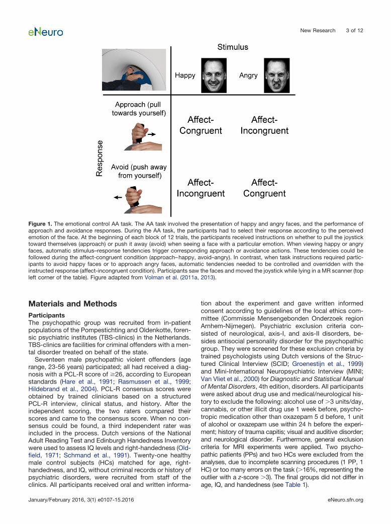
Figure 1. The emotional control AA task. The AA task involved the presentation of happy and angry faces, and the performance ofapproach and avoidance responses. During the AA task, the participants had to select their response according to the perceivedemotion of the face. At the beginning of each block of 12 trials, the participants received instructions on whether to pull the joysticktoward themselves (approach) or push it away (avoid) when seeing a face with a particular emotion. When viewing happy or angryfaces, automatic stimulus–response tendencies trigger corresponding approach or avoidance actions. These tendencies could befollowed during the affect-congruent condition (approach–happy, avoid–angry). In contrast, when task instructions required partic-ipants to avoid happy faces or to approach angry faces, automatic tendencies needed to be controlled and overridden with theinstructed response (affect-incongruent condition). Participants saw the faces and moved the joystick while lying in a MR scanner (topleft corner of the table). Figure adapted from Volman et al. (2011a, 2013).
New Research 3 of 12
January/February 2016, 3(1) e0107-15.2016 eNeuro.sfn.org
ProcedureTwo test sessions took place. During the first session,right-handedness, IQ, MINI, and SCID were assessed.During the second session, participants completed sev-eral questionnaires upon arrival in the laboratory, includ-ing the State-Trait Anxiety Inventory (STAI) to measureanxiety levels (Spielberger, 1983). Next, they providedsaliva for the testosterone measurement. Afterward, par-ticipants were positioned in the 1.5 T MR scanner andfamiliarized with the task setup. Immediately after this, thefMRI session started with the AA task (duration, 30 min)followed by another task (not included in this report). Aftera short break outside the scanner, the anatomical scan(duration, 5 min) and an unrelated task were acquired inthe side-by-side 3 T MR scanner.
Experimental taskThe AA task consisted of 24 blocks (with 12 trials perblock and a baseline period of 21-24 s) during whichparticipants had to respond to visually presented faceseither by pulling a joystick toward themselves (approach)or by pushing it away from themselves (avoid; Fig. 1). Theparticipants had to categorize faces as happy, angry, andneutral (filler items), based on their affective expressions.During each block, two of the three affective expressionswere presented as stimuli, because only two responsescould be given to categorize the stimulus. This resulted insix different block types each used four times, represent-ing the affect (happy–angry, happy–neutral, angry–neu-tral) � movement (approach–avoid) combinations. At thestart of each block, participants received written instruc-tions regarding the required response mapping. The af-fect � movement combinations were pseudorandomlyand evenly distributed (with no affect combination repe-tition), and the combination of the first block was coun-terbalanced across participants. Within each block,affective expressions and gender types were pseudoran-domly presented, avoiding three or more sequential pre-sentations of the same expression/gender, and twopresentations of the same facial model. Each face waspresented for 100 ms, preceded by a 300 ms blankscreen, and followed by the participant’s response, ablank screen, and by a pseudorandom intertrial interval(ITI; 1-3 s). A baseline period of 21-24 s preceded eachblock. The faces were from 36 models (18 male) obtainedfrom several databases (Ekman and Friesen, 1976; Ma-tsumoto and Ekman, 1988; Lundqvist et al., 1998; Marti-nez and Benavente, 1998), each showing all expressions.
The pictures were in grayscale, matched for brightnessand contrast values, and displayed against a black back-ground. To exclude influence from hair and nonfacialcontours, the faces were trimmed. Joystick displace-ments of �80% along the sagittal plane within 2 s fromstimulus presentation were marked as valid responses.Invalid responses were signaled for 1 s with written feed-back stating “you did not move your joystick far enough.”After moving the joystick, participants had to return to thestarting position (defined as the central area extending20% along the sagittal plane) before the end of the ITI.Otherwise, visual feedback indicated “return the joystickto the starting position,” and the ITI was repeated afterparticipants returned the joystick. The training at the be-ginning consisted of six blocks; one block of eight trialsfor each of the six affect � movement combinations.Different visual stimuli were used during the training andscanning blocks.
Materials and apparatusThe fMR images were acquired on a 1.5 T MRI scanner(Avanto, Siemens Medical Systems) with an eight-channelhead coil using a multiecho generalized autocalibratingpartially parallel acquisitions (GRAPPA) sequence [Poseret al., 2006; repetition time (TR), 2.14 ms; five echo times(TEs), 9.4/21/33/44/56 ms; 34 transversal slices; ascend-ing acquisition; distance factor, 17%; effective voxel size,3.3 � 3.3 � 3.5 mm; field of view (FOV), 212 mm].High-resolution anatomical images were acquired on a 3T MRI scanner with a 32-channel head coil using a mag-netization prepared rapid gradient echo sequence (TR,2300 ms; TE, 3.03 ms; 192 sagittal slices; voxel size, 1.0� 1.0 � 1.0 mm; FOV, 256 mm).
An MR-compatible joystick (Fiber Optic Joystick, Cur-rent Designs; sampling rate, 550 Hz) was placed on par-ticipants’ abdomens to ensure comfortable push-and-pullmovements (Fig. 1). Participants wore MR-compatibleheadphones to reduce scanner noise (Commander XGMRI Audio System, Resonance Technologies). Stimuliwere projected at the center of a screen, viewed via amirror above the participant’s head, with a visual angle of4° � 6° (width � height). Stimuli presentation and acqui-sition of joystick positions were controlled by a PC run-ning Presentation version 13 (http://www.neurobs.com).
Salivary measurementsParticipants filled two Salicaps (IBL) with saliva for testos-terone measurement, which were stored at �25°C. Tes-tosterone concentration was measured using competitivechemiluminescence immunoassay with a sensitivity of0.0025 ng/ml (IBL International, Tecan). Intra-assay andinterassay coefficients are between 10% and 12%. Tocontrol variables influencing testosterone levels, partici-pants were instructed to refrain from any food, cigarettes,and drinks (except water) for 1 h before the experiment.
Behavioral analysisBehavioral data was analyzed using MATLAB version 7.9(MathWorks) and PASW Statistics 18 (SPSS Inc.). First, toobtain a precise measure of movement onset [reactiontime (RT)], the joystick movement for each trial was re-
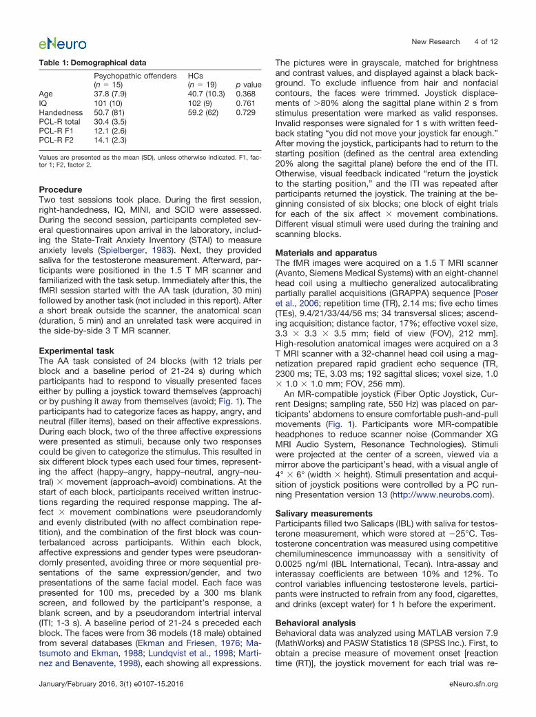
Table 1: Demographical data
Psychopathic offenders(n � 15)
HCs(n � 19) p value
Age 37.8 (7.9) 40.7 (10.3) 0.368IQ 101 (10) 102 (9) 0.761Handedness 50.7 (81) 59.2 (62) 0.729PCL-R total 30.4 (3.5)PCL-R F1 12.1 (2.6)PCL-R F2 14.1 (2.3)
Values are presented as the mean (SD), unless otherwise indicated. F1, fac-tor 1; F2, factor 2.
New Research 4 of 12
January/February 2016, 3(1) e0107-15.2016 eNeuro.sfn.org
constructed using the joystick displacement measure-ments. Excluded trials showed a joystick movement in thewrong direction, an extreme RT (�150 or �1500 ms),peak velocity (�0.1 cm/s), or movement time (�400 ms);or an error rate of above chance level in a block (in thatcase, the whole block was excluded). RTs and testoster-one levels were log transformed to obtain a normal dis-tribution. Second, following previous studies (Roelofset al., 2009; Volman et al., 2011b), we conducted three-way repeated-measures ANOVA (ANCOVArm) on themean RT and error rates, with factors group (PP, HC),movement (approach, avoid), and valence (happy, angry),including standardized testosterone and STAI state ascovariate. A measure of anxiety (STAI) was included toaccount for the effects of psychopathy type (e.g., primaryvs secondary); and the possible effects on emotionalbehavior, hormonal levels, amygdala, and prefrontal cor-tex functioning (Freitas-Ferrari et al., 2010; Koenigs et al.,2011; Giltay et al., 2012; Fouche et al., 2013). The �-levelwas set at p � 0.05.
Functional MRI dataSingle-subject analysesImaging data were preprocessed and analyzed using SPM8(Statistical Parametric Mapping; http://www.fil.ion.ucl.ac.uk/spm). The first four volumes of each participant’s datasetwere discarded to allow for T1 equilibration. Given the mul-tiecho GRAPPA MR sequence (Poser et al., 2006), headmotion parameters were estimated on MR images with theshortest TE (9.4 ms), since these are least affected by pos-sible artifacts. These motion correction parameters, esti-mated using a least-squares approach with six rigid bodytransformation parameters (translations, rotations), were ap-plied to the five echo images collected for each excitation.After spatial realignment, the five echo images were com-bined into a single MR volume using an optimized echoweighting method (Poser et al., 2006). The time series foreach voxel was temporally realigned to the first slice in time.The T1-weighted image was spatially coregistered to themean of the functional images. The fMRI time series weretransformed and resampled at an isotropic voxel size of 2mm into standard Montreal Neurological Institute (MNI)space by unified segmentation and normalization using thecoregistered T1-weighted image (Ashburner and Friston,2005). The normalized functional images were spatiallysmoothed using an isotropic 8 mm full-width at half-maximum Gaussian kernel.
The fMRI time series of each subject were further ana-lyzed using an event-related approach in the context ofgeneral linear model, including the following effects: ap-proach–happy, approach–neutral, approach–angry, avoid–happy, avoid–neutral, and avoid–angry. Trials excluded frombehavioral analyses and periods of instructions or feed-back were modeled as regressors. Vectors describing thetime of picture presentation (onset) and RT of each event(duration) were convolved with the canonical hemody-namic response function. Potential confounding effects ofresidual head movement were modeled using original,squared, cubic, first-order, and second-order derivativesof the movement correction parameters (Lund et al.,
2005). Three further regressors, describing the timecourse of signal intensities of white matter, CSF, and theportion of the MR image outside the skull were alsoadded. This procedure accounts for image intensity shiftsdue to hand movements within or near the magnetic fieldof the scanner (Verhagen et al., 2006). Finally, fMRI timeseries were high-pass filtered (cutoff 120 s). Temporalautocorrelation was modeled as a first-order autoregres-sive process.
Group analysesConsistent effects across participants and between groupswere tested using a random-effects multiple regressionanalysis that included six contrast images (approach–happy, approach–neutral, approach–angry, avoid–happy,avoid–neutral, avoid–angry) per participant. Together, theseimages represented the estimated cerebral effects from 12conditions of the experimental design [group (PP, HC) �valence (happy, neutral, angry) � response (approach,avoid)]. Standardized log-transformed testosterone andstandardized STAI state levels were included in the multipleregression analysis as condition-specific [group (PP, HC) �valence (happy, neutral, angry) � response (approach,avoid)] regressors, generating another 12 regressors pervariable.
All analyses assessed the congruency effect, reflectingtask-related differences of affect-incongruent (approach–angry, avoid–happy) versus affect-congruent trials (ap-proach–happy, avoid–angry; Roelofs et al., 2009; Volmanet al., 2011b). We considered two effects. First, to test forgeneral effects of congruency, we performed an analysison the congruency effect over both groups and for eachgroup separately. When assessing the effects of onegroup explicitly, we also tested whether those effectswere specific to that group and were significantly weakerin the other group (at p � 0.05 uncorrected) by maskingthe statistical map describing the congruency effect in thefirst group (using multiple comparisons correction, seebelow) with the statistical map describing the group �congruency contrast. Second, to test whether testoster-one differentially modulated the control of emotionallyrelevant actions in the groups, we performed a group �congruency contrast on the regressor parametrizing inter-individual differences in testosterone on task-related con-ditions. If such an interaction is present, the testosteronemodulation on the congruency effect of each group sep-arately is considered. In addition to whole-brain analyses,we used a volume of interest (VOI) on coordinates previ-ously found to be modulated by testosterone during thecongruency effect in healthy students (two 8-mm-radiusspheres centered on the following MNI coordinates: x,�30; y, 58; and z, 2; and x, 32; y, 54; and z, 8; Volmanet al., 2011b).
The reported activations are corrected for multiplecomparisons using familywise error (FWE) correction. Forwhole-brain analyses, we made inferences at cluster level(FWE: p � 0.05, corresponding to a cluster size of �140on the basis of intensity threshold, p � 0.001). For VOIanalyses, we made inferences at voxel-level (FWE cor-rected, p � 0.05; Worsley et al., 1996; Friston, 1997).Anatomical inference is drawn by superimposing SPM
New Research 5 of 12
January/February 2016, 3(1) e0107-15.2016 eNeuro.sfn.org
showing significant signal changes on structural imagesof participants. For anatomical accuracy, we report onlyactivation peaks in gray matter. Anatomical landmarkswere identified using the atlas of Duvernoy et al. (1991).Brodmann areas (BAs) were assigned by superimposingsignificant SPM on the SPM anatomy toolbox (Eickhoffet al., 2005) and MRIcron template (http://www.mccauslandcenter.sc.edu/mricro/mricron/).
Connectivity analysesThe aim of the following analysis was to test whetherinter-regional coupling of the aPFC (see Results) with theamygdala and other brain regions during the congruencyeffect was different between the groups and modulatedby testosterone. To test for these effects, we used thepsychophysiological interactions (PPIs) method (Fristonet al., 1997). More specifically, we tested for significantdifferences between the regression coefficients of eachvoxel over the right aPFC during the affect-incongruentversus the affect-congruent conditions. To select voxelsto be included in the VOI, we used the following anatom-ical constraints (Stephan et al., 2010): for each participant,selected voxels fell within a sphere with a radius of 4 mmaround the peak voxel corresponding to the activatedcluster of the congruency effect over both groups (coor-dinates: x, 30; y, 58; z, 14; see Results). Participant spe-cific contrast images were generated describing the PPIbetween the time courses of the right aPFC VOI andaffect-incongruent versus affect-congruent conditions.Group differences and testosterone modulations on task-related coupling between the aPFC and other regionswere then assessed using a multiple regression design onparticipant-specific contrast images with their corre-sponding testosterone (log-transformed, standardized)and STAI state (standardized) levels as subject- andgroup-specific regressors. In addition to whole-brain analy-ses, we assessed significant voxel-level effects (FWE cor-rected for multiple comparisons, p � 0.05) within theamygdala, defined on the Automated Anatomical Labelingatlas (Tzourio-Mazoyer et al., 2002) using the WFU PickAtlastool (Maldjian et al., 2003).
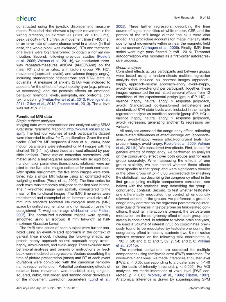
ResultsBehavioral resultsFifteen psychopathic criminals (PPs; PCL-R score of �26,according to European standards (Rasmussen et al.,1999; Hare, 2003; Hildebrand et al., 2004) and 19 HCs (fordemographics, see Table 1) were included in the analy-ses. Participants performed the task accurately and con-sistently (error rates: PPs, 7.9%; HCs, 7.3%; omissions:PPs, 1.6%; HCs, 1.5%; undefined responses: PPs, 0.9%;HCs, 0.3%; Table 2).
A significant movement � valence interaction for theRTs indicated that, over groups, participants respondedmore slowly during affect-incongruent (approach–angry,avoid–happy) than during affect-congruent trials (ap-proach–happy, avoid–angry; F(1,29) � 10.4, p � 0.003; Fig.2). This congruency effect replicates the behavioral resultsfrom previous fMRI studies (Roelofs et al., 2009; Volmanet al., 2011b, 2013). Furthermore, there were main effects
of movement (F(1,29) � 26.3, p � 0.001) and valence (F(1,29)
� 28.7, p � 0.001), reflecting the slowing of avoidancemovements and responses to angry faces in general(Table 2). There were no significant effects involvinggroup, including no main effect (p � 0.3). The congruencyeffect correlated positively (without corrections for multi-ple comparisons) with the PCL-R total score (p � 0.048, R� 0.517, respectively). Excluding anxiety from the analy-ses did not affect the outcomes. Moreover, when includ-ing the neutral conditions in the analyses, the movement� valence (happy, neutral, angry) interaction for RTs re-mained significant (F(1,28) � 5.5, p � 0.010), showing thatneutral approach–avoidance effects are intermediarycompared with happy and angry (Table 2).
For the error rates, the three-way ANCOVArm showedmain effects of movement (F(1,29) � 27.5, p � 0.001),valence (F(1,29) � 25.9, p � 0.001), and testosterone (F(1,29)
� 4.6, p � 0.040), and a valence � testosterone interac-tion (F(1,29) � 4.3, p � 0.047). There were no other signif-icant effects for the error rates (p � 0.15).
Table 2: RTs and error rates for each group and factor of theAA task
Psychopathic offenders HCsApproach Avoid Approach Avoid
Errors (%)Happy 3.2 (0.9) 8.9 (1.8) 2.4 (0.8) 7.7 (1.1)Neutral 6.1 (1.3) 5.8 (1.1) 7.1 (1.4) 5.2 (1.0)Angry 10.1 (2.2) 13.1 (2.1) 9.6 (1.8) 11.6 (1.8)RT (ms)Happy 554 (25) 625 (35) 553 (23) 603 (25)Neutral 666 (28) 687 (31) 639 (21) 668 (24)Angry 630 (25) 665 (33) 620 (24) 630 (23)
Values are presented as the mean (SE).
Figure 2. Behavioral results. Mean RTs (�SEM) for the affect-congruent and affect-incongruent conditions of the AA task forthe healthy control subjects and psychopathic offenders. Thegroups were significantly slower to provide affect-incongruentresponses (approach–angry; avoid–happy) than affect-congruent responses (approach–happy; avoid–angry), with nosignificant group differences.
New Research 6 of 12
January/February 2016, 3(1) e0107-15.2016 eNeuro.sfn.org
Endogenous testosterone levels [median (SD): PPs, 101pg/ml (70 pg/ml); HCs, 90 pg/ml (46 pg/ml)] and stateanxiety levels [STAI mean (SD): PPs, 32 (8); HCs, 32 (5)]did not differ between groups (p � 0.4), and showed nocorrelations with psychopathy (PCL-R) scores or witheach other (p � 0.1).
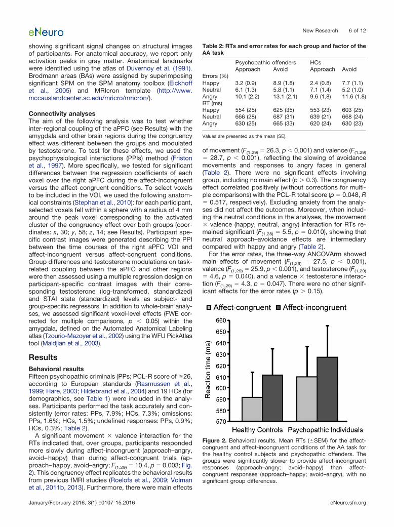
fMRI resultsMultiple regression analysesTo assess the two main questions of this study, we iso-lated cerebral structures showing stronger responsesduring affect-incongruent than affect-congruent trials(congruency effect), and cerebral structures in which thecongruency effect was modulated by testosterone levels.
The results showed a significant congruency effectacross groups in the aPFC [ROI analysis: MNI coordinates(x, y, z): (30, 58, 14) and (�30 58 10); pFWE � 0.001 and0.036; t � 4.46 and 3.43; for further details, see Table 3].As expected, this effect was driven by the healthy controlgroup, and it was significantly weaker in the psychopathicoffenders [pFWE � 0.001 and 0.040; t � 4.58 and 3.40, onthe congruency effect in healthy control subjects masked
implicitly by group (HC � PP) � congruency interaction].The implicit masking demonstrates that the group � con-gruency interaction is also significant at puncorrected � 0.05within the significant voxels corrected for multiple com-parisons on the HC congruency effect. The psychopathygroup showed no significant congruency effect in thisregion (pFWE � 0.3). There was also a significant congru-ency effect across groups in the right superior parietallobule (whole-brain analysis); this effect was driven mainlyby the psychopathy group (Table 3).
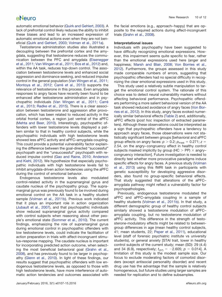
Critically, testosterone modulated the congruency ef-fect in the aPFC differently in psychopathic offenders andhealthy control subjects (whole-brain analysis on testos-terone � group � congruency: MINI coordinates (x, y, z):(30, 58, 12); pFWE � 0.001; t � 5.10; for all details, seeTable 3). Post hoc analyses revealed that, in the psychop-athy group, congruency effects decreased as testoster-one levels increased [MNI coordinates (x, y, z): (32, 56, 10)and (�30, 58, 8); pFWE � 0.002 and 0.015; t � 4.34 and3.74]. The modulatory effect of testosterone on congru-ency was absent in the healthy control subjects (pFWE �
Table 3: Clusters showing significantly larger activity for the affect-incongruent vs the affect-congruent conditions (emotion-control effect)
Anatomical region Putative BA Side x y z Voxels (n) p value t valueWhole-brain effectsCongruency effect over groupsCuneus 18 R/L �16 �96 24 634 �0.001 4.85SPL/Superior occipital gyrus 7/19 L �30 �76 34 254 0.004 4.37IFG 45/47 L �52 22 �10 223 0.007 4.37Cuneus 18 R 18 �98 16 190 0.016 4.52Congruency effect for psychopathy groupSPL/superior occipital gyrus 7/19 R/L 8 �82 38 925 �0.001 4.98Angular gyrus 39/19 L �30 �72 34 337 0.001 4.35Superior temporal gyrus 42 L �32 �32 6 214 0.009 4.51SPL 7 R 28 �74 46 158 0.034 4.58Cerebellum L �24 �66 �38 146 0.046 4.63Negative testosterone modulation of group
(psychopathic offenders � healthy control subjects)� congruency interaction
aPFC 10 R 30 58 12 391 �0.001 5.10Supramarginal gyrus 40 R 54 �42 54 325 0.001 4.66Caudate nucleus R 10 10 2 273 0.002 4.69Putamen/Insula L �34 6 �10 176 0.022 5.22Cerebellum R 18 �76 �38 188 0.016 5.09Negative testosterone modulation of
Congruency effect in psychopathySupramarginal gyrus 40 R 52 �40 54 657 �0.001 5.32Precentral/superior frontal gyrus 6 R/L 6 22 66 471 �0.001 5.65Caudate nucleus R 6 6 4 228 0.006 4.28VOI on bilateral aPFCCongruency effect over groups 10 R 30 58 14 31 0.001 4.46
10 L �30 58 10 5 0.036 3.43Congruency effect in healthy control subjects 10 R 32 58 14 12 0.001 4.58
10 L �34 52 4 2 0.040 3.40Negative testosterone modulation of group
(psychopathic offenders � healthy control subjects)� congruency interaction
10 R 30 58 12 145 �0.001 5.1010 L �24 56 6 15 0.010 3.87
Negative testosterone modulationof congruency effect in psychopathy
10 R 32 56 10 77 0.002 4.3410 L �30 58 8 17 0.015 3.74
Coordinates are defined in MNI (x, y, z) space. The p values represent the FWE cluster-level corrected values for the whole-brain analyses and FWE voxel-level corrected values for the VOI analyses. IFG, Inferior frontal gyrus; L, left; R, right; SPL, superior parietal lobule.
New Research 7 of 12
January/February 2016, 3(1) e0107-15.2016 eNeuro.sfn.org
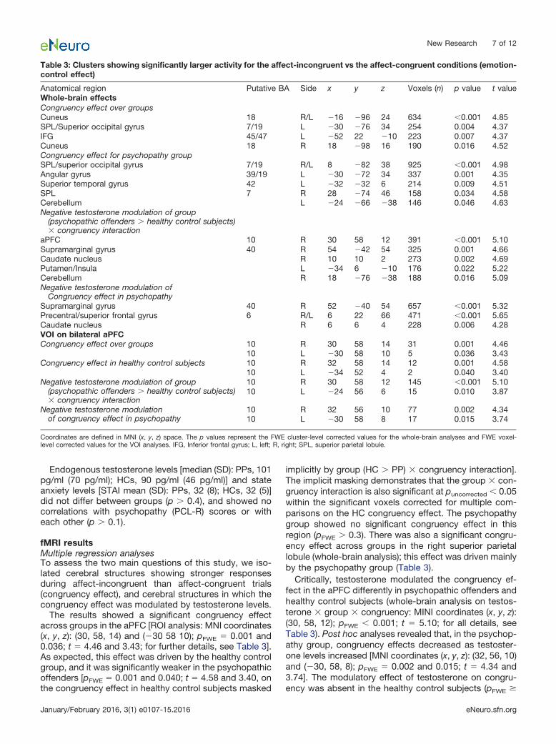
0.05; Fig. 3A–C). The whole-brain analysis also showed aneffect in the right caudate nucleus and right inferior su-pramarginal gyrus, driven by reduced congruency effectsas a function of testosterone in the psychopathy group(Fig. 3D–F; Table 3).
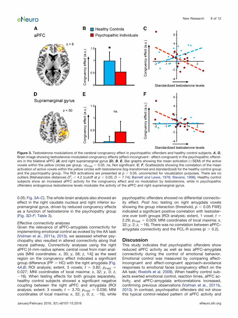
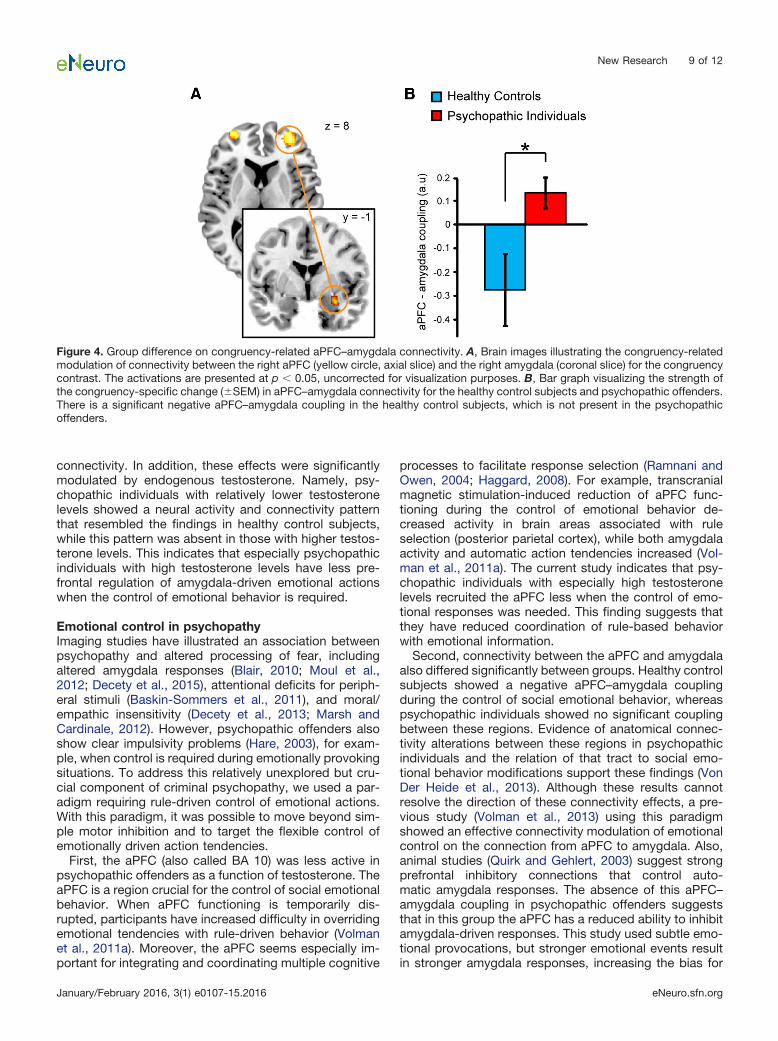
Effective connectivity analysesGiven the relevance of aPFC–amygdala connectivity forimplementing emotional control as evoked by the AA task(Volman et al., 2011a, 2013), we assessed whether psy-chopathy also resulted in altered connectivity along thatneural pathway. Connectivity analyses using the rightaPFC [4-mm-radius sphere; central voxel from main anal-ysis (MNI coordinates: x, 30; y, 58; z, 14)] as the seedregion on the congruency effect indicated a significantgroup difference (PP � HC) with the right amygdala (Fig.4A,B; ROI analysis; extent, 3 voxels; t � 3.82; pFWE �0.027; MNI coordinates of local maxima: x, 32; y, 0; z,�16). When testing effects for both groups separately,healthy control subjects showed a significant negativecoupling between the right aPFC and amygdala (ROIanalysis; extent: 3 voxels, t � 3.70; pFWE � 0.036; MNIcoordinates of local maxima: x, 32; y, 0; z, �16), while
psychopathic offenders showed no differential connectiv-ity effect. Post hoc testing on right amygdala voxelsshowing the group interaction (threshold, p � 0.05 FWE)indicated a significant positive correlation with testoster-one over both groups (ROI analysis; extent, 1 voxel; t �2.29; pFWE � 0.029; MNI coordinates of local maxima: x,32; y, 2; z, �16). There was no correlation between aPFC–amygdala connectivity and the PCL-R scores (p � 0.2).
DiscussionThis study indicates that psychopathic offenders showreduced aPFC activity as well as less aPFC–amygdalaconnectivity during the control of emotional behavior.Emotional control was measured by comparing affect-incongruent and affect-congruent approach–avoidanceresponses to emotional faces (congruency effect on theAA task; Roelofs et al. 2009). When healthy control sub-jects exerted emotional control, reaction times, aPFC ac-tivity, and aPFC–amygdala anticorrelations increased,confirming previous observations (Volman et al., 2011b,2013). In contrast, psychopathic offenders did not showthis typical control-related pattern of aPFC activity and
Figure 3. Testosterone modulations of the cerebral congruency effect in psychopathic offenders and healthy control subjects. A, D,Brain image showing testosterone-modulated congruency effects (affect-incongruent�affect-congruent) in the psychopathic offend-ers in the bilateral aPFC (A) and right supramarginal gyrus (D). B, E, Bar graphs showing the mean activation (�SEM) of the activevoxels within the yellow circles per group. �pFWE � 0.05. ns, Not significant. C, F, Scatterplots showing the correlation of the meanactivation of active voxels within the yellow circles with testosterone (log-transformed and standardized) for the healthy control groupand the psychopathy group. The ROI activations are presented at p � 0.05, uncorrected for visualization purposes. There are nooutliers [Mahalanobis distances D2
i � 4.2 (cutoff at p � 0.05; D � 7.74); Barnett and Lewis, 1978; Stevens, 1996]. Healthy controlsubjects show an increased aPFC activity for the congruency effect and no modulation by testosterone, while in psychopathicoffenders endogenous testosterone levels modulate the activity of the aPFC and right supramarginal gyrus.
New Research 8 of 12
January/February 2016, 3(1) e0107-15.2016 eNeuro.sfn.org
connectivity. In addition, these effects were significantlymodulated by endogenous testosterone. Namely, psy-chopathic individuals with relatively lower testosteronelevels showed a neural activity and connectivity patternthat resembled the findings in healthy control subjects,while this pattern was absent in those with higher testos-terone levels. This indicates that especially psychopathicindividuals with high testosterone levels have less pre-frontal regulation of amygdala-driven emotional actionswhen the control of emotional behavior is required.
Emotional control in psychopathyImaging studies have illustrated an association betweenpsychopathy and altered processing of fear, includingaltered amygdala responses (Blair, 2010; Moul et al.,2012; Decety et al., 2015), attentional deficits for periph-eral stimuli (Baskin-Sommers et al., 2011), and moral/empathic insensitivity (Decety et al., 2013; Marsh andCardinale, 2012). However, psychopathic offenders alsoshow clear impulsivity problems (Hare, 2003), for exam-ple, when control is required during emotionally provokingsituations. To address this relatively unexplored but cru-cial component of criminal psychopathy, we used a par-adigm requiring rule-driven control of emotional actions.With this paradigm, it was possible to move beyond sim-ple motor inhibition and to target the flexible control ofemotionally driven action tendencies.
First, the aPFC (also called BA 10) was less active inpsychopathic offenders as a function of testosterone. TheaPFC is a region crucial for the control of social emotionalbehavior. When aPFC functioning is temporarily dis-rupted, participants have increased difficulty in overridingemotional tendencies with rule-driven behavior (Volmanet al., 2011a). Moreover, the aPFC seems especially im-portant for integrating and coordinating multiple cognitive
processes to facilitate response selection (Ramnani andOwen, 2004; Haggard, 2008). For example, transcranialmagnetic stimulation-induced reduction of aPFC func-tioning during the control of emotional behavior de-creased activity in brain areas associated with ruleselection (posterior parietal cortex), while both amygdalaactivity and automatic action tendencies increased (Vol-man et al., 2011a). The current study indicates that psy-chopathic individuals with especially high testosteronelevels recruited the aPFC less when the control of emo-tional responses was needed. This finding suggests thatthey have reduced coordination of rule-based behaviorwith emotional information.
Second, connectivity between the aPFC and amygdalaalso differed significantly between groups. Healthy controlsubjects showed a negative aPFC–amygdala couplingduring the control of social emotional behavior, whereaspsychopathic individuals showed no significant couplingbetween these regions. Evidence of anatomical connec-tivity alterations between these regions in psychopathicindividuals and the relation of that tract to social emo-tional behavior modifications support these findings (VonDer Heide et al., 2013). Although these results cannotresolve the direction of these connectivity effects, a pre-vious study (Volman et al., 2013) using this paradigmshowed an effective connectivity modulation of emotionalcontrol on the connection from aPFC to amygdala. Also,animal studies (Quirk and Gehlert, 2003) suggest strongprefrontal inhibitory connections that control auto-matic amygdala responses. The absence of this aPFC–amygdala coupling in psychopathic offenders suggeststhat in this group the aPFC has a reduced ability to inhibitamygdala-driven responses. This study used subtle emo-tional provocations, but stronger emotional events resultin stronger amygdala responses, increasing the bias for
Figure 4. Group difference on congruency-related aPFC–amygdala connectivity. A, Brain images illustrating the congruency-relatedmodulation of connectivity between the right aPFC (yellow circle, axial slice) and the right amygdala (coronal slice) for the congruencycontrast. The activations are presented at p � 0.05, uncorrected for visualization purposes. B, Bar graph visualizing the strength ofthe congruency-specific change (�SEM) in aPFC–amygdala connectivity for the healthy control subjects and psychopathic offenders.There is a significant negative aPFC–amygdala coupling in the healthy control subjects, which is not present in the psychopathicoffenders.
New Research 9 of 12
January/February 2016, 3(1) e0107-15.2016 eNeuro.sfn.org
automatic emotional behavior (Quirk and Gehlert, 2003). Alack of prefrontal control likely reduces the ability to inhibitthese biases and lead to an increased expression ofautomatic emotional actions even when they are not ben-eficial (Quirk and Gehlert, 2003; Volman et al., 2011a).
Testosterone administration studies also illustrated adecoupling between the prefrontal cortex and the amy-gdala, suggesting that testosterone reduces the commu-nication between the PFC and amygdala (Eiseneggeret al., 2011; Van Wingen et al., 2011; Bos et al., 2012) and,within the AA task, reduces top-down control. The asso-ciation between testosterone levels and enhanced socialaggression and dominance seeking, and reduced impulsecontrol in the general population (Van Wingen et al., 2011;Montoya et al., 2012; Carré et al., 2013) supports therelevance of testosterone in this process. Even amygdalaresponses to angry faces have recently been found to beenhanced after testosterone administration and in psy-chopathic individuals (Van Wingen et al., 2011; Carréet al., 2013; Radke et al., 2015). There is a clear associ-ation between testosterone and aggression after provo-cation, which has been related to reduced activity in theorbital frontal cortex, a region just ventral of the aPFC(Mehta and Beer, 2010). Interestingly, psychopathic of-fenders with lower testosterone levels displayed a pat-tern similar to that in healthy control subjects, while thepsychopathic individuals with high testosterone levelsshowed less aPFC activity and aPFC–amygdala coupling.This could provide a potential vulnerability factor explain-ing the difference between the goal-directed “successful”psychopath and the “unsuccessful” psychopath with re-duced impulse control (Gao and Raine, 2010; Andersonand Kiehl, 2012). We hypothesize that especially psycho-pathic individuals with high testosterone levels fail toinhibit amygdala-driven action tendencies using the aPFCduring the control of emotional behavior.
Endogenous testosterone levels also modulatedcontrol-related activity in the supramarginal gyrus andcaudate nucleus of the psychopathy group. The supra-marginal gyrus was previously found to be involved duringemotional control on the AA task in a healthy studentsample (Volman et al., 2011b). Previous work indicatedthat it plays an important role in action organization(Jubault et al., 2007), and that psychopathic individualsshow reduced supramarginal gyrus activity comparedwith control subjects when reasoning about other peo-ple’s emotional state (Sommer et al., 2010). The currentfindings, emphasizing the role of supramarginal gyrusduring emotional control in psychopathic offenders withlow testosterone levels, could indicate the facilitation ofaction preparation in trials with affect-incongruent stimu-lus–response mapping. The caudate nucleus is importantfor incorporating predicted action outcome, when select-ing the most beneficial behavioral goal (Grahn et al.,2008), and has previously found to be larger in psychop-athy (Glenn et al., 2010). In light of these findings, ourresults suggest that psychopathic offenders with low en-dogenous testosterone levels, as opposed to those withhigh testosterone levels, have more interference of auto-matic action tendencies and outcomes associated with
the facial emotions (e.g., approach–happy) that are op-posite to the required actions during affect-incongruenttrials (Grahn et al., 2008).
Interpretational issuesIndividuals with psychopathy have been suggested tohave difficulty recognizing emotional expressions. How-ever, this impairment seems quite specific to fear, ratherthan the emotional expressions used here (anger andhappiness; Marsh and Blair, 2008; Von Borries et al.,2012). Furthermore, the groups assessed in this studymade comparable numbers of errors, suggesting thatpsychopathic offenders had no special difficulty in recog-nizing the clear emotional expressions used in this study.
This study used a relatively subtle manipulation to tar-get the emotional control system. The rationale of thischoice was to detect neural vulnerability markers withoutaffecting behavioral performance. Psychopathic offend-ers performing a more salient behavioral version of the AAtask showed reduced avoidance of angry faces (Von Bor-ries et al., 2012). In this study, angry faces evoked numer-ically similar behavioral effects (Table 2) and, additionally,aPFC effects (post hoc inspection of extracted parame-ters). Although these observations could be interpreted asa sign that psychopathic offenders have a tendency toapproach angry faces, those observations were not sta-tistically significant between groups [behavioral and aPFCgroup effects on angry faces: p � 0.2; pFWE � 0.271; z �2.54, on the angry–congruency effect in healthy controlsubjects masked implicitly by group (HC � PP) � angry–congruency interaction]. Future investigation is needed todirectly test whether more provocative paradigms inducespecific effects for angry faces. A previous study (Volmanet al., 2013) using this fMRI task in participants withgenetic susceptibility for developing aggressive disor-ders, also found no group-specific behavioral effects.That study suggested that alterations of the aPFC–amygdala pathway might reflect a vulnerability factor forpsychopathologies.
Previously, endogenous testosterone modulated theaPFC and aPFC–amygdala coupling in a sample ofhealthy students (Volman et al., 2011b). In that study, adifferent demographic group of healthy control subjectssimilarly showed a testosterone modulation of aPFC–amygdala coupling, but no testosterone modulation ofaPFC activity. This difference in the strength of testo-sterone-modulatory effects might be related to between-group differences in age (mean healthy control subjects,41; mean students, 22; Peper et al., 2011), educationallevel (staff of forensic psychiatric institute vs universitystudents), or general anxiety [STAI trait, lower in healthycontrol subjects of the current study; mean (SD): 29 (4.4)and 34 (6.9), respectively; t(37) � �2.605; p � 0.014]. Alimitation of this study is the modest sample size. Ourfocus to exclude moderating factors of comorbid disor-ders (except antisocial personality disorder) and recentdrug use has the advantage that the sample is relativelyhomogeneous, but future studies using larger samples areneeded for replication and to define subsamples.
New Research 10 of 12
January/February 2016, 3(1) e0107-15.2016 eNeuro.sfn.org
ConclusionPsychopathic offenders showed reduced aPFC activityand aPFC–amygdala connectivity during control of emo-tional actions, suggesting a decreased coordinationof emotional information during rule-driven behavior.Moreover, endogenous testosterone modulated the in-volvement of these neural mechanisms. Psychopathic of-fenders with high testosterone levels showed lessinvolvement of the aPFC, aPFC–amygdala connectivity,supramarginal gyrus, and caudate nucleus, whereas psy-chopathic individuals with low testosterone levels re-cruited the aPFC in a fashion similar to that of healthycontrol subjects. These findings suggest that a lack ofprefrontal control during emotional actions may explainenhanced impulsivity in psychopathic offenders duringemotionally provoking situations. They outline a neuroen-docrine model underlying impulsive emotional behavior inpsychopathy and support the relevance of assessing apotential imbalance in testosterone function to guidetreatment. It remains to be seen whether these neuroen-docrine alterations of emotional control are also present inhighly impulsive or antisocial individuals.
ReferencesAnderson NE, Kiehl KA (2012) The psychopath magnetized: insights
from brain imaging. Trends Cogn Sci 16:52-60. CrossRef MedlineAshburner J, Friston KJ (2005) Unified segmentation. Neuroimage
26:839-851. CrossRef MedlineBarnett V, Lewis T (1978) Outliers in statistical data. New York: Wiley.Baskin-Sommers AR, Curtin JJ, Newman JP (2011) Specifying the
attentional selection that moderates the fearlessness of psycho-pathic offenders. Psychol Sci 22:226-234. CrossRef Medline
Blair RJR (2010) Neuroimaging of psychopathy and antisocial be-havior: a targeted review. Curr Psychiatry Rep 12:76-82. CrossRefMedline
Blair RJR (2013) Psychopathy: cognitive and neural dysfunction.Dialogues Clin Neurosci 15:181–190.
Blair RJR, Mitchell D, Blair K (2005) The psychopath: emotion and thebrain. Oxford, UK: Blackwell.
Bos PA, Panksepp J, Bluthé RM, van Honk J (2012) Acute effects ofsteroid hormones and neuropeptides on human social-emotionalbehavior: a review of single administration studies. Front Neuroen-docrinol 33:17-35. CrossRef Medline
Carré JM, Hyde LW, Neumann CS, Viding E, Hariri AR (2013) Theneural signatures of distinct psychopathic traits. Soc Neurosci8:122-135. CrossRef Medline
Chen M, Bargh JA (1999) Consequences of automatic evaluation:immediate behavioral predispositions to approach or avoid thestimulus. Pers Soc Psychol Bull 25:215-224. CrossRef
Cornell DG, Warren J, Hawk G, Stafford E, Oram G, Pine D (1996)Psychopathy in instrumental and reactive violent offenders. J Con-sult Clin Psychol 64:783-790. Medline
Craig MC, Catani M, Deeley Q, Latham R, Daly E, Kanaan R, Pic-chioni M, McGuire PK, Fahy T, Murphy DGM (2009) Altered con-nections on the road to psychopathy. Mol Psychiatry 14:946-953.CrossRef Medline
Decety J, Skelly LR, Kiehl KA (2013) Brain response to empathy-eliciting scenarios involving pain in incarcerated individuals withpsychopathy. JAMA Psychiatry 70:638-645. CrossRef Medline
Decety J, Chen C, Harenski CL, Kiehl KA (2015) Socioemotionalprocessing of morally-laden behavior and their consequences onothers in forensic psychopaths. Hum Brain Mapp 36:2015-2026.CrossRef Medline
Dolan M, Anderson IM, Deakin JF (2001) Relationship between 5-HTfunction and impulsivity and aggression in male offenders withpersonality disorders. Br J Psychiatry 178:352-359.
Duvernoy HM, Cabanis EA, Vannson JL (1991) The human brain:surface, three-dimensional sectional anatomy and MRI. Vienna:Springer.
Eickhoff SB, Stephan KE, Mohlberg H, Grefkes C, Fink GR, AmuntsK, Zilles K (2005) A new SPM toolbox for combining probabilisticcytoarchitectonic maps and functional imaging data. Neuroimage25:1325-1335. CrossRef Medline
Eisenegger C, Haushofer J, Fehr E (2011) The role of testosterone insocial interaction. Trends Cogn Sci 15:263-271. CrossRef Medline
Ekman P, Friesen WV (1976) Pictures of facial affect. Palo Alto, CA:Consulting Psychologist.
Enter D, Spinhoven P, Roelofs K (2014) Alleviating social avoidance:effects of single dose testosterone administration on approach-avoidance action. Horm Behav 65:351-354. CrossRef Medline
Fouche JP, van Der Wee NJ, Roelofs K, Stein DJ (2013) Recentadvances in the brain imaging of social anxiety disorder. HumPsychopharmacol 28:102-105. CrossRef Medline
Freitas-Ferrari MC, et al. (2010) Neuroimaging in social anxiety dis-order: a systematic review of the literature. Prog Neuropsychop-harmacol Biol Psychiatry 34:565-580. CrossRef Medline
Friston KJ (1997) Testing for anatomically specified regional effects.Hum Brain Mapp 5:133-136. Medline
Friston KJ, Buechel C, Fink GR, Morris J, Rolls E, Dolan RJ (1997)Psychophysiological and modulatory interactions in neuroimaging.Neuroimage 6:218-229. CrossRef Medline
Gao Y, Raine A (2010) Successful and unsuccessful psychopaths: aneurobiological model. Behav Sci Law 28:194-210. CrossRefMedline
Giltay EJ, Enter D, Zitman FG, Penninx BW, van Pelt J, Spinhoven P,Roelofs K (2012) Salivary testosterone: associations with depres-sion, anxiety disorders, and antidepressant use in a large cohortstudy. J Psychosom Res 72:205-213. CrossRef
Glenn AL, Raine A, Yaralian PS, Yang Y (2010) Increased volume ofthe striatum in psychopathic individuals. Biol Psychiatry 67:52-58.CrossRef Medline
Grahn JA, Parkinson JA, Owen AM (2008) The cognitive functions ofthe caudate nucleus. Prog Neurobiol 86:141-155. CrossRef Med-line
Groenestijn MAC, Akkerhuis GW, Kupka RW, Schneider N, NolenWA (1999) Gestructureerd klinisch interview voor de vaststellingvan DSM-IV as-I stoornissen (SCID-I). Lisse, The Netherlands:Swets Test.
Haggard P (2008) Human volition: towards a neuroscience of will.Nat Rev Neurosci 9:934-946. CrossRef Medline
Hare RD (2003) The Hare psychopathy checklist-revised, Ed 2. To-ronto, Canada: Multi-Health Systems.
Hare RD, Hart SD, Harpur TJ (1991) Psychopathy and the DSM-IVcriteria for antisocial personality disorder. J Abnorm Psychol 100:391-398. Medline
Hildebrand M, De Ruiter C, Nijman H (2004) PCL-R psychopathypredicts disruptive behavior among male offenders in a Dutchforensic psychiatric hospital. J Interpers Violence 19:13-29. Cross-Ref
Jubault T, Ody C, Koechlin E (2007) Serial organization of humanbehavior in the inferior parietal cortex. J Neurosci 27:11028-11036.CrossRef Medline
Koenigs M, Baskin-Sommers A, Zeier J, Newman JP (2011) Investi-gating the neural correlates of psychopathy: a critical review. MolPsychiatry 16:792-799. CrossRef Medline
Lund TE, Nørgaard MD, Rostrup E, Rowe JB, Paulson OB (2005)Motion or activity: their role in intra- and inter-subject variation infMRI. Neuroimage 26:960-964. CrossRef Medline
Lundqvist D, Flykt A, Öhman A (1998) The Karonlinska direct emo-tional faces-KDEF: CD ROM. Solna, Sweden: Department of Clin-ical Neuroscience, Psychology Section, Karolinska Institute.
Maldjian JA, Laurienti PJ, Kraft RA, Burdette JH (2003) An automatedmethod for neuroanatomic and cytoarchitectonic atlas-based in-terrogation of fMRI data sets. Neuroimage 19:1233-1239. Medline
New Research 11 of 12
January/February 2016, 3(1) e0107-15.2016 eNeuro.sfn.org
Malterer MB, Glass SJ, Newman JP (2008) Psychopathy and traitemotional intelligence. Pers Individ Dif 44:735-745. CrossRef Med-line
Marsh AA, Blair RJ (2008) Deficits in facial affect recognition amongantisocial populations: a meta-analysis. Neurosci Biobehav Rev32:454-465. CrossRef Medline
Marsh AA, Cardinale EM (2012) When psychopathy impairs moraljudgments: neural responses during judgments about causingfear. Soc Cogn Affect Neurosci 9:3-11 CrossRef Medline
Martinez AM, Benavente R (1998) The AR face database. Barcelona,Spain: Computer Vision Center, Universidad Autònoma de Barce-lona 24.
Matsumoto D, Ekman P (1988) Japanese and Caucasian facial ex-pressions of emotion (JACFEE) [slides]. San Francisco: Universityof California, Human Interaction Laboratory.
Mehta PH, Beer J (2010) Neural mechanisms of the testosterone-aggression relation: the role of orbitofrontal cortex. J Cogn Neu-rosci 22:2357-2368. CrossRef Medline
Montoya ER, Terburg D, Bos PA, van Honk J (2012) Testosterone,cortisol, and serotonin as key regulators of social aggression: areview and theoretical perspective. Motiv Emot 36:65-73. Cross-Ref Medline
Motzkin JC, Newman JP, Kiehl KA, Koenigs M (2011) Reducedprefrontal connectivity in psychopathy. J Neurosci 31:17348-17357. CrossRef Medline
Moul C, Killcross S, Dadds MR (2012) A model of differentialamygdala activation in psychopathy. Psychol Rev 119:789-806.CrossRef Medline
Oldfield RC (1971) The assessment and analysis of handedness: theEdinburgh inventory. Neuropsychologia 9:97-113. Medline
Patrick CJ, Hicks BM, Krueger RF, Lang AR (2005) Relations be-tween psychopathy facets and externalizing in a criminal offendersample. J Pers Disord 19:339-356.
Poser BA, Versluis MJ, Hoogduin JM, Norris DG (2006) BOLD con-trast sensitivity enhancement and artifact reduction with multiechoEPI: parallel-acquired inhomogeneity-desensitized fMRI. MagnReson Med 55:1227-1235.
Peper JS, van den Heuvel MP, Mandl RC, Pol HE, van Honk J (2011)Sex steroids and connectivity in the human brain: a review ofneuroimaging studies. Psychoneuroendocrinology 36:1101-1113.CrossRef Medline
Price CJ, Friston KJ (1999) Scanning patients with tasks they canperform. Hum Brain Mapp 8:102-108. Medline
Quirk GJ, Gehlert DR (2003) Inhibition of the amygdala: key topathological states? Ann N Y Acad Sci 985:263-272. Medline
Radke S, Volman I, Mehta P, van Son V, Enter D, Sanfey A, Toni I, deBruijn ERA, Roelofs K (2015) Testosterone biases the amygdalatoward social threat approach. Sci Adv 1:e1400074. CrossRefMedline
Ramnani N, Owen AM (2004) Anterior prefrontal cortex: insights intofunction from anatomy and neuroimaging. Nat Rev Neurosci 5(3):184-194. CrossRef Medline
Rasmussen K, Storsaeter O, Levander S (1999) Personality disor-ders, psychopathy, and crime in a Norwegian prison population.Int J Law Psychiatry 22:91-97. Medline
Roelofs K, Minelli A, Mars RB, van Peer J, Toni I (2009) On the neuralcontrol of social emotional behavior. Soc Cogn Affect Neurosci4:50-58. CrossRef Medline
Schmand B, Bakker D, Saan R, Louman J (1991) The Dutch ReadingTest for Adults: a measure of premorbid intelligence level. TijdschrGerontol Geriatr 22:15-19.
Sommer M, Sodian B, Döhnel K, Schwerdtner J, Meinhardt J, HajakG (2010) In psychopathic patients emotion attribution modulatesactivity in outcome-related brain areas. Psychiatry Res 182:88-95.CrossRef Medline
Spielberger CD (1983) Manual for the state-trait anxiety inventory(STAI-form Y). Palo Alto, CA: Consulting Psychologists.
Stålenheim EG, Eriksson E, von Knorring L, Wide L (1998) Testos-terone as a biological marker in psychopathy and alcoholism.Psychiatry Res 77:79-88. Medline
Stephan KE, Penny WD, Moran RJ, den Ouden HEM, Daunizeau J,Friston KJ (2010) Ten simple rules for dynamic causal modeling.Neuroimage 49:3099-3109. CrossRef Medline
Stevens J (1996) Applied multivariate statistics for the social sci-ences, Ed 3. Mahwah, NJ: Lawrence Erlbaum Associates.
Tzourio-Mazoyer N, Landeau B, Papathanassiou D, Crivello F, EtardO, Delcroix N, Mazoyer B, Joliot M (2002) Automated anatomicallabeling of activations in SPM using a macroscopic anatomicalparcellation of the MNI MRI single-subject brain. Neuroimage15:273-289. CrossRef Medline
Van Vliet IM, Leroy H, Van Megen HJM (2000) De MINI-Internationaalneuropsychiatrisch interview: een kort gestructureerd diagnos-tisch interview voor DCM-IV en ICD-10 psychiatrische stoornissen.Leiden, The Netherlands: LUMC.
Van Wingen G, Ossewaarde L, Bäckström T, Hermans EJ, FernándezG (2011) Gonadal hormone regulation of the emotion circuitry inhumans. Neuroscience 191:38-45. CrossRef Medline
Verhagen L, Grol MJ, Dijkerman HC, Toni I (2006) Studying visually-guided reach-to-grasp movements in an MR-environment. Neuro-image 31:S45.
Volman I, Roelofs K, Koch S, Verhagen L, Toni I (2011a) Anteriorprefrontal cortex inhibition impairs control over social emotionalactions. Curr Biol 21:1766-1770. CrossRef Medline
Volman I, Toni I, Verhagen L, Roelofs K (2011b) Endogenous testos-terone modulates prefrontal-amygdala connectivity during socialemotional behavior. Cereb Cortex 21:2282-2290.
Volman I, Verhagen L, Den Ouden HEM, Fernandez G, Rijpkema M,Franke B, Toni I, Roelofs R (2013) Reduced serotonin transporteravailability decreases prefrontal control of the amygdala. J Neu-rosci 33:8974-8979. CrossRef
Von Borries AKL, Volman I, De Bruijn ERA, Bulten BH, Verkes RJ,Roelofs K (2012) Psychopaths lack the automatic avoidance ofsocial threat: relation to instrumental aggression. Psychiatry Res200:761-766. CrossRef Medline
Von Der Heide RJ, Skipper LM, Klobusicky E, Olson IR (2013)Dissecting the uncinate fasciculus: disorders, controversies and ahypothesis. Brain 136:1692-1707. CrossRef Medline
Walters GD (2003) Predicting institutional adjustment and recidivismwith the psychopathy checklist factor scores: a meta-analysis.Law Hum Behav 27:541-558. Medline
Worsley KJ, Marrett S, Neelin P, Vandal AC, Friston KJ, Evans AC(1996) A unified statistical approach for determining significantsignals in images of cerebral activation. Hum Brain Mapp 4:58-73.CrossRef Medline
New Research 12 of 12
January/February 2016, 3(1) e0107-15.2016 eNeuro.sfn.org