Embed Size (px)
Citation preview

Testing the interaction of flavonoids with proteindisulfide isomerase PDIA3 and the effects on proteinbiological properties
Kanski, Ines
Master's thesis / Diplomski rad
2015
Degree Grantor / Ustanova koja je dodijelila akademski / stručni stupanj: University of Zagreb, Faculty of Pharmacy and Biochemistry / Sveučilište u Zagrebu, Farmaceutsko-biokemijski fakultet
Permanent link / Trajna poveznica: https://urn.nsk.hr/urn:nbn:hr:163:378527
Rights / Prava: In copyright
Download date / Datum preuzimanja: 2021-12-05
Repository / Repozitorij:
Repository of Faculty of Pharmacy and Biochemistry University of Zagreb

Ines Kanski
Testing the interaction of flavonoids with
protein disulfide isomerase PDIA3 and the
effects on protein biological properties
DIPLOMA THESIS
University of Zagreb, Faculty of Pharmacy and Biochemistry
Zagreb, 2015.

This thesis has been registered for the Molecular Biology with Genetic Engineering course and
submitted to the University of Zagreb, Faculty of Pharmacy and Biochemistry. The
experimental work was conducted at the Department of Biochemical Sciences at Sapienza
University of Rome, under the expert guidance of Full Professor Fabio Altieri, Ph.D. and
supervision of Assistant Professor Olga Gornik, Ph.D.
I would like to thank my mentor Assistant Professor Olga Gornik, Ph.D. for taking time and
helping me write this thesis. Also, I would like to express my deep gratitude to my Italian co-
mentor Full Professor Fabio Altieri, Ph.D. for accepting me in his laboratory as well as for his
professional and valuable advices, patience, compliance and assistance. I would like to thank
my Italian colleagues for giving me warm welcome and making great working atmsphere.
I wish to give special and endless thank to my parents for supporting me in all my decisions
and during the overall education.
Finally, I would like to thank my family, friends and boyfriend for their support, understanding
and patience.

Table of contents
1. Introduction .............................................................................................................. 1
1.1. Protein disulfide isomerase family (PDI) ......................................................... 1
1.2. Endoplasmic reticulum protein 57 (ERp57) ..................................................... 2
1.2.1. ERp57 in the endoplasmic reticulum ........................................................... 3
1.2.2. ERp57 in the cytosol .................................................................................... 4
1.2.3. ERp57 in the nucleus ................................................................................... 4
1.2.4. ERp57 on cell membrane ............................................................................. 5
1.3. Flavonoids ........................................................................................................ 5
1.3.1. Eupatorin ...................................................................................................... 7
1.3.2. Apigenin and apigenin-7-glucoside ............................................................. 8
1.3.3. Kaempferol ................................................................................................... 9
1.3.4. Naringenin .................................................................................................. 10
1.3.5. 3-O-Methylquercetin .................................................................................. 10
1.3.6. Cyanidin ..................................................................................................... 11
1.3.7. Genistein .................................................................................................... 11
1.3.8. Luteolin-7-glucoside .................................................................................. 12
2. Aim of the project .................................................................................................. 13
3. Materials and methods ........................................................................................... 14
3.1. Expression and purification of ERp57, its abb'fragment and a' domain ........ 15
3.1.1. ERp57 ......................................................................................................... 15
3.1.2. Abb' fragment ............................................................................................. 17
3.1.3. a' domain .................................................................................................... 17
3.2. Electrophoresis techniques ............................................................................. 18
3.2.1. SDS-PAGE ................................................................................................. 18
3.3. Spectrofluorimetry .......................................................................................... 19
3.3.1. Phenomenon of fluorescence ..................................................................... 19

3.3.2. Quenching .................................................................................................. 20
3.3.2.1. Fluorescence quenching of ERp57 by flavonoids ................................... 21
3.4. Activity assay ................................................................................................. 22
3.5. Protein stability ............................................................................................... 23
3.6. EMSA (Electrophoretic mobility shift assay) ................................................ 25
4. Results and discussion ........................................................................................... 27
4.1. Purification of ERp57 ..................................................................................... 27
4.2. Purification of abb' fragment of ERp57 .......................................................... 29
4.3. Purification of a' domain of ERp57 ................................................................ 31
4.4. Quenching effects of flavonoids in interaction with ERp57 .......................... 33
4.5. Effects of flavonoids on protein activity ........................................................ 37
4.6. Effects of flavonoids on protein stability ....................................................... 40
4.7. Effects of flavonoids on protein-DNA binding .............................................. 43
5. Conclusion ............................................................................................................. 45
6. References .............................................................................................................. 47
7. Sažetak/Summary .................................................................................................. 52
7.1. Sažetak ............................................................................................................ 52
7.2. Summary ......................................................................................................... 54

1 Introduction
1
1. Introduction
The protein PDIA3 or ERp57 (Endoplasmic Reticulum protein 57) is a part of the family
of disulfide isomerase (PDI). The proteins belonging to this family are located predominantly
in the endoplasmic reticulum (ER) but this is not their only location. These proteins are
characterized by multiple domains, each structurally similiar to thioredoxin, a small protein and
ubiquitous responsible for a variety of cellular redox processes. Unlike the thioredoxin,
members of the protein disulfide isomerase family may have two or three active sites,
characterized by two vicinal cysteins (-C-X-X-C-). The catalytic activity of ERp57 is not
manifested only in reduction and formation of disulfide bonds, but also for the isomerization of
the disulfide bonds that are formed during the correct folding of newly synthesized proteins
(Turano et al, 2011).
As well as in the endoplasmic reticulum, which is reponsible for the proper protein
folding, ERp57 was found in other cellular compartments such as the cytosol, the cell surface,
in the mitochondria and nucleus.
In the recent years some studies have shown hyper-expression of ERp57 in the cells which
are stressed. This stressing conditions cause protein misfolding and other degenerations, but
ERp57 with its chaperonic and redox activities may help to reduce these harmful effects.
1.1. Protein disulfide isomerase family (PDI)
Protein disulfide isomerase is a member of the thioredoxin superfamily of redox proteins,
mainly localized in the endoplasmic reticulum and characterized by the presence of a
thioredoxin-like folding domain. PDIs constitute a family of structurally related enzymes which
catalyze disulfide bonds formation, reduction or isomerization of newly synthesized proteins in
the lumen of the ER. They act also as chaperones and are part of quality-control system for the
correct folding of the proteins in the same subcellular compartment (Turano et al 2002).
PDI consists of four thioredoxin-like domains (a, b, b', a'), a linker (x) and an acidic C-
terminal extension. Only a and a' contain the catalytic site with –Cys-X-X-Cys- motive. The b'
domain provides the primary peptide or non-native protein binding site but other domains also
contribute to the binding. It is seen that the a and a' domains isolated can catalyze thiol-disulfide
exchange reactions, but only in combination with b domains isomerization reactions are
possible.

1 Introduction
2
Figure 1. a) Representation of PDI domains; b) Protein structure of yeast PDI with catalytic
sites in green
A great amount of proteins are part of this family in humans. The number and the
arrangement of the thioredoxin-like domains with the specific sequence of the catalytic active
site can be used to define different members of this family.
1.2. Endoplasmic reticulum protein 57 (ERp57)
One of the family members of the protein disulfide isomerase is PDIA3, also known as
GRP57 (glucose-regulated protein 58), ERp57, ER-60, ERp58, Q2 and 1,25D3-MARRS. It is
coded by the gene PDIA3 on chromosome 15, it weighs about 57 kDa and is formed by 505
amino acids of which first 24 represent the signal peptide (Turano et al 2011). The protein is
mainly localozed in endoplasmic reticulum and has an established function as a chaperon and
disulfide-rearrangements enzyme which helps proper folding in newly synthesized proteins
(Coe and Michalak, 2010).
ERp57 consists of four domains named a, b, b', a', each one characterized by a
thioredoxin-like active site with alternating alpha-helices and beta-strands. The first and the
fourth domain, that is a and a', are catalytic domains containing the thioredoxin-like active site
sequence -Cys-Gly-His-Cys-, whereas domains b and b' are not only necessary for the full
activity of ERp57, but also provide the binding sites for calreticulin and calnexin (Turano et al,
2011). Three-dimensional structure of ERp57 shows a U-shape structure with catalytic sites of

1 Introduction
3
the two active thioredoxin domains facing each other. Interior surface is hydrophobic and
contributes to ability to bind unfolded proteins and catalyze the formation of correct disulfides
(Cygler et al, 2006).
Figure 2. Linear and three dimensional model of ERp57 structure
Apart from localization in endoplasmic reticulum, ERp57 is also present in the cytosol,
in the nucleus, in mitochondria and on the cell surface.
1.2.1. ERp57 in the endoplasmic reticulum
The endoplasmic reticulum (ER) provides an environment that allows the oxidative
folding and post-translational modifications of protein that enter the secretory pathway (Jessop
et al, 2007). The first function of ERp57 highlighted within ER has been to participate in the
proper folding of newly synthesized glycoproteins and to control that were secreted into the cell
membrane or positioned correctly. This requires the interaction of ERp57 with calnexin (CNX)
and calreticulin (CRT), which are responsible for recognizing and binding newly synthesized
glycoproteins (High, 2000). These endoplasmic chaperones determine if the proteins are to be
released from the endoplasmic reticulum to be expressed, or alternatively, if they are to be sent

1 Introduction
4
to the proteosome for degradation (Bedard et al, 2005). Glycoproteins do not directly interact
with ERp57 but are first bound to the lectins which in turn mediate the binding to ERp57
(Cohen-Doyle, 2002).
ERp57 is also involved in the assembly of the major histocompatibility (MHC) class I
complex in human cells. MHC class I complex has the function to bind short antigenic peptides
derived from cytosol and to present them on the cell surface of cytotoxic T-lymphocytes,
permitting the detection and elimination of pathogen-infected cells. Mature MHC class I is
composed of two subunits: a trans-membrane heavy chain (HC) and a small soluble protein, β2-
microglobulin. The function of ERp57 is to allow the formation of the correct disulfide bonds
in the heavy chain permitting its association with β2-microglobulin. Later, ERp57 participates
in forming of the peptide-loading complex (PLC) responsible for adding peptide fragments on
the MHC class I complex (Cohen-Doyle, 1996).
1.2.2. ERp57 in the cytosol
The presence of the ERp57 protein in the cytosol is important for the interaction of this
protein with the retinoic acid receptor and STAT3. STAT3 belogs to the STAT (Signal
Transducer and Activator of Transcription) protein family. In response with cytokines and
growth factors, STAT family members are phosphorylated by receptor-associated kinases and
then form homo- or hetero-dimers that translocate to the cell nucleus, where they act as
transcription activators (Darnell, 1997). All cytoplasmic proteins can be imported in nucleus if
they possess a specific aminoacid sequence recognized by the nuclear import system. Since
STAT3 is devoid of such nuclear localization signal, it is thought that STAT3 uses interaction
with ERp57 to enter the nucleus (Altieri et al, 2002).
1.2.3. ERp57 in the nucleus
Initially the idea that the protein could have a nuclear localization was not considered
because it was thought there was a contamination from the endoplasmic reticulum, where the
protein is normally present. However, further studies confirmed the presence of ERp57 in the
nucleus. It was also shown that several proteins can interact with ERp57 within the nuclear
compartment, such as APE/Ref-1, Ku80, Ku70 and nuclear matrix proteins (Altieri et al, 2006).
Further proof of ERp57 nuclear localization was the discovery that ERp57 can be associated
with DNA, showing preference for AT-rich sequence and S/MAR (scaffold/matrix associated
regions)-like regions (Coppari et al, 2002).

1 Introduction
5
ERp57's DNA-binding properties are strongly dependent on the redox state of the protein.
The a' domain is resposible for the binding properties and it has been proved that the oxidized
form of the domain is the competent one (Grillo et al, 2002).
ERp57 can regulate gene expression contributing to the correct folding of multiprotein
complexes that are directly involved in DNA binding or maintaining the appropriate redox state
of transcription factors (Altieri et al, 2002).
1.2.4. ERp57 on cell membrane
Numerous studies have shown presence of the ERp57 protein on the cell surface, alone
or in complex with membrane proteins (Hirano et al, 1995). One of the possible roles for the
oxidoreductase ERp57 activity on cell membrane is to participate in gamete fusion, where
ERp57 is located on the acrosome of the sperm (Ellerman et al, 2006). ERp57 was reported as
the cell surface receptor for the vitamin D metabolite 1,25-dihydroxyvitamin D3
[1,25(OH)2D3] and thus was also called 1,25D3-MARRS (Membrane Associated, Rapid
Response Steroid binding) receptor (Khanal and Nemere, 2007).
1.3. Flavonoids
Flavonoids are broad class of molecules with low molecular weight characterized by
flavone core. This group of naturally occurring abundantly present compounds are distributed
in leaves, cortex, seeds and flowers of plants where they have various functions: protection
from ultraviolet radiation, pathogens and herbivores (Harborne and Wiliams, 2000). They are
present mainly in fruits, vegetables, wine, tea and cocoa. Flavonoids were discovered by Nobel
Prize laureate Albert Szent Gyorgyi in the year 1930.
Flavonoids are the most important plant pigments for flower coloration, producing yellow
or red/blue pigmentation in petals designed to attract pollinator animals. In higher plants, they
are involved in UV filtration and symbiotic nitogen fixation. They may act as a chemical
messenger or physiological regulator and they can also act as cell cycle inhibitors (Barile et al,
2008).
Many of their health benefits is attributed to their anti-oxidant and chelating ability. They
are capable of scavenging hydroxyl radicals, superoxide anions and lipid peroxy radicals. They
can also participate in the regeneration of other antioxidants, such as vitamin E. They are used
in prevention and treatment of cardiovascular diseases due to their ability to inhibit lipid
peroxidation. It has been reported that they also show anti-bacterial, anti-inflamatory, anti-

1 Introduction
6
allergic, anti-mutagenic, anti-viral, anti-neoplastic, anti-thrombotic and vasodilatory effects
(Bast et al, 2001; Jovanovic et al, 1998; Allan and Miller, 1996).
Figure 3. General structure of flavonoids
The basic structure of flavonoids consists of two benzene rings, A and B, linked through
a heterocyclic pyrane ring, C. They differ in their structure from each other at the ring C. Dietary
flavonoids differ in the arrangements of hydroxyl, methoxy, glycosidic group and in the
conjugation between A- and B- rings. Hydroxyl groups are added, methylated, sulfonated or
glucuronised during metabolism (Kumar and Pandey, 2013; Plaza et al, 2014; Heim et al, 2002).
The in vivo anti-oxidant activity of flavonoids and their metabolites mainly depends on the
arrangement of functional groups on the skeleton of the base structure. According to the
oxidation form of the central ring (C) and the different substitutions, flavonoids are classified
in various classes: flavone, isoflavone, flavonol, flavanone, anthocyanin and flavans (catechins,
biflavans).

1 Introduction
7
1.3.1. Eupatorin
Eupatorin or 5,3'-dihydroxy-6,7,4'-trimethoxy-flavone is extracted from the leaves of
Orthosiphon stamineus Benth (Lamiaceae). This plant has been used in Indonesia and Malaysia
for hundreds of years for healing effect kidney stones, atherosclerosis, rheumatism, gout and
diabetes.
Figure 4. Molecular structure of eupatorin
It is a free radical scavenger, possesses an anti-oxidant, anti-inflammatory and anti-tumor
effect (Yam et al, 2010). Among other abilities, eupatorin can block the cell cycle arresting
cells in G2/M phase, can induce apoptosis, destroy the mitotic spindle and inhibit tumor
angiogenesis. All these features make this molecule an excellent candidate for future studies as
a possible antitumor therapeutic strategy. In addition, it shows significant inhibiting activity on
various kinases such as EGFRs (epidermal growth factor receptor), PDGFR (platelet derived
growth factor receptor), VEGFR (vascular endothelial growth factor receptor) and CDKs
(cyclin-dependent kinases). All these anti-proliferative activities of eupatorin were tested on
different tumor cell lines, such as cells of adenocarcinoma of the cervix (HeLa), breast cancer
cells (MDA-MB468) and finally those of gastric adenocarcinoma (B16F10). It was observed
that almost all beneficial effects were reached at very low concentrations, in micromolar range,
and doses that are cytotoxic to cancer cells do not kill healthy cells instead. Thus it has been
hypothesized the fact that eupatorin is able to differentiate between cancer cells and normal
cells (Dolečková et al, 2012; Salmela et al, 2012).

1 Introduction
8
1.3.2. Apigenin and apigenin-7-glucoside
Apigenin (4',5,7-trihydroxyflavone) is a common dietary flavonoid abundantly present in
fruits and vegetables such as oranges and grapefruit, but also in parsley and onions. However,
major source of apigenin is dry extract of Matricaria chamomilla (Asteraceae) flowers which
is consumed in form of a tea. It also exsits as a dimer and is extracted from the buds and flowers
of Hypericum perforatum (Hypericaceae). In nature it is present as apigenin-7-O-glucoside
(apigetrin-found in dandelion coffee) and in various acetylated derivatives.
Figure 5. Molecular structure of apigenin
Figure 6. Molecular structure of apigenin-7-glucoside
Many of the biological effects of apigenin are associated with its anti-oxidant power. It
also has anti-mutagenic, anti-viral and purgative effect. It seems to give good results in recent
studies on breast cancer, as well as cervix, colon, lungs and prostate cancers. Another biological

1 Introduction
9
effect induced by apigenin is the reduction of plasma levels of low density lipoprotein (LDL),
inhibiton of platelet aggregation and the reduction of cell proliferation (Shukla and Gupta,
2010). Apigenin has a good inflammatory activity with many different targets, including the
inhibition of the NF-κB pathway as one of the most known mechanisms (Kang et al, 2011).
However, further research is required before apigenin could be brought to the clinical trials, but
it has the potential for further investigation and development as a cancer chemopreventive
and/or therapeutic agent.
1.3.3. Kaempferol
Kaempferol (3,5,7-trihydorxy-2-(4-hydroxyphenyl)-4H-1-benzopyran-4-one) is a yellow
compound and phytoestrogen found in many edible plants (tea, broccoli, cabbage, kale, beans,
endive, leek, tomato, strawberries and grapes) and in plants or botanical products commonly
used in traditional medicine (e.g. Ginkgo biloba, Tilia spp, Equisetum spp, Moringa oleifera,
Sophora japonica and propolis).
Figure 7. Molecular structure of kaempferol
Its anti-oxidant/anti-inflammatory effects have been demonstrated in various disease
models, including those for encephalomyelitis, diabetes, asthma and carcinogenesis.
Kaempferol act as a scavenger of free radicals and superoxide radicals as well as preserve the
activity of various anti-oxidant enzymes such as catalase, glutathione peroxidase, and
glutathione-S-transferase (Rajendran et al, 2014). More detailed studies should be performed
to confirm anti-cancer effects of kaempferol for clinical use (Seung-Hee and Kyung-Chul,
2013).

1 Introduction
10
1.3.4. Naringenin
Naringenin (5,7-dihyroxy-2-(4-hydroxyphenyl)chroman-4-one) is flavan found in
grapefruits, oranges and tomatoes (Vallverdu-Queralt et al, 2012).
Figure 8. Molecular structure of naringenin
Naringenin showed lipid lowering properties and it can inhibit VLDL secretion from
cultured hepatocytes in a manner resembling insulin. For these reasons it represents a promising
therapeutic approach for metabolic syndrome (Mulvihill et al, 2009). It has an inhibitory effect
on the human cytochrome P450 isoform CYP1A2 (Fuhr, 1993) so may have effect on
pharmacokinetics of several drugs.
1.3.5. 3-O-Methylquercetin
The 3-O-methylquercetin, also called quercetin-3-O-methyl-ether, is one of quercetin
derivatives that is frequently found in plants used in medicine. There are a few data in the
literature on the the presence this molecule in nature and its biological activity.
Figure 9. Molecular structure of 3-O-methylquercetin

1 Introduction
11
A study performed on mice FL83B hepatocytes in culture showed that this molecule
reduces the formation of ROS induced by copper and protect the cells from cell death caused
by the presence of copper. This effect is due to an increase in biosynthesis of GSH and
superoxide dismutase (SOD) after the ROS formation and the activation of the PI3K/Akt and
MAP/Erk pathways (Tseng et al, 2012).
1.3.6. Cyanidin
Cyanidin belongs to a group of anthocyanins and is water-soluble pigment. The natural
electron deficiency of anthocyanins makes these compounds particulary reactive, rendering
them also very sensitive to pH and temperature changes. Cyanidin is present in berries
(blackberry, blueberry, cherry, cranberry, elderberry and others) but also in other fruits
including apples, pears, peaches and plums.
Figure 10. Molecular structure of cyanidin
Literature data confirm its antioxidant activity and biological properties with a potential
beneficial role in human health.
1.3.7. Genistein
Genistein (4’,5,7-trihydroxyisoflavone) is a phytoestrogen with a wide variety of
pharmacological effects in animal cells, including tyrosine kinase inhibition. It is found in lupin,
fava beans, soybeans, kudzu and psoralea. Dietary genistein ingestion has been linked to a wide
range of potential beneficial effects that include chemoprevention of breast and prostate
cancers, cardiovascular disease and post-menopausal ailments (Kaufman et al, 1997; Dixon RA
and Ferreira D, 2002).

1 Introduction
12
Figure 11. Molecular structure of genistein
However, it may stimulate existing breast tumor growth and antagonize the effects of
tamoxifen (de Lemos, 2001).
1.3.8. Luteolin-7-glucoside
Luteolin-7-glucoside, also known as glucoluteolin and cynaroside is a flavone found in
Campanula persicifolia and C. rotundifolia, Teucrium gnaphalodes and in artichokes (Teslov
and Teslov, 1972; Nüβlein and Kreis, 2005).
Figure 12. Molecular structure of luteolin-7-glucoside
Cynaroside is a flavonoid compound that exhibits anti-oxidative capabilities. A study
showed pretreatment of H9c2 cardiomyoblasts with cynaroside significantly reduced the
apoptotic rate enhancing the endogenous anti-oxidative activity of superoxide dismutase,
glutathione peroxidase, and catalase, thereby inhibiting intracellular reactive oxygen species
(ROS) generation (Sun X et al, 2011).
2 Aim of the project
13
2. Aim of the project
Protein PDIA3/ERp57 is an important protein with multiple functions and is distributed
in several cellular compartments. ERp57 is involved in various diseases and has a potential to
be a pharmacological target. This is further supported by the obsevartion that ERp57 can
interact with flavonoids, known for their antioxidant capacity.
Following a previous research performed in this laboratory, where the binding of ERp57
to catechins, a class of flavonoid, as well as some derivatives of quercetin was investigated, the
aim of this project is to reach a deeper understanding of the interaction between ERp57 and
other classes of flavonoids (particulary kaempferol, eupatorin, cyanidin, naringenin, apigenin,
genistein, 3-O-methylquercetin, apigenin-7-glucoside and luteolin-7-glucoside).
The goal is to understand how different substitutions on the flavonoid basic structure can
modify the interaction with ERp57 and have also effects on protein activity, stability and DNA-
binding capability. Fluorescence quenching is used to measure flavonoid binding to ERp57,
while electrophoretic mobility shift assay (EMSA) is used to evaluate flavonoid effect on
ERp57-DNA interaction. Influence of mentioned flavonoids on protein activity is performed
using a substrate modified with a fluorescent dye, dieosin glutathione disulfide, and the effect
on protein stability was tested by differential scanning fluorimetry (DSF).

3 Materials and methods
14
3. Materials and methods
Instruments and materials:
PCR instrument GenAmp PCR System 2400 (Perkin Elmer, USA)
Laboratory cetrifuge (Hettich, Germany)
Anion exchange column Macro-Prep Q (BioRad, USA)
Affi-Prep Heparin column (BioRad, USA)
Spectrofluorometer FluoroMax (HORIBA Scientific, Japan)
Automatic single-channel pipettes, adjustable volume (Eppendorf, Germany)
Test tubes Falcon (BD Biosciences, USA)
Analytical balance (Mettler, USA)
Chemidoc MPTM Imaging System (BioRad, USA)
Standards, reagents and other chemicals:
plasmid pET21 (Clonetech Laboratories, USA)
LB Broth powder (Sigma-Aldrich, St Louis, USA)
Yeast extract (Sigma-Aldrich, St Louis, USA)
IPTG isopropyl-β-thiogalactopyranoside (Sigma-Aldrich, St Louis, USA)
ammonium sulphate (Sigma-Aldrich, St Louis, USA)
DTT dithiothreitol (Sigma-Aldrich, St Louis, USA)
PBS buffer (Sigma-Aldrich, St Louis, USA)
Coomassie Brilliant Blue color (BioRad, USA)
Tris 1 M, pH 8.0 (Sigma-Aldrich, St Louis, USA)

3 Materials and methods
15
3.1. Expression and purification of ERp57, its abb'fragment and a'
domain
3.1.1. ERp57
Bacterial BL21 cells were used for protein expression because they have a high capacity
to synthesize proteins if stimulated with IPTG (isopropyl-β-thiogalactopyranoside). BL21 were
first transformed with a pET21 plasmid containing coding sequence of the protein of interest
and then grown on LB agar supplemented with ampicilin. Later a single colony was picked-up
and grown as following:
Preinoculum (in a small flask previuosly autoclaved) with 100ml of YT medium,
100μL of ampicilin (100 mg/mL) and one colony picked from the agar plate with
a sterile stick
Cells were grown at 37oC with shaking for 24 hours
Inoculum: 2 flasks each containing 50mL preinoculum added to 450mL YT,
450μL ampicilin (100 mg/mL)
Incubation of inoculum (2h) at 30oC until A600 reaches 0,6 OD (optical density)
Adding 400μL of IPTG 1mM in each flask, expression of the protein is being
induced
Cells were left to grow for other 5 hours at 30oC and centrifugated at 5000rpm at
4oC for 15 min
Cells harvested by centrifugation were resuspended in NEN buffer (20mM Tris-
HCl pH 8.0, 100mM NaCl, 0.5mM EDTA) containing 0.25% TritonX-100, 5mM
DTT and 1mM PMSF (phenyl-methyl-sulfonyl fluorid), using 5 mL for every 125
mL of culture
Cell suspension was subjected to lysis by sonication using Ultrasonic Device
Hielscher UP100H for 20 seconds with a pause of 20sec in between, 6 times
repeated (the process is done on ice due to production of warmth that can cause
damage to the sample) and cleared by centrifugation at 12000rpm for 15min at
4oC
Supernatant was collected and left on ice while pellet was resuspended with half
of the resuspension buffer used above and the sonication procedure was repeated
as previously described

3 Materials and methods
16
Supernatant collected after second sonification was united with the first one and
NEN buffer added up to 100mL
At this point begins precipitation with ammonium sulfate in two steps:
1. precipitation of bacterial proteins that are insoluble in 30% saturation solution
of ammonium sulfate by adding 176 g/L. After incubation under stirring
conditions at 4oC for 2h and then centrifugation at 12000rpm for 20min at 4oC,
pellet was discarded and supernatant subjected to next step
2. precipitation of proteins that are insoluble in 75% saturation solution of
ammonium sulfate by adding additional 340 g/L respect to the initial volume.
Mixing under stirring conditions and centrifugation steps are repeated but at
this point pellet is collected (supernatant is thrown away)
The next step of purification is dialysis in which buffer Tris HCl 20 mM pH 8.0,
NaCl 40 mM (40 mL of Tris 1 M pH 8.0, 4.68 g NaCl and water to fill up to 2L)
was used
After 24 hours of dialysis, buffer was changed to Tris HCl 20 mM pH 8.0, NaCl
20 mM (40 mL of Tris 1 M ph 8.0 and 2.34 g NaCl and water to fill up to 2L) and
dialysis step is repeated
After dialysis and centrifugation at 10000 rpm, 10 min, supernatant was loaded
on an anion-exchange column Macro-Prep Q (Biorad) which was then washed in
Tris HCl 20 mM pH 8.0, NaCl 20 mM buffer.Elution was done with Tris HCl
20mM pH 8.0, NaCl 150 mM (flow rate 1 mL/min)
Purification quality was tested by SDS electrophoresis
Eluted material was dialysed against Tris HCl 20 mM pH 8.0, NaCl 20 mM
overnight
Dialyzed proteinswere loaded on Affi-Prep Heparin column (which was washed
with the same buffer used for dialysis) and eluted using a NaCl gradient from 20
mM to 1 M in Tris HCl 20 mM pH 8.0 buffer (gradient elution in 60 min, flow
rate 1 mL/min)
Collected fractions were analyzed using SDS gel electrophoresis and those
containing ERp57 were collected and dialyzed against Tris HCl 20 mM pH 8.0,
NaCl 20 mM overnight
Ion-exchange chromatography was repeated for the third time (anion-exchange
column Macro-Prep Q washed in Tris HCl 20 mM pH 8.0, NaCl 20 mM). This

3 Materials and methods
17
time the elution was done with a NaCl gradient from 20 mM to 250 mM in Tris
HCl 20 mM pH 8.0 (elution 35 min, flow 1 mL/min)
At the end, purification quality was tested by SDS gel electrophoresis and quantity
of protein by spectrophotometer measurement
3.1.2. Abb' fragment
To express the protein portion of ERp57 corresponding to abb' domains, the same
procedure described for the whole protein was followed, with some minor differences. The first
sammonium sulphate precipitation step was performed with a50%saturation ammonium
sulphate solution instead of 30%. Dialysis stepafter ammonium sulphate precipitation was
performed using the same buffer both times (Tris HCl 20 mM pH 8.0, NaCl 20 mM). The
fractination on the first anion-exchange column and on the Heparin column were performed as
described for the whole protein, but the second fractination step on the anion-exchange column
was not required since the collected protein was pure enough.
3.1.3. a' domain
Bacterial BL21 cells were transformed with a pET29 plasmid containing the coding
sequence of the protein of interest and then grown on LB agar supplemented with kanamycin.
Similar to abb’ fragment, the same procedure described for the whole protein was
followed, with some differences. After the two steps of ammonium sulphate precipitation (same
as for the ERp57; 30 and 75%), a first dialysis step in Tris HCl 20 mM pH 8.0, NaCl 20 mM
(40 mL of 1 M Tris pH 8.0 and 2,34 g NaCl with water to fill up to 2 L) was carried out. The
day after the dialysis buffer was changed to Tris HCl 20 mM pH 8.0, NaCl 40 mM (40 mL of
1M Tris pH 8.0 and 4,68 g NaCl with water to fill up to 2 L) and left on dialysis for another
two days. The dialyzed solution was centrifuged at 12000 rpm at 4˚C for 10 min and the
supernatant was loaded on an anion-exchange Macro-Prep Q (Biorad) column which was
washed in Tris HCl 20 mM pH 8.0, NaCl 40 mM and elution was performed with the same
buffer. The a’ domain does not bind to the column, so it is found in flow through.Eluate after
the elution with 1 M NaCl was also collected. The protein in flow through was further purified
using an Heparin Affi-Prep column. After loading, the column was washed with Tris HCl 20
mM pH 8.0, NaCl 20 mM and eluted using a linear gradient from 20 mM to 1 M NaCl in 20
mM Tris HCl, pH 8.0. The obtained fractions were analyzed by SDS electrophoresis to evaluate
the quality of the purification and those containing the a’ domain were collected and protein
concentration was evaluated by spectrophotometer measurement.

3 Materials and methods
18
3.2. Electrophoresis techniques
Electrophoresis is relatively simple, rapid and highly sensitive tool to study the properties
of proteins. The separation of proteins by electrophoresis is based on the fact that charged
molecules will migrate through a matrix upon application of an electric field provided by
immersed electrodes. Generally the sample runs in a support matrix such as agarose or
polyacrylamide gel.
3.2.1. SDS-PAGE
Poly-acrylamide gel is widely used to separate proteins. SDS-PAGE electrophoresis uses
a discontinuous polyacrylamide gel as a support medium and the detergent sodium dodecyl
sulfate (SDS) to denature the proteins allowing protein separation strictly by their size. This
procedure denaturates proteins and consequently cannot be used to analyze native proteins
(Hames, 2002). The negative charge of bound SDS molecules overwhelmes the intrisic charge
of a protein and thereby gives all proteins a uniform negative charge density. SDS-denaturated
proteins therefore migrate as polyanions through the polyacrylamid gel toward the positive
electrode (the anode) according to their size. At the end of the electrophoretic separation,
smaller proteins will be found near the bottom of the gel and larger proteins near the top
(Goodman, 2008).
Protein samples were solubilized in Loading buffer 1x final concentration (Loading
buffer 4x: SDS 4%, DTT 100 mM, Tris HCl 125 mM pH 6.8 and bromophenol blue as indicator
of migration). Glycerol serves to burden sample and facilitates loading into the well, SDS
denaturates proteins and DTT reduces disulfide bonds.
Protein Quadcolor (4.6-300 kDa) is used as molecular weight marker.
Polyacrylamide gel consists of two parts:
Stacking gel that represents the upper part of the gel and its function is to concentrate
the protein samples which are loaded it wells. It consists of stacking buffer,
acrylamide, SDS, APS (ammonium persulfate, used as an initiator for gel formation),
water and TEMED (N, N, N', N'-tetramethylethylendiamine, that stabilizes free
radicals and improves polymerization).
Running gel constitutes the lower part and its function is to separate proteins of the
different samples based on their molecular weight. It consists of the same ingredients
as stacking gel but in different quantity. According to desired porosity concentration

3 Materials and methods
19
of acrylamide varies: higher concentration results smaller pores, consequently
capacity to separate proteins is bigger.
Table 1. Composition of 10% polyacryamide gel
Running Stacking
Acrylamide 4 mL 0.5 mL
Lower/Upper
buffer 3 mL 1.25 mL
Water 4.9 mL 3.25 mL
SDS 120 μL 50 μL
APS 100 μL 50 μL
Temed 10 μL 5μL
After running electrophoresis at 200 V, 50 min, gel is washed with bidistilled water and
colored with Coomassie Brilliant Blue overnight. The next day gel is washed again and once
decolored is dried in gel-dryer.
3.3. Spectrofluorimetry
3.3.1. Phenomenon of fluorescence
Luminescence is the emission of light from any substance, and occurs from electronically
excited states. Luminescence is formally divided into two categories, fluorescence and
phosphorescence, depending on the nature of the excited state (Lakowicz, 2006).
Fluorescence is an analytically important emission phenomenon in which atoms or
molecules are excited by absorption of a beam of electromagnetic radiation. The excited species
then relax to the ground state, giving up their excess energy as photon. The wavelength of
absorbed radiation must be at lower values (higher energy) then the emitted (fluoresced)
wavelength. The difference between these two wavelengths is known as Stokes shift and in
general the best results are obtained from compounds involving large shifts (Walker and
Wilson, 2005).

3 Materials and methods
20
Figure 13. Jablonski diagram
State S0 is called the ground state of the fluorophore (fluorescent molecule) and S1 is its
first (electronically) excited state. A molecule in state S1 can relax by various competing
pathways. It can undergo to 'non-radiative relaxation' in which the excitation energy is
dissipated by heat (vibrations) of the solvent. Excited organic molecules can also relax via
conversion to a triplet state, which may subsequently relax via phosphorescence or by a
secondary non-radiative relaxation step. Relaxation of an S1 state can also occur through
interaction with a second molecule through fluorescence quenching.
Most of the intrinsic fluorescence emissions of a folded protein are due to excitation of
tryptophan residues (three residues in ERp57). Tryptophan is one of the best fluorophores, with
a wavelength of maximum absorption of 280 nm and emission ranging from 300 nm to 350 nm
depending on the polarity of the local environment. Furthermore, tryptophan fluorescence is
strongly influenced by proximity of other residues (i.e., nearby protonated groups such as Asp
or Glu can cause quenching of Trp fluorescence).
3.3.2. Quenching
Quenching is a process that decreases the fluorescence intensity of a given substance.
Most commonly, quenching of fluorophores occurs by one of two mechanisms: contact
quenching and FRET quenching. In contact quenching, the fluorophore and quencher are in
sufficiently close proximity to allow for direct electronic interaction of the excited state of the
fluorophore with the quencher molecule. When the distance between the fluorophore and
quencher is increased, generally to the range of 20-100 Å, the alternative mechanism of FRET
(Förster resonance energy transfer) quenching is observed.

3 Materials and methods
21
Presentation of quenching data is usually in the form of the Stern-Volmer plot:
𝐹˳
𝐹= 1 + Ksv [𝑄]
F0-fluorescence intensity in the absence of quencher
F-fluorescence intensity in the presence of quencher
Ksv-Stern-Volmer quenching constant
[Q]-concentration of quenching agent
The quenching effect of flavonoids was calculated using the following equation:
𝑄 = 1 −𝐹
𝐹˳× 100%
Q-percentage of quenching
F0-fluorescence intensity in the absence of quencher
F-fluorescence intensity in the presence of quencher
3.3.2.1. Fluorescence quenching of ERp57 by flavonoids
Spectroscopic measurements were recorded using a luminescence spectrometer
Fluoromax in 1cm quartz cuvette. Quantitative analyses of the potential interaction between
individual flavonoids and ERp57 were performed by fluorimetric titration. Briefly, solution of
ERp57 (0.5μM) was titrated in quartz cuvette with successive additions of flavonoid (1mM
solution), equal to 2, 4, 6, 8, 10 and 20μM in the cuvette. Flourescence emission spectra were
recorded from 300 to 400nm with excitation at 290nm under continuous stirring. All
experiments were carried out at 25˚C. Flourescence intensity was read at protein emission
maximum of 338nm. Flourescence spectra of flavonoids diluted in buffer (0-20 μM) were
recorded as blanks under the same experimental conditons and substracted from the
corresponding flavonoid-ERp57 system to correct the flourescence background. Readings were
performed three times for each concentration and three times for the whole titration analysis.
Flavonoids used in experiments are pure commercially available Sigma
Aldrichsubstances: kaempferol (60010), eupatorin (E4660), cyanidin (79457), naringenin
(52186), 3-O-Methylquercetin (90081), apigenin (A3145), genistein (G6649), luteolin-7-
glucoside (49968), apigenin-7-glucoside (44692). They were dissolved in DMSO in oreder to
yield 40mM stock solution. Stock solutions were preserved on -20˚C.

3 Materials and methods
22
Solutions used in measurements are:
Buffer PBS (10 mM, pH 7.4) adjusted by DTT (0.1 mM) and EDTA (0.2mM);
prepared just before the use
An aliquot of ERp57 (72μM) dissolved in PBS in order to yield solution with
ERp57 at a final concentration of 0.5μM
Flavonoids (kaempferol, eupatorin, cyanidin, naringenin, 3-O-Methylquercetin,
apigenin, genistein, apigenin-7-glucoside, luteolin-7-glucoside; 40mM stock
solution) were diluted by 50%PBS/50% ethanol to obtain 1mM solutions used for
titration
Prior dilution the aliquot of protein stock solution was incubated with 0.1M DTT for 30
min to allow complete reduction of disulfide bonds (reduced protein).
3.4. Activity assay
The activity assays were made with the whole ERp57 protein. The reductase activity of
ERp57 was monitored by using fluorogenic substrate compound,dieosin glutathione disulfide
(Di-E-GSSG).This substrate was obtained by a reaction between oxidized glutathione and
eosine-isothiocyanate, followed by chromatographic separation on Sephadex column to resolve
free eosin from modified glutathion.Di-E-GSSG has low fluorescence which
increasessignificantly with reduction of its disulfide bonds by ERp57 reductase activity (the
fluorescence of Di-E-GSSG is lower than the fluorescence of two molecules of E-GSH, reduced
glutathione linked to eosin). To analyze variation in the reductase activity of ERp57 in the
presence of flavonoids the variation of Di-E-GSSG fluorescence was followed compared to
protein alone. The emission of Di-E-GSSG was detected with Fluoromax fluorometer in 10 mm
disposable cuvettes.
Solutions used in measurements are:
Buffer: PBS 10 mM pH 7.4, EDTA 0.2 mM
Flavonoid stocks: prepared by dissolving the substances in DMSO to a concentration
of 40 mM and stored at -20oC. For the analysis was used 1:40 dilution (5 μL of 40
mM solution + 195 μL of PBS/ethanol 50:50) to use flavonoids of a final
concentration of 1 mM.

3 Materials and methods
23
Schematic protocol for measuring protein activity:
Control: 1977.5 μL of buffer + 22.5 μL of protein ERp57 44.4 μM (final
concentration 0.5 μM) + 40 μL Di-E-GSSG 7.4 μM (final concentration 150 nM) +
10 μL DTT 1 mM (final concentration 0.5 μM). Monitor fluorescence for 180 sec
with excitation peak at 525 nm and reading performed at 545 nm at 25oC. Repeat
measurement 3 times. Blank was obtained using the same solution except the
addition of ERp57 (fluorescence increase was neglegible).
Samples with flavonoids: 1937.5 μL of buffer + 22.5 μL of protein ERp57 44.4 μM
(final concentration 0.5 μM) + 40 μL of flavonoid 1 mM (final concentration 20 μM).
Incubation for 2 minutes. Add 40 μL Di-E-GSSG 7.4 μM + 10 μL DTT 1 mM.
Monitor fluorescence for 180 sec with excitation peak at 525 nm and reading
performed at 545 nm at 25oC. Repeat measurement 3 times.
3.5. Protein stability
Differential scanning fluorimetry (DSF) is a rapid and inexpensive screening method to
indentify low-molecular–weight ligands that bind and stabilize purified proteins. The
temperature at which a protein unfolds is measured by an increase in the fluorescence of a dye
with high affinity for hydrophobic parts of the protein, which are exposed as the protein unfolds.
DSF is best performed using a conventional real-time PCR instrument. Ligand from stock
solutions are added to a solution containing protein and dye, distributed into the wells of the
PCR plate and fluorescence intensity measured as the temperature is gradually raised.
The stability of a protein is related to its Gibbs free energy of unfolding (ΔG) which is
temperature-dependent. The stability of most proteins decreases with the temperature; as the
temperature increases, the ΔG decreases and becomes zero at equilibrium where the
concentrations of folded and unfolded protein are equal. At this point, the temperature is
considered as melting temperature (Tm). If a compound binds to a protein, the free energy
contribution of ligand binding in most cases results in an increase in the Tm. It has been shown
that the stabilizing effect of compounds upon binding is proportional to the concentration and
affinity of the ligands. The fluorescence intensity is plotted as a function of temperature; this
generates a sigmoidal curve that can be described by a two-state transition. The inflection point
of the transition curve (Tm) is calculated using the Boltzmann equation:
𝑦 = 𝐿𝐿 +(𝑈𝐿 − 𝐿𝐿)
1 + 𝑒𝑇𝑚−𝑥
𝑎

3 Materials and methods
24
where the LL and UL are the values of minimum and maximum intensities, respectively, and a
denotes the slope of the curve within Tm. The simplest way to calculate Tm values is to
determine the maximum of the first derivative; this possibility is offered by many software
packages, such as Graph Pad Prism 5.
The dye with the most favorable properties for DSF is SYPRO orange, owing to its high
signal-noise ratio. The SYPRO has excitation at 492 nm and emission at 610 nm (Niesen et al,
2007)
Figure 14. Representation of the process of protein denaturation and fluorescence variation of
SYPRO
At the beginning when the protein is still in its stable conformation there is low
fluorescence emission because the SYPRO hasn't bound the protein yet. As the temperature
increases the protein begins to expose its hydrophobic sites that allow SYPRO to bind. This is
pointed out by a significant increase in fluorescence at around 610 nm as a result of binding of
the protein with SYPRO. Once the maximum peak intensity is reached, there is a gradual
decrease in fluorescence that can be explained by the release of SYPROfrom the protein caused
by the aggregation and precipitation of the latter.

3 Materials and methods
25
Preparation of the oxidized protein for PCR-melting: protein ERp57 was diluted in buffer
TBS (20 mM Tris/HCl, 150 mM NaCl) to yield the final concentration of 1 μM. Afterwards,
the SYPRO orange commercial stock solution was added in a 1:1000 dilution.
Preparation of the flavonoids stock solutions:
1. From the initial solution of 40 mM in DMSO it was prepared 400 μM intemediate
solution in buffer made of 4 parts TBS:EtOH (50:50)/6 parts TBS to have final concentration
of 50 μM in the well.
2. From the intermediate solution of 400 μM it was prepared 160 μM solution in buffer
made of 4 parts TBS:EtOH/6 parts TBS to obtain the final concentration of 20 μM in the well.
3. From the intermediate solution of 400 μM it was prepared 40 μM solution in buffer
made of 4 parts TBS:EtOH/6 parts TBS to obtain the final concentration of 5 μM in the well.
4. From the 160 μM solution it was prepared 16 μM solution in buffer made of 4 parts
TBS:EtOH/6 parts TBS to obtain the final concentration of 2 μM in the well.
5. From the 40 μM solution it was prepared 4 μM solution in buffer made of 4 parts
TBS:EtOH/6 parts TBS to obtain the final concentration of 0.5 μM in the well.
Schematic protocol for loading PCR plate:
In each well it was added 35 μL of stock protein + TBS buffer + SYPRO orange
Afterwards, 5 μL of different intermediate solutions of flavonoids were added to each
well to yield concentrations of 0.5 μM, 2 μM, 5 μM, 20 μM, 50 μM. In protein control
test it was added 5 μL of buffer made of 4 parts TBS:EtOH/6 parts TBS.
3.6. EMSA (Electrophoretic mobility shift assay)
The gel electrophoresis mobility shift assay (EMSA) is used to detect protein complexes
with nucleic acids. In the classical assay, solutions of protein and nucleic acid are combined
and the resulting mixtures are subjected to electrophoresis under native conditions through
polyacrylamide or agarose gel.It is based on the observation that the electrophoretic mobility
of a protein-nucleic acid complex is typically less than that of the free nucleic acid (Hellman
and Fried, 2007). A DNA fragment labeled with flourscein was used allowing its visuakization
in absence of other dyes as ethidium bromide. Fluorescein emits fluorescence upon excitation
with UV light or wavelengths in the blue spectra. If a protein binds to DNA segment its mass
increases causing the delay in its electrophoretic migration compared to the free DNA fragment.

3 Materials and methods
26
For the separation run it was used a 5% acrylamide gel which possesses mesh wide enough to
allow migration of the free DNA and the DNA-protein complex.
Polyacrylamide gel 5%:
Acrylamide-bisacrylamide (30%) 1.5 mL
10X TBE (Tris-Borate-EDTA) 0.3 mL
Bi-distilled water 10.2 mL
10% APS (Ammonium persulfate) 100 μL
TEMED (N,N,N′,N′-Tetramethylethylenediamine) 10 μL
Running buffer: TBE 0,025%
10X TBE 25 mL
Bi-distilled water 975 mL
To test the effects of flavonoids on DNA-binding capability of ERp57, an 80 bp DNA
fragment, AT rich, was analyzed in presence of the isolated a' domain of ERp57 (D4 domain)
which is mainly responsible for the DNA-binding properties of ERp57.
In each well wereloaded 10 μL of solutions prepared as followed:
1. Free DNA: 1 μL DNA 5 ng/μL + 3 μL binding buffer 3X + 6 μL H2O
2. DNA + D4 domain of ERp57: 1 μL DNA 5 ng/μL + 2 μL D4 81 μM + 3 μL binding
buffer 3X + 4 μL H20
3. DNA + D4 domain of ERp57 in reduced form: 1 μL DNA 5 ng/μL + 3 μL binding
buffer 3X + 2 μL D4 81 μM + 1 μL DTT 0.1 M + 3 μL H20
4. DNA + D4 domain of ERp57 + flavonoids in different concentrations: 1 μL DNA 5
ng/μL + 3 μL binding buffer 3X + 2 μL D4 81 μM + 1 μL of flavonoids in final
concentrations of 25 μM and 50 μM
Following a pre-run of 20 minutes at 140 V, the solutions were loaded and
electrophoresis was performed for 10 minutes at 80 V and then at 110 V for 20-25 min.
Images were captured on Chemidoc MP TM Imaging System (BIO-RAD).

4 Results and discussion
27
4. Results and discussion
4.1. Purification of ERp57
ERp57 was cloned using PCR into the expression vector pET21,expressed in E.coli
BL21, and later purified as described in Chapter 3.
The cells were lysed by sonication with Ultrasonic Sonicator and clarified lysate was
fractioned by ammonium sulfate precipitation (30% and 75%). Samples taken during the
purification were analysed by SDS-PAGE to ensure that the protein is actually present in the
75% saturation precipitate.
Figure 15. SDS-PAGE analysis (10% polyacrylamide gel) of fractions obtained after lysis and
ammonium sulphate precipitation. P: unsoluble proteins, S: soluble extract, 30% and 75% ammonium
sulfate precipitation, M: protein markers
Purification of the ERp57 protein required several passages through chromatographic
column. Firstly, protein solution was loaded on the ion-exchange column Macro-Prep Q
(Biorad), from which the greatest amount of ERp57 protein was elute in 150 mM NaCl.This
eluate was then diluted with Tris HCl buffer to lower the salt concentration and loaded on
Heparin Affi-Prep column. Proteins were eluted with a NaCl gradient from 20 mM to 1 M
NaCl. The fractions collected were analysed by SDS-PAGE electrophoresis which showed that
fractions 27 to 33 have the greatest amount of ERp57 (Figure 16.). Fractions were collected and
dialyzed to lower salt concentration.

4 Results and discussion
28
Figure 16. SDS-PAGE electrophoretic analysis (10%) of fractions eluted from the heparin column
Following dialysis, the solution was passed through another anion-exchange column and
fractions obtained by passing through elution gradient of NaCl from 20 mM to 250 mM were
analyzed by SDS-PAGE, as shown in Figure 17.
Figure 17. SDS-PAGE electrophoretic analysis (10%) of fractions eluted from the second anion-
exchange column
Fractions 18-21 were collected, dialyzed and after spectrophotometric quantification used
for further experiments (ε= 45000 M-1cm-1).
4 Results and discussion
29
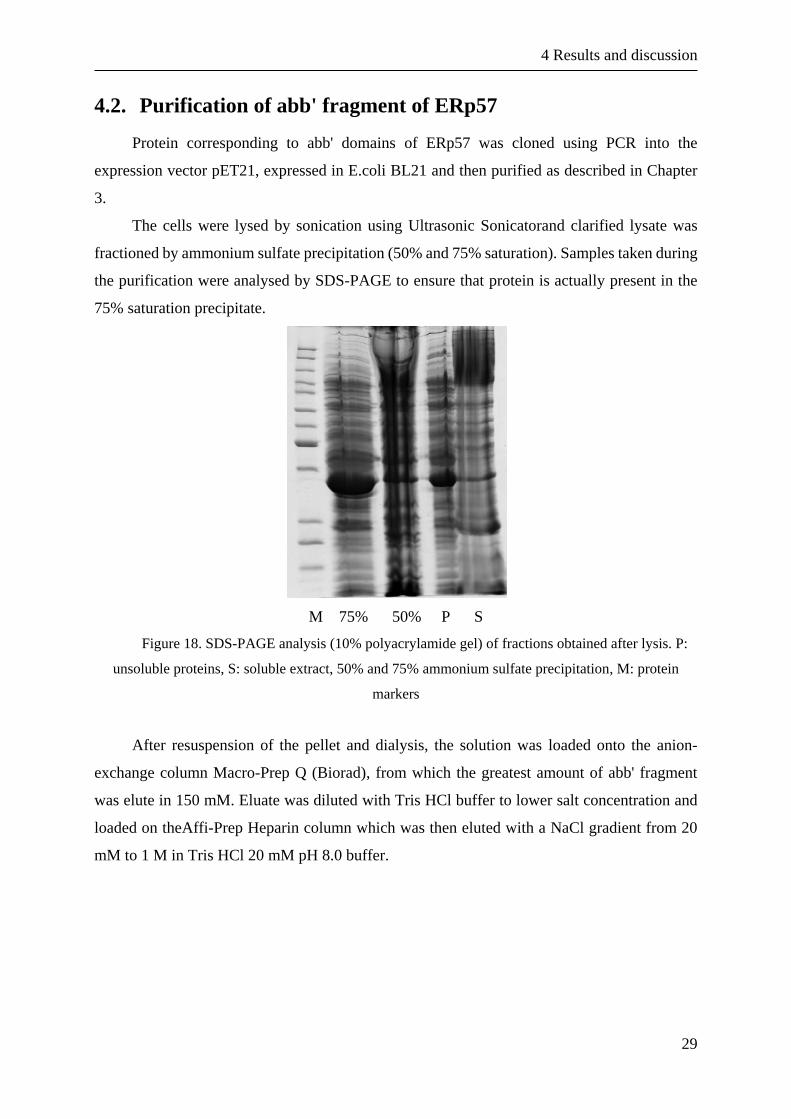
4.2. Purification of abb' fragment of ERp57
Protein corresponding to abb' domains of ERp57 was cloned using PCR into the
expression vector pET21, expressed in E.coli BL21 and then purified as described in Chapter
3.
The cells were lysed by sonication using Ultrasonic Sonicatorand clarified lysate was
fractioned by ammonium sulfate precipitation (50% and 75% saturation). Samples taken during
the purification were analysed by SDS-PAGE to ensure that protein is actually present in the
75% saturation precipitate.
M 75% 50% P S
Figure 18. SDS-PAGE analysis (10% polyacrylamide gel) of fractions obtained after lysis. P:
unsoluble proteins, S: soluble extract, 50% and 75% ammonium sulfate precipitation, M: protein
markers
After resuspension of the pellet and dialysis, the solution was loaded onto the anion-
exchange column Macro-Prep Q (Biorad), from which the greatest amount of abb' fragment
was elute in 150 mM. Eluate was diluted with Tris HCl buffer to lower salt concentration and
loaded on theAffi-Prep Heparin column which was then eluted with a NaCl gradient from 20
mM to 1 M in Tris HCl 20 mM pH 8.0 buffer.

4 Results and discussion
30
Figure 19. Heparin chromatogram of elution of abb' fragment of ERp57
P S FT 12 13 14 15 16 M
17 18 19 20 21 22 23 24 25 26 27 28 29 30 pur
Figure 20. SDS-PAGE electrophoretic analysis (10%) of fractions eluted from the anion-
exchange column (P-pellet, S-supernatant, FT-flow through, pur-purified domain)

4 Results and discussion
31
Electrophoretic analysis showed that the greatest amounts of protein corresponding to
abb' fragment of ERp57 is bound to the heparin column and is eluted from fraction 24 to 28.
These fractions were collected, dialyzed and after spectrophotometric quantification used for
further experiments.
4.3. Purification of a' domain of ERp57
Protein corresponding to a' domain of ERp57 was produced by E.coli BL21 as described
in Chapter 3. The cells were lyzed by sonication and the solution was precipitated in ammonium
sulphate, first at 30% and subsequently at 75%.
Figure 21. SDS-PAGE analysis (12% polyacrylamide gel) of fractions obtained after cell lysis and
ammonium sulphate precipitation
In figure 21. it is demonstrated that the domain a’ (which has a molecular weight of 18284
Da) is found in the pellet after precipitation at 75% ammonium sulphate. This pellet was
resuspended in and dialyzed in the same buffer over the night to remove the ammonium
sulphate. The dialyzed solution was centrifuged and the supernatant was passed on to anion-
exchange Macro-Prep Q column.

4 Results and discussion
32
Figure 22.SDS-PAGE analysis after anion-exchange chromatography
As shown in Figure 22. the a’ domain of ERp57 does not bind to the anion-exchange
column so we find it in the flow through. The flow through is then passed over the Heparin
Affi-Prep column and eluted with NaCl gradient from 20 mM to 1 M. The fractions were
analysed by SDS electrophoresis.
Figure 23. SDS-PAGE analysis (12% polyacrylamide gel after heparin column and gradient elution)
Electrophoretic analysis showed that fractions containing the highest amount of a’
domain are 21, 22, 23. These fractions were collected, dialyzed and after spectrophotometric
quantification(ε= 14565M-1cm-1) used for further experiments.

4 Results and discussion
33
4.4. Quenching effects of flavonoids in interaction with ERp57
To learn more about structural features relevant for interaction of flavonoids with ERp57,
fluorescence spectroscopy analysis with ERp57 protein in reduced conditions was performed.
To expand the analysis on flavonoids-ERp57 interaction already started in the laboratory,
additional flavonoids which differs in size, substitutions and the presence of a sugar moiety
were tested.
As mentioned in Chapter 3, ERp57 protein has an intrinisc fluorescence due to its three
tryptophan residues (one in each redox active a and a' domains and one localized in the b'
domain). Therefore fluorescence quenching studies may be used to determine the possible
interactions between protein and other molecules. Moreover, to better analyze the interaction
and obtain some quantitative parameters, fluorescence quenching of the protein was followed
using different concentrations of flavonoids.
The quenching effect on protein intrinsic fluorescence was calculated using the following
equation:
𝑄 = 1 −𝐹
𝐹˳× 100%
Q: percentage of quenching
F: intensity of fluorescence in presence of quencher (flavonoids)
F0: intensity of fluorescence in absence of quencher
Figure 24. Decrease in fluorescence intensity of ERp57 with increasing concentrations of
eupatorin (2, 4, 6, 8, 10, 15 and 20 μM).

4 Results and discussion
34
In figure 26. it is demonstrated how increasing concentrations of eupatorin reduce the
fluorescence intensity of the protein ERp57, highlighting in this way the existence of an
interaction between the protein and the molecule. The same fluorescence analysis using
increasing concentrations has been performed with all other flavonoid molecules.
Figure 25. Decrease in intrinsic fluorescence intensity of ERp57 in presence of increasing
concentrations of naringenin (2, 4, 6, 8, 10, 15 and 20 μM).
In figure 25. it is shown the effect of naringenin on fluorescence intensity of ERp57. It is
apparent that naringenin has much less quenching effect.
Figure 26. Comparison of quenching effect of different flavonoids (all at 20μM concentration)
interacting with ERp57 (average of three measurements with error bars).

4 Results and discussion
35
More specific data that quantify the interaction between the flavonoids and the whole
protein ERp57 may be obtained by the Stern-Volmer equation, obtaining the Ksv(Stern-Volmer
quenching constant), and by using a different derived equation that allows us to estimate the Kd
(dissociation constant).
Stern-Volmer equation:
𝐹˳
𝐹= 1 + Ksv [𝑄]
[Q]: quencher concentration
F: intensity of fluorescence in presence of quencher (flavonoids)
F0: intensity of fluorescence in absence of quencher
Ksv: Stern-Volmer quenching constant
Figure 27. Graphic analyses of data based on the quenching effect of eupatorin at increasing
concentrations on ERp57 for calculating Ksv.
Equation used to calculate the dissociation constant (Kd):
log10
(𝐹0 − 𝐹)
𝐹= 𝑛 log10 𝐾𝑎 − 𝑛 log10
1
[𝑄𝑡] − (𝐹0 − 𝐹)[𝑃𝑡]
𝐹0
y = 165598x + 0.7667R² = 0.9945
0.0000
0.5000
1.0000
1.5000
2.0000
2.5000
3.0000
3.5000
4.0000
4.5000
0.00E+00 5.00E-06 1.00E-05 1.50E-05 2.00E-05 2.50E-05
F 0/F
[Q]/M

4 Results and discussion
36
F0: fluorescence intensity in the absence of quencher
F: fluorescence intensity in the presence of quencher
Ka: binding constant (reverse from Kd)
[Qt]: final concentration of quenching agent
[Pt]: final concentration of protein
n: number of binding sites
Figure 28. Graphic analysis of data for calculating Kd
An important parameter that needs to be taken into consideration during this test is the
concentration of the quencher. Some flavonoids show absorbance in the wavelength range used
for quenching analysis. It is then possible that, at higher concentrations, they absorb part of
light and/or emitted radiation and therefore alter the quantum of fluorescence and the shape of
the spectrum. This phenomenon is due to the fact that the absorption occurs in the spectral range
of overlap between the absorption and emission spectrum. To overcome this problem, solutions
with absorbance less than 0.1 OD should be used. Therefore, the absorbance (at 290 and 338
nm) of flavonoids in highest concentration used in experiments (20 µM) was assessed. The
obtained results showed that certain flavonoids (eupatorin, apigenin) at the highest
concentration have absorbance values greater than 0.1 OD to at least one of two wavelengths.
Thus, the value of the dissociation constant was calculated using only the first 5 concentrations
of the molecules, that is up to 10 μM, where the absorbance of the flavonoid was lower.
y = -1.2174x + 6.2298R² = 0.9995
-1.0000
-0.8000
-0.6000
-0.4000
-0.2000
0.0000
0.2000
0.4000
0.6000
4.5000 4.7000 4.9000 5.1000 5.3000 5.5000 5.7000 5.9000
log(
F0-F
)/F
log(1/([Q]-[P](F0-F)/F0))

4 Results and discussion
37
Table 2. Summary table of binding parameters of ERp57 flavonoids interaction
Kd (μM) Ksv (M-1) Quenching (%)
Eupatorin 7,6 16559 57,4
Apigenin 11,3 11893 46,6
Apigenin-7-glucoside 21,0 53477 30,9
3-O-MethylQuercetin 23,3 53821 31,7
Luteolin-7-glucoside 28,8 36634 24,3
Kaempferol 30,0 39349 24,0
Cyanidin 34,2 33682 23,3
Genistein 37,0 27326 20,6
Naringenin 48,2 19540 46,4
From obtained data arises that eupatorin and apigenin show the highest affinity for ERp57
(dissociation constant is the smallest) and naringenin shows the lowest affinity (dissociation
constant is the highest).
4.5. Effects of flavonoids on protein activity
Catalytic activity of ERp57 requires binding and conformational changes of the substrate
and all 4 domains of the protein are necessary to guarantee correct function of the protein. This
process can be altered with the presence of small compounds such as flavonoids whose binding
near the catalytic site can prevent correct interaction ERp57-substrate and that way reducing its
activity.
Possible effect of flavonoids on reductase activity of the protein was tested using
fluorogenic compound:dieosin glutathione disulfide. As described in Chapter 3, mixture
consisting flavonoids and protein in buffer together with catalytic concentration of DTT and
eosin-labeled glutathione was tested. Because of the reductase activity of ERp57 the disulfide
bridge of glutathione is cleaved and this reduction causes an increase in fluorescence due to
free eosin excitation at 525 nm.

4 Results and discussion
38
Figure 29. Decrease of fluorescence following the inhibiton of ERp57 reductase activity by eupatorin
It is evident that the rate of increase in fluorescence of dienosin glutathion disulfide in the
presence of flavonoid in the incubation mixture is less than that observed for the protein ERp57
alone. This probably indicates that because of the interaction between flavonoid and the protein
makes the latter is unable to reduce disulfide bond in the oxidized eosin-labeled glutathione and
therefore the fluorescence emission due to the release of reduced glutathion linked eosin is
lower. The increase of fluorescence is thus an indirect measure of the concentration of reduced
glutathione which in turn is an indirect measure of the reductase activity of ERp57. Changes in
fluorescence consequently indicate the possible effects, positive or negative, of the molecules
on the catalytic activity of the protein.
The effects of flavonoids on the acitivity of protein ERp57 were tested at concentration
of 20 µM for all molecules. The data were processed by extrapolating for each substance the
slope of the tangent to the curve of fluorescence in a time interval from 0 to 15 seconds as
shown in Figure 30.
0
2000
4000
6000
8000
10000
12000
0 50 100 150 200
Flu
ore
sce
nce
/cp
s
Time/s
ERp57
ERp57+eupatorin

4 Results and discussion
39
Figure 30. Linearization of the fluorescence curve in the first 15 seconds for the calculation of the
slope of the tangent to the curve
From these data was calculated percentage of protein activity and reported in histograms
with error bars.
Figure 31. Comparison of flavnoid effect (20 μM) on ERp57 reductase activity
Unlike the quenching, in this case high concentrations of flavonoids did not interfere with
the fluorescence analysis since data were recorded in visible range (in this case particulary at
525 and 544 nm), wavelengths at which these molecules don't give phenomenon of absorption.
For this, no precautions were used during the calculation of the reductase activity.
y = 93.897x + 2245.8R² = 0.9743
y = 61.263x + 832.85R² = 0.9659
0
500
1000
1500
2000
2500
3000
3500
4000
0 5 10 15 20
Flu
ore
sce
nce
/cp
s
Time/s
ERp57
ERp57+eupatorin
0
20
40
60
80
100
120
140
% a
ctiv
ity
% activity

4 Results and discussion
40
4.6. Effects of flavonoids on protein stability
Another test carried out was to examine the effect of flavonoids on the protein stability.
According to protocol given in Chapter 3, ERp57protein was treated with increasing
concentrations of flavonoids in the presence of a fluorescent compound (SYPRO orange). The
temperature at which a protein unfolds is measured by an increase in the fluorescence of a dye
with the affinity for hydrophobic parts of the protein, which are exposed as the protein unfolds.
Temperature at which there is half of fluorescence increase is the melting temperature
(Tm), which corresponds to the inflection point of the transition curve. Tm value can be
calculated fitting data with the Boltzmann equation. An increase in the Tm was observed for
ERp57 protein incubated in the presence of selected flavonoids, and the effect seems to be
related to the flavonoid concentration. This observation support a stabilization effect of these
substances on ERp57.
Figure 32. Changes in SYPRO orange fluorescence with the increase in temperature observed
for the protein alone and in presence of different flavonoid concentrations (0.5, 2, 5, 20, 50 μM)
By analyzing data obtained in the test we can detect that some molecules show also a
change in maximum fluorescence intensity when used in higher concentrations. We can
hypothesize that these molecules could interact with multiple binding sites and that some of
these sites are also close to or are the binding sites for the SYPRO. In this way SYPRO appeared
unable to bind the protein and therefore the emission fluorescence appears reduced. However,
further analysis of that phenomenon isn't possible because we don't have an estimate number
of binding sites and their relative affinity.

4 Results and discussion
41
Figure 33. Increase in melting temperature (Tm) of ERp57 by increasing concentrations of kaempferol
Variations in melting temperature of ERp57 due to the effects of flavonoids are shown in
Figure 34.
Figure 34. Effects of flavonoids in different concentrations on protein ERp57 stability
As demonstrated in diagram, kaempferol has the highest stabilizing effect, followed by
cyanidin and apigenin. Naringenin and eupatorin didn't show stabilizing effect, furthermore,
eupatorin even showed a slight destabilizing effect on the protein. Genistein is the only
exception were melting temperature decreases with the increase of flavonoid concentration.
-0.5
0
0.5
1
1.5
2
2.5
3
3.5
4
4.5
∆ t
em
pe
ratu
re
Flavonoids
50 μM
20 μM
5 μM

4 Results and discussion
42
Figure 35.Comparison of temperature and fluorescence dependence on kaempferol concentrations
Looking into temperature and fluorescence trends gives better inside to flavonoid and
SYPRO binding. Fitting with a log-dose response curve the observed melting temperature
obtained from stability assay against the flavonoid concentration is possible estimate a Kdvalue
that needs to be taken with caution for two reasons. First is that we don't know the exact number
of binding sites for each flavonoid on the protein. We could guess from the fluorescence
quenching data analysis but the equation assumes the same affinity for each binding site.
Secondly, it is observed maximum fluorescence decreasing with the increasing of flavonoid
concentrations which could mean that the flavonoids and SYPRO share the same binding sites
on the protein.
Table 3. Comparison of Kd obtained by the DSF (Kd1) and quenching fluorescence (Kd2)
Kd1 (µM) Kd2 (µM)
Apigenin 2,6 11,3
Cyanidin 43,0 34,2
Kaempferol 5,4 30,0
Moreover, fluorescence quenching assay experiments were performed at a constant
temperature while the increase in the temperature in DSF analysis will result in a protein
conformational change and new binding sites could be introduced. In any case, kaempferol not
only stabilizes the protein but also interferes with SYPRO binding. In addition, Kd values

4 Results and discussion
43
estimated from the DSF data analysis are in the same range as those obtained from fluorescence
quenching data.
4.7. Effects of flavonoids on protein-DNA binding
The interaction between protein ERp57 and an 80 bp DNA fragment in the presence of
all tested flavonoids was studied by EMSA as described in Chapter 3. The 80 bp DNA fragment
was previously produced and its final concentration after purification was 50 ng/μL. 5 ng of
DNA were used for each 10 μL incubation mixture, to yield a final concentration of 0.5 ng/μL,
which was then loaded on a 5% polyacrylamide gel.
To analyze the interaction of the protein with DNA, it was carried out a test comparing
the mobility of a free DNA solution with a DNA solution containing the a' domain, which
contains the DNA binding site. To evaluate which molecules interfere with the DNA-binding
activity of the protein, EMSA test with all molecules at 20 µM and then 50 µM was carried out.
Flavonoids tested were: genistein, naringenin, kaempferol, cyanidin, apigenin-7-
glucoside, luteolin-7-glucoside.
Figure 36. Image obtained from EMSA assay. All flavonoids are in 20 µM concentrations.In order
from the left to right are: DNA, DNA+a', DNA+a'+DTT, DNA+a'+genistein, DNA+a'+naringenin,
DNA+a'+kaempferol, DNA+a'+cyanidin, DNA+a'+apigenin-7-glucoside, DNA+a'+luteolin-7-
glucoside, DNA+a'+EGCG, DNA+a'+ellagic acid
As controls were used DNA fragment alone, to have a signal without shift due to protein
binding, and the DNA fragment with the protein, to obtain a signal of DNA completely bound
to the protein. For negative control, the protein was treated with DTT since protein reduction

4 Results and discussion
44
abolishes its DNA binding activity. As it was previously demonstrated by Trnková at al. (2013),
epigallocatechin gallate (EGCG) is able to interfere with DNA-domain a' interaction, with an
80% inhibition in the DNA binding activity, so EGCG was used as a positive control.
None of the tested flavonoids showed a significant DNA-binding inhibiton, and
consequently a quantitave analysis wasn't performed. Knowing from previous research (Grillo
et al, 2002) that binding site for DNA is localized on a' domain of the protein (a β-sheet before
the active site), we can exclude this part of the protein as a binding site for the flavonoids.

5 Conclusion
45
5. Conclusion
In this thesis the importance of orientation of the B ring was tested. Apigenin has the B
ring binded on C2 position while in genistein it is on C3. Also, it was tested the importance of
free rotation of the B ring. In naringenin B ring has free rotation while it is blocked in cyanidin
and genistein (cyanidin also has different C ring arrangement). Naringenin showed the highest
dissociation constant (48 µM) and lowest binding affinity to ERp57. Genistein showed a
slightly better Kd (37 µM) as well as cyanidine (34 µM). From these data we can conclude that
a free rotation of the B ring, as well as a different position on the C ring, has a bad influence on
binding of flavonoid to the protein. Apigenin is proved to be the molecule with the best binding
abilities, with Kd=11 µM, and for this reason we can conclude that a B ring linked to C2 position
and the absence of the free rotation are essential for binding to ERp57.
Furthermore, influence of additional hydroxyl group in position C3 was tested. As already
mentioned, apigenin has a dissociation constant of 11 µM in contrast to kaempferol which
showed 30 µM dissociation constant. This result confirms previous conclusion made in this
laboratory that optimal number of hydroxyl groups for a better binding is 3.
Additionally, the importance of different methyl position was tested. Eupatorin, molecule
with the lowest dissociation constant (7 µM) and best binding affinity has 2 hydroxyl- groups
and 3 methoxyl- groups while 3-O-methylquercetin has 4 hydroxyl- and 1 methoxyl- showed a
higher Kd (23 µM). From previous research quercetin (which has 5 hydroxyl- groups) showed
a Kd equal to 23µM, so we can conclude that addition of onemethoxyl- group does not increase
the binding affinity to ERp57. Probably the higher affinity observed for eupatorin is related to
the presence of more than one methoxyl-group.
Finally, it was tested presence of a sugar moiety on A7 position. We previously observed
that the addition of a sugar moiety on C3 position (rutin, isoquercetin and quercetin) does not
have an effect on binding properties as compared to the corresponding un-glycosilated
flavonoid, quercetin. In this case, apigenin-7-glucoside (Kd=21 µM) showed lower binding
affinity than apigenin (11µM) while the affinity of luteolin-7-glucoside was even worse (29
µM). This can be explained by the fact that glucose, which is hydrophilic and rich in hydroxyl-
groups, has a negative effect on flavonoid binding to the ERp57 protein. Considering that

5 Conclusion
46
luteolin-7-glucoside has one more hydroxyl- group than apigenin-7-glucoside, this is an
agreement with the lower affinity observed for this molecule (29µM vs. 21 µM).
In conclusion, apigenin and eupatorin remain the lead compounds with the best properties
for binding to ERp57. Further experiments with additional compounds, as well as Quantitative
Structure-Activity Relationship (QSAR) analysis on all tested molecules, will provide
information to define the best properties for binding with high affinity to ERp57 and identify
compound to be used as a selective inhibitor/modulator of ERp57 biological activities.

6 References
47
6. References
Allan L, Miller ND. Antioxidant flavonoids: structure, function and clinical usage. Alternative
Medicine Review, 1996, 1(2), 103-111.
Altieri F, D'Amrosio C, Grillo C, Maceroni M, Merluzzi S, Scaloni A, Turano C. Cooperative
activity of Ref1/APE and ERp57 in reductive activation of transcription factors. Free
Rad Biol Med, 2006, 41, 1113-1123.
Altieri F, Coppari S, Ferraro A, Turano, C. Proteins of the PDI family: unpredicted non-ER
locations and functions. J Cell Physiol, 2002, 193, 154-163.
Barile E, Curi P, Dolci M, Galeotti F, Lanzotti V. Flavonoids from carnation (Dianthus
caryophyllus) and their antifungal activity. Photochemistry Letters, 2008, 1, 44-48.
Bast A, Haenen GR, Heijnen CG, van Acker FA, van der Vijgh WJ. Flavonoids as peroxynitrite
scavanger: the role of the hydroxyl group. Toxicol In Vitro, 2001, 15(1), 3-6.
Bedard K, Michalak M, Opas M, Szabo E. Celular functions of endoplasmic reticulum
chaperones calreticulin, calnexin and ERp57. International Review of Citology, 2005,
245, 91-121.
Coe H, Michalak M. ERp57, a multifunctional endoplasmic reticulum resident oxidoreductase.
Int J Biochem Cell Biol, 2010, 42(6), 796-9.
Cohen-Doyle MF, Leach MR, Thomas DY, Williams DB. Localization of the lectin, ERp57
binding and polypeptide binding sides of calnexin and calreticulin. J Biol Chem, 2002,
277, 29686-29697.
Cohe-Doyle MF, Jackson MR, Peterson MA, Vassilakos A, Williams DB. The molecular
chaperone calnexin facilitates folding and assembly of class I histocompatibility
molecules. EMBO J, 1996, 15, 1495-1506.
Coppari S, Altieri F, Ferraro A, Chichiarelli S, Eufemi M, Turano C. Nuclear localization and
DNA interaction of protein disulfide isomerase ERp57 in mammalian cells. J Cell
Biochem. 2002, 85, 325-333.

6 References
48
Cygler M, Gehring K, Kozlov G, Maattanen P, Nagar B, Pollock S, Schrag JD, Thomas DY.
Crystal structure od bb' domains of the protein disulfide isomerase ERp57. Structure,
2006, 14, 1331-1339.
Darnell JE Jr. STATs and gene regulation. Science, 1997, 277, 1630-1635.
Dolečková I, Rárová L, Grúz J, Vondrusová M, Strnad M, Kryštof V. Antiproliferative and
antiangiogenic effects of flavone eupatorin, an active constituent of chloroform extract
of Orthosiphon stamineus leaves. Fitoterapia, 2012, 83 (6), 1000-1007.
Ellerman DA, Myles DG, Primakoff P. A role for sperm surface protein disulfide isomerase
activity in gamete fusion: evidence for the participation of ERp57. Developmental Cell,
2006, 10, 831-837.
Ellgaard L, Ruddock L. W. The human protein disulphide isomerase family: substrate
interactions and functional properties. EMBO Rep, 2005, 6(1), 28-32.
Fuhr U, Klittich K, Staib AH. Inhibitory effect of grapefruit juice and its bitter prinicpal,
naringenin, on CYP1A2 dependent metabolism of caffeine in man. Br J Clin Pharmacol,
1993, 35(4), 431-436.
Goodman SR. Medical Cell Biology. Elsevier Academic Press publication, London, 2008, p12.
Grillo C, Coppari S, Turano C, Altieri F. The DNA-binding activity of protein disulfide
isomerase ERp57 is associated with the a' domain. Biochem. Biophys. Res. Commun,
2002, 295, 67-73.
Hames BD. Gel electrophoresis of proteins, a practical approach. Oxford University Press,
2002, p1.
Harborne JB, Williams CA. Advances in flavonoid research since 1992. Phytochemistry. 2000,
55(6), 481-504.
Heim KE, Tagliaferro AR, Bobilya DJ. Flavonoid antioxidants: chemistry, metabolism and
structure-activity relationship. J Nutr Biochem, 2002, 13(10), 572-584.
Hellman LM, Fried MG. Electrophoretic Mobility Shift Assay (EMSA) for Detecting Protein-
Nucleic Acid Interactions. Nat Protoc, 2007, 2(8), 1849-1861.

6 References
49
High S, Lecomte FJ, Russell SJ, Abell BM, Oliver JD. Glycoprotein folding in the endoplasmic
reticulum: a tale of three chaperones? FEBS Lett, 2000, 476(1-2), 38-41.
Hirano N, Shibasaki F, Sakai R, Tanaka T, Nishida J, Yazaki Y, Takenawa T, Hirai H.
Molecular cloning of the human glucose-regulated protein ERp57/GRP58, a thiol-
dependent reductase. Identification of its secretory form and inducible expression by the
oncogenic transformation. Eur. J. Biochem. 1995, 234, 336-342.
Jessop CE, Chakravarthi S, Garbi N, Hämmerling GJ, Lovell S, Bulleid NJ. ERp57 is essential
for efficient folding of glycoproteins sharing common structural domains. EMBO J,
2007, 26, 28-40.
Jovanovic SV, Steenken S, Simic MG, Hara Y. Antioxidant properties of flavonoids: reduction
potentials and electron transfer reactions of flavonoid radicals. Flavonoids in Health and
Disease, 1998, p.137.
Kang OH, Lee JH, Kwon DY. Apigenin inhibits release of inflammatory mediators by blocking
the NF-κB activation pathways in the HMC-1 cells. Immunopharmacol Immunotoxicol,
2011, 33, 473-479.
Khanal RC, Nemere I. The ERp57/GRp58/1,25D3-MARRS receptor: multiple functional roles
in diverse cell systems. Curr Med Chem, 2007, 14, 1087-1093.
Kumar S, Pandey AK. Chemistry and biological activities of flavonoids: an overview. Scientific
World Journal, 2013, 2013.
Lakowicz JR. Principles of Fluorescence Spectroscopy. Springer, 2006, p1.
Mulvihill EE, Allister EM, Brian GS, Telford DE, Sawyez CG, Edwards JY et al. Naringenin
prevents dyslipidemia, apolipoprotein B overproduction, and hyperinsulinemia in LDL-
receptor-null mice with diet-induced insulin resistance. Diabetes, 2009, 58(10), 2198-
2210.
Niesen FH, Berglund H, Vedadi M. The use of differential scanning fluorimetry to detect ligand
interaction that promote protein stability. Nat Protoc, 2007, 2, 2212-2221.

6 References
50
Plaza M, Pozzo T, Liu J, Gulshan-Ara KZ, Turner C, Nordberg-Karlsson E. Substituent effects
on in vitro antioxidizing properties, stability, and solubility in flavonoids. J Agric Food
Chem, 2014, 62 (15), 3321-3333.
Rajendran P, Rengarajan T, Nandakumar N, Palaniswami R, Nishigaki Y, Nishigaki I.
Kaempferol, a potential cytostatic and cure for inflammatory disorders. Eur J Med Chem,
2014, 86, 103-112.
Salmela AL, Pouwels J, Kukkonen-Macchi A, Waris S, Toivonen P, Jaakkola K et al. The
flavonoid eupatorin inactivates the mitotic checkpoint leading to polyploidy and
apoptosis. Exp Cell Res, 2012, 318, 578-592.
Seung-Hee K, Kyung-Chul C. Anti-cancer effect and underlying mechanism(s) of kaempferol,
a phytoestrogen, on the regulation of apoptosis in diverse cancer cell models. Toxicol
Res, 2013, 29(4), 229-234.
Shukla S, Gupta S. Apigenin: a promissing molecule for cancer prevention. Pharm Res, 2010,
27 (6), 962-978.
Trnková, L., Ricci, D., Grillo, C., Colotti, G., Altieri, F. Green tea catechins can bind and
modify ERp57/PDIA3 activity. Biochim Biophys Acta. 2013, 1830(3), 2671-2682.
Tseng HL, Li CJ, Huang LH, Chen CY, Tsai CH, Lin CN, Hsu HY. Quercetin 3-O-methyl ether
protects FL83B cells from copper induced oxidative stress through the PI3K/Akt and
MAPK/Erk pathway. Toxicol Appl Pharmacol, 2012, 264(1), 104-113.
Turano C, Coppari S, Altieri F, Ferraro A. Proteins of the PDI family: unpredicted non-ER
locations and functions. J Cell Physiol,2002, 193(2), 154-163.
Turano C, Gaucci E, Grillo C, Chichiarelli S. ERp57/GRP58: a protein with multiple functions.
Cell Mol Biol Lett. 2011, 42(6), 796-799.
Vallverdu-Queralt A, Odriozola-Serrano I, Oms-Oliu G, Lamuela-Raventos RM, Elez-
Martinez P, Martin-Belloso O. Change in the polyphenol profile of tomato juices
processed by pulsed electric fields. J Agric Food Chem, 2012, 60 (38), 9667-9672.
Walker J, Wilson K. Principles and Techniques of Biochemistry and Molecular Biology.
University Press, Cambridge, 5th edition, p471.

6 References
51
Yam MF, Lim V, Salman IM, Ameer OZ, Ang LF, Rosidah N et al. HPLC and anti-
inflammatory studies of the flavonoid rich chloroform extract fraction of Orthosiphon
stamineus leaves. Molecules, 2010, 15, 4452-4456.

7 Sažetak/Summary
52
7. Sažetak/Summary
7.1. Sažetak
UVOD
Protein PDIA3 odnosno ERp57 je protein iz obitelji protein disulfid izomeraza (engl.
protein disulfide isomerase, PDI) koja se sastoji od različitih proteina s višestrukom
organizacijom domena. Strukturu ERp57 obilježava prisutnost četiriju domena nazvanih a, b,
b' i a'. Domene a i a' sadrže tioredoksinu sličnu sekvencu aktivnog mjesta dok su domene b i
b' redoks inaktivne. Trodimenzionalna struktura ERp57 pokazuje da protein ima oblik slova U,
s katalitičkim, tioredoksinskim domenama koje su okrenute jedna prema drugoj. Zbog svoje
šaperonske, redoks i izomerazne aktivnosti sudjeluje u smanjenju štetnih posljedica u
staničnom stresu.
Flavonoidi su skupina prirodnih spojeva sveprisutnih u našoj prehrani. Široko su
rasprostranjeni u biljkama (voće, povrće, mahunarke, žitarice) te u pićima poput čaja, vina i
piva. Prema kemijskoj strukturi flavonoidi pripadaju velikoj skupini prirodnih polifenolnih
spojeva kojima je svojstvena prisutnost hidroksilnih skupina na aromatskoj jezgri (polihidroksi
fenoli). Derivati su 1,3-difenilpropana (C6-C3-C6). Flavonoidi pokazuju spazmolitski,
diuretski, protuupalni i dijaforetski učinak. Njihovo djelovanje može biti i antialergijsko,
antimikrobno, antiviralno, estrogeno, antihepatotoksično, te trenutno najistraživanije-
antioksidativno.
OBRAZLOŽENJE TEME
Protein ERp57 je povezan s raznim patološkim stanjima i oboljenjima te ima potencijal
biti terapijskom metom, posebice imajući na umu da može stupiti u interakciju s polifenolima
koji su poznati po svojim pozitivnim učincima na zdravlje.
Cilj ovog diplomskog rada bio je istražiti vezanje odabranih flavonoida (eupatorin,
apigenin, apigenin-7-glukozid, kempferol, naringenin, 3-O-metilkvercetin, genistein, cijanidin,
luteolin-7-glukozid) s proteinom ERp57. U tu svrhu korištena je fluorescencijska
spektrofotometrija i učinak gašenja fluorescencije. Nadalje, istražen je utjecaj flavonoida na
disulfidno reduktaznu aktivnost proteina, njegovu stabilnost te interakciju ERp57-DNA.

7 Sažetak/Summary
53
MATERIJALI I METODE
Transformirane bakterijske BL21 (koristeći plazmidni vektor pET21 i pET29) stanice
upotrijebljene su za ekspresiju proteina ERp57 i njegove a' domene. Nakon rasta jedna kolonija
je prenesena te je prema protokolu provedena ekspresija i purifikacija proteina ERp57, odnosno
domene a'. Kvaliteta purifikacije provjerena je SDS elektroforezom, a količina proteina
određena spektroskopski.
Kvantitativne analize potencijalne interakcije između pojedinog flavonoida i ERp57
izvedene su fluorimetrijskom titracijom pomoću učinka gašenja fluorescencije.
Reduktazna aktivnost proteina ERp57 i utjecaj flavonoida na aktivnost proteina testirana
je koristeći dieozin glutation disulfid. Reduktaznom aktivnosti proteina ERp57 dolazi do
redukcije molekule dieozin gluation disulfida te povećanja fluorescenije.
Ispitivanje stabilnosti proteina i utjecaj flavonoida na njegovu stabilnost provedena je
pomoću diferencijalne pretražne fluorimetrije koristeći real-time PCR instrument.
Utjecaj flavonoida na interakciju ERp57-DNA ispitan je elektroforezom (EMSA)
pomoću fluoresceina koji se veže na DNA.
REZULTATI I RASPRAVA
Eupatorin i apigenin pokazali su najveći afinitet vezanja za ERp57 (Kd= 7,6 i 11,3μM
redom), dok je naringenin pokazao najmanji (Kd= 48,2 μM).
Interakcijom eupatorina, cijanidina i luteolin-7glukozida sa ERp57 dolazi do smanjenja
reduktazne aktivnosti proteina (59%, 78% i 94%), dok ostali flavonoidi nemaju značajan utjecaj
na njegovu aktivnost.
Apigenin i cijanidin imaju stabilizacijski efekt na protein te su dobivene konstante
disocijacije otprilike u istom rangu kao i one dobivene quenching fluorescencijom.
Ni jedan od testiranih flavonoida nije pokazao značajnu inhibiciju vezanja proteina
ERp57 na DNA.
ZAKLJUČAK
U ovom radu potvrđena je važnost hidroksilne skupine za interakciju flavonoida sa
proteinom ERp57 (max. broj za optimalno vezanje je 3).Također je uočena važnost nedostatka
slobodne rotacije B prstena flavonoida za vezanje s proteinom. Dodatak šećera na A7 poziciju
djeluje nepovoljno na vezanje flavonoida i proteina ERp57.

7 Sažetak/Summary
54
7.2. Summary
The protein PDIA3 or ERp57 (Endoplasmic Reticulum protein 57) is a member of the
family of disulfide isomerase (PDI). The protein is mainly localized within the endoplasmic
reticulum and has an established function as a chaperon and disulfide-rearrangements enzyme.
Flavonoids are a broad class of molecules characterized by flavone core. They are found
in leaves, cortex, seeds and flowers of plants where they hace various functions. Most of their
health benefits are attributed to their anti-oxidant and chelating abitily.
ERp57 is involved in various diseases and has a potential to be a pharmacological target.
The aim of this thesis was to reach a deeper understanding of the interaction between ERp57
and selected flavonoids (eupatorin, apigenin, apigenin-7-glucoside, kaempferol, naringenin,
cyanidin, genistein, 3-O-methylquercetin, luteolin-7-glucoside). The goal is to understand how
different substitutions on the flavonoid basic structure can modify the interaction with the
protein.
Using fluorescence quenching of ERp57 by flavonoids it was calculated dissociation
constant. Eupatorin and apigenin showed lowest Kd (7,6 and 11,3μM respectively) and
therefore highest affinity to bind ERp57.
Eupatorin, cyanidin and luteolin-7-glucoside decreased reductase activity of the protein,
while other flavonoids didn't show significant effect on it.
Apigenin and cyanidin showed positive effect on protein ERp57 stability.
None of the tested flavonoids showed a significant DNA-binding inbibiton.
In this thesis the importance of hydroxyl group for flavonoid binding to ERp57 is
confirmed. Also, it is noticed the importance of the lack of free rotation of the flavonoid B-ring
to bind with the protein. Addition of sugar moiety to A7 position adversely effects the binding
of flavonoids and the protein ERp57. Taking into account fluorescence quenching results and
the effects on protein activity and stability, we can conclude that eupatorin and apigenin the
compound with the best properties for binding to ERp57.

Temeljna dokumentacijska kartica
Sveučilište u Zagrebu
Farmaceutsko-biokemijski fakultet
Zavod za biokemiju i molekularnu biologiju
A. Kovačića 1, 10000 Zagreb, Hrvatska
Diplomski rad
INTERAKCIJA FLAVONOIDA S PROTEINOM ERp57
Ines Kanski
SAŽETAK
U okviru ovog diplomskog rada istražena je interakcija između odabranih flavonoida (eupatorin,
apigenin, apigenin-7-glukozid, kempferol, naringenin, 3-O-metilkvercetin, cijanidin, genistein,
luteolin-7-glukozid) sa proteinom ERp57 koji spada u skupinu disulfid-izomeraza (PDI). Za
procjenu vezanja flavonoida sa proteinom ERp57 korištena je fluorescencijska spektrofotometrija i
učinak gašenja fluorescencije, dok je za ispitivanje utjecaja flavonoida na vezanje ERp57-DNA
korištena migracijska elektroforeza (EMSA). Također je testiran utjecaj flavonoida na aktivnost i
stabilnost proteina.
Apigenin i eupatorin pokazali su najmanju konstantu disocijacije, odnosno najveći afinitet za ERp57
te ih to čini najboljim molekulama za vezanje na protein ERp57. Potvrđena je važnost hidroskilne
skupine za interakciju flavonoida sa proteinom.
Rad je pohranjen u Središnjoj knjižnici Sveučilišta u Zagrebu Farmaceutsko-biokemijskog fakulteta.
Rad sadrži: 54 stranica, 36 grafičkih prikaza, 3 tablice i 44 literaturnih navoda. Izvornik je na engleskom jeziku.
Ključne riječi: Flavonoidi, apigenin, eupatorin, ERp57, gašenje fluorescencije
Mentor: Dr. sc. Olga Gornik, docentica Sveučilišta u Zagrebu Farmaceutsko-biokemijskog fakulteta.
Ocjenjivači: Dr. sc. Olga Gornik, docentica Sveučilišta u Zagrebu Farmaceutsko-biokemijskog fakulteta.
Dr. sc. Sandra Šupraha Goreta, docentica Sveučilišta u Zagrebu Farmaceutsko-biokemijskog
fakulteta.
Dr. sc. Suzana Inić, docentica Sveučilišta u Zagrebu Farmaceutsko-biokemijskog fakulteta.
Rad prihvaćen: rujan 2015.

Basic documentation card
University of Zagreb
Faculty of Pharmacy and Biochemistry
Department of Biochemistry and Molecular Biology
A. Kovačića 1, 10000 Zagreb, Croatia
Diploma thesis
TESTING THE INTERACTION OF FLAVONOIDS WITH PROTEIN DISULFIDE
ISOMERASE PDIA3 AND THE EFFECTS ON PROTEIN BIOLOGICAL PROPERTIES
Ines Kanski
SUMMARY
In this thesis, interaction between a protein that belongs to the family of protein disulfide isomerases
(PDI)- protein ERp57, and chosen flavonoids- eupatorin, apigenin, apigenin-7-glucoside,
kaempferol, naringenin, 3-O-Methylquercetin, cyanidin, genistein and luteolin-7-glucoside was
tested. Interaction between ERp57 and flavonoids was assessed using fluorescence quenching
method, while electrophorectic mobility shift assay (EMSA) was used to evaluate flavonoid effect
on ERp57-DNA interaction. Flavonoid influence on protein activity and protein stability was also
tested.
Apigenin and eupatorin showed the lowest dissociation constant and the highest binding affinity to
ERp57, making them the best compounds to be used as selective inhibitors/modulators of ERp57
biological activities.
The thesis is deposited in the Central Library of the University of Zagreb Faculty of Pharmacy and Biochemistry.
Thesis includes: 54 pages, 36 figures, 3 tables and 44 references. Original is in English.
Keywords: Flavonoids, apigenin, eupatorin, ERp57, fluorescence quenching
Mentor: Olga Gornik, Ph.D. Assistant Professor, University of Zagreb Faculty of Pharmacy and
Biochemistry
Reviewers: Olga Gornik, Ph.D. Assistant Professor, University of Zagreb Faculty of Pharmacy and
Biochemistry
Sandra Šupraha Goreta, Ph.D. Assistant Professor, University of Zagreb Faculty of Pharmacy
and Biochemistry
Suzana Inić, Ph.D. Assistant Professor, University of Zagreb Faculty of Pharmacy and
Biochemistry
The thesis was accepted: September 2015.