Embed Size (px)
Citation preview
Veterinary Vaccine Services
O-0310708
Testing Services for Veterinary Vaccines
www.bioreliance.com
ContentsIntroduction _____________________________________________________________________________________ 1
General requirements ___________________________________________________________________________ 1
Viral extraneous agents __________________________________________________________________________ 1
Non-viral extraneous agents ______________________________________________________________________ 1
Custom testing ________________________________________________________________________________ 1
Cell Banks ________________________________________________________________________________________ 4
MCS and WCS direct testing ______________________________________________________________________ 4
MCS and WCS extract testing _____________________________________________________________________ 5
Mammalian Vaccines ______________________________________________________________________________ 6
Neutralizing the virus ___________________________________________________________________________ 6
In vitro assay __________________________________________________________________________________ 6
Avian Vaccines ____________________________________________________________________________________ 7
Neutralizing the virus ___________________________________________________________________________ 8
Test for extraneous viruses using embryonated hens’ eggs (Ph Eur, CVMP) _________________________________ 8
In vitro assays:__________________________________________________________________________________ 9
Test in chick kidney cells (Ph Eur, CVMP) ________________________________________________________ 9
Test for Avian leucosis viruses (Ph Eur, CVMP) ____________________________________________________ 9
Test for Reticuloendotheliosis virus (REV) (CVMP) __________________________________________________ 9
Test for Chick anaemia virus (CAA) (CVMP) ______________________________________________________ 9
Test for extraneous agents using chicks (Ph Eur, CVMP) ________________________________________________ 9
Fish Vaccines ____________________________________________________________________________________ 11
Vaccines Based on Insect Cell Lines/Viruses ___________________________________________________________ 11
Other Substances of Animal Origin__________________________________________________________________ 12
Preparation of test substances ___________________________________________________________________ 12
In vitro assay __________________________________________________________________________________ 12
Bovine serum _________________________________________________________________________________ 12
Endpoint Tests __________________________________________________________________________________ 13
Staining for cytopathic effects ___________________________________________________________________ 13
Haemadsorption assay _________________________________________________________________________ 13
Immunofluorescence assay ______________________________________________________________________ 13
Other tests ___________________________________________________________________________________ 13
References ______________________________________________________________________________________ 13
Testing Services for Veterinary Vaccines 1
www.bioreliance.com
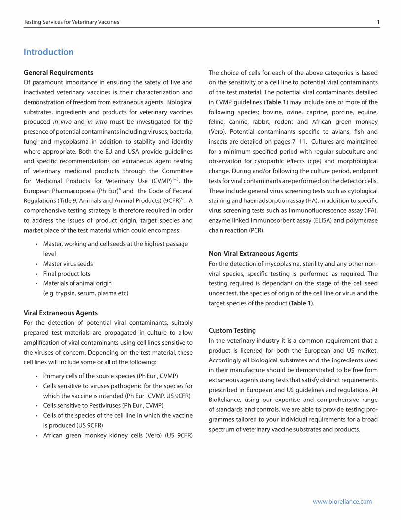
Introduction General RequirementsOf paramount importance in ensuring the safety of live and inactivated veterinary vaccines is their characterization and demonstration of freedom from extraneous agents. Biological substrates, ingredients and products for veterinary vaccines produced in vivo and in vitro must be investigated for the presence of potential contaminants including; viruses, bacteria, fungi and mycoplasma in addition to stability and identity where appropriate. Both the EU and USA provide guidelines and specific recommendations on extraneous agent testing of veterinary medicinal products through the Committee for Medicinal Products for Veterinary Use (CVMP)1–3, the European Pharmacopoeia (Ph Eur)4 and the Code of Federal Regulations (Title 9; Animals and Animal Products) (9CFR)5 . A comprehensive testing strategy is therefore required in order to address the issues of product origin, target species and market place of the test material which could encompass:
• Master, working and cell seeds at the highest passage level
• Master virus seeds• Final product lots• Materials of animal origin
(e.g. trypsin, serum, plasma etc)
Viral Extraneous AgentsFor the detection of potential viral contaminants, suitably prepared test materials are propagated in culture to allow amplification of viral contaminants using cell lines sensitive to the viruses of concern. Depending on the test material, these cell lines will include some or all of the following:
• Primary cells of the source species (Ph Eur , CVMP)• Cells sensitive to viruses pathogenic for the species for
which the vaccine is intended (Ph Eur , CVMP, US 9CFR)• Cells sensitive to Pestiviruses (Ph Eur , CVMP)• Cells of the species of the cell line in which the vaccine
is produced (US 9CFR)• African green monkey kidney cells (Vero) (US 9CFR)
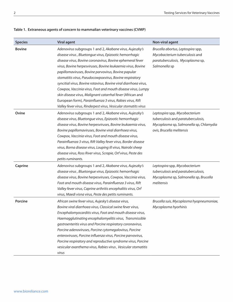
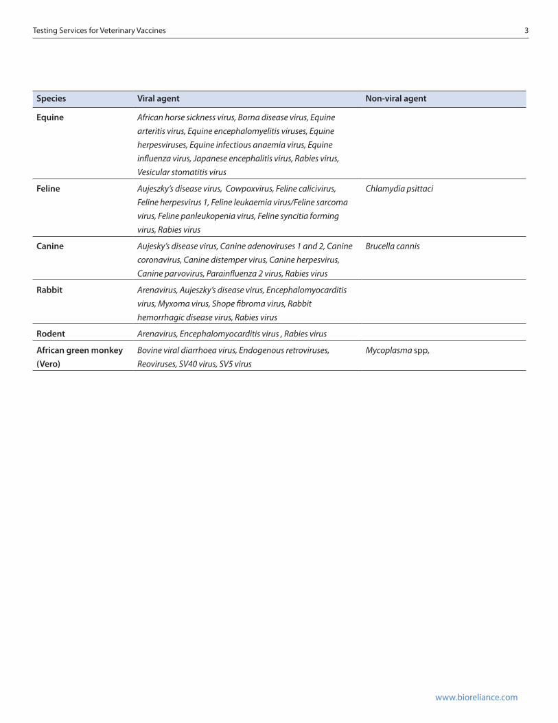
The choice of cells for each of the above categories is based on the sensitivity of a cell line to potential viral contaminants of the test material. The potential viral contaminants detailed in CVMP guidelines (Table 1) may include one or more of the following species; bovine, ovine, caprine, porcine, equine, feline, canine, rabbit, rodent and African green monkey (Vero). Potential contaminants specific to avians, fish and insects are detailed on pages 7–11. Cultures are maintained for a minimum specified period with regular subculture and observation for cytopathic effects (cpe) and morphological change. During and/or following the culture period, endpoint tests for viral contaminants are performed on the detector cells. These include general virus screening tests such as cytological staining and haemadsorption assay (HA), in addition to specific virus screening tests such as immunofluorescence assay (IFA), enzyme linked immunosorbent assay (ELISA) and polymerase chain reaction (PCR).
Non-Viral Extraneous AgentsFor the detection of mycoplasma, sterility and any other non-viral species, specific testing is performed as required. The testing required is dependant on the stage of the cell seed under test, the species of origin of the cell line or virus and the target species of the product (Table 1).
Custom TestingIn the veterinary industry it is a common requirement that a product is licensed for both the European and US market. Accordingly all biological substrates and the ingredients used in their manufacture should be demonstrated to be free from extraneous agents using tests that satisfy distinct requirements prescribed in European and US guidelines and regulations. At BioReliance, using our expertise and comprehensive range of standards and controls, we are able to provide testing pro-grammes tailored to your individual requirements for a broad spectrum of veterinary vaccine substrates and products.
2 Testing Services for Veterinary Vaccines
www.bioreliance.com
Table 1. Extraneous agents of concern to mammalian veterinary vaccines (CVMP)
Species Viral agent Non-viral agent
Bovine Adenovirus subgroups 1 and 2, Akabane virus, Aujeszky’s disease virus , Bluetongue virus, Epizootic hemorrhagic disease virus, Bovine coronavirus, Bovine ephemeral fever virus, Bovine herpesviruses, Bovine leukaemia virus, Bovine papillomaviruses, Bovine parvovirus, Bovine papular stomatitis virus, Pseudocowpoxvirus, Bovine respiratory syncitial virus, Bovine rotavirus, Bovine viral diarrhoea virus, Cowpox, Vaccinia virus, Foot and mouth disease virus, Lumpy skin disease virus, Malignant catarrhal fever (African and European form), Parainfluenza 3 virus, Rabies virus, Rift Valley fever virus, Rinderpest virus, Vesicular stomatits virus
Brucella abortus, Leptospira spp, Mycobacterium tuberculosis and paratuberculosis, Mycoplasma sp, Salmonella sp
Ovine Adenovirus subgroups 1 and 2, Akabane virus, Aujeszky’s disease virus, Bluetongue virus, Epizootic hemorrhagic disease virus, Bovine herpesviruses, Bovine leukaemia virus, Bovine papillomaviruses, Bovine viral diarrhoea virus, Cowpox, Vaccinia virus, Foot and mouth disease virus, Parainfluenza 3 virus, Rift Valley fever virus, Border disease virus, Borna disease virus, Louping ill virus, Nairobi sheep disease virus, Ross River virus, Scrapie, Orf virus, Peste des petits ruminants.
Leptospira spp, Mycobacterium tuberculosis and paratuberculosis, Mycoplasma sp, Salmonella sp, Chlamydia ovis, Brucella melitensis
Caprine Adenovirus subgroups 1 and 2, Akabane virus, Aujeszky’s disease virus , Bluetongue virus, Epizootic hemorrhagic disease virus, Bovine herpesviruses, Cowpox, Vaccinia virus, Foot and mouth disease virus, Parainfluenza 3 virus, Rift Valley fever virus, Caprine arthritis encephalitis virus, Orf virus, Maedi visna virus, Peste des petits ruminants.
Leptospira spp, Mycobacterium tuberculosis and paratuberculosis, Mycoplasma sp, Salmonella sp, Brucella melitensis
Porcine African swine fever virus, Aujesky’s disease virus, Bovine viral diarrhoea virus, Classical swine fever virus, Encephalomyocarditis virus, Foot and mouth disease virus, Haemagglutinating encephalomyelitis virus, Transmissible gastroenteritis virus and Porcine respiratory coronavirus, Porcine adenoviruses, Porcine cytomegalovirus, Porcine enteroviruses, Porcine influenza virus, Porcine parvovirus, Porcine respiratory and reproductive syndrome virus, Porcine vesicular exanthema virus, Rabies virus , Vesicular stomatitis virus
Brucella suis, Mycoplasma hyopneumoniae, Mycoplasma hyorhinis
Testing Services for Veterinary Vaccines 3
www.bioreliance.com
Species Viral agent Non-viral agent
Equine African horse sickness virus, Borna disease virus, Equine arteritis virus, Equine encephalomyelitis viruses, Equine herpesviruses, Equine infectious anaemia virus, Equine influenza virus, Japanese encephalitis virus, Rabies virus, Vesicular stomatitis virus
Feline Aujeszky’s disease virus, Cowpoxvirus, Feline calicivirus, Feline herpesvirus 1, Feline leukaemia virus/Feline sarcoma virus, Feline panleukopenia virus, Feline syncitia forming virus, Rabies virus
Chlamydia psittaci
Canine Aujesky’s disease virus, Canine adenoviruses 1 and 2, Canine coronavirus, Canine distemper virus, Canine herpesvirus, Canine parvovirus, Parainfluenza 2 virus, Rabies virus
Brucella cannis
Rabbit Arenavirus, Aujeszky’s disease virus, Encephalomyocarditis virus, Myxoma virus, Shope fibroma virus, Rabbit hemorrhagic disease virus, Rabies virus
Rodent Arenavirus, Encephalomyocarditis virus , Rabies virus
African green monkey (Vero)
Bovine viral diarrhoea virus, Endogenous retroviruses, Reoviruses, SV40 virus, SV5 virus
Mycoplasma spp,
4 Testing Services for Veterinary Vaccines
www.bioreliance.com
Cell BanksMaster working cell seeds (MCS), working cell seeds (WCS) and WCS at the highest passage level for the production of vaccines for veterinary use must be characterized to preclude extrane-ous agents and adverse properties.
Testing includes microscopy, sterility, mycoplasma, viruses, species identity, karyology and tumorigenicity (Table 2). Tests are carried out for freedom from contaminating viruses directly on cultures of MCS and WCS and also by inoculating MCS and WCS extracts onto suitable detector cell lines.
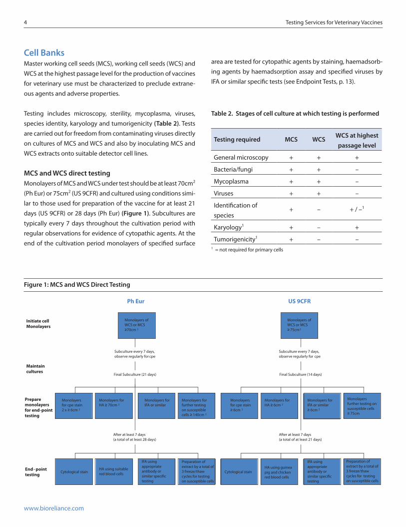
MCS and WCS direct testingMonolayers of MCS and WCS under test should be at least 70cm2 (Ph Eur) or 75cm2 (US 9CFR) and cultured using conditions simi-lar to those used for preparation of the vaccine for at least 21 days (US 9CFR) or 28 days (Ph Eur) (Figure 1). Subcultures are typically every 7 days throughout the cultivation period with regular observations for evidence of cytopathic agents. At the end of the cultivation period monolayers of specified surface
area are tested for cytopathic agents by staining, haemadsorb-ing agents by haemadsorption assay and specified viruses by IFA or similar specific tests (see Endpoint Tests, p. 13).
Figure 1: MCS and WCS Direct Testing
for
2
Ph Eur
Monolayers of WCS or MCS ≥70cm 2
Subculture every 7 days, observe regularly for cpe
Final Subculture (21 days)
Initiate cell Monolayers
Maintain cultures
Prepare monolayersfor end-point testing
Monolayers for HA ≥ 70cm 2
After at least 7 days (a total of at least 28 days)
End - point testing
Monolayersfor cpe stain 2 x ≥ 6cm 2
Monolayers for IFA or similar
Monolayers for further testing on susceptible cells ≥ 140cm 2
Cytological stainHA using suitable red blood cells
IFA using appropriate antibody or similar speci�c testing
Preparation of extract by a total of 3 freeze/thaw cycles for testing on susceptible cells
US 9CFR
Subculture every 7 days, observe regularly for cpe
Final Subculture (14 days)
Monolayers for HA ≥ 6cm 2
After at least 7 days (a total of at least 21 days)
Monolayersfor cpe stain ≥ 6cm 2
Monolayers for IFA or similar ≥ 6cm 2
Monolayersfurther testing on susceptible cells ≥ 75cm
Cytological stainHA using guinea pig and chicken red blood cells
IFA using appropriate antibody or similar speci�c testing
Preparation of extract by a total of 3 freeze/thaw cycles for testing on susceptible cells
Monolayers of WCS or MCS ≥ 75cm 2
Table 2. Stages of cell culture at which testing is performed
Testing required MCS WCS WCS at highest passage level
General microscopy + + +
Bacteria/fungi + + –
Mycoplasma + + –
Viruses + + –
Identification of species
+ – + / –1
Karyology1 + – +
Tumorigenicity1 + – –1 = not required for primary cells
Testing Services for Veterinary Vaccines 5
www.bioreliance.com
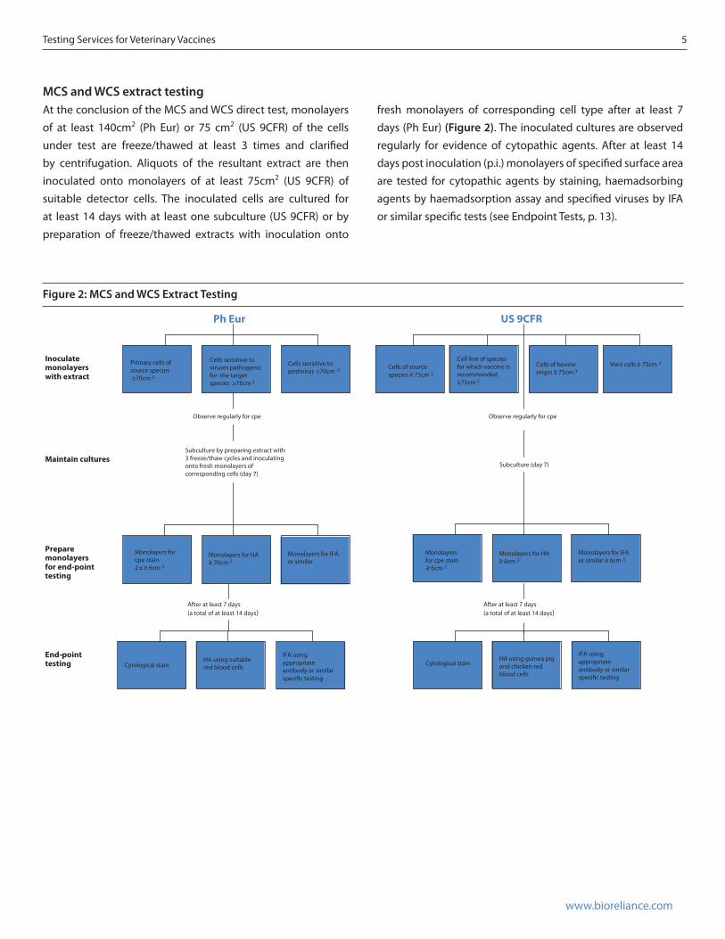
MCS and WCS extract testing At the conclusion of the MCS and WCS direct test, monolayers of at least 140cm2 (Ph Eur) or 75 cm2 (US 9CFR) of the cells under test are freeze/thawed at least 3 times and clarified by centrifugation. Aliquots of the resultant extract are then inoculated onto monolayers of at least 75cm2 (US 9CFR) of suitable detector cells. The inoculated cells are cultured for at least 14 days with at least one subculture (US 9CFR) or by preparation of freeze/thawed extracts with inoculation onto
Ph Eur
Cells sensitive to viruses pathogenic for the target species ≥70cm 2
Observe regularly for cpe
Subculture by preparing extract with 3 freeze/thaw cycles and inoculating onto fresh monolayers of corresponding cells (day 7)
After at least 7 days (a total of at least 14 days)
Inoculate monolayerswith extract
Maintain cultures
Prepare monolayersfor end-pointtesting
End-point-testing
Primary cells of source species ≥70cm 2
Cells sensitive to pestivirus ≥70cm 2
Monolayers for HA ≥ 70cm 2
Monolayers for cpe stain 2 x ≥ 6cm 2
Monolayers for IFA or similar
IFA using appropriate antibody or similar speci�c testing
Cytological stainHA using suitable red blood cells
Cell line of species for which vaccine is recommended ≥75cm 2
Cells of bovine origin ≥ 75cm 2
Vero cells ≥ 75cm 2
US 9CFR
Cells of source species ≥ 75cm 2
Observe regularly for cpe
Subculture (day 7)
Monolayers for HA ≥ 6cm 2
Monolayersfor cpe stain ≥ 6cm 2
Monolayers for IFA or similar ≥ 6cm 2
After at least 7 days (a total of at least 14 days)
IFA using appropriate antibody or similar speci�c testing
Cytological stainHA using guinea pig and chicken red blood cells
Figure 2: MCS and WCS Extract Testing
fresh monolayers of corresponding cell type after at least 7 days (Ph Eur) (Figure 2). The inoculated cultures are observed regularly for evidence of cytopathic agents. After at least 14 days post inoculation (p.i.) monolayers of specified surface area are tested for cytopathic agents by staining, haemadsorbing agents by haemadsorption assay and specified viruses by IFA or similar specific tests (see Endpoint Tests, p. 13).
6 Testing Services for Veterinary Vaccines
www.bioreliance.com
Ph Eur
Cells sensitive toviruses pathogenicfor the target species ≥70 cm2
Observe regularly for cpe
Subculture every 7 days by preparing extract with 3 freeze/thaw cycles and inoculating onto fresh monolayers of corresponding cells
Cells sensitive topestivirus ≥70 cm2
Test at 10 vaccine doses/ml if possible
Inoculatemonolayers
Maintain
Primary cells of source species ≥70cm2
Monolayers forHA ≥ 70cm2
Monolayers forcpe stain 2 × ≥ 6cm2
Monolayers for IFA or similar
After at least 7 days (a total of at least 28 days)
IFA using appropriateantibody or similarspeci�c testing
Cytological stainHA using suitablered blood cells
Prepare monolayersfor end-pointtesting
End-pointtesting
Final subculture (day 21)
IFA using appropriateantibody or similarspeci�c testing
Cytological stainHA using guineapig and chickenred blood cells
Monolayers forcpe stain ≥ 6cm2
Monolayers forHA ≥ 6cm2
Monolayers for IFA or similar ≥ 6cm2
After at least 7 days (a total of at least 14 days)
Subculture (day 7)
Observe regularly for cpe
US 9CFR
Cell line of thespecies of cells inwhich the MSV ispropogated ≥ 75 cm2
Vero cells ≥75 cm2
At least 1ml per cell monolayer
Cell line of species for whichthe vaccine isrecommended ≥ 75 cm2
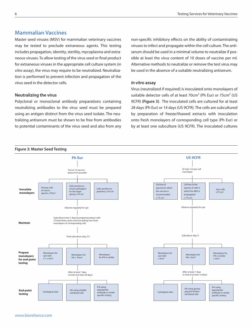
Figure 3: Master Seed Testing
Mammalian VaccinesMaster seed viruses (MSV) for mammalian veterinary vaccines may be tested to preclude extraneous agents. This testing includes propagation, identity, sterility, mycoplasma and extra-neous viruses. To allow testing of the virus seed or final product for extraneous viruses in the appropriate cell culture system (in vitro assay), the virus may require to be neutralized. Neutraliza-tion is performed to prevent infection and propagation of the virus seed in the detector cells.
Neutralizing the virusPolyclonal or monoclonal antibody preparations containing neutralizing antibodies to the virus seed must be prepared using an antigen distinct from the virus seed isolate. The neu-tralizing antiserum must be shown to be free from antibodies to potential contaminants of the virus seed and also from any
non-specific inhibitory effects on the ability of contaminating viruses to infect and propagate within the cell culture. The anti-serum should be used in a minimal volume to neutralize if pos-sible at least the virus content of 10 doses of vaccine per ml. Alternative methods to neutralize or remove the test virus may be used in the absence of a suitable neutralizing antiserum.
In vitro assayVirus (neutralized if required) is inoculated onto monolayers of suitable detector cells of at least 70cm2 (Ph Eur) or 75cm2 (US 9CFR) (Figure 3). The inoculated cells are cultured for at least 28 days (Ph Eur) or 14 days (US 9CFR). The cells are subcultured by preparation of freeze/thawed extracts with inoculation onto fresh monolayers of corresponding cell type (Ph Eur) or by at least one subculture (US 9CFR). The inoculated cultures
Testing Services for Veterinary Vaccines 7
www.bioreliance.com
are observed regularly for evidence of cytopathic agents. After at least 28 (Ph Eur) or 14 (US 9CFR) days p.i. monolayers of specified surface area are tested for cytopathic agents by staining, haemadsorbing agents by haemadsorption assay and specified viruses by IFA or similar specific tests (See Endpoint Tests p. 13).
Avian VaccinesMaster seed viruses (MSV) and cell seeds for avian veterinary vaccines may be tested to preclude extraneous agents. This testing includes that detailed for mammalian vaccines with some additional specifications: any chickens, embryos and tissue cultures used in production of avian vaccines must be derived from specific pathogen free (SPF) chicken flocks.
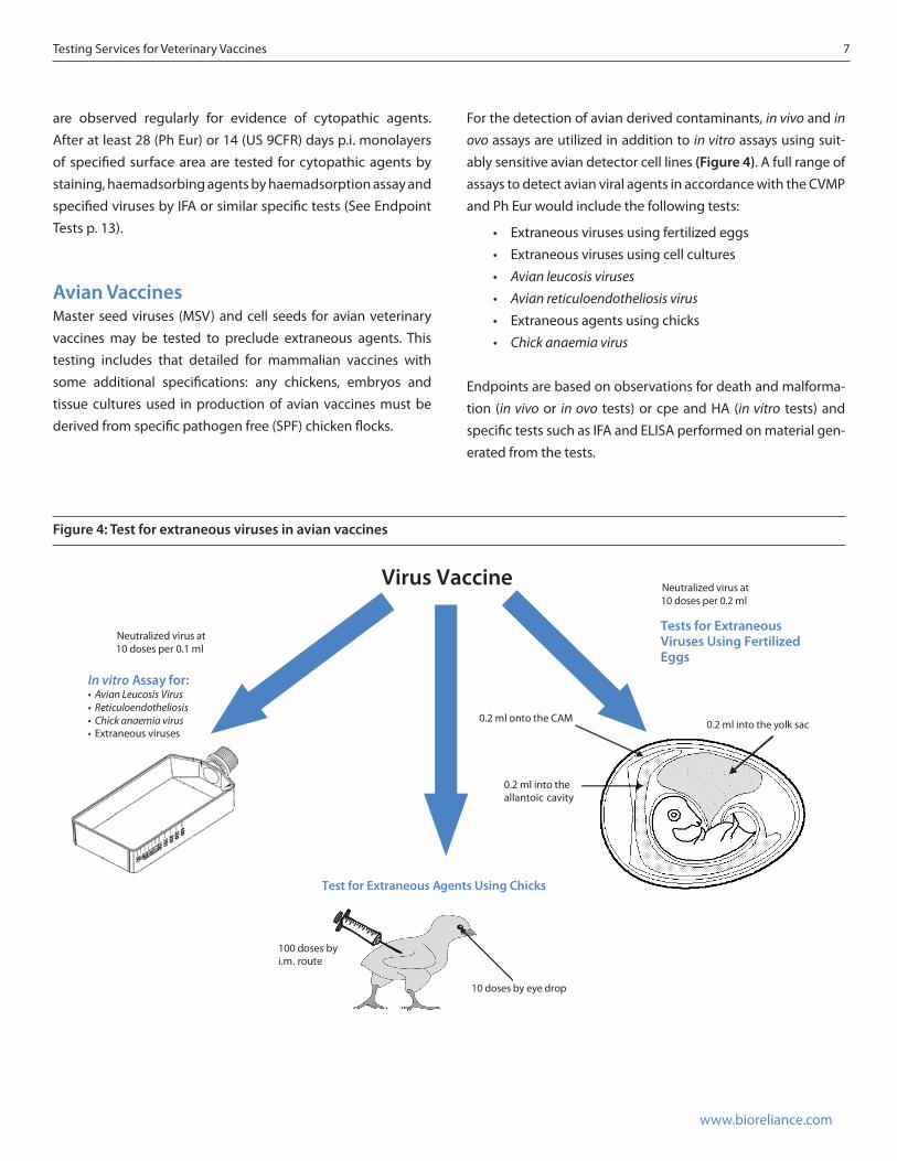
For the detection of avian derived contaminants, in vivo and in ovo assays are utilized in addition to in vitro assays using suit-ably sensitive avian detector cell lines (Figure 4). A full range of assays to detect avian viral agents in accordance with the CVMP and Ph Eur would include the following tests:
• Extraneous viruses using fertilized eggs• Extraneous viruses using cell cultures• Avian leucosis viruses• Avian reticuloendotheliosis virus• Extraneous agents using chicks• Chick anaemia virus
Endpoints are based on observations for death and malforma-tion (in vivo or in ovo tests) or cpe and HA (in vitro tests) and specific tests such as IFA and ELISA performed on material gen-erated from the tests.
Figure 4: Test for extraneous viruses in avian vaccines
allantoic cavity
i.m
Virus Vaccine
Neutralized virus at 10 doses per 0.1 ml
Neutralized virus at 10 doses per 0.2 ml
0.2 ml onto the CAM
0.2 ml into the
Tests for ExtraneousViruses Using Fertilized Eggs
In vitro Assay for:• Avian Leucosis Virus• Reticuloendotheliosis• Chick anaemia virus• Extraneous viruses
10 doses by eye drop
. route100 doses by
0.2 ml into the yolk sac
Test for Extraneous Agents Using Chicks
8 Testing Services for Veterinary Vaccines
www.bioreliance.com
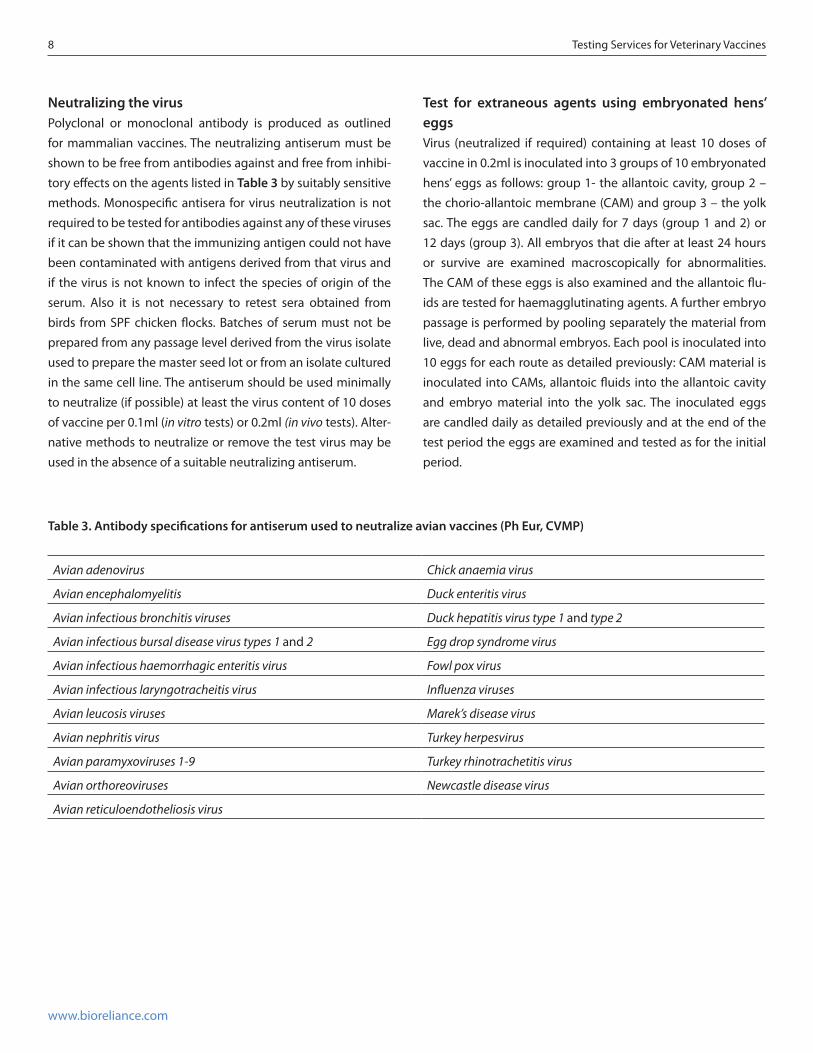
Table 3. Antibody specifications for antiserum used to neutralize avian vaccines (Ph Eur, CVMP)
Avian adenovirus Chick anaemia virus
Avian encephalomyelitis Duck enteritis virus
Avian infectious bronchitis viruses Duck hepatitis virus type 1 and type 2
Avian infectious bursal disease virus types 1 and 2 Egg drop syndrome virus
Avian infectious haemorrhagic enteritis virus Fowl pox virus
Avian infectious laryngotracheitis virus Influenza viruses
Avian leucosis viruses Marek’s disease virus
Avian nephritis virus Turkey herpesvirus
Avian paramyxoviruses 1-9 Turkey rhinotrachetitis virus
Avian orthoreoviruses Newcastle disease virus
Avian reticuloendotheliosis virus
Neutralizing the virusPolyclonal or monoclonal antibody is produced as outlined for mammalian vaccines. The neutralizing antiserum must be shown to be free from antibodies against and free from inhibi-tory effects on the agents listed in Table 3 by suitably sensitive methods. Monospecific antisera for virus neutralization is not required to be tested for antibodies against any of these viruses if it can be shown that the immunizing antigen could not have been contaminated with antigens derived from that virus and if the virus is not known to infect the species of origin of the serum. Also it is not necessary to retest sera obtained from birds from SPF chicken flocks. Batches of serum must not be prepared from any passage level derived from the virus isolate used to prepare the master seed lot or from an isolate cultured in the same cell line. The antiserum should be used minimally to neutralize (if possible) at least the virus content of 10 doses of vaccine per 0.1ml (in vitro tests) or 0.2ml (in vivo tests). Alter-native methods to neutralize or remove the test virus may be used in the absence of a suitable neutralizing antiserum.
Test for extraneous agents using embryonated hens’ eggsVirus (neutralized if required) containing at least 10 doses of vaccine in 0.2ml is inoculated into 3 groups of 10 embryonated hens’ eggs as follows: group 1- the allantoic cavity, group 2 – the chorio-allantoic membrane (CAM) and group 3 – the yolk sac. The eggs are candled daily for 7 days (group 1 and 2) or 12 days (group 3). All embryos that die after at least 24 hours or survive are examined macroscopically for abnormalities. The CAM of these eggs is also examined and the allantoic flu-ids are tested for haemagglutinating agents. A further embryo passage is performed by pooling separately the material from live, dead and abnormal embryos. Each pool is inoculated into 10 eggs for each route as detailed previously: CAM material is inoculated into CAMs, allantoic fluids into the allantoic cavity and embryo material into the yolk sac. The inoculated eggs are candled daily as detailed previously and at the end of the test period the eggs are examined and tested as for the initial period.
Testing Services for Veterinary Vaccines 9
www.bioreliance.com
In vitro assaysTest in chick kidney cells (Ph Eur, CVMP)Virus (neutralized if required) is inoculated onto 5 replicate monolayers of at least 25cm2 of chick kidney cells. The inocu-lated cells are cultured for at least 21 days with subculture at 4 to 7 day intervals. Each subculture is performed with pooled cells and fluids from all 5 monolayers that have undergone one freeze/thaw cycle. The extract is then inoculated onto fresh monolayers of chick kidney cells. The inoculated cultures are observed regularly for evidence of cytopathic agents. At the end of the culture period monolayers of specified surface area are tested for cytopathic agents by staining, haemadsorbing agents by haemadsorption assay and for haemagglutinating agents by haemagglutination assay.
Test for Avian leucosis viruses (Ph Eur, CVMP)Virus (neutralized if required) is inoculated onto 5 replicate monolayers of at least 50cm2 of primary or secondary chick embryo fibroblasts that are known to be susceptible to sub-groups A, B and J of Avian leucosis viruses (support the growth of exogenous but not endogenous Avian leucosis viruses). The inoculated cells are cultured for at least 9 days with subculture at 3 to 4 day intervals. Cells are retained from each subculture and at the end of the culture period are tested for group spe-cific Avian leucosis antigen by ELISA assay.
Test for Reticuloendotheliosus virus (REV) (CVMP)Virus (neutralized if required) is inoculated onto 5 repli-cate monolayers of at least 25cm2 of primary or second-ary chick or duck embryo fibroblasts. The inoculated cells are cultured for at least 10 days with subculture twice at 3 to 4 day intervals. At the end of the culture period mono-layers of specified surface area are tested for REV by IFA.
Test for Chick anaemia virusDNA is extracted from an appropriate volume of virus and tested by polymerase chain reaction (PCR) to detect Chick anaemia virus. The PCR assay allows the detection of a nucleic acid target molecule and uses two target specific oligonucle-otide primers that flank a target DNA sequence. The internal sequence is amplified by repeated cycles of heat denaturation of the DNA template, annealing the primers to their comple-mentary sequences on each DNA strand and extension of the annealed primers with a thermostable Taq DNA polymerase. The amplified sequences are identified by hybridization with an oligonucleotide probe specific for the target DNA.
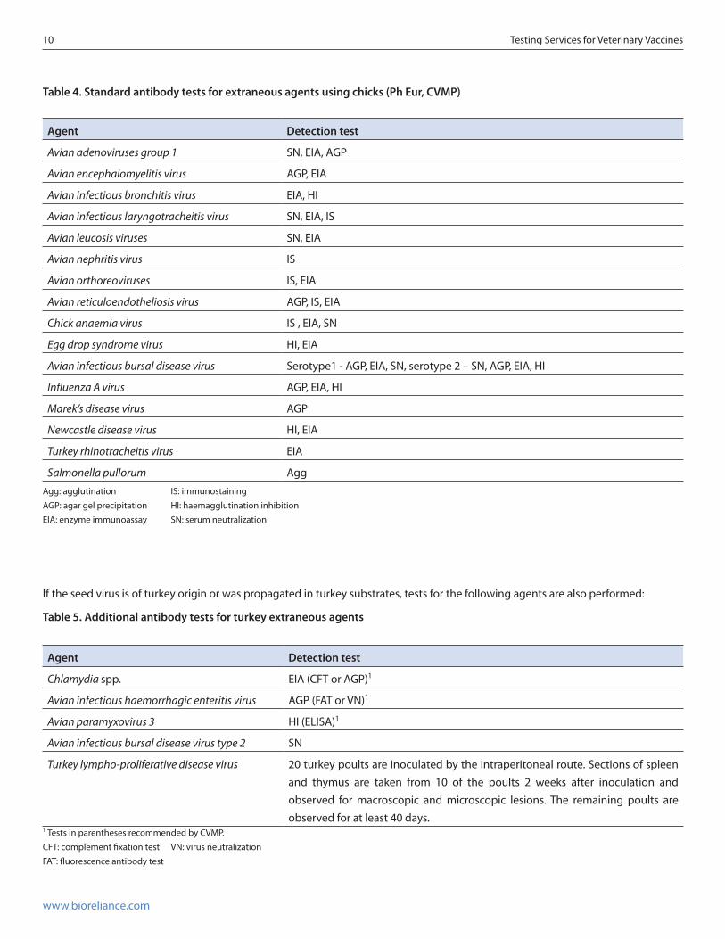
Test for extraneous agents using chicks (Ph Eur, CVMP)At least 10 chicks that are 2 weeks old (older birds may be used if the seed virus is pathogenic for birds of this age) are inocu-lated with virus (neutralized if required). Virus is inoculated at 100 doses by the intramuscular route and 10 doses by eye-drop. Repeat inoculations are performed 2 weeks later. The chicks are observed for a period of 5 weeks. Serum is collected from each chick at the end of the test period and is tested for antibodies to extraneous agents using specific tests listed in Table 4 and also those listed in Tables 5, 6 and 7 if required.
10 Testing Services for Veterinary Vaccines
www.bioreliance.com
Table 4. Standard antibody tests for extraneous agents using chicks (Ph Eur, CVMP)
Agent Detection test
Avian adenoviruses group 1 SN, EIA, AGP
Avian encephalomyelitis virus AGP, EIA
Avian infectious bronchitis virus EIA, HI
Avian infectious laryngotracheitis virus SN, EIA, IS
Avian leucosis viruses SN, EIA
Avian nephritis virus IS
Avian orthoreoviruses IS, EIA
Avian reticuloendotheliosis virus AGP, IS, EIA
Chick anaemia virus IS , EIA, SN
Egg drop syndrome virus HI, EIA
Avian infectious bursal disease virus Serotype1 - AGP, EIA, SN, serotype 2 – SN, AGP, EIA, HI
Influenza A virus AGP, EIA, HI
Marek’s disease virus AGP
Newcastle disease virus HI, EIA
Turkey rhinotracheitis virus EIA
Salmonella pullorum AggAgg: agglutination IS: immunostainingAGP: agar gel precipitation HI: haemagglutination inhibitionEIA: enzyme immunoassay SN: serum neutralization
Table 5. Additional antibody tests for turkey extraneous agents
Agent Detection test
Chlamydia spp. EIA (CFT or AGP)1
Avian infectious haemorrhagic enteritis virus AGP (FAT or VN)1
Avian paramyxovirus 3 HI (ELISA)1
Avian infectious bursal disease virus type 2 SN
Turkey lympho-proliferative disease virus 20 turkey poults are inoculated by the intraperitoneal route. Sections of spleen and thymus are taken from 10 of the poults 2 weeks after inoculation and observed for macroscopic and microscopic lesions. The remaining poults are observed for at least 40 days.
1 Tests in parentheses recommended by CVMP.CFT: complement fixation test VN: virus neutralizationFAT: fluorescence antibody test
If the seed virus is of turkey origin or was propagated in turkey substrates, tests for the following agents are also performed:
Testing Services for Veterinary Vaccines 11
www.bioreliance.com
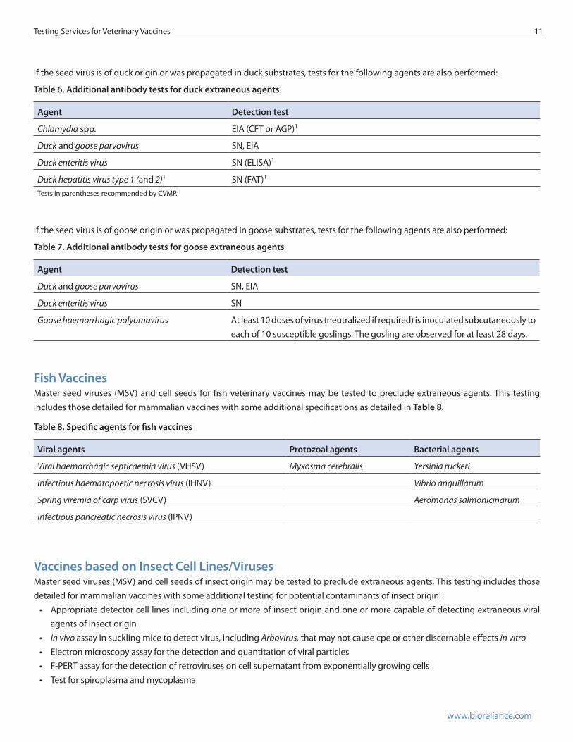
Table 6. Additional antibody tests for duck extraneous agents
Agent Detection test
Chlamydia spp. EIA (CFT or AGP)1
Duck and goose parvovirus SN, EIA
Duck enteritis virus SN (ELISA)1
Duck hepatitis virus type 1 (and 2)1 SN (FAT)1
1 Tests in parentheses recommended by CVMP.
Table 7. Additional antibody tests for goose extraneous agents
Agent Detection test
Duck and goose parvovirus SN, EIA
Duck enteritis virus SN
Goose haemorrhagic polyomavirus At least 10 doses of virus (neutralized if required) is inoculated subcutaneously to each of 10 susceptible goslings. The gosling are observed for at least 28 days.
Table 8. Specific agents for fish vaccines
Viral agents Protozoal agents Bacterial agents
Viral haemorrhagic septicaemia virus (VHSV) Myxosma cerebralis Yersinia ruckeri
Infectious haematopoetic necrosis virus (IHNV) Vibrio anguillarum
Spring viremia of carp virus (SVCV) Aeromonas salmonicinarum
Infectious pancreatic necrosis virus (IPNV)
Fish VaccinesMaster seed viruses (MSV) and cell seeds for fish veterinary vaccines may be tested to preclude extraneous agents. This testing includes those detailed for mammalian vaccines with some additional specifications as detailed in Table 8.
If the seed virus is of duck origin or was propagated in duck substrates, tests for the following agents are also performed:
If the seed virus is of goose origin or was propagated in goose substrates, tests for the following agents are also performed:
Vaccines based on Insect Cell Lines/VirusesMaster seed viruses (MSV) and cell seeds of insect origin may be tested to preclude extraneous agents. This testing includes those detailed for mammalian vaccines with some additional testing for potential contaminants of insect origin:
• Appropriate detector cell lines including one or more of insect origin and one or more capable of detecting extraneous viral agents of insect origin
• In vivo assay in suckling mice to detect virus, including Arbovirus, that may not cause cpe or other discernable effects in vitro• Electron microscopy assay for the detection and quantitation of viral particles• F-PERT assay for the detection of retroviruses on cell supernatant from exponentially growing cells• Test for spiroplasma and mycoplasma
12 Testing Services for Veterinary Vaccines
www.bioreliance.com
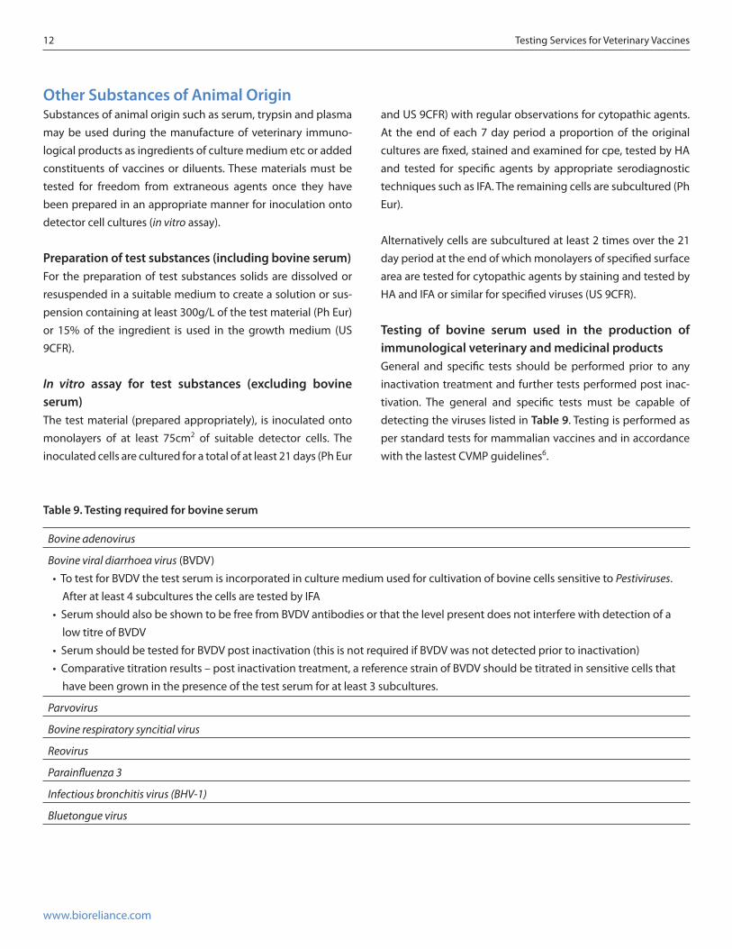
Table 9. Testing required for bovine serum
Bovine adenovirus
Bovine viral diarrhoea virus (BVDV)•TotestforBVDVthetestserumisincorporatedinculturemediumusedforcultivationofbovinecellssensitivetoPestiviruses.
After at least 4 subcultures the cells are tested by IFA•SerumshouldalsobeshowntobefreefromBVDVantibodiesorthatthelevelpresentdoesnotinterferewithdetectionofa
low titre of BVDV•SerumshouldbetestedforBVDVpostinactivation(thisisnotrequiredifBVDVwasnotdetectedpriortoinactivation)•Comparativetitrationresults–postinactivationtreatment,areferencestrainofBVDVshouldbetitratedinsensitivecellsthat
have been grown in the presence of the test serum for at least 3 subcultures.
Parvovirus
Bovine respiratory syncitial virus
Reovirus
Parainfluenza 3
Infectious bronchitis virus (BHV-1)
Bluetongue virus
Other Substances of Animal OriginSubstances of animal origin such as serum, trypsin and plasma may be used during the manufacture of veterinary immuno-logical products as ingredients of culture medium etc or added constituents of vaccines or diluents. These materials must be tested for freedom from extraneous agents once they have been prepared in an appropriate manner for inoculation onto detector cell cultures (in vitro assay).
Preparation of test substances (including bovine serum)For the preparation of test substances solids are dissolved or resuspended in a suitable medium to create a solution or sus-pension containing at least 300g/L of the test material (Ph Eur) or 15% of the ingredient is used in the growth medium (US 9CFR).
In vitro assay for test substances (excluding bovine serum)The test material (prepared appropriately), is inoculated onto monolayers of at least 75cm2 of suitable detector cells. The inoculated cells are cultured for a total of at least 21 days (Ph Eur
and US 9CFR) with regular observations for cytopathic agents. At the end of each 7 day period a proportion of the original cultures are fixed, stained and examined for cpe, tested by HA and tested for specific agents by appropriate serodiagnostic techniques such as IFA. The remaining cells are subcultured (Ph Eur).
Alternatively cells are subcultured at least 2 times over the 21 day period at the end of which monolayers of specified surface area are tested for cytopathic agents by staining and tested by HA and IFA or similar for specified viruses (US 9CFR).
Testing of bovine serum used in the production of immunological veterinary and medicinal productsGeneral and specific tests should be performed prior to any inactivation treatment and further tests performed post inac-tivation. The general and specific tests must be capable of detecting the viruses listed in Table 9. Testing is performed as per standard tests for mammalian vaccines and in accordance with the lastest CVMP guidelines6.
Testing Services for Veterinary Vaccines 13
www.bioreliance.com
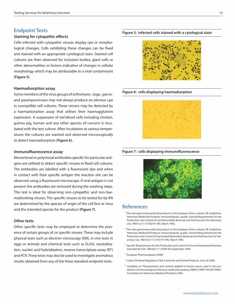
Endpoint TestsStaining for cytopathic effectsCells infected with cytopathic viruses display cpe or morpho-logical changes. Cells exhibiting these changes can be fixed and stained with an appropriate cytological stain. Stained cell cultures are then observed for inclusion bodies, giant cells or other abnormalities or lesions indicative of changes in cellular morphology which may be attributable to a viral contaminant (Figure 5).
Haemadsorption assaySome members of the virus groups of orthomyxo-, toga-, parvo- and paramyxoviruses may not always produce an obvious cpe in susceptible cell cultures. These viruses may be detected by a haemadsorption assay that utilises their haemagglutinin expression. A suspension of red blood cells including chicken, guinea pig, human and any other species of concern is incu-bated with the test culture. After incubation at various temper-atures the cultures are washed and observed microscopically to detect haemadsorption (Figure 6).
Immunofluorescence assayMonoclonal or polyclonal antibodies specific for particular anti-gens are utilized to detect specific viruses in fixed cell cultures. The antibodies are labelled with a fluorescent dye and when in contact with their specific antigen the reaction site can be observed using a fluorescent microscope. If viral antigen is not present the antibodies are removed during the washing steps. This test is ideal for observing non-cytopathic and non-hae-madsorbing viruses. The specific viruses to be tested for by IFA are determined by the species of origin of the cell line or virus and the intended species for the product (Figure 7).
Other testsOther specific tests may be employed to determine the pres-ence of certain groups of, or specific viruses. These may include physical tests such as electron microscopy (EM), in vivo tests in eggs or animals and chemical tests such as ELISA, neutraliza-tion, nucleic acid hybridization, reverse transcriptase assay (RT) and PCR. These tests may also be used to investigate anomalous results obtained from any of the three standard endpoint tests.
References1 The rules governing medicinal products in the European Union, volume 7B, Guidelines,
Veterinary Medicinal Products, Immunologicals, quality. General Requirements for the Production and Control of Live Mammalian Bacterial and Viral Vaccines for Veterinary Use. 7BIm1a (111/3182/91-EN), March 1992.
2 The rules governing medicinal products in the European Union, volume 7B, Guidelines, Veterinary Medicinal Products, Immunologicals, quality. General Requirements for the Production and Control of Inactivated Mammalian Bacterial and Viral Vaccines for Vet-erinary Use. 7BIm1b (111/3181/91-EN), March 1992.
3 Specific Requirements for the Production and Control of Live and Inactivated Vaccines Intended for Fish. 7BIm9a (111/3590-92) September 1994.
4 European Pharmacopoeia (2008)
5 Code of Federal Regulations Title 9, Animals and Animal Products, June 30 2008
6 Guideline on “Requirements and controls applied to bovine serum used in the pro-duction of immunological veterinary medicinal products, EMEA/CVMP/743/00-FINAL”. Committee for Veterinary Medicinal Products 2002.
Figure 6: cells displaying haemadsorption
Figure 5: infected cells stained with a cytological stain
Figure 7: cells displaying immunofluorescence
www.bioreliance.comNorth America Toll Free: 800 553 5372 Tel: 301 738 1000
Europe & International Tel: +44 (0)141 946 9999 • Japan Tel: +03 5796 7430Email: [email protected]
©2012 Sigma-Aldrich Co. LLC. All rights reserved. BioReliance and SAFC are trademarks of Sigma-Aldrich Co. LLC or its Affiliates, registered in the US and other countries. O-0310708
BioReliance Corp.14920 Broschart RoadRockville, Maryland 20850Tel: 800.553.5372 Fax: 301.610.2590Email: [email protected]
BioReliance Ltd.Todd CampusWest of Scotland Science ParkGlasgow, Scotland G20 0XATel: 44 (0) 141 946.9999 Fax: 44 (0) 141 946.0000Email: [email protected]
BioReliance Ltd.Innovation ParkHillfoots RoadStirling, Scotland FK9 4NFTel: 44 (0) 141 946.9999 Fax: 44 (0) 141 946.0000Email: [email protected]
BioReliance, K.K.c/o Sigma-Aldrich Japan K.K.Tennoz Central Tower 4F2-2-24 Higashi-ShinagawaShinagawa-kuTokyo 140-0002, JapanTel: +81 (0)3 5796 7430Fax: +81 (0)3 5796 7435Email: [email protected]
BioReliance Ltd. c/o Sigma-Aldrich Chemicals Pvt Ltd102 Alpha BuildingHiranandani GardensPowai, Mumbai 400076Tel: +91 22 40872364Fax: +91 22 25797589Email: [email protected]


![1999 [Advances in Veterinary Medicine] Veterinary Vaccines and Diagnostics Volume 41 __ Evaluation of risks and benefits](https://img.pdfslide.us/doc/110x75/613ca5e89cc893456e1e7b63/1999-advances-in-veterinary-medicine-veterinary-vaccines-and-diagnostics-volume.jpg)