Embed Size (px)
Citation preview
C034-D
Testing for Coronary Artery Disease
Please visit the UHN Patient Education website for more health information: www.uhnpatienteducation.ca© 2015 University Health Network. All rights reserved. This information is to be used for informational purposes only and is not intended as a substitute for professional medical advice, diagnosis or treatment. Please consult your health care provider for advice about a specific medical condition. A single copy of these materials may be reprinted for non-commercial personal use only.
Author: CardiologyRevised: 01/2015Form: D-5111
Read this guide to learn about the tests you might have in the hospital.
These tests include:
• An electrocardiogram (ECG)
• An echocardiogram (echo)
• An exercise stress test
• A nuclear scan
• A heart angiogram (heart catheterization)
What tests can I expect when I am in the hospital?
If you have heart problems, you may have to be admitted to a hospital. During your stay in hospital, you may have a number of tests.
The tests explained below help your health care team understand your heart problem better.
Electrocardiogram (ECG)
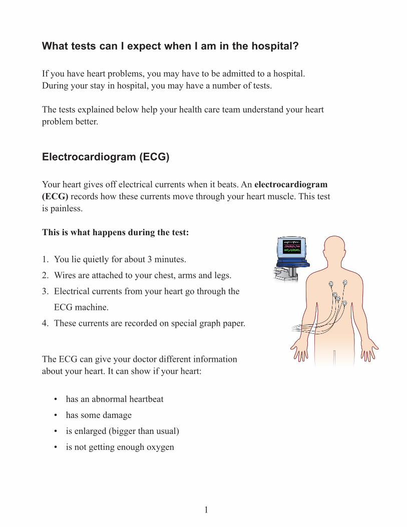
Your heart gives off electrical currents when it beats. An electrocardiogram (ECG) records how these currents move through your heart muscle. This test is painless.
This is what happens during the test:
1. You lie quietly for about 3 minutes.
2. Wires are attached to your chest, arms and legs.
3. Electrical currents from your heart go through the
ECG machine.
4. These currents are recorded on special graph paper.
The ECG can give your doctor different information about your heart. It can show if your heart:
• has an abnormal heartbeat
• has some damage
• is enlarged (bigger than usual)
• is not getting enough oxygen
1
Echocardiogram
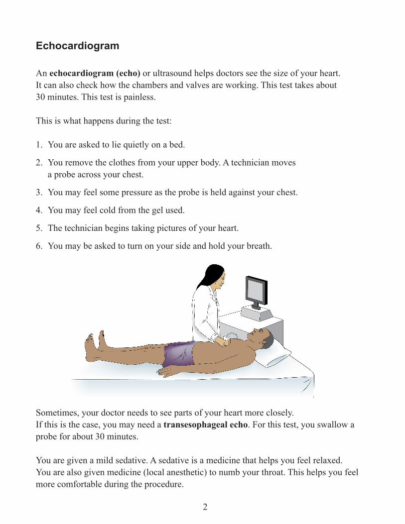
An echocardiogram (echo) or ultrasound helps doctors see the size of your heart. It can also check how the chambers and valves are working. This test takes about 30 minutes. This test is painless.
This is what happens during the test:
1. You are asked to lie quietly on a bed.
2. You remove the clothes from your upper body. A technician moves a probe across your chest.
3. You may feel some pressure as the probe is held against your chest.
4. You may feel cold from the gel used.
5. The technician begins taking pictures of your heart.
6. You may be asked to turn on your side and hold your breath.
Sometimes, your doctor needs to see parts of your heart more closely. If this is the case, you may need a transesophageal echo. For this test, you swallow a probe for about 30 minutes.
You are given a mild sedative. A sedative is a medicine that helps you feel relaxed. You are also given medicine (local anesthetic) to numb your throat. This helps you feel more comfortable during the procedure.
2
Exercise Stress Test
An exercise stress test measures how your heart deals with the stress of physical activity. You are connected by wires to an ECG monitor. It checks your heart’s electrical activity and your blood pressure.
How to prepare for the test:
For 48 hours (2 days) before the test:
1. Do not drink caffeinated beverages such as coffee, tea or Coca-Cola. Caffeinated beverages can affect the results of the test and make them inaccurate.
On the day you have your test:
2. Wear comfortable walking shoes and loose clothing.3. Do not eat heavy foods.4. Do not drink a lot of liquid.
Ask you doctor if you should take your medicines on the day of your test.
3
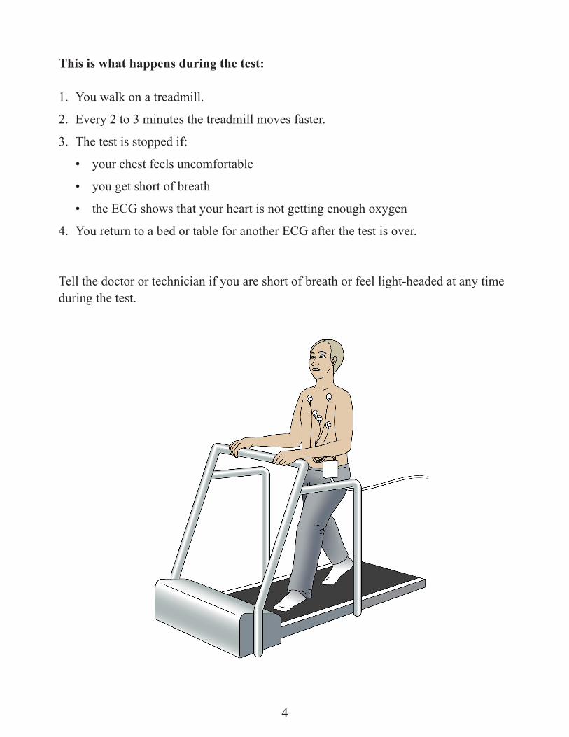
This is what happens during the test:
1. You walk on a treadmill.
2. Every 2 to 3 minutes the treadmill moves faster.
3. The test is stopped if:
• your chest feels uncomfortable
• you get short of breath
• the ECG shows that your heart is not getting enough oxygen
4. You return to a bed or table for another ECG after the test is over.
Tell the doctor or technician if you are short of breath or feel light-headed at any time during the test.
4
Nuclear Scans
A nuclear scan checksthebloodflowtotheheartmuscle.Itlooksforadamaged heart muscle and checks how well your heart pumps blood to your body.
In a nuclear scan, a small amount of radioactive material is injected into a vein. Radioactive material gives off energy. This material is not harmful to you.
There are 3 types of nuclear scans:
1. Exercise thallium scan/cardiolite
2. Persantine thallium scan/cardiolite
3. Multiple Gated Acquisition scan (MUGA)
Read the section below to learn about each scan.
1. Exercise thallium scan/cardiolite
The exercise thallium scan shows your cardiologist the areas of your heart that do not get enough oxygen during exercise.
This is what happens during the test:
1. A small amount of a radioactive material called thallium (or cardiolite) is injected into your bloodstream. This travels through your coronary arteries into your heart muscle.
2. You walk on a treadmill.3. A special camera measures the amount of radioactive material that reaches the
heart muscle while you exercise. 4. After walking on the treadmill, you lie on a table or bed. 5. You will get another scan. This is done while you are resting, about 4 hours after
the treadmill.
5
2. Persantine thallium scan/cardiolite
This test shows your cardiologist the areas of your heart that do not get enough oxygen during exercise. If you are not able to walk on a treadmill, your doctor may choose this test instead of an exercise thallium scan.
Instead of walking on a treadmill, the doctor injects a medicine called persantine. This medicine has the same effect on your heart as exercise.
This test helps your cardiologist to see:
• the size of your heart
• wherethereispoorbloodflow
• if or where your heart muscle is damaged
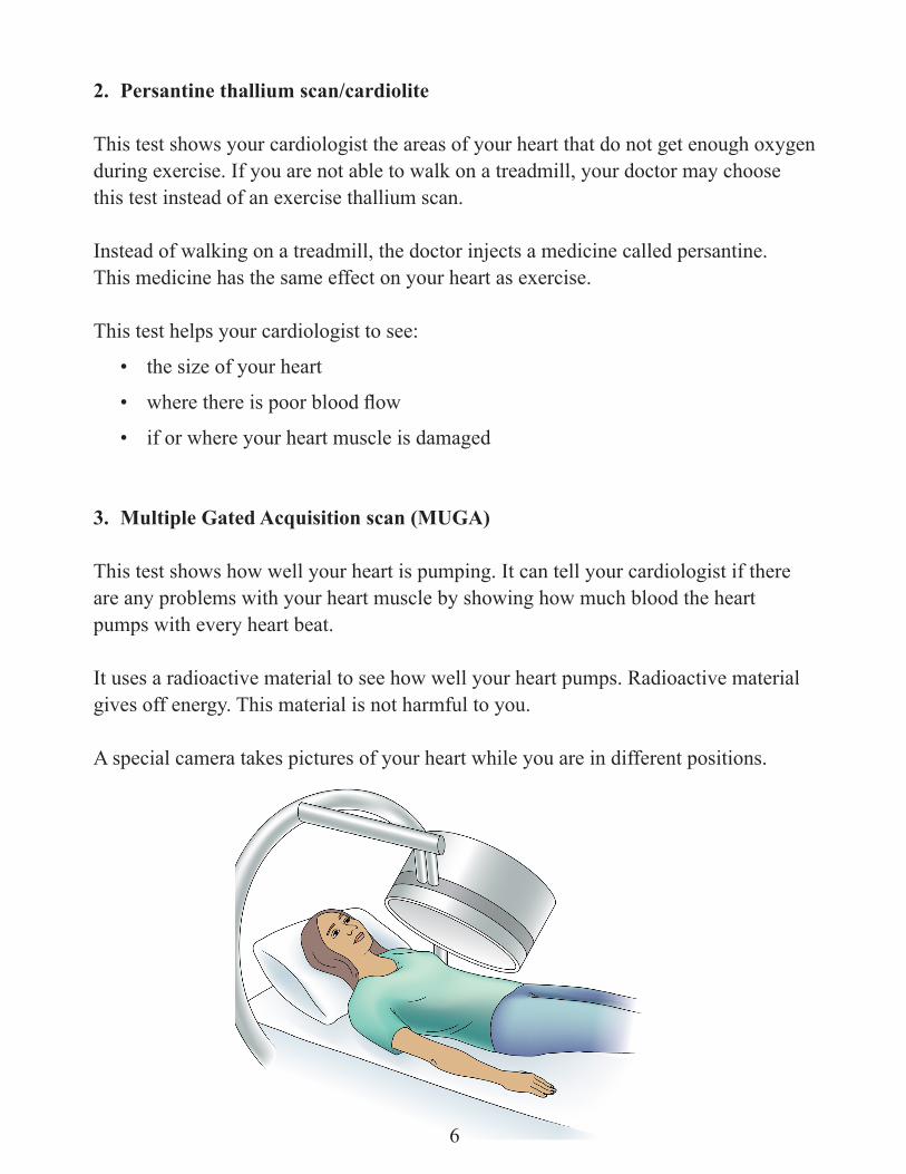
3. Multiple Gated Acquisition scan (MUGA)
This test shows how well your heart is pumping. It can tell your cardiologist if there are any problems with your heart muscle by showing how much blood the heart pumps with every heart beat.
It uses a radioactive material to see how well your heart pumps. Radioactive material gives off energy. This material is not harmful to you.
A special camera takes pictures of your heart while you are in different positions.
6
Heart angiogram (heart catheterization)
A heart angiogram (sometimes called a coronary angiogram) is a test to see if there are problems in different parts of your heart. This test can help the doctor see if there are problems:
• with any valves in your heart
• in any chambers in your heart
• with the main blood vessels of the heart (aorta or pulmonary)
• with thebloodflowinyourcoronaryarteries(ifthereisfattybuildup)
During a heart angiogram, dye is put through a catheter (a small tube) into the different partsofyourheart.Thenx-raysaretaken.Thishelpstofindanyproblems.
This test takes about 30 minutes to 1 hour. It takes place in a cardiac catheterization laboratory or “cath lab” for short.
How to prepare for the test:
If your heart angiogram is in the morning: • Do not to eat or drink anything after midnight the night before.
If your heart angiogram is in the afternoon: • You may be able to eat a light breakfast.
Please ask your nurse or doctor whether you should eat breakfast.
7
This is what happens during the test:
1. You are asked to lie on a table under an x-ray camera.
2. Your doctor puts a small amount of freezing in your groin or arm. Your skin may burn or sting as the freezing is injected.
3. Once the skin is numb, your doctor puts a needle into your artery. A small tube (also called an introducer sheath) is then put over the artery.
4. Another longer tube is moved along the artery until it reaches the heart. This tube helps the doctor to put smaller tubes and wires into your heart.
5. Once the tube is in your heart, your doctor injects x-ray dye into the tube. You do not feel the tube as it travels to your heart. You may feel a brief warm or hot feeling in your chest when the dye is injected.
6. Atechniciantakespictureswithanx-raycamera.Thiscamerarecordstheflowof dye in your coronary arteries. Pictures may also be taken of the pumping chamber (left ventricle) of your heart.
7. When the doctor has enough pictures, the tube and needle are removed.
8. You are then moved to a recovery area. The longer tube is removed. A clamp is put on the needle site. This prevents bleeding and allows the needle site to form a seal.
9. When the clamp is taken off, a bandage is placed over the needle site.
10. You are brought back to your room on the Cardiology ward. You will rest in bed for about 4 hours if your groin was injected (also called femoral approach). You will rest for 2 hours if your arm was injected (also called radial approach).
11. You are asked not to bend the affected leg for 4 hours. The head of your bed is raised to 30 degrees.
8
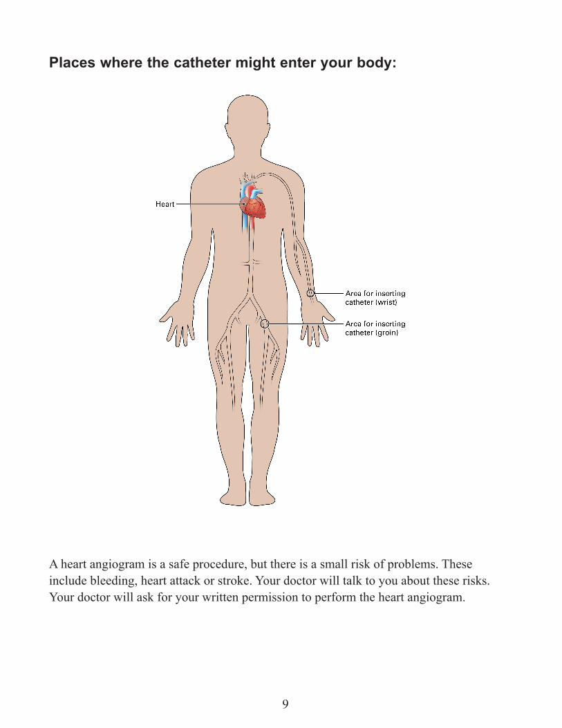
Places where the catheter might enter your body:
A heart angiogram is a safe procedure, but there is a small risk of problems. These include bleeding, heart attack or stroke. Your doctor will talk to you about these risks. Your doctor will ask for your written permission to perform the heart angiogram.
9

Write any notes or questions here
_______________________________________________________________________
_______________________________________________________________________
_______________________________________________________________________
_______________________________________________________________________
_______________________________________________________________________
_______________________________________________________________________
_______________________________________________________________________
_______________________________________________________________________
______________________________________________________________________
_______________________________________________________________________
_______________________________________________________________________
_______________________________________________________________________
_______________________________________________________________________
_______________________________________________________________________
_______________________________________________________________________
_______________________________________________________________________
_______________________________________________________________________
_______________________________________________________________________
_______________________________________________________________________
_______________________________________________________________________
_______________________________________________________________________
_______________________________________________________________________
_______________________________________________________________________
_______________________________________________________________________
_______________________________________________________________________
_______________________________________________________________________
10
This resource was written by:
Jeannine Costigan, RN, MScN, ACNPRuth Elias, MSc, RDJanice Jickling, RN, MScN, ACNPMin Hong, BSC. PharmJane MacIver, RN, MScNHeather Moon, BHSc OTNicki Morris, BHSc PTDiane Neil-Pollinger, MSW, RSWPeter Nielsen, RN, BSCN, MNGraham Reid, PhDJohn Ross, MD, FRCP©
Reviewed and revised by CICU Patient Education Committee, 2004Reviewed and revised by Zelia Souter (PCC), Pauline Glaves (PCC) and Rob Fuerte (APNE), 2014
Reviewed by the Patient and Family Education Program