Embed Size (px)
Citation preview
Pergamon Tetrahedron Letters, Vol, 38, No. 7, pp. 1223-1226, 1997 Copyright © 1997 Elsevier Science Ltd
Printed in Great Bdtain. All tights reserved PII: S0040-4039(97)00022-I 0040-4039/97 $17.00 + 0.00
T e r p e p t i n , A N o v e l M a m m a l i a n C e l l C y c l e Inh ib i to r ,
P r o d u c e d by A s p e r g i l l u s t e r r e u s 95F-1
Terumi Kagamizonoa.b, Noriyoshi Sakaib, Koshi Araib, Kimie Kobinataa, and Hiroyuki Osadaa.*
aThe Institute of Physical and Chemical Research (RIKEN), 2-1 Hirosawa, Wako-shi, Saitama, 351-01, Japan
b Medicinal Research Laboratories, Taisho Pharmaceutical Co., Ltd, 1-403 Yoshino-eho, Ohmiya-shi, Saitama, 330, Japan
Abstract: Terpelxin, a novel peptide having cell cycle inhibitory activity, was isolated from the cultured broth of Aspergillus terreus 951:-1 and its structure was elucidated by spectral analyses. Terpeptin inhibited the cell cycle progression of mouse tsFI210 cells in the G2/M phase with a minimum inhibitory concentration of 62.5 p-M. © 1997, Elsevier Science Ltd. All rights reserved.
The bioassay using mouse cdc2 mutant cell line, tsFl'210 cell, is a useful and practical assay method for the
screening of new mammalian cell cycle inhibitors of microbial origin t. Using this assay, we have found several
new cell cycle inhibitors, tryprostatin A, B2. 3, spirotryprostatins A, B4, 5 and cyclotryprostatins A-D6 in the
fermentation broth of a fungus, Aspergillusfumigatus BM939.
In the course of continuous screening, we found a fungal strain 95F-1 that produced a new cell cycle
inhibitor and we have now isolated a novel compound named
terpeptin (1). Herein, the isolation, the structure elucidation and
biological activity of I are described.
The producing strain 95F-1 was isolated from a soil
sample collected at Naha City, Okinawa, Japan and identified as
Aspergillus terreus 95F-1 through a taxonomic study.
The strain was cultured at 28 °C for 72 hours in a medium
Cltj Y
!
containing 2.5% glucose, 2% soybean, 0.5% polypeptone, 0,3% yeast extract, 1% soluble starch, 0.5% meat
extract, 0,2% NaCI, 0.05% KH2PO4 and 0.05% MgSO4 (pH 5.8).
The whole broth (7L) was extracted with 3.5 L acetone to give an aqueous acetone solution. The acetone
solution was evaporated under reduced pressure to remove acetone and the obtained residue was extracted with
EtOAc to afford an EtOAc extract (18.5 g). This extract was separated by successive column chromatographies
on silica gel and Sephadex LH-20 (MeOH). The active fractions containing terpeptin were further purified by
preparative HPLC on an ODS column with eluent CH3CN-H20 (75:25) to give a yellow amorphous solid of 1
(53.3 rag).
1223
1224
Terpeptin (1)7 gave a [M+] at rrdz 480 in ElMS and had the molecular formula C28I-I~N403 established by
elemental analysis. The IR spectrum of 1 suggested the presence of amide groups (3312, 1627 cm-t). The UV
spectrum revealed the presence of an indole chromophore in 1 with the absorption maxima at 230 and 285 nm,
like those of tryprostatinsZ3. In the tH NMR spectrumS, 1 showed signals due to a 1,2-disubstituted benzene
ring (8 7.30 d, J=8 Hz, 7-H; 8 7.24 d, J=8 Hz, 4-H; 8 7.03 td, J=8, 2 Hz, 6-H; 8 6.94 m, 5-H), an N-methyl (8
3.06 s, 13-CH3), three tertiary methyl groups (8 1.65 s, 29-H3; 8 1.68 s, 28-H3; 8 1.79 s, 18-H3), four
secondary methyl groups (5 0.67 d, J=6 Hz, 24-H3; 8 0.70 d, J--6 Hz, 21-H3; 8 0.82 d, J=7 Hz, 20-H3; 8 0.85
d, J--6 Hz, 23-H3), three exchangeable protons (6 8.05 d, J=8 Hz, 16-H; 8 8.81 d, J=l I Hz, 10-H; 8 10.94 s, l-
H), two oxymethines (54.40 t, J=9 Hz, 15-H; 8 4.74 d, J=l 1 Hz, 12-H), and three olefinic protons (5 5.31 m,
26-H; 8 5.79 d, J=7 Hz, 8-H; 8 6.76 t,J=10 Hz, 9-H) along with signals due to one methylene and two
methines. The t3C NMR spectrum of 18, analyzed by the DEPT method, indicated the presence of three amide
carbonyls (8 167.6 s, C-11; 8 168.9 s, C-17; 8 172.6 s, C-14) and five quaternary sp2 carbons (5 136.6 s, C-2; 8
132.3 s, C-27; 8 126.9 s, C-3a; 8 105.1 s, C-3; 8 135.6 s, C-7a).
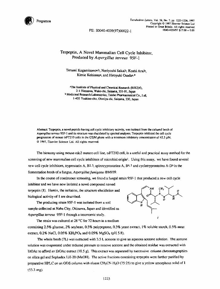
Detailed analyses of tH-IH COSY, HMQC and NOESY spectra of 1, coupled with the structural infor -
mation from the UV and IR spectra, led us to postulate the presence of the partial structures A-E in l(Fig.l).
The cis-relation of As olefin, in the partial structure B, for example, was established by the J8,9 value ( 7 Hz )
and the NOE observed between 8-H and 9-H in the NOESY spectrum.
HMBC experiment on 1 allowed the attribution of all quaternary carbons and supported these partial
structures. For instance, C-2, C-3, C-3a and C-7a in the partial structure A were assigned on the basis of the
HMBC correlations (5-H, 7-H/C-3a; 4-H, 6-H/C-7a; 1-H/C-2,C-3; 4-H/C-3). The assignments of the
quaternary sp 2 carbons and three amide carbonyls in the partial structures C, D and E were also performed
according to the long-range couplings shown by solid line arrows on those partial structures in Fig. 1.
Accordingly, partial structures D and E were identified as N- methyl valine and N-acetyi valine residues,
respectively. . / " - ~ ~ gL H '~" "~" ' ~ ~ "
.L %L\3a\ ~ 29
, , ,: ...½, H, /
A B
23 24 0 ¢f'~H
CH V I g .
. ,
6' I1"~"~ ,4:,_ h k , GHS '~19 ~1"13 ~ 2o 21
D E
Fig.l Partial structures for !
C
III-IH COSY
ItMBC ~- NO F.S Y
1225
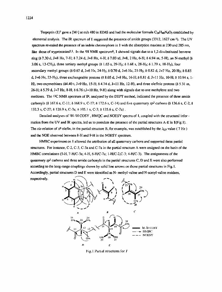
A further extensive inspection of the HMBC spectrum of I revealed the connections of the partial
structures. Namely, the correlation between 25-1-I2 and C-2 indicated that the isoprenyl moiety (partial structure
C) attached to C-2 in the partial structure A. Similarly, the partial structure B was connected to partial structure A
at the C-3 position according to the cross peaks between H-8 and C-2, C-3a and between H-9 and C-3 in the
HMBC spectrum. Furthermore, the connectivities of the amino acid moieties, partial structures D and E, were
shown by the long-range correlations; 9-H, 10-H~ C-11, 13-CH3 ~ C-14 as shown in Fig. 2. This was also
supported by the NOEs observed between 10-H and 12-H and between 13-CH3 and 15-H, respectively, in the
NOESY spectrum. Thus the planar structure of I was deduced.
5
6
L~3~ ~ 20 21
, I '° " ,o d II I ~ 9xXa~ N 12 I 18
~ l ~//-i~ ( :H2 122 O ~ 7 a ~N za i 23j,"',,...
7 H ~ ~' ~ " "24 26 ~ 29 ~ m - - NOESY
= HMBC
28
Fig.2 Planar structure of I
Recently, new diketopiperadines named tryprostatins2, 3 have been isolated from Aspergillusfumigatus as
novel cell cycle inhibitors, but no peptide similar to I has been reported. The structure of I consists of an
isoprenyl group, an indole moiety which was derived from a tryptophan by decarboxylation and further
dehydrogenation at the C-8 and C-9 positions, and two unusual amino acids, N-methyl valine and N-acetyl
valine. The compounds with an indole and amino acid moieties connecting straightly, such as in hemiasteflins 9
or chondriamidesl0, are rare among natural products. It is of great biogenetic and biological interest that 1, a
novel peptide, has been isolated for the first time as a new G2/M inhibitor of the mammalian cell cycle. In the
randomly cultured assayl, terpeptin inhibited the cell cycle progression of mouse tsFT210 cells in the G2/M
phase with a minimum inhibitory concentration of 62.5 ~tM. Detailed studies on the biological activity and
stereochemistry of lwill be reported elsewhere.
Acknowledgment:
We thank Dr. C. B. Cui and Dr. T. Nakata for helpful suggestion for structure elucidation. We also thank
Ms. R. Onose for bioassay and Mrs. T. Chijimatsu for NMR measurements.
1226
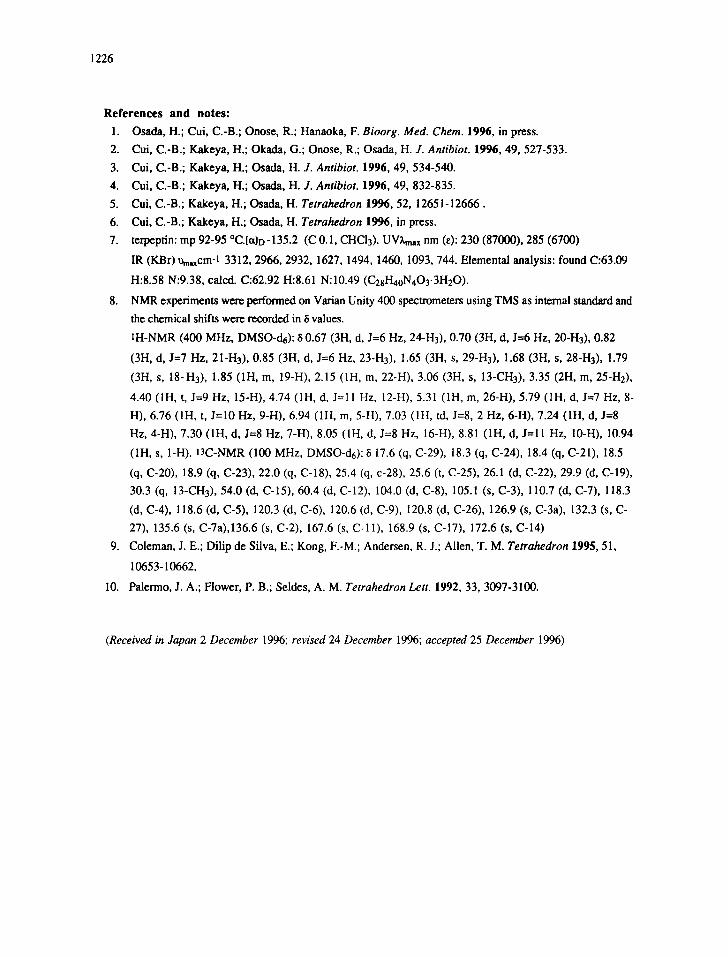
References and notes:
1. Osada, H.; Cui, C.-B.; Onose, R.; Hanaoka, F. Bioorg. Med. Chem. 1996, in press.
2. Cui, C.-B.; Kakeya, H.; Okada, G.; Onose, R.; Osada, H. J. Antibiot. 1996, 49, 527-533.
3. Cui, C.-B.; Kakeya, H.; Osada, H. J. Antibiot. 1996, 49, 534-540.
4. Cui, C.-B.; Kakeya, H.; Osada, H. J. Antibiot. 1996, 49, 832-835.
5. Cui, C.-B.; Kakeya, H.; Osada, H. Tetrahedron 1996, 52, 12651-12666.
6. Cui, C.-B.; Kakeya, H.; Osada, H. Tetrahedron 1996, in press.
7. terpeptin: mp 92-95 °C.[ct]D-135.2 (C 0.1, CHCI3). UVkmax nm (~): 230 (87000), 285 (6700)
IR (KBr)~xcm-I 3312, 2966, 2932, 1627, 1494, 1460, 1093, 744. Elemental analysis: found C:63.09
H:8.58 N:9.38, calcd. C:62.92 H:8.61 N:10.49 (C28Hn0N4Oy3H20).
8. NMR experiments were performed on Varian Unity 400 spectrometers using TMS as internal standard and
the chemical shifts were recorded in 8 values.
1H-NMR (400 MHz, DMSO-d6): 8 0.67 (3H, d, J=6 Hz, 24-H3), 0.70 (3H, d, J=6 Hz, 20-H3), 0.82
(3H, d, J=7 Hz, 21-H3), 0.85 (3H, d, J=6 Hz, 23-H3), 1.65 (3H, s, 29-H3), 1.68 (3H, s, 28-H3), 1.79
(3H, s, 18-H3), 1.85 (1H, m, 19-H), 2.15 (1H, m, 22-H), 3.06 (3H, s, 13-CH3), 3.35 (2H, m, 25-H2),
4.40 (1H, t, J=9 Hz, 15-H), 4.74 (1H, d, J=ll Hz, 12-H), 5.31 (1H, m, 26-H), 5.79 (1H, d, J=7 Hz, 8-
H), 6.76 (1H, t, J=10 Hz, 9-H), 6.94 (1H, m, 5-H), 7.03 (1H, td, J=8, 2 Hz, 6-H), 7.24 (1H, d, J--8
Hz, 4-H), 7.30 (1H, d, J=8 Hz, 7-H), 8.05 (1H, d, J=8 Hz, 16-H), 8.81 (1H, d, J=l l Hz, 10-H), 10.94
(1H, s, I-H). 13C-NMR (100 MHz, DMSO-d6): ~ 17.6 (q, C-29), 18.3 (q, C-24), 18.4 (q, C-21), 18.5
(q, C-20), 18.9 (q, C-23), 22.0 (q, C-18), 25.4 (q, c-28), 25.6 (t, C-25), 26.1 (d, C-22), 29.9 (d, C-19),
30.3 (q, 13-CH3), 54.0 (d, C-15), 60.4 (d, C-12), 104.0 (d, C-8), 105.1 (s, C-3), 110.7 (d, C-7), 118.3
(d, C-4), 118.6 (d, C-5), 120.3 (d, C-6), 120.6 (d, C-9), 120.8 (d, C-26), 126.9 (s, C-3a), 132.3 (s, C-
27), 135.6 (s, C-7a),136.6 (s, C-2), 167.6 (s, C-11), 168.9 (s, C-17), 172.6 (s, C-14)
9. Coleman, J. E.; Dilip de Silva, E.; Kong, F.-M.; Andersen, R. J.; Allen, T. M. Tetrahedron 1995, 51,
10653-10662.
I0. Palermo, J. A.; Flower, P. B.; Seldes, A. M. Tetrahedron Lett. 1992, 33, 3097-3100.
(Received in Japan 2 December 1996; revised 24 December 1996; accepted 25 December 1996)


![Index [] · 2011-03-23 · Index 937 Ascochyta rabiei 507 Aspergillus nidulans 755 Aspergillus ochraceus 135, 158 Aspergillus terreus 506, 755 Aspidosperma alkaloids 106 – fragmentation](https://img.pdfslide.us/doc/110x75/5e2b9234e715b60f857d1b3b/index-2011-03-23-index-937-ascochyta-rabiei-507-aspergillus-nidulans-755-aspergillus.jpg)