Embed Size (px)
Citation preview

Terminology
Resource File
Version 2 1July 2012

Version 2 2July 2012
Terminology Resource File
This resource file has been compiled and designed by the Northern Assistant Transfusion Practitioner group which was formed in 2008 and who later identified the need for such a file. This resource file is aimed at Assistant Transfusion Practitioners to help them understand the medical terminology and its relevance which they may encounter in the patient’s medical and nursing notes. The resource file will not include all medical complaints or illnesses but will incorporate those which will need to be considered and appreciated if a blood component was to be administered. The authors have taken great care to ensure that the information contained in this document is accurate and up to date. Authors: Jackie Cawthray Carron Fogg Julia Llewellyn Gillian McAnaney Lorna Panter Marsha Whittam Edited by: Denise Watson Document administrator: Janice Robertson ACKNOWLEDGMENTS We would like to acknowledge the following people for providing their valuable feedback on this first edition: Tony Davies Transfusion Liaison Practitioner Rose Gill Transfusion Practitioner Marie Green Transfusion Practitioner Tina Ivel Transfusion Practitioner Terry Perry Transfusion Specialist Janet Ryan Transfusion Practitioner Dr. Hazel Tinegate Consultant Haematologist Reviewed July 2012 Next review due July 2013

Contents
Page no.
Abbreviation list 6 Abdominal Aortic Aneurysm (AAA) 7 Acidosis 7 Activated Partial Thromboplastin Time (APTT) 7 Acquired Immune Deficiency Syndrome / 7 Human Immunodeficiency Virus (AIDS / HIV) Alcoholic Liver Disease (ALD) 7 Allogeneic blood components 8 Alzheimer’s disease 8 Amniocentesis 8 Anaemia 9 Angina Pectoris 9 Ante partum haemorrhage 9 Antibiotic 10 Antibody 10 Anticoagulants 10 Anti-D Ig (Immunoglobulin) 10 Antigen 10 Antihistamine 10 Apheresis 10 Arteries 10 Aspirin 11 Asthma – see Hypoxia 19 Beriplex – see Warfarin 29 Bleeding 11 Blood 11 Blood Groups 12 Bone marrow 12 Buffy coats 13 Cell Salvage 13 Cerebral haemorrhage 13 Cerebral infarct 13 Chronic Obstructive Pulmonary Disease (COPD) – see Hypoxia 19 Clinical Pathology Accreditation (CPA) 13 Coagulation 13 Consent 13 Crohn’s disease – see Digestive System 15 Crossmatch 13 Cryoprecipitate 13 Cytomegalovirus (CMV) 13 Dabigatran 14 Deep Vein Thrombosis (DVT) 14 Dextrose Solution 14 Dialysis 14 Digestive system 15 Disseminated Intravascular Coagulation (DIC) 15 Electrocardiogram (ECG) – see Angina Pectoris 9 Electronic issue 15 Emphysema – see Hypoxia 19 Erythrocytes – see Red Cells 26 Erythropoietin (EPO) – see Dialysis 14 EU Directive 15 Factor VIIa 16 Factor VIII 16
Version 2 3July 2012

Foetal cells 16 Ferritin 16 Fibrinogen 16 Fresh Frozen Plasma (FFP) 16 Good Manufacturing Practice (GMP) 16 Granulocytes 17 Haematinic 17 Haematocrit 17 Haematopoiesis – see Bone Marrow 12 Haemodynamically stable 17 Haemoglobin 17 Haemoglobinopathies 17 Haemolysis 17 Haemolytic Disease of the Newborn (HDN) 17 Haemorrhage 18 Haemostasis 18 Health Service Circulars (HSC) 18 Hemiplegia 18 Heparin 18 Hodgkin’s and Non Hodgkin’s Lymphoma 18 Human Albumin Solution 18 Hypertension – see Haemodynamically stable 17 Hypotension – see Haemodynamically stable 17 Hypothermia 19 Hypoxia 19 Intra-operative 20 Intra-uterine transfusion (IUT) 21 Intravascular 21 Intravenous 21 Iron 21 Iron Overload 21 Irradiated (Gamma or X-ray) components 21 Jaundice 21 Jehovah’s Witnesses 22 Kleihauer 22 Left Ventricular Failure (LVF) 22 Leucocytes 22 Leucodepletion 22 Leukaemia 22 Lymph nodes and lymphatic system 23 Maximum Surgical Blood Order Schedule (MSBOS) 23 Medicines and Healthcare products Regulatory Agency (MHRA) 23 Myelodysplasia 23 Myeloma 23 Myocardial Infarction (MI) – see Angina Pectoris 9 National Health Service (NHS) 23 National Patient Safety Agency (NPSA) 23 NHS Blood and Transplant (NHSBT) 23 NHS Litigation Authority (NHSLA) 23 Non Steroidal Anti-Inflammatory Drug (NSAID) 23 Observations 23 Obstetrics – see Post Partum Haemorrhage 25 Octaplex – see Warfarin 29 Oxygen saturation 24 Pancytopenia 24 Paracetamol – see Pyrexial 25 Platelets 24 Pneumonia – see Hypoxia 19 Pneumothorax – see Hypoxia 19 Post-operative 24
Version 2 4July 2012

Post Partum Haemorrhage (PPH) 25 Pre-operative 25 Prophylaxis 25 Prothrombin Complex Concentrate (PCC) - see Warfarin 29 Prothrombin Time (PT) 25 Pulmonary Embolus (PE) – see Hypoxia 19 Pulmonary oedema – see Left Ventricular Failure 22 Pyrexial 25 Red cells 26 Renal function – see Dialysis 14 Rigors – see Pyrexial 26 Rivaroxaban 26 Safer Practice Notice (SPN) 26 Safety of Blood, Tissues and Organs (SaBTO) 26 Sarcoma 26 Sepsis 26 Serious Adverse Blood Reactions and Events (SABRE) 26 Serious Hazards of Transfusion (SHOT) 27 Special requirements 27 Spleen 27 Splenectomy – see Spleen 27 Splenomegaly – see Spleen 27 Stem cells – see Bone Marrow 12 Suffixes/Prefixes 7 Tachycardia 27 Thrombocytes – see Platelets 24 Thrombocytopenia 27 Thrombus 27 Traceability (cold chain) 27 Tranexamic Acid 27 Transfusion Associated Circulatory Overload (TACO) 28 Transfusion Associated Graft versus Host Disease (TA-GvHD) 28 Transfusion Related Acute Lung Injury (TRALI) 28 Transfusion Transmitted Infection (TTI) 28 Variant Creutzfeldt-Jakob Disease (vCJD) 28 Vascular 28 Vasoconstriction 28 Vasodilation 28 Vasovagal syncope (fainting) 29 Veins 29 Venepuncture 29 Venesection 29 Vitamin K 29 Warfarin 29
Version 2 5July 2012

ABBREVIATION LIST AAA Abdominal Aortic Aneurysm APTT Activated Partial Thromboplastin Time AIDS / HIV Acquired Immune Deficiency Syndrome / Human Immunodeficiency Virus ALD Alcoholic Liver Disease (ALD) BCSH British Committee for Standards in Haematology BSQR Blood Safety and Quality Regulations COPD Chronic Obstructive Pulmonary Disease CMV Cytomegalovirus CPA Clinical Pathology Accreditation DVT Deep Vein Thrombosis DIC Disseminated Intravascular Coagulation ECG Electrocardiogram EPO Erythropoietin FFP Fresh Frozen Plasma FDP Fibrinogen Degradation Products g/L Gram per litre GMP Good Manufacturing Practice HAS Human Albumin Solution Hb Haemoglobin HDFN Haemolytic Disease of the Foetus and Newborn HDN Haemolytic Disease of the Newborn HSC Health Service Circulars IDA Iron Deficient Anaemia Ig Immunoglobulin IUT Intra Uterine Transfusion IV Intravenous LVF Left Ventricular Failure MHRA Medicines and Healthcare products Regulatory Agency MI Myocardial Infarction MSBOS Maximum Surgical Blood Order Schedule NHL Non Hodgkins Lymphoma NHS National Health Service NPSA National Patient Safety Agency NHSBT NHS Blood and Transplant NHSLA NHS Litigation Authority NSAID Non Steroidal Anti-Inflammatory Drug PCC Prothrombin Complex Concentrate PT Prothrombin Time PE Pulmonary Embolus PPH Post Partum Haemorrhage PR Per Rectum PV Per Vagina Rh Rh system rAAA Ruptured Abdominal Aortic Aneurysm SPN Safer Practice Notice SaBTO Advisory Committee for the Safety of Blood, Tissues and Organs SABRE Serious Adverse Blood Reactions and Events SHOT Serious Hazards of Transfusion TACO Transfusion Associated Circulatory Overload TA-GvHD Transfusion Associated Graft versus Host Disease TRALI Transfusion Related Acute Lung Injury TTI Transfusion Transmitted Infection vCJD Variant Creutzfeldt-Jakob Disease
Version 2 6July 2012

Prefixes Suffixes
a/an- without -ectomy to remove Ante- before -itis inflammation Anti- against -lysis destruction of cells Dys- difficult -ology study Endo- within -orrhoea flowing Haem/haemo/haemato- denotes blood -oscopy to look Hyper- high -ostomy opening Hypo- low -otomy to cut Intra- within / inside -penia lack of Macro- large - cyte cell Mega- large Micro- small Pan- all Poly- many Post- after Pre- before Abdominal Aortic Aneurysm (AAA)
• An Abdominal Aortic Aneurysm (also known as a triple A) is a dilation (ballooning) of the abdominal aorta due to disease or congenital deficiency. A ruptured Abdominal Aortic Aneurysm (rAAA) causes severe internal bleeding which can be life threatening.
Acidosis
• A condition in which the acidity of body fluids and tissues is abnormally high. Acidosis may occur during haemorrhage and resuscitation and must be corrected as it contributes to the bleeding tendency.
Activated Partial Thromboplastin Time (APTT)
• Is a measure of the part of the clotting process which involves coagulation factors VIII, IX, XII and X, V. The normal range for clotting is between 30-40 seconds. This test is used mainly to monitor the control of heparin therapy.
Acquired Immune Deficiency Syndrome / Human Immunodeficiency Virus (AIDS / HIV)
• A virus which can be transmitted in blood, semen, vaginal secretions and saliva that can cause AIDS. The virus multiplies in the lymph nodes throughout the asymptomatic period (a time when the patient does not show any signs or symptoms of the virus). AIDS appears when the lymph nodes can no longer contain the virus and the immune system collapses. AIDS cripples the immune system by interfering with the activity of helper T cells (cells that produce cytotoxic T cells that destroy cancerous and virus-infected cells). Severe weight loss, night sweats, swollen lymph nodes and frequent infections characterize the syndrome. The risk of getting HIV from a blood transfusion is approximately 1 in 5 million.
Alcoholic Liver Disease (ALD)
• The liver is an accessory organ making up part of the digestive system. It is the largest gland in the body and one of the most important having both metabolic and regulatory roles. The liver breaks down ingested food so that it can be absorbed into the blood stream and inflammation of the liver is known as hepatitis. A complication of ALD is that liver failure interferes with the manufacture of clotting factors and if bleeding occurs it can be difficult to control due to deranged coagulation.
Version 2 7July 2012
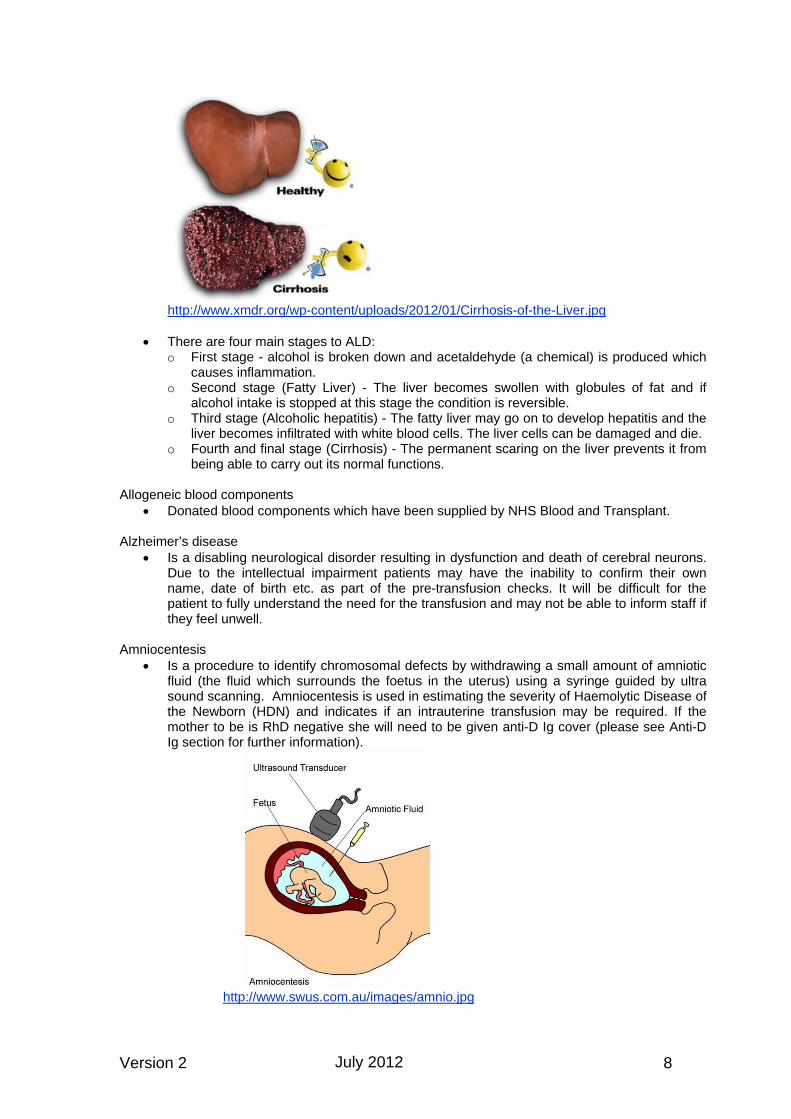
http://www.xmdr.org/wp-content/uploads/2012/01/Cirrhosis-of-the-Liver.jpg
• There are four main stages to ALD:
o First stage - alcohol is broken down and acetaldehyde (a chemical) is produced which causes inflammation.
o Second stage (Fatty Liver) - The liver becomes swollen with globules of fat and if alcohol intake is stopped at this stage the condition is reversible.
o Third stage (Alcoholic hepatitis) - The fatty liver may go on to develop hepatitis and the liver becomes infiltrated with white blood cells. The liver cells can be damaged and die.
o Fourth and final stage (Cirrhosis) - The permanent scaring on the liver prevents it from being able to carry out its normal functions.
Allogeneic blood components
• Donated blood components which have been supplied by NHS Blood and Transplant. Alzheimer’s disease
• Is a disabling neurological disorder resulting in dysfunction and death of cerebral neurons. Due to the intellectual impairment patients may have the inability to confirm their own name, date of birth etc. as part of the pre-transfusion checks. It will be difficult for the patient to fully understand the need for the transfusion and may not be able to inform staff if they feel unwell.
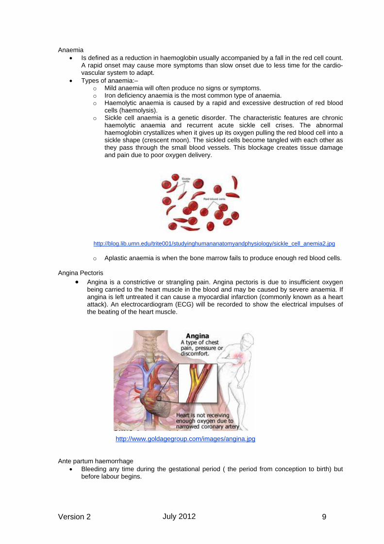
Amniocentesis
• Is a procedure to identify chromosomal defects by withdrawing a small amount of amniotic fluid (the fluid which surrounds the foetus in the uterus) using a syringe guided by ultra sound scanning. Amniocentesis is used in estimating the severity of Haemolytic Disease of the Newborn (HDN) and indicates if an intrauterine transfusion may be required. If the mother to be is RhD negative she will need to be given anti-D Ig cover (please see Anti-D Ig section for further information).
http://www.swus.com.au/images/amnio.jpg
Version 2 8July 2012
Anaemia • Is defined as a reduction in haemoglobin usually accompanied by a fall in the red cell count.
A rapid onset may cause more symptoms than slow onset due to less time for the cardio-vascular system to adapt.
• Types of anaemia:– o Mild anaemia will often produce no signs or symptoms. o Iron deficiency anaemia is the most common type of anaemia. o Haemolytic anaemia is caused by a rapid and excessive destruction of red blood
cells (haemolysis). o Sickle cell anaemia is a genetic disorder. The characteristic features are chronic
haemolytic anaemia and recurrent acute sickle cell crises. The abnormal haemoglobin crystallizes when it gives up its oxygen pulling the red blood cell into a sickle shape (crescent moon). The sickled cells become tangled with each other as they pass through the small blood vessels. This blockage creates tissue damage and pain due to poor oxygen delivery.
http://blog.lib.umn.edu/trite001/studyinghumananatomyandphysiology/sickle_cell_anemia2.jpg o Aplastic anaemia is when the bone marrow fails to produce enough red blood cells.
Angina Pectoris
• Angina is a constrictive or strangling pain. Angina pectoris is due to insufficient oxygen being carried to the heart muscle in the blood and may be caused by severe anaemia. If angina is left untreated it can cause a myocardial infarction (commonly known as a heart attack). An electrocardiogram (ECG) will be recorded to show the electrical impulses of the beating of the heart muscle.
http://www.goldagegroup.com/images/angina.jpg
Ante partum haemorrhage
• Bleeding any time during the gestational period ( the period from conception to birth) but before labour begins.
Version 2 9July 2012

Antibiotic • Antibiotic drugs are used to treat infections which are caused by bacteria and to prevent
infections when the immune system is compromised. Many haematology patients will be on various types of drugs including antibiotics due to the nature of the disease. Patients with neutropenia (a low white cell count) may require intravenous (IV) antibiotics. Intravenous antibiotics are part of the treatment regime for Transfusion Transmitted Infections (TTI) and must not be mixed with blood components. If both antibiotics and blood components are required at the same time then two separate cannulas are required.
Antibody
• Naturally occurring antibodies to A and/or B antigens are found in the plasma of patients whose red cells lack the corresponding antigen. In the ABO blood group system, antibodies form early in life to the antigen which is lacking, so group A individuals have anti-B, group B have anti-A, group O have anti-A and anti-B, whereas group AB people have neither. In other blood group systems, the antibodies to the missing antigens are not formed unless stimulated to do so by either a transfusion of blood containing that antigen, or by pregnancy, when the foetus has the antigen the mother is lacking. This is most commonly seen in the Rh system, where the mother is RhD negative, carrying an RhD positive baby. There are many other antigens on red cells other than A and B, some of which can cause a transfusion reaction.
Anticoagulants
• An anticoagulant drug prevents the blood from clotting and is taken to treat blood clots and overly thick blood. Heparin and warfarin are the most frequently used anticoagulant drugs.
Anti-D Ig (Immunoglobulin)
• Prophylactic anti-D Ig is routinely offered to RhD negative mothers who are pregnant as a single dose at 28 weeks or two separate doses at 28 and 34 weeks (depending on hospital policy). Anti-D is administered intra-muscularly and is necessary as a small amount of the baby’s blood could pass into the mother’s blood stream and the mother may produce antibodies to the baby’s red cells if the baby is RhD positive. Anti-D Ig is also given following sensitising events that may cause feto-maternal bleeding e.g. threatened or spontaneous abortion, termination of pregnancy, closed abdominal injury / trauma, intrauterine death.
Antigen
• Antigens are substances found on the surface of cells which induce antibody production and interact with them in a specific way stimulating an immune response.
Antihistamine
• Antihistamine drugs are used to block the effects of histamine (a chemical that is present in cells and released during an allergic reaction).
Apheresis
• Apheresis is a medical process whereby the donated blood is passed through a cell separator so that one component is collected and the remainder is given back to the donor. This process is commonly used for the collection of platelets.
Arteries
• Arteries are blood vessels that carry blood away from the heart. They are tubes with thick, elastic, muscular walls which are able to withstand high pressure of blood flow. Their structure helps to even out the peaks and troughs of blood pressure caused by the heartbeat to keep the blood flowing at a constant pressure.
Version 2 10July 2012

http://www.mynews.in/News/dailyimage/news/Heart--300--1.png Aspirin
• Aspirin is an analgesic (medication administered to relieve pain) but is also used to prevent blood clots. e.g. after coronary stenting / angioplasty. Aspirin therapy can cause defective platelet function, resulting in an abnormal bleeding time and increases the risk of bleeding which in extreme cases may require a platelet transfusion.
Asthma – see Hypoxia Beriplex – see Warfarin Bleeding:
• Epistaxis is bleeding from the nose. The mucosa lining the nasal cavity consists of thin walled veins and is prone to injury. Nosebleeds can range from being very light to profuse, which can result in a haemorrhage requiring transfusion or cauterization (direct application of a heated instrument) to stop the bleeding. If blood is swallowed this can result in the patient vomiting back up the blood.
• Haematemesis is the vomiting of blood as a result of bleeding from the oesophagus, stomach or duodenum which has the appearance of coffee grounds (sometimes referred to as ‘Coffee Ground Vomiting’).
• Haematoma is a collection of blood outside the blood vessel and within the tissues. • Haematuria is blood in the urine which may / may not be visible. • Haemoptysis is the coughing up of blood or of blood-stained sputum from the bronchi,
larynx, trachea, or lungs. • Per Rectum (PR) is bleeding from stomach, bowel or rectum, also known as blood in
stools. Melaena refers to the passing of black tarry stools. • Per Vagina (PV) is bleeding from the vagina. • Variceal bleeding (Varices) in general refers to distended veins which may leak blood.
Oesophageal varices can occur in Alcoholic Liver Disease. Blood
• Blood delivers oxygen and nutrients to the body’s cells, collects waste, distributes hormones, spreads heat around the body to control temperature and helps fight infection and heal injuries. Blood is made up of red cells, white cells, platelets and plasma.
Version 2 11July 2012

Blood Groups
• These are determined by the presence or absence of genetically determined antigens located on the surface of red blood cells. The four main blood groups are A, B, AB and O. The ABO system is the most important blood group system in transfusion practice. The ABO system is the only one in which the antigens present on the red cells can usually be predicted from knowledge of the antibodies present in the plasma. Apart from the ABO system the next most important clinically significant blood grouping system is the Rh system. This consists of the following antigens; D, C, c, E, e, with the D antigen being the most clinically important antigen of the Rh system. It defines the individuals blood group as being either RhD positive or Rh negative. There are many more antigens that might need to be taken into consideration when blood is required for a patient.
• A group and screen is a blood test to establish the patients ABO, RhD status, and antibody screen to ensure that the patient receives the correct blood components. If the wrong blood group is given the patient may have a transfusion reaction which could result in death.
Bone marrow
There are two types of marrow – red – where red blood cells, white blood cells, and platelets are produced and yellow which consist of mainly fat cells.
• Bone marrow is the flexible tissue found in the hollow interior of bones and in adults it is the large bones that produce the new blood cells.
http://www.daviddarling.info/images/bone_marrow.gif
• Stem cells are the precursor cells to all the blood cells produced by the bone marrow and can be stimulated by drug therapy. Stem cells can be harvested before chemotherapy and later reinfused (transplanted) in order to treat patients with haematology / oncology type diseases. If successful, the transplant will lead to normal blood cell production.
• Haematopoiesis is the formation and development of the blood cells from stem cells.
Version 2 12July 2012

Buffy coats
• The buffy coat is a granulocyte and platelet layer that forms between red cells and plasma when a pack of whole blood is centrifuged. Platelet concentrates can be processed from the buffy coat.
Cell Salvage
• Cell salvage is the collecting of blood that is lost during or shortly after an operation, which may then be given back to you.
• Intra-operative cell salvage is a procedure in which the patient’s own blood that is lost during surgery is collected, washed and processed in a cell salvage machine to enable the red cells to be returned to the patient. There is no risk of the patient acquiring antibodies as they are receiving their own blood back. What must be remembered is that most of the white blood cells, platelets, plasma, and clotting factors are removed during the process and patients receiving significant quantities of salvaged red cells may well need plasma and platelet support. Intra-operative cell salvage is a method used extensively for vascular, orthopaedic, cardiac and trauma surgery and is growing in use in Obstetrics and other specialties. The process can be adapted to be a continuous and closed cycle and thereby accepted by some Jehovah’s Witnesses.
• Post Operative Cell Salvage is the reinfusion of a patient’s own blood which has been collected by wound drainage after an operation (usually used in Orthopaedic surgery following hip and/or knee operations). Unlike intra-operative cell salvage this blood is not filtered or washed.
Cerebral haemorrhage
• Bleeding from cerebral vessels into the tissue of the brain. Cerebral infarct
• Is a localised area of dead tissue in the brain due to an inadequate blood supply.
Chronic Obstructive Pulmonary Disease (COPD) - see Hypoxia Clinical Pathology Accreditation (CPA)
• All National Health Service and private laboratories are regularly inspected against CPA standards, which ensures that agreed standards of practice are adhered to.
Coagulation
• Blood is converted from a liquid to a solid state (blood clot). Consent
• In March 2010 SaBTO initiated a public consultation on patient consent for blood transfusion. The purpose was to consult widely on the options for undertaking consent for blood component transfusion and the potential operational challenges if documented consent were to be mandated. This consultation process resulted in 14 SaBTO recommendations for consent for component blood transfusion and can be accessed via the following link:
http://www.transfusionguidelines.org.uk/Index.aspx?Publication=BBT&Section=22&pageid=7691 Crohn’s disease – see Digestive System Crossmatch
• The process of testing the patient’s plasma against donor red cells. Prior to transfusion, it is essential to determine the patient’s ABO and RhD status, and detect any potentially harmful antibodies so that suitable blood can be selected. There are many other antigens on red cells other than A and B, some of which can cause a transfusion reaction. Blood issued for patients can be fully crossmatched, group specific or flying squad (emergency).
o Fully crossmatched – following testing to determine the ABO group, Rh status and identifying any antibodies the patient’s blood sample is tested against the unit of blood selected, either by manual or automated techniques, to ensure it is fully compatible.This is the safest blood anyone can receive.
Version 2 13July 2012

o Group specific - A patient’s blood sample is tested to determine the correct ABO group and Rh status but has not been tested for antibodies. This would be used in an emergency situation when there is not enough time to wait for fully crossmatched blood.
o Flying squad blood (emergency O neg) – O negative red cells are classified as the universal donor and can be used for all red cell groups. This blood is only to be used in absolute emergency situations when there is no time to wait for either group specific or fully crossmatched blood.
Cryoprecipitate
• Cryoprecipitate is produced after controlled thawing of frozen plasma to precipitate high molecular weight proteins, including factor VIIIc, von Willebrand factor and fibrinogen. It may be useful in disseminated intravascular coagulation (DIC) and may be indicated when the plasma fibrinogen is less than 1g/L. In massive transfusion the level should be maintained above 1.5 g/L
Cytomegalovirus (CMV)
• Cytomegalovirus (CMV) is a type of herpes virus. Transfusion of CMV positive components can cause infection in immunosuppressed patients. In March, 2012 the Advisory Committee on the Safety of Blood, Tissues and Organs (SaBTO) released a position statement on CMV testing of blood components. The statement notes that leucodepletion should be considered to offer sufficient CMV protection. There are notable exceptions where the risk of transmission remains the same, but the consequences of infection could be more severe. The full statement can be seen at http://www.dh.gov.uk/health/2012/03/sabto/
Dabigatran
• Dabigatran is a new type of anticoagulant used to prevent harmful clots occurring in your blood. It prevents the blood from clotting as quickly or as effectively as normal as it interferes with chemicals needed to make clots or clotting factors. Dabigatran is a new type of anticoagulant and works in a slightly different way to warfarin. People who take dabigatran do not need to have regular blood tests. However, they still need to have occasional blood tests to make sure their kidneys are working well. In addition, for most people the dose of dabigatran remains the same throughout treatment.
Deep Vein Thrombosis (DVT)
• A blood clot in one of the deep veins of the body, often in the legs. Patients are anticoagulated with heparin in the first instance and warfarin longer term. If the patient needs an operation while anticoagulated, there is an increased risk of bleeding.
Dextrose Solution
• Dextrose solution is used to increase a patient’s blood volume. It can cause haemolysis so must never be mixed with blood components and it may interfere with haemostasis by affecting platelet function.
Dialysis
• Renal function is an indication of the state of the kidneys. The kidneys are paired organs situated at the upper rear of the abdominal cavity on either side of the spinal column. Their functions include filtering waste products from the blood which is then excreted along with excess water as urine.
• Dialysis is used as a replacement for kidney function in patients with renal failure. There are two types of dialysis; haemodialysis and peritoneal dialysis, both of which work on the same principle of replacing kidney function by removing waste products from the blood. In kidney failure, the kidneys don’t remove salts, waste and water and the body can’t maintain safe levels of sodium, potassium and other minerals. Dialysis also helps control blood pressure, which can rise or fall dangerously, due to an imbalance of salts and minerals.
• Patients with renal failure are often anaemic because the kidneys do not produce enough erythropoietin (EPO), which stimulates the bone marrow to produce red blood cells. Before the availability of therapeutic EPO, patients were given frequent transfusions, with all the associated complications.
Version 2 14July 2012

Digestive system
• The digestive tract consists of a long tube of hollow organs which runs from the mouth to the anus and includes the oesophagus, stomach, small intestine and large intestine. The liver, gall bladder and pancreas produce secretions for digestion that drain into the small intestine. An adult digestive tract is about 30ft long.
http://mybioversa.com/yahoo_site_admin/assets/images/GI-system.16514627_std.jpg
• Bleeding can occur almost anywhere along the length of the digestive tract and can be due to iron deficiency, warfarin therapy or coagulation disorders.
• Bleeding in the upper part can be due to peptic ulcers, oesophageal varices, Mallory-Weiss tears, gastritis, oesophagitis or benign tumours and cancer. In the lower digestive tract, causes of bleeding include diverticulitis, colitis, Crohn’s disease, haemorrhoids, fissures, angiodysplasia, polyps and cancer. If bleeding is severe and/or prolonged, a transfusion may be necessary to improve the haemoglobin level.
Disseminated Intravascular Coagulation (DIC)
• Disseminated intravascular coagulation (DIC) results from the activation of coagulation and fibrinolytic systems, leading to the consumption of platelets, coagulation proteins and fibrinogen. It may be provoked by tissue damage due to trauma or by underperfusion, hypothermia, sepsis or obstetric complications. There may be severe microvascular bleeding with or without thrombotic complications. Diagnosis is by clinical signs of unexpected bleeding or thrombosis, low fibrinogen or platelets, prolonged prothrombin time (PT) and raised fibrinogen degradation products (FDP) or D-dimers (indicative of reduced clotting function). Treatment includes transfusion of red cells, fresh frozen plasma, cryoprecipitate, platelets and, occasionally, factor VIIa.
Electrocardiogram (ECG) – see Angina Pectoris
Electronic issue
• Electronic issue involves the selection and issuing of units without direct serological testing between the patient and the donor red cells. Electronic issue is not recommended for neonates as blood issued has to be compatible with antibodies from the mother that may be present in the baby’s blood. Hospitals using electronic issue must comply with the British Committee for Standards in Haematology (BCSH) national guidelines “The specification and use of information technology systems in blood transfusion practice”.
Emphysema – see Hypoxia Erythrocytes – see Red Cells Erythropoietin (EPO) – see Dialysis
Version 2 15July 2012

EU Directive • Two EU Directives - 2002/98/EC and 2004/33/EC - have been transposed into UK law
through the Blood Safety and Quality Regulations (BSQR) 2005 (Statutory Instrument 2005/50 and Statutory Instrument 2005/1098). These regulations set standards for quality and safety in the collection, testing, processing, storage and distribution of human blood components and came into force on 8 February 2005. Compliance is compulsory and will be monitored by the MHRA.
Factor VIIa
• Recombinant factor VIIa was developed to assist in managing haemophilia patients that have antibodies to coagulation factors VIII or IX and to control massive haemorrhage (surgical, traumatic or obstetric bleeding).
• The criteria for use of recombinant factor Vlla include: o severe haemorrhage where there is a reasonable prospect of long-term recovery
e.g. multiple trauma o focal bleeding points have been dealt with o effective replacement of coagulation factors and platelets has been shown by
laboratory tests (fibrinogen / platelets). Other adverse factors such as heparin, hypothermia, acidosis, hypocalcaemia, should have been excluded or corrected.
Factor VIII
• Is a blood protein and a deficiency of factor VIll causes the inherited bleeding disorder known as haemophilia. To reduce the potential risk of infection resulting from repeated administration of fractionated plasma derivatives patients receive recombinant factor VIII. In severe haemophilia A, treatment with factor VIII must be provided as soon as possible after bleeding has occurred.
Foetal cells
• A foetus receives its blood supply from the mother via the placenta. As most of the oxygen is used up by the mother, gas exchange in the placenta is much less efficient than in the lungs. Foetal haemoglobin (Hb) needs to bind with oxygen at a higher afinity than the maternal blood if the foetus is to receive enough oxygen. At birth approx 50-95% of the infants Hb is foetal Hb, and is normally between 17 to 18g/dL. After 6 months of age this declines, adult Hb becomes dominant and in most adults no foetal cells should be detected.
Ferritin
• Is the principal tissue storage form of iron and levels of ferritin are linked to iron status. A raised serum ferritin indicates iron overload whilst a low serum ferritin would indicate iron deficiency anaemia.
Fibrinogen
• Fibrinogen is a protein that is present in the blood and converted into fibrin during the blood clotting process. It is produced by the liver and can be raised in inflammatory reactions, pregnancy and stress.
Fresh Frozen Plasma (FFP)
• Plasma is the fluid component of blood, separated from a donation of whole blood, frozen and then stored for up to two years as FFP. Once thawed FFP must be transfused within 24 hours if stored according to cold chain requirements.
• FFP can be used in cases of massive transfusion to replace coagulation factors. Non–UK pathogen-reduced FFP is recommended for the treatment of Thrombotic Thrombocytopenic Purpura and FFP is not to be used to reverse the effects of warfarin.
• Children born after January 1996 should only receive Methylene Blue treated FFP sourced from outside the UK to reduce the risk of transmission of variant Creutzfeldt-Jakob Disease (vCJD).
Good Manufacturing Practice (GMP)
• The aim of GMP is to assure the quality of the product for the safety, well being and protection of the patient. Compliance with GMP is a statutory requirement for the National
Version 2 16July 2012

Blood Service and Hospital Transfusion Laboratories and inspections are performed in the UK by the Medicines and Healthcare products Regulatory Agency (MHRA).
Granulocytes
• These are mainly given to neutropenic cancer patients developing bacterial sepsis unresponsive to conventional antibiotic therapy for at least 24-48 hours. They are collected from apheresis donors who may / may not have been stimulated by steroids and/or growth factors. Granulocyte preparations can only be stored for 24 hours at 20-24°C. They need to be cross-matched with the recipient's plasma because of the large number of red cells they contain and need to be irradiated because of the large number of lymphocytes present.
Haematinic
• A drug that increases the amount of haemoglobin in the blood e.g. oral iron supplements. Haematocrit
• This represents the percentage of whole blood that is made up of cells and the remainder by plasma.
Haematopoiesis – see Bone Marrow Haemodynamically stable
• The heart is part of the cardiovascular system and is approximately the size of a clenched fist. It is divided into two sides, the right side pumps blood to the lungs the left side pumps blood around the body.
• Haemodynamically stable means that blood flow or the circulation are not changing and the cardiac output and blood pressure are steady. Haemodynamic instability is a state requiring drug or mechanical support to maintain a normal blood pressure or adequate cardiac output. Hypertension is persistently raised blood pressure and may cause headaches and visual disturbances. It is particularly common in men and middle-aged to elderly patients. Hypotension is low blood pressure and it may be caused by shock in cases such as disease or serious injury.
Haemoglobin • Haemoglobin (Hb) is a protein molecule and the pigment which gives red blood cells their
colour. Haemoglobin is needed to carry oxygen around the body to supply the tissues. Haemoglobinopathies
• A genetic defect that results in an abnormality in the production of haemoglobin. Haemolysis
• Haemolysis is the breakdown of red cells faster than the marrow can compensate. Haemolysis can be inherited e.g. spherocytosis, acquired e.g. auto immune disease or as a result of a transfusion reaction.
Version 2 17July 2012

Haemolytic Disease of the Newborn (HDN) (or HDFN - Haemolytic Disease of the Foetus and Newborn)
• Haemolytic Disease of the Newborn (HDN) is the result of red cell alloimmunisation (the body mounts an immunological response against red cell antigens from another individual) IgG antibodies formed as a result of alloimmunisation pass from the maternal circulation across the placenta and into the circulation of the foetus. The level of antibodies rise and start to destroy the baby’s blood cells, leading to HDN (see diagram below). The anti-D antibody is responsible for most cases of severe HDN although anti-c, anti-E, anti-K and other antibodies are occasionally found. Although antibodies against the ABO blood group system are the most frequent cause of HDN this is usually mild. Prophylactic anti-D Ig is offered to RhD negative mothers during pregnancy and after birth if the baby is RhD positive to prevent HDN in future pregnancies.
http://legacy.owensboro.kctcs.edu/gcaplan/anat2/notes/14_23.jpg Haemorrhage
• The loss of blood from the circulatory system which can occur internally, where blood leaks from blood vessels inside the body or externally, either through a natural opening such as the nose, or through a break in the skin.
Haemostasis
• The stopping of bleeding through the complex clotting process or administration of clotting products.
Health Service Circulars (HSC)
• A formal communication from the Department of Health, directed at NHS executives, which contain a requirement for action. E.g. HSC 2007/001 Better Blood Transfusion, Safe and Appropriate Use of Blood.
Hemiplegia
• A condition in which one side of the body is paralysed which may be congenital or from illness or stroke. The affected limbs should be avoided for blood samples, infusions and transfusions.
Heparin
• Heparin is an anticoagulant initially used to treat patients with deep vein thrombosis (DVT) or pulmonary embolism (PE). It is then followed by warfarin therapy to reduce the risk of recurrence. Heparin does not cross the placenta so is the favoured drug used if anticoagulation is required during pregnancy.
Hodgkin’s and Non Hodgkin’s Lymphoma (NHL)
• Both Hodgkin's and non-Hodgkin's lymphoma are cancers that originate in a type of white blood cell known as a lymphocyte, an important component of the body's immune system. Both of these malignancies may cause similar symptoms, but the conditions themselves are different. The distinction between Hodgkin's and non-Hodgkin's lymphoma is made upon examination of the cancerous material. The type of abnormal cells identified in the sample determines how a lymphoma is classified. Non-Hodgkin's lymphoma is much more common than Hodgkin's lymphoma. Patients with Hodgkin’s lymphoma need to have irradiated blood components at all stages of the disease.
Version 2 18July 2012

Human Albumin Solution (HAS)
• Human albumin solution is a licensed pharmaceutical product that is derived from human plasma. Human albumin solution is indicated for restoring and maintaining the circulating blood volume. It is available as 4-5% and 20% solutions but care must be taken with the 20% solution as there is a high risk of fluid overload with its use.
Hypertension - see Haemodynamically stable Hypotension - see Haemodynamically stable Hypothermia
• A condition in which the body’s core temperature drops below that required for normal metabolism and body functions which is defined as 35°C (95°F). Hypothermia can precipitate DIC so it is important to keep the patient warm and use a blood warmer if a fast rate of transfusion is required e.g. major haemorrhage.
Hypoxia
• The paired lungs along with the trachea and bronchi form the lower respiratory tract. The left lung consists of two lobes the right lung has three. The primary bronchi enter the lung getting smaller and smaller eventually ending with the alveoli (air sacs). The walls of the alveoli are largely thin single cells. Their function is to facilitate gas exchange, oxygen passing from the air into the capillary blood and carbon dioxide leaving the blood to be exhaled. Hyperventilating is breathing faster and / or deeper than is necessary (over breathing).
http://www.childrenscolorado.org/imgs/KidsHealth/image/ial/images/206/206_image.gif
Hypoxia is a condition in which the body is deprived of adequate oxygen supply and can be referred to as generalised hypoxia (body as a whole) or tissue hypoxia (a region of the body). Hypoxia may be a sign of a transfusion reaction but can also be due to:
• Asthma which is an allergic reaction characterised by smooth muscle spasms in the bronchi resulting in wheezing and difficulty breathing.
• Chronic Obstructive Pulmonary Disease (COPD) is a disease which leads to damaged airways in the lungs, causing them to become narrower and making it harder for air to get in and out of the lung.
• Emphysema is a respiratory disease caused by toxic vapours, like cigarette smoke. • Pneumonia is inflammation of the lungs due to a viral or bacterial infection. The symptoms
are usually fever, chills, shortness of breath and a cough that produces yellow-green sputum and occasionally blood.
Version 2 19July 2012

http://www.nhlbi.nih.gov/health/health-topics/topics/pnu/causes.html
• Pneumothorax is a collection of air in the chest cavity causing chest pain or shortness of breath. If the air continues to leak it may produce a tension pneumothorax which can be life threatening. A small pneumothorax may disappear without treatment, if not the air must be removed using a tube with a one-way valve.
http://images.medicinenet.com/images/illustrations/pneumothorax.jpg
• A pulmonary embolus is a foreign or abnormal particle circulating in the blood, such as a bubble of air, a blood clot, or cholesterol plaque. A pulmonary embolus occurs when the pulmonary artery is blocked by an embolus and it can be life threatening.
Intra-operative
• Intra-operative is defined as the physical and psychological care given to the patient in the anaesthetic room and theatre until transfer to the recovery room. Patients may need transfusions as part of their care given pre, during or post operation.
Version 2 20July 2012

Intra-uterine Transfusion (IUT) • A transfusion of red cells to a baby whilst in utero. IUT is used to treat severe foetal
anaemia due to HDN, sickle cell disease and parvovirus infection. It is given through the umbilical vessels or the intrahepatic portion of the umbilical vein.
Intravascular
• Intravascular means within the blood vessels. Patients are cannulated and a giving set is attached to the cannula. This allows for an infusion to be given directly into a vein e.g. a blood transfusion.
Intravenous
• Intravenous (IV) refers to anything which is administered through a vein, such as an intravenous injection, infusion or a transfusion.
Iron
• Iron is part of the haemoglobin molecule in red blood cells and normally the body recycles the iron freed from the breakdown of red blood cells. When iron stores are low, haemoglobin cannot be produced in sufficient quantities and the patient becomes anaemic - iron deficient anaemia (IDA). If a patient is bleeding they lose iron as part of the overall blood loss.
Iron overload
• With each unit of blood transfused the patient receives approximately 250mg of iron but the body is unable to excrete excess iron which can lead to additional health problems for transfusion dependant patients. The body will start to deposit the excess iron around the heart, liver, and endocrine organs.which can lead to cardiac and liver problems and diabetes. To prevent this happening transfusion dependent patients will usually be given iron chelation therapy, so that the chelated iron can be excreted from the body either in the urine or faeces.
Irradiated (Gamma or X-ray) components
• Cellular blood components (red cells and platelets) are treated with either gamma or x-ray irradiation to prevent any live white blood cells engrafting and proliferating in the patient’s bone marrow after transfusion.
• Irradiated components are given to patients with a suppressed / immunocompromised immune system to prevent transfusion-associated graft versus-host-disease (TA-GvHD).
• Red cells may be irradiated at any time up to 14 days after collection, and therefore stored for a further 14 days from irradiation. If red cells are requested for a patient at risk from hyperkalaemia (high levels of potassium) e.g. intrauterine or exchange transfusion, it is recommended that red cells be transfused within 24 hours of irradiation (BCSH, 2010).
Jaundice
• One cause of jaundice is the result of red blood cell breakdown in cases of haemolytic anaemia, which may be due to an inherited disorder, e.g. sickle cell disease, or acquired as a result of a transfusion reaction or haemolytic disease of the newborn. The patient may need a transfusion whilst haemolysis is occurring.
Version 2 21July 2012

Jehovah’s Witnesses • Jehovah’s Witnesses are not opposed to surgery or medicine, but for deeply-held reasons
of religious faith decline allogeneic blood transfusion, therefore request non-blood medical and surgical management. Jehovah’s Witnesses do not accept the use of blood components, however, when it comes to blood ‘fractions’ e.g. albumin, anti-D Ig, this is a personal decision for each patient.
Kleihauer
• A Kleihauer test is performed on RhD negative women who are carrying or deliver a RhD positive baby, or who suffer a potentially sensitising event while pregnant, to determine how much foetal blood has crossed the placenta into the mother’s circulation. The result is used to calculate how much more than the standard dose of anti-D immunoglobulin is required to clear the foetal cells and prevent the mother producing her own anti-D, which could lead to HDN in any future pregnancies where the baby is RhD positive.
Left Ventricular Failure (LVF)
• Left ventricular failure is heart failure in which the left ventricle fails to contract forcefully enough to maintain a normal cardiac output and peripheral perfusion. Pulmonary oedema is an accumulation of fluid in the lungs which is usually due to left sided heart failure. It can also be due to a chest infection or the inhalation of irritant gases or any of the causes of generalised oedema. Transfusing too much or too quickly can cause left ventricular failure so medical staff need to consider the use of a diuretic e.g. furosemide.
http://www.beliefnet.com/healthandhealing/images/si1619.jpg Leucocytes
• Leucocytes (also known as white blood cells), are nucleated cells of the blood. They can be divided into neutrophils, eosinophils, basophils, lymphocytes and monocytes, all of which play a role in defending the patient against infection. Neutrophils, monocytes, eosinophils and basophils are phagocytes; they engulf and destroy foreign material and damaged cells, whereas lymphocytes produce antibodies.
Leucodepletion
• Leucodepletion is the removal of white blood cells (leucocytes) by filtration and in 1999 it was introduced in the UK for all blood components as a precautionary measure against variant Creutzfeldt-Jakob Disease (vCJD). Another benefit of leucodepletion is that it reduces the incidence of febrile reactions due to antibodies against white cells
Leukaemia
• Is a cancer of the white blood cells. The bone marrow starts to produce large numbers of abnormal white cells, disrupting production of the normal blood cells and affecting the vital functions that these blood cells carry out.
Version 2 22July 2012
Lymph nodes and lymphatic system • A lymph node is a small circular ball-shaped organ of the immune system distributed widely
throughout the body and linked by lymphatic vessels. The lymphatic system is an extensive drainage system that returns water and proteins from various tissues back in to the bloodstream. If the lymph nodes (in the axilla) have been affected due to cancer or treatment you must use the unaffected arm to take a blood sample or administer a blood transfusion.
Maximum Surgical Blood Order Schedule (MSBOS)
• An agreed tariff of the quantity of red cells ordered by type of surgical procedure, set by an individual trust.
Medicines and Healthcare products Regulatory Agency (MHRA)
• An executive agency of the Department of Health. They are the UK regulatory authority who work with and monitor the UK blood services, healthcare providers and other relevant organisations to improve blood quality and safety.
Myelodysplasia
• A bone marrow disorder where the bone marrow does not function normally and produces an insufficient number of normal blood cells.
Myeloma
• A type of cancer that develops from cells in the bone marrow called plasma cells. Myocardial Infarction (MI) - see Angina Pectoris National Health Service (NHS)
• The National Health Service (NHS) began on July 5th 1948 with the aim of providing good healthcare to all, regardless of wealth. For the first time hospitals, doctors, nurses, pharmacists, opticians and dentist were brought under one organisation.
National Patient Safety Agency (NPSA)
• The aim of the NPSA was to lead and contribute to improved, safe patient care by informing, supporting and influencing healthcare organisations and individuals working in the health sector. On 1 June, 2012 the key functions and expertise for patient safety transferred to the NHS Commissioning Board Authority (NHS CBA) from the NPSA http://www.commissioningboard.nhs.uk/2012/05/31/npsa-transfer/
NHS Blood and Transplant (NHSBT)
• NHS Blood and Transplant is a Special Health Authority, dedicated to saving and improving lives through a wide range of services to the NHS. One main service is the provision of blood components to NHS trusts and independent hospitals in England and North Wales.
NHS Litigation Authority (NHSLA)
• The aim of the NHSLA is to promote the highest possible standards of patient care to minimise the suffering resulting from any adverse event.
Non steroidal anti-inflammatory drug (NSAID)
• Non steroidal anti-inflammatory drugs (NSAID) have a lasting analgesic; antipyretic property and an anti-inflammatory affect e.g. Ibuprofen. They are contra-indicated if the patient is sensitive to aspirin or any other NSAID or has a coagulation defect as they can increase the chances of bleeding and gastro-intestinal side effects.
Observations
• Blood Pressure is the pressure exerted by moving blood on the vessel wall. A value above 140/90 mmHg in those below 50 years and 160/95 mmHg in those above 60 years is abnormal.
• Pulse is the wave form of blood passing through an artery as a consequence to cardiac contraction.
Version 2 23July 2012

Version 2 24July 2012
• Respiration rate is normally 12-18 breaths per minute. As the body exchanges gases (oxygen and carbon dioxide) in the lungs, oxygen from the air is breathed in and carbon dioxide is exhaled. When physical exertion takes place, we will not only take more breaths but also a greater volume. The heart will also beat faster to help deliver the increased oxygen. Patients may require additional oxygen therapy to supplement oxygen intake in order to correct hypoxia and relieve breathlessness. The oxygen can be administered through either a mask or nasal cannula.
• Temperature - The normal core temperature of the body is 37˚C (98.6˚F) but the reading will depend on how and where it is taken:
o Orally – thermometer placed under the tongue. o Axillary - thermometer placed under the armpit and the reading is 1˚C
lower than oral temperature. o Rectal – thermometer placed up the rectum is considered more accurate
than both orally and axillary temperature and the reading is 1˚C higher than oral temperature.
o Tympanic - an infrared probed is placed inside the ear. Care must be taken as poor technique can result in inaccurate readings.
In blood transfusion, observations should be recorded before the start of each transfusion, then again 15 minutes after the transfusion begins (to detect an acute transfusion reaction) and lastly within 60 minutes of the transfusion ending (to detect a delayed transfusion reaction).
Obstetrics – see Post partum haemorrhage Octaplex – see Warfarin Oxygen saturation
• Oxygen saturation is the oxygen content divided by oxygen capacity expressed in volume per cent. A probe is put on the patient’s clean finger and a machine records the percentage of oxygen the patient has in their haemoglobin. The best level of oxygen is 100% but as the amount of oxygen in the blood decreases so does the reading. A reading of less that 95% should be a warning sign (unless the patient is known to have low oxygen levels previously). If the reading is less than 95% then inform a registered nurse as soon as possible and if the reading falls below 90% then you must get help immediately.
Pancytopenia
• A reduction in the number of red cells, white cells and platelets which may be due to an over active spleen, leukaemia, aplastic anaemia, transfusion reaction or following chemotherapy.
Paracetamol – see Pyrexial
Platelets
• Platelets (also called thrombocytes) play an essential role in arresting bleeding and are needed for the blood to clot. Most platelets are administered prophylactically (to prevent) bleeding rather than to stop bleeding. Platelets may be given if a patient has a low platelet count and/or is bleeding. Unlike other blood components, platelets need to be stored at 22˚C and must be agitated to prevent them clumping. Storage at 22˚C increases the risk of bacterial contamination. Since May 2011, platelet donations have been tested for bacterial contamination which has allowed extension of the shelf life from five to seven days.
Pneumonia – see Hypoxia Pneumothorax – see Hypoxia Post-operative
• The time after an operation. Patient observations are carried out and possibly further blood samples are taken e.g. full blood count to check for anaemia due to surgical blood loss.

Post Partum Haemorrhage (PPH) • Obstetrics is the branch of medicine dealing with childbirth, puerperium and management of
childbirth. A PPH is excessive bleeding following childbirth and is described as either primary or secondary.
o A primary PPH is a loss of blood estimated to be more than 500 mls, from the genital tract, within 24 hours of delivery (the most common obstetric haemorrhage). A minor PPH is an estimated blood loss of up to 1000 mls and a major PPH is an estimated blood loss over 1000 mls.
o A secondary PPH is defined as abnormal bleeding from the genital tract, from 24 hours after delivery until 6 weeks postpartum.
Pre-operative
• The time before an operation takes place. To ensure the patient is fit for surgery all necessary samples are taken and results obtained e.g. full blood count, blood group, crossmatch.
Prophylaxis
• Prophylaxis is a treatment that focuses on prevention of disease e.g. anti-D Ig offered to RhD negative mothers during pregnancy and again after birth if the baby is RhD positive.
Prothrombin Complex Concentrate – see Warfarin Prothrombin Time (PT)
• Prothrombin time (PT) is a coagulation test that measures the part of the clotting process involving factors VII, X, V, prothrombin and fibrinogen and detects any deficiencies. It is used to monitor warfarin therapy
http://ctarz.wordpress.com/2009/02/25/figurative-representations/ Pulmonary Embolus (PE) – see Hypoxia Pulmonary oedema – see Left Ventricular Failure Pyrexial
• Pyrexia means feverish and is one way the body shows something is wrong e.g. a sign of disease. A normal temperature is around 37˚C and it is important to record the patient’s temperature before, during and after transfusion. Recording observations would allow a transfusion reaction to be detected and treated appropriately. Paracetamol is a drug that has analgesic (to relieve pain) and antipyretic properties (reduces body temperature). It is commonly used to treat a mild temperature rise associated with a transfusion. A rigor is an episode of shaking or exaggerated shivering which can occur with a high fever.
Version 2 25July 2012

Red cells • Red blood cells (also known as erythrocytes) are the most common type of blood cell and
are responsible for delivering oxygen to the body tissues via the blood flow through the circulatory system. These cells' cytoplasm is rich in haemoglobin, and is responsible for the blood's red colour.
http://www.sciencequiz.net/jcscience/jcbiology/circulatorysystem/red_blood_cells.jpg
Renal function – see dialysis
Rigors – see Pyrexial Rivaroxaban
• Rivaroxaban is used to prevent harmful clots occurring in your blood. Rivaroxaban works by preventing your blood from clotting as quickly or as effectively as normal. It does this by blocking a substance in your blood which is involved in the development of blood clots, called factor Xa.
Safer Practice Notice (SPN)
• An advice or notification from the NPSA on patient safety issues that are important and have a specific timeline for implementation e.g. SPN 14, Right patient, right blood.
Safety of Blood, Tissues and Organs (SaBTO)
• The advisory committee on the safety of blood, tissues and organs. They are responsible for advising the UK government ministers and health department on the most appropriate ways to ensure the safety of blood, cells, tissues and organs for transfusion / transplantation.
Sarcoma
• A cancer of the connective tissue (bone, cartilage, fat). Soft tissue sarcoma is used to describe tumours of soft tissue, which includes elements that are in connective tissue, but not derived from it (i.e. muscles and blood vessels).
Sepsis
• Sepsis is a condition in which the body is fighting a severe infection that has spread via the bloodstream and can lead to DIC.
Serious Adverse Blood Reactions and Events (SABRE)
• The UK Blood Safety and Quality Regulations (2005) require that serious adverse events and serious adverse reactions related to blood and blood components are reported to the MHRA, the UK Competent Authority for blood safety. The web-based reporting system is known as SABRE – Serious Adverse Blood Reactions and Events which allows reporters to electronically submit reports directly to the MHRA and SHOT.
Serious Hazards of Transfusion (SHOT)
• SHOT (Serious Hazards of Transfusion) is the United Kingdom’s independent, professionally led, confidential enquiry into the serious hazards of transfusion. SHOT
Version 2 26July 2012

collects information on transfusion reactions and adverse events from all healthcare organisations in the United Kingdom that are involved in the process of blood transfusion.
Special requirements
• Some patients may require a blood component with special requirements to prevent complications associated with transfusion e.g. irradiated blood components are needed to prevent the development of transfusion-associated graft-versus-host disease (TA-GvHD).
Spleen
• The spleen is responsible for cleaning your blood, destroying old red blood cells and fighting infection. A splenectomy is the surgical removal of the spleen and splenomegaly is an abnormally large spleen.
Splenectomy – see Spleen Splenomegaly – see Spleen
Stem cells – see Bone Marrow Tachycardia
• Tachycardia is an increased heart rate and can be a symptom of a transfusion reaction. Thrombocytes – see Platelets Thrombocytopenia
• Thrombocytopenia means a low platelet count. The normal range is 150-350x109/L, but bleeding seldom occurs with platelet counts less than 50x109/L and is usually minor with counts exceeding 10x109/L. Spontaneous bleeding and bleeding requiring medical attention usually indicate that a platelet count is less than10x109/L. However, clinical bleeding often does not correlate with the platelet count because other factors, such as the integrity of the endothelium (single layer of cells that line the blood vessels) and the function of the residual platelets, also affect haemostasis. Thrombocytopenia can be caused by a number of factors including various illnesses, medication and alcoholism.
Thrombus
• A thrombus is a blood clot, the end product of the coagulation cascade. It is desirable in cases of injury, but not in instances of thrombosis. If a thrombus is formed in a blood vessel and becomes detached, it is then known as an embolus.
Traceability and the Cold chain
• The Blood Safety and Quality Regulations state that every blood component must be traced to its final fate; either to the patient who received it or if it was otherwise disposed of. This tracking enables action to be taken if a patient or donor subsequently develops a transfusion transmissible infection or a blood component is implicated in a transfusion reaction. Cold chain records provide evidence that blood components are not left out of controlled storage for longer than the specified length of time before being given to a patient and allows lab staff to decide whether or not an unused unit can safely be returned to stock.
Tranexamic acid
• Tranexamic acid is given to stop or reduce heavy bleeding. When you bleed, your body forms clots to stop the bleeding. Tranexamic acid works by stopping the clots from breaking down and so reduces the bleeding.
Transfusion Associated Circulatory Overload (TACO)
• When too much fluid is transfused or the transfusion is too rapid, acute left ventricular failure (LVF) may occur. Patients may show signs of cardiac failure before transfusion, so need to be transfused slowly (but still within 4 hours of the blood component leaving controlled storage) and may require a diuretic, e.g. furosemide. The transfusion should be
Version 2 27July 2012

stopped and the patient treated with diuretics and oxygen. Volume overload due to fluid shift is a special risk with 20% albumin solutions.
Transfusion-Associated Graft-versus-Host Disease (TA-GvHD)
• TA-GvHD is a rare, but serious complication which is invariably fatal. It is caused by the engraftment and proliferation of transfused donor white cells, which damage the patient’s lymphoid tissue and bone marrow. The symptoms of fever, rash, liver dysfunction, diarrhoea and pancytopaenia, appear 1-6 weeks post transfusion. Particularly at risk are immunocompromised patients, e.g. due to certain malignancies and treatments, babies having intra-uterine or exchange transfusion and those receiving bone marrow transplants. The risk of TA-GvHD can be greatly reduced by giving these patients irradiated blood components.
Transfusion Related Acute Lung Injury (TRALI)
• TRALI is associated with an acute, usually severe, transfusion reaction which involves difficult or laboured breathing (dyspnoea) with oxygen deficiency (hypoxia) and may involve chills, fever and a non-productive cough. The condition is classically characterised by the production of nodules on the lung (bilateral pulmonary infiltrates). These occur within 6 hours of transfusion, with no apparent cause other than the transfusion. TRALI causes severe, occasionally fatal, transfusion reactions, associated with white blood cell incompatibility, due to antibodies in the donor plasma, often white cell antibodies. NHSBT should be notified as the donor may need to be permanently removed from the donor panel.
Transfusion Transmitted Infection (TTI)
• All blood donations are tested by NHSBT for hepatitis B, hepatitis C, HIV, syphilis, human T-cell lymphotropic virus and cytomegalovirus. Malaria is a possible TTI, so people are not allowed to donate for a period of time after visiting endemic malarial areas. Other infections may also be transmitted by transfusion, but those mentioned above are the known major risks. Platelets pose a higher risk of infection than either red cells (stored at 4°C) or FFP (frozen) because they are stored at 22˚C and so are a perfect breeding ground for bacteria.
Variant Creutzfeldt-Jakob Disease (vCJD)
• First identified in 1996, vCJD is thought to be caused by a prion, and is identical to the Bovine Spongiform Encephalopathy found in animals. It causes behaviour disorders, depression and anxiety, followed by dementia and death within 6-24 months of transmission. It can be transmitted by transfusion, but leucodepletion, introduced in 1999, significantly reduces the risk. To date there is no diagnostic test available. Anyone born after 1st Jan 1996 should be given FFP imported from countries with a low risk of vCJD.
Vascular
• Vascular relates to or is supplied with blood vessels. Vasoconstriction
• Contracting of the muscular wall of the blood vessels causing narrowing of the blood vessels.
Vasodilation
• Relaxation of the muscular wall of the blood vessels resulting in a widening of the actual internal diameter.
Vasovagal syncope (fainting)
• The vagus nerve (nerve used to help control automatic functions such as heartbeat) reduces the rate at which the heart beats and lowers the blood pressure causing the patient to ‘faint’. It can be caused by injury or profuse bleeding.
Version 2 28July 2012

www.umm.edu/neurosciences/vagus_nerve.htm Veins
• A blood vessel carrying blood toward the heart. Venepuncture
• Insertion of a needle into a vein to withdraw a blood sample. Venesection
• The removal of a quantity of blood e.g. from a patient who is producing too much as in haemochromatosis (an hereditary disorder in which there is an excessive absorption of iron from the gastro-intestinal tract) and polycythaemia (an increase in the packed cell volume in the blood), or as part of the blood donation process.
Vitamin K
• One of two naturally occurring fat-soluble vitamins that help in the clotting of blood. Vitamin K is given to reduce the effect of warfarin (see below).
Warfarin
• Warfarin is an anticoagulant drug taken to prevent the blood from clotting and to treat blood clots and overly thick blood. Warfarin can be prescribed for either a 3-6 month period or for life depending on the indication for its use. Prothrombin Complex Concentrate (PCC) is recommended for the urgent reversal of warfarin, rather than Fresh Frozen Plasma. Beriplex and Octaplex are the names of PCC used in hospitals.
Version 2 29July 2012

Version 2 30July 2012
References
Bird, M. (2008), Medical Terminology and Clinical Procedures, 3rd Ed. National Services for Health Improvement. British National Formulary (2007), BMJ Publishing Group, London & RPS Publishing, London. British Committee for Standards in Haematology, Blood Transfusion Task Force, (2004), Guidelines for the use of fresh frozen plasma, cryoprecipitate and cryosupernatant, The British Society for Haematology, 126, 11-28. British Committee for Standards in Haematology, Blood Transfusion Task Force, (2003), Guidelines for compatibility procedures in blood transfusion laboratories, Transfusion Medicine, 2004, 14, 59-73 British Committee for Standards in Haematology, Blood Transfusion Task Force, (2010), Guidelines on the use of irradiated blood , British Journal of Haematology, 152, 35-51. Chisholm, M et al (1999), Oxford Handbook of Clinical Haematology. Oxford: Oxford University Press. Dougherty, L. & Lister, S. (2008), The Royal Marsden Hospital Manual of Clinical Nursing Procedures, 7th Ed. West Sussex: Wiley-Blackwell. Eastham, RD, Slade, RR. (1992) Clinical Haematology, 7th Ed. Oxford: Butterworth-Heinemann Ltd. Green, D. & Ludlam, C. (2004) Bleeding Disorders, Health Press. Health Protection Agency http://www.hpa.org.uk Hoffbrand, A, Moss, P & Pettit,J. (2006), Essential Haematology, 5th Ed. Oxford: Blackwell Publishing Ltd. Hoffbrand, A, Moss, P & Pettit,J. (2001), Essential Haematology, 4th Ed. Oxford: Blackwell Publishing Ltd. Hughes-Jones, NC, Wickramasinghe, SN, Hatton, C. (2007), Haematology. 7th Ed. Oxford: Blackwell Publishing Ltd. Jehovah’s Witnesses Our Views on Health Care, (2009) Hospital Information Services for Jehovah’s Witnesses, London Klein H. G & Anstee D.J, (2005) Mollison’s Blood Transfusion in Clinical Medicine, 11th Ed, Blackwell Publishing, Oxford. Learoyd, P. (2003), An Introduction to Blood Group Serology and Transfusion. Leeds National Blood Service. Martin, E.A. (Ed.) (2008), Minidictionary for Nurses, 6th Ed. Oxford University Press. , McCelland, DBL. (Ed.) (2007), Handbook of Transfusion Medicine, 4th Ed. London: TSO. National Patient Safety Agency, http://www.npsa.nhs.uk NHS Litigation Authority, http://www.nhsla.com Parker S, (2007) The Human Body Book, Dorling Kindersley Limited. London Peters, M. (2007), Illustrated Medical Dictionary, 2nd Ed. London: Dorling Kindersley Limited. Smart, T. (2001), Human Body. London: Dorling Kindersley. Silverthorn DU (2006), Human Physiology: An Integrated Approach. Fourth edition. San Francisco: Pearson Benjamin Cummings. Springhouse (2005), Anatomy & Physiology made Incredibly Easy (2 ed). Lippincott Williams & Wilkins. London. Steiner, S. (2003), Quick Medical Terminology, 4th Ed. New Jersey: John Wiley & Sons. http://en.wikipedia.org/ www.baxterhealthcare.co.uk http://hypertextbook.com/facts/LenaWong.shtml http://www.patient.co.uk/