Embed Size (px)
Citation preview
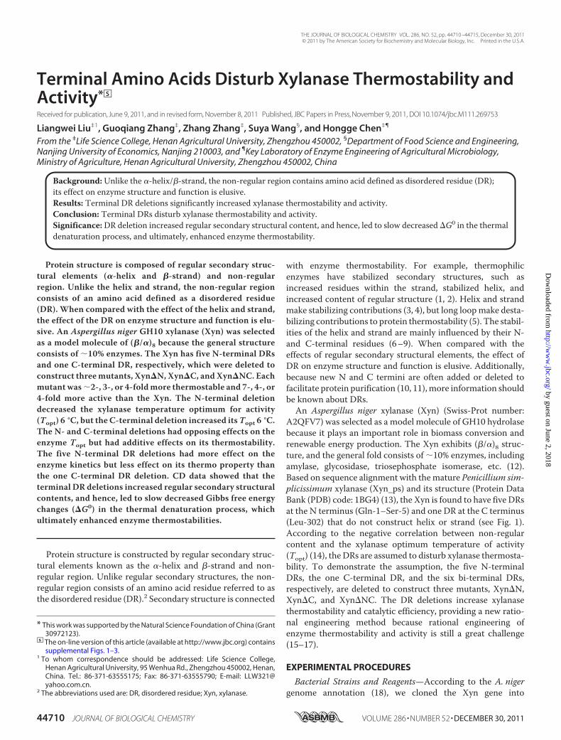
Terminal Amino Acids Disturb Xylanase Thermostability andActivity*□S
Received for publication, June 9, 2011, and in revised form, November 8, 2011 Published, JBC Papers in Press, November 9, 2011, DOI 10.1074/jbc.M111.269753
Liangwei Liu‡1, Guoqiang Zhang‡, Zhang Zhang‡, Suya Wang§, and Hongge Chen‡¶
From the ‡Life Science College, Henan Agricultural University, Zhengzhou 450002, §Department of Food Science and Engineering,Nanjing University of Economics, Nanjing 210003, and ¶Key Laboratory of Enzyme Engineering of Agricultural Microbiology,Ministry of Agriculture, Henan Agricultural University, Zhengzhou 450002, China
Background:Unlike the �-helix/�-strand, the non-regular region contains amino acid defined as disordered residue (DR);its effect on enzyme structure and function is elusive.Results: Terminal DR deletions significantly increased xylanase thermostability and activity.Conclusion: Terminal DRs disturb xylanase thermostability and activity.Significance:DR deletion increased regular secondary structural content, and hence, led to slow decreased �G0 in the thermaldenaturation process, and ultimately, enhanced enzyme thermostability.
Protein structure is composed of regular secondary struc-tural elements (�-helix and �-strand) and non-regularregion. Unlike the helix and strand, the non-regular regionconsists of an amino acid defined as a disordered residue(DR). When compared with the effect of the helix and strand,the effect of the DR on enzyme structure and function is elu-sive. An Aspergillus niger GH10 xylanase (Xyn) was selectedas a model molecule of (�/�)8 because the general structureconsists of �10% enzymes. The Xyn has five N-terminal DRsand one C-terminal DR, respectively, which were deleted toconstruct threemutants, Xyn�N, Xyn�C, and Xyn�NC. Eachmutant was�2-, 3-, or 4-foldmore thermostable and 7-, 4-, or4-fold more active than the Xyn. The N-terminal deletiondecreased the xylanase temperature optimum for activity(Topt) 6 °C, but the C-terminal deletion increased itsTopt 6 °C.The N- and C-terminal deletions had opposing effects on theenzyme Topt but had additive effects on its thermostability.The five N-terminal DR deletions had more effect on theenzyme kinetics but less effect on its thermo property thanthe one C-terminal DR deletion. CD data showed that theterminal DR deletions increased regular secondary structuralcontents, and hence, led to slow decreased Gibbs free energychanges (�G0) in the thermal denaturation process, whichultimately enhanced enzyme thermostabilities.
Protein structure is constructed by regular secondary struc-tural elements known as the �-helix and �-strand and non-regular region. Unlike regular secondary structures, the non-regular region consists of an amino acid residue referred to asthe disordered residue (DR).2 Secondary structure is connected
with enzyme thermostability. For example, thermophilicenzymes have stabilized secondary structures, such asincreased residues within the strand, stabilized helix, andincreased content of regular structure (1, 2). Helix and strandmake stabilizing contributions (3, 4), but long loopmake desta-bilizing contributions to protein thermostability (5). The stabil-ities of the helix and strand are mainly influenced by their N-and C-terminal residues (6–9). When compared with theeffects of regular secondary structural elements, the effect ofDR on enzyme structure and function is elusive. Additionally,because new N and C termini are often added or deleted tofacilitate protein purification (10, 11),more information shouldbe known about DRs.An Aspergillus niger xylanase (Xyn) (Swiss-Prot number:
A2QFV7) was selected as a model molecule of GH10 hydrolasebecause it plays an important role in biomass conversion andrenewable energy production. The Xyn exhibits (�/�)8 struc-ture, and the general fold consists of �10% enzymes, includingamylase, glycosidase, triosephosphate isomerase, etc. (12).Based on sequence alignment with themature Penicillium sim-plicissimum xylanase (Xyn_ps) and its structure (Protein DataBank (PDB) code: 1BG4) (13), the Xyn is found to have five DRsat the N terminus (Gln-1–Ser-5) and one DR at the C terminus(Leu-302) that do not construct helix or strand (see Fig. 1).According to the negative correlation between non-regularcontent and the xylanase optimum temperature of activity(Topt) (14), the DRs are assumed to disturb xylanase thermosta-bility. To demonstrate the assumption, the five N-terminalDRs, the one C-terminal DR, and the six bi-terminal DRs,respectively, are deleted to construct three mutants, Xyn�N,Xyn�C, and Xyn�NC. The DR deletions increase xylanasethermostability and catalytic efficiency, providing a new ratio-nal engineering method because rational engineering ofenzyme thermostability and activity is still a great challenge(15–17).
EXPERIMENTAL PROCEDURES
Bacterial Strains and Reagents—According to the A. nigergenome annotation (18), we cloned the Xyn gene into
* This work was supported by the Natural Science Foundation of China (Grant30972123).
□S The on-line version of this article (available at http://www.jbc.org) containssupplemental Figs. 1–3.
1 To whom correspondence should be addressed: Life Science College,Henan Agricultural University, 95 Wenhua Rd., Zhengzhou 450002, Henan,China. Tel.: 86-371-63555175; Fax: 86-371-63555790; E-mail: [email protected].
2 The abbreviations used are: DR, disordered residue; Xyn, xylanase.
THE JOURNAL OF BIOLOGICAL CHEMISTRY VOL. 286, NO. 52, pp. 44710 –44715, December 30, 2011© 2011 by The American Society for Biochemistry and Molecular Biology, Inc. Printed in the U.S.A.
44710 JOURNAL OF BIOLOGICAL CHEMISTRY VOLUME 286 • NUMBER 52 • DECEMBER 30, 2011
by guest on June 2, 2018http://w
ww
.jbc.org/D
ownloaded from
pET20b(�) (Novagen, Shanghai, China). The Xyn has 302 res-idues (Swiss-Prot number: A2QFV7), consisting of the residuelength of mature Xyn_ps (Swiss-Prot number: P56588) (13).According to the Xyn_ps structure (1BG4) and sequence align-ment, the Xyn has five N-terminal DRs (Gln-1, Ala-2, Ser-3,Val-4, and Ser-5) and one C-terminal DR (Leu-302) (see Fig. 1).The five N-terminal DRs, the one C-terminal DR, and the sixbi-terminal DRs, respectively, were deleted to construct threemutants, Xyn�N, Xyn�C, and Xyn�NC. Molecular reagentswere Pfu polymerase, restriction endonucleases (NdeI/XhoI),T4 DNA ligase, DNA and protein marker, etc. (Takara Inc.,Dalian, China).Construction of theMutants—The pET20b(�)-xyn served as
template for the construction of deletion mutants (see Fig. 1).The genes xyn, xyn�n, xyn�c, and xyn�nc were amplified byusing the primers p1/p2, p3/p2, p1/p4, and p3/p4, respectively,with underlined letters showing NdeI and XhoI restrictionsites. The sequences used are as follows: p1, ggaattccatatgcag-gcttcagtgagtattga; p2, ccaaattactcgaggagagcatttgcgatagc; p3,ggaattccatatgattgataccaaattcaaggc; p4, ccaaattactcgagagcattt-gcgatagcag. PCR was carried out by using 3.0 �l of pET20b-xynas template, 1.0 �l of each of the related primers, 0.5 �l of Pfupolymerase, 4.0 �l of dNTPs, and 1� buffer. PCR conditionswere: 4 min at 94 °C, 30 cycles (1 min at 94 °C; 1 min at 47 °C; 1min at 72 °C), 10 min at 72 °C.The amplified genes were cloned into pET20b(�) and
digested with NdeI and XhoI to delete redundant restrictionendonuclease sites (supplemental Fig. 1). After transformingEscherichia coli BL21(DE3)-competent cells, the recombinantplasmids were extracted and sequenced to confirm geneaccuracy by using an ABI 3730 automated DNA sequencer(Invitrogen Biotechnology, Shanghai, China). The accuratetransformants containing pET20b-xyn�n, pET20b-xyn�c,pET20b-xyn�nc, and pET20b-xyn were grown, induced, andcollected to extract xylanases according to standard protocols(19). Because the xylanases had C-terminal His6 tags, the xyla-nases were purified by using Co2� binding resin (AmershamBiosciences). Active fractions were pooled and further purifiedby using Sephadex G-25. The xylanases were detected using12% polyacrylamide SDS-PAGE and stained with CoomassieBrilliant Blue G-250. Protein concentration was measured by aSpectrophotometer ND-1000 at 280 nm using derivatization(NanoDrop Technologies, Wilmington, DE).Assay of Xylanase Property—Properties of the mutant xyla-
nases were assayed in parallel with the Xyn for three independ-ent reactions, and the data were averaged. Xylanase Topt wasdetermined from 30 to 60 °C, and pHopt was determined frompH 2.6 to 7.0 in imidazole-biphthalate buffer. After incubationat 50 °C for a 10-min interval from0 to 100min, residual activitywas assayed and expressed as a ratio in the percentage of theuntreated xylanase activity. The data were fitted to the Arrhe-nius function (y � A � e�kt) to calculate the thermal inactiva-tion half-life for activity (t1⁄2) to indicate thermostability (Origin,version 8.0). Xylanase kinetics was assayed at optimal condi-tions by reaction for 5 min using birch wood xylan at concen-trations from 2.5 to 50 mg/ml (Sigma-Aldrich, Shanghai,China). The data were fitted with the Hill function (y�Vmax �[S]/(Km� [S]) to calculatemaximal activity (Vmax) andKm (Ori-
gin). Xylanase standard activity was determined by the dinitro-salicylic acid method described previously (19).Far-UV Circular Dichroism (CD) Spectra—The xylanase CD
spectra were determined on a JASCO J810 spectropolarimeterflushed with nitrogen gas. Each protein sample was scannedthree times, and the data were averaged and used for reportedCD spectra, which were recorded from 195 to 260 nm using a0.1-cm path-length cuvette at a scan speed of 50 nm/min,response time of 2 s, bandwidth of 2 nm, and pitch of 0.2 nm.For thermodynamic analysis, the thermal denaturation CDspectra were recorded at 220 nm from 30 to 80 °C upon heatingat a constant rate of 2 °C/min. The molar ellipticities were cal-culated according to the following equation
��� �� � MW
100 � l � C
where [�], �, l, andC are themolar and observed ellipticity, pathlength (in centimeters), and molar protein concentration,respectively. Thermal denaturation curves were fitted to amodified van’t Hoff equation using a reversible two-state pro-tein unfolding model (20). The spectra data were fitted to thenative and denatured transition baselines to obtain thermalmelting temperature (Tm), enthalpy change (�H0), entropychange (�S), and Gibbs’ free energy change (�G0) values (Sig-maPlot, Version 10.0).
RESULTS
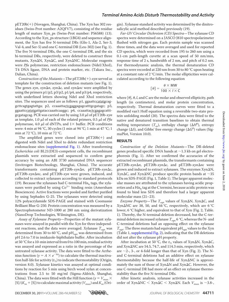
Construction of the Deletion Mutants—The DR-deletedgenes produced specific DNA bands at �1.3 kb on gel electro-phoresis (Fig. 1). After we confirmed the accuracies of theextracted recombinant plasmids, the transformants containingpET20b-xyn�n, pET20b-xyn�c, and pET20b-xyn�nc weregrown and induced to extract xylanases. The enzymes Xyn�N,Xyn�C, and Xyn�NC produce specific protein bands at �35kDa on SDS-PAGE (Fig. 1, Table 1). The larger apparentmolec-ular masses are attributed to the xylanases having acidic prop-erties and aHis6 tag at theC termini, because acidic proteinwasfound to bind less SDS and therefore had a larger apparentmolecular mass (21–23).Enzyme Property—The Topt values of Xyn�N, Xyn�C, and
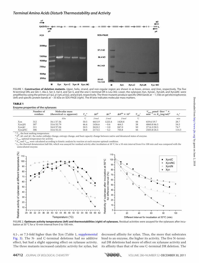
Xyn�NC are 38, 50, and 44 °C, respectively, which are 6 °Clower, 6 °C higher, and equivalent to that of Xyn (Fig. 2, Table.1). Thereby, the N-terminal deletion decreased, but the C-ter-minal deletion increased xylanaseTopt 6 °C, whereas theN- andC-terminal deletions had an opposing effect on the xylanaseTopt. The threemutants had equivalent pHopt values to the Xyn(Table 1, supplemental Fig. 2), indicating that the DR deletionsdid not alter the xylanase pH property.After incubation at 50 °C, the t1⁄2 values of Xyn�N, Xyn�C,
and Xyn�NC are 54.5, 74.7, and 114.3 min, respectively, whichare �2-, 3-, or 4-fold longer than that of Xyn (Fig. 2). The N-and C-terminal deletions had an additive effect on xylanasethermostability because the half-life of Xyn�NC is approxi-mately the sum of those of Xyn�N and Xyn�C. However, theone C-terminal DR had more of an effect on xylanase thermo-stability than the five N-terminal DRs.After kinetic analysis, xylanase activities increased in the
order of Xyn�NC Xyn�C Xyn�N. Each Vmax is �3.8-,
Terminal Amino Acids Disturb Thermostability and Activity
DECEMBER 30, 2011 • VOLUME 286 • NUMBER 52 JOURNAL OF BIOLOGICAL CHEMISTRY 44711
by guest on June 2, 2018http://w
ww
.jbc.org/D
ownloaded from
4.1-, or 7.3-fold higher than the Xyn (Table 1, supplementalFig. 3). The N- and C-terminal deletions had no additiveeffect, but had a slight opposing effect on xylanase activity.The three mutants increased catalytic activity for xylan, but
decreased affinity for xylan. Thus, the more that substratesbind to an enzyme, the higher its activity. The five N-termi-nal DR deletions had more of effect on xylanase activity andits affinity than that of the one C-terminal DR deletion. The
FIGURE 1. Construction of deletion mutants. Upper, helix, strand, and non-regular region are shown in as boxes, arrows, and lines, respectively. The fiveN-terminal DRs are Gln-1, Ala-2, Ser-3, Val-4, and Ser-5, and the one C-terminal DR is Leu-302. Lower, the xylanases Xyn, Xyn�C, Xyn�N, and Xyn�NC wereamplified by using the primers p1/p2, p1/p4, p3/p2, and p3/p4, respectively. The three mutants produce specific DNA bands at �1.3 kb on gel electrophoresis(left) and specific protein bands at �35 kDa on SDS-PAGE (right). The M lane indicates molecular mass markers.
TABLE 1Enzyme properties of the xylanases
Number ofresidues
Molecular mass(theoretical vs. apparent) Tm
a �Sb �Cb �H0b � 103 Toptc
Vmax �mol � liter�1 �min�1 vs. Km(mg/ml)d t1⁄2e
kDa °C J/mol J/mol J/mol °C minXyn 312 34.1/37.56 50.5 4415.9 1225.4 1428.8 44 659.6/19.7 28.7Xyn�N 307 33.6/35.79 46.4 1456.6 �0.6 464.5 38 4860.8/46.5 54.5Xyn�C 311 34.0/37.04 50.5 2618.2 �2.0 847.0 50 2714.3/30.5 74.7Xyn�NC 306 33.6/35.55 50.8 2173.5 �4.2 703.8 44 2503.8/26.5 114.3
aTm, the heat melting temperature.b H0, �S, and �C, the molar enthalpy change, entropy change, and heat capacity change between native and denatured states of enzyme.c Topt, optimal temperature for activity.d Km and Vmax were calculated according to kinetic analysis by reaction at each enzyme optimal condition.e t1⁄2, the thermal denaturation half-life, which was assayed for residual activity after incubation at 50 °C for a 10-min interval from 0 to 100 min and was compared with theunincubated enzyme.
FIGURE 2. Optimum activity temperatures (left) and thermostabilities (right) of xylanases. Residual activities were assayed for the xylanases after incu-bation at 50 °C for a 10-min interval from 0 to 100 min.
Terminal Amino Acids Disturb Thermostability and Activity
44712 JOURNAL OF BIOLOGICAL CHEMISTRY VOLUME 286 • NUMBER 52 • DECEMBER 30, 2011
by guest on June 2, 2018http://w
ww
.jbc.org/D
ownloaded from
affinity of Xyn�NC was higher than those of Xyn�N andXyn�C.CD Analysis—According to the thermal denaturation CD
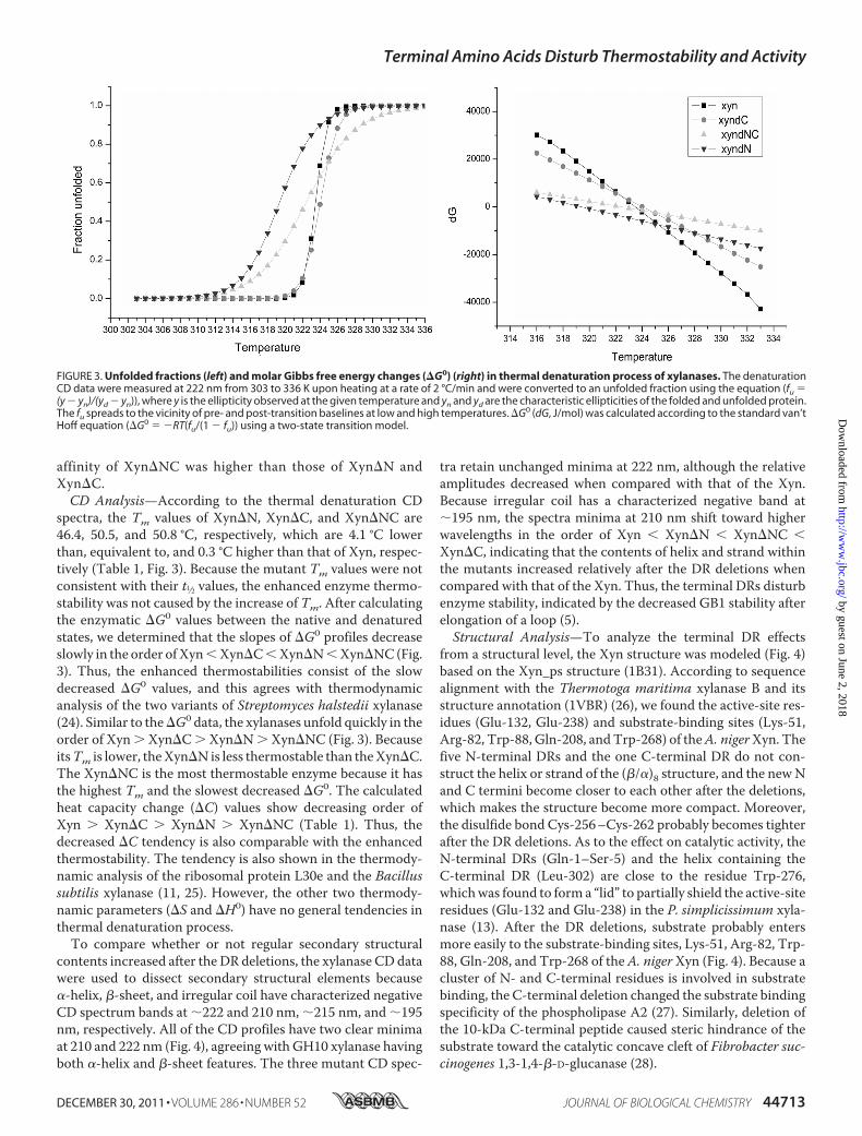
spectra, the Tm values of Xyn�N, Xyn�C, and Xyn�NC are46.4, 50.5, and 50.8 °C, respectively, which are 4.1 °C lowerthan, equivalent to, and 0.3 °C higher than that of Xyn, respec-tively (Table 1, Fig. 3). Because the mutant Tm values were notconsistent with their t1⁄2 values, the enhanced enzyme thermo-stability was not caused by the increase of Tm. After calculatingthe enzymatic �G0 values between the native and denaturedstates, we determined that the slopes of �G0 profiles decreaseslowly in the order ofXynXyn�CXyn�NXyn�NC(Fig.3). Thus, the enhanced thermostabilities consist of the slowdecreased �G0 values, and this agrees with thermodynamicanalysis of the two variants of Streptomyces halstedii xylanase(24). Similar to the�G0 data, the xylanases unfold quickly in theorder of Xyn Xyn�C Xyn�N Xyn�NC (Fig. 3). BecauseitsTm is lower, theXyn�N is less thermostable than theXyn�C.The Xyn�NC is the most thermostable enzyme because it hasthe highest Tm and the slowest decreased �G0. The calculatedheat capacity change (�C) values show decreasing order ofXyn Xyn�C Xyn�N Xyn�NC (Table 1). Thus, thedecreased �C tendency is also comparable with the enhancedthermostability. The tendency is also shown in the thermody-namic analysis of the ribosomal protein L30e and the Bacillussubtilis xylanase (11, 25). However, the other two thermody-namic parameters (�S and �H0) have no general tendencies inthermal denaturation process.To compare whether or not regular secondary structural
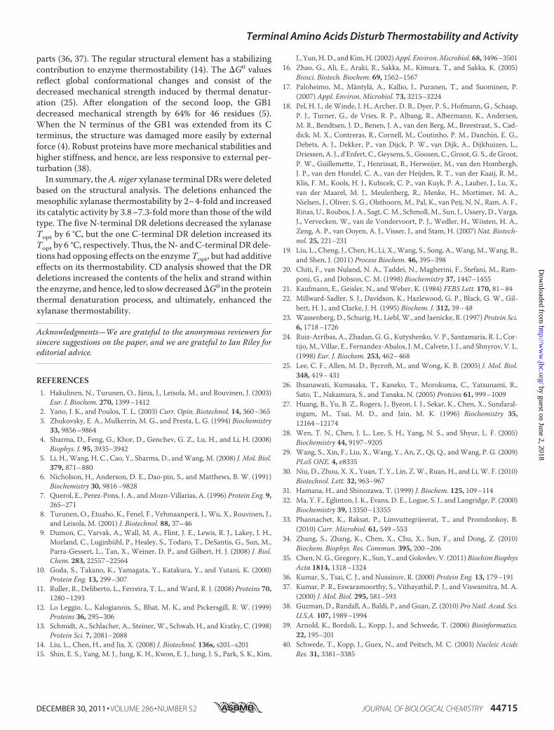
contents increased after the DR deletions, the xylanase CD datawere used to dissect secondary structural elements because�-helix, �-sheet, and irregular coil have characterized negativeCD spectrum bands at �222 and 210 nm, �215 nm, and �195nm, respectively. All of the CD profiles have two clear minimaat 210 and 222 nm (Fig. 4), agreeing with GH10 xylanase havingboth �-helix and �-sheet features. The three mutant CD spec-
tra retain unchanged minima at 222 nm, although the relativeamplitudes decreased when compared with that of the Xyn.Because irregular coil has a characterized negative band at�195 nm, the spectra minima at 210 nm shift toward higherwavelengths in the order of Xyn Xyn�N Xyn�NC Xyn�C, indicating that the contents of helix and strand withinthe mutants increased relatively after the DR deletions whencompared with that of the Xyn. Thus, the terminal DRs disturbenzyme stability, indicated by the decreased GB1 stability afterelongation of a loop (5).Structural Analysis—To analyze the terminal DR effects
from a structural level, the Xyn structure was modeled (Fig. 4)based on the Xyn_ps structure (1B31). According to sequencealignment with the Thermotoga maritima xylanase B and itsstructure annotation (1VBR) (26), we found the active-site res-idues (Glu-132, Glu-238) and substrate-binding sites (Lys-51,Arg-82, Trp-88, Gln-208, andTrp-268) of theA. nigerXyn. Thefive N-terminal DRs and the one C-terminal DR do not con-struct the helix or strand of the (�/�)8 structure, and the newNand C termini become closer to each other after the deletions,which makes the structure become more compact. Moreover,the disulfide bond Cys-256–Cys-262 probably becomes tighterafter the DR deletions. As to the effect on catalytic activity, theN-terminal DRs (Gln-1–Ser-5) and the helix containing theC-terminal DR (Leu-302) are close to the residue Trp-276,whichwas found to form a “lid” to partially shield the active-siteresidues (Glu-132 and Glu-238) in the P. simplicissimum xyla-nase (13). After the DR deletions, substrate probably entersmore easily to the substrate-binding sites, Lys-51, Arg-82, Trp-88, Gln-208, and Trp-268 of theA. nigerXyn (Fig. 4). Because acluster of N- and C-terminal residues is involved in substratebinding, the C-terminal deletion changed the substrate bindingspecificity of the phospholipase A2 (27). Similarly, deletion ofthe 10-kDa C-terminal peptide caused steric hindrance of thesubstrate toward the catalytic concave cleft of Fibrobacter suc-cinogenes 1,3-1,4-�-D-glucanase (28).
FIGURE 3. Unfolded fractions (left) and molar Gibbs free energy changes (�G0) (right) in thermal denaturation process of xylanases. The denaturationCD data were measured at 222 nm from 303 to 336 K upon heating at a rate of 2 °C/min and were converted to an unfolded fraction using the equation (fu �(y � yn)/(yd � yn)), where y is the ellipticity observed at the given temperature and yn and yd are the characteristic ellipticities of the folded and unfolded protein.The fu spreads to the vicinity of pre- and post-transition baselines at low and high temperatures. �G0 (dG, J/mol) was calculated according to the standard van’tHoff equation (�G0 � �RT(fu/(1 � fu)) using a two-state transition model.
Terminal Amino Acids Disturb Thermostability and Activity
DECEMBER 30, 2011 • VOLUME 286 • NUMBER 52 JOURNAL OF BIOLOGICAL CHEMISTRY 44713
by guest on June 2, 2018http://w
ww
.jbc.org/D
ownloaded from

DISCUSSION
Previously, either the entire domain of the enzyme was trun-cated completely, or terminal residues of the enzyme were suc-cessively deleted (27–33). The present study deleted the sixterminal DRs based on structural analysis, respectively, and thedeletions significantly increased the xylanase thermostabilityand activity. Similar to our data, the N-glycanase increaseddeglycosylation activity after deletion of the N-terminal helix(29). After successive truncation analysis, the seven N-terminalresidues and the three C-terminal residues were shown to beunnecessary for the Clostridium thermocellum lichenase cata-lytic activity (30). As to enzyme thermostability, the successivedeletion of terminal residues decreased the phosphoribosyl-transferase thermostability (31); probably, its structure andconformation were disturbed by the successive deletions. Sim-ilar to our data, the extra four N-terminal residues decreasedthe lysozyme thermostability and its refolding rate (10). Dele-tion of the four C-terminal glycine-rich repeats and the domainincreased enzyme thermostability, substrate binding affinity,and activity, respectively (28, 30, 32). Residue mutations hap-pened generally in the N termini of the 15 most thermostablexylanases, showing that the N-terminal region was more sus-ceptible to thermal unfolding (9). The five N-terminal muta-tions might confer structural stability, and hence, prevent theoverall thermal unfolding of themesophilic Streptomyces oliva-ceovirdis xylanase (34). TheN andC termini were found to playvery prominent roles in the inactive and active fold switch of theB. subtilis xylanase (11). Although commonly used in facilitat-ing protein purification by adding or deleting N- or C-terminalresidues, newly created residues interfered with protein struc-
ture and function (11). Thus, in functional analysis, the proteinshould be complete and have no extra residues, as well as beingfree of unnecessary residues encoded by redundant endonu-clease sites in expression vectors whenever possible.Topt, t1⁄2, and Tm were assayed to describe the different ther-
mal features of the xylanases. Topt reflects enzymatic activity ata certain temperature. An enzyme with a higher Topt is com-monly regarded as having a higher thermostability (12), and it isoften described by the organism growth temperature optimum.t1⁄2 reflects the thermal resistance of an enzyme at a certain tem-perature; therefore, it is more suitable to indicate enzyme ther-mostability. When compared with the Xyn, the Xyn�N had alowerTopt, but a longer t1⁄2. The difference indicates that the twoparameters are not always consistent, especially formutant andwild type of a same enzyme, and that the two parameters con-nect with different structural features. Unlike the two previousparameters, the Tm values were assayed to show 50% of thexylanases being thermally inactivated after continuous incuba-tion from 30 to 80 °C upon heating at a rate of 2 °C/min; there-fore, the Tm values of three mutants are higher than their Toptvalues. Moreover, the Photinus pyralis (firefly) luciferase Tmreflects a thermal deactivation intermediate state that includesreversible active and inactive states (35).Because the five N-terminal DRs (Gln-1–Ser-5) and the one
C-terminal DR (Leu-302) do not form regular secondary struc-tural elements, the DR deletions increased contents of the helixand strand within the enzyme, and thus, led to slow decreased�G0 tendency in the thermal denaturation process. Thermo-philic proteins had more stable helixes and larger residue frac-tions within helical conformations than mesophilic counter-
FIGURE 4. CD spectra (left) and structure (right) of xylanase. The CD spectra (left) were determined from 195 to 260 nm. Protein concentrations were 0.699,0.789, 0.697, and 0.777 mg/ml for the xylanases, Xyn, Xyn�C, Xyn�N, and Xyn�NC, respectively. Similar to the Xyn, the three mutants exhibit broad CD spectrabands with minima at 210 and 222 nm, consisting of (�/�)8 structure of GH10 xylanase having compact features of �-helix and �-sheet. Thereby, the threedeletion mutants fold properly at room temperature. Because irregular coil has a characterized negative band at 195 nm, the shifts of minima at 210 nm towardhigher wavelength show that the irregular coil contents of the xylanase decrease after the DR deletions, and therefore, the contents of strand and helix increaserelatively in the order of Xyn Xyn�N Xyn�NC Xyn�C, according to CD reference spectra by Dr. J. T. Yang (JASCO software package). The A. niger Xynstructure (right) was modeled by using the Swiss-model software (39, 40), with the �-sheet shown with an arrow, the �-helix shown with spiral ribbon, and theirregular coil shown as a line. According to sequence alignment with the T. maritima xylanase B and its structure (1VBR) (26), we found active-site residues(Glu-132, proton donor, and Glu-238, nucleophile) and substrate-binding sites of the A. niger Xyn. The N-terminal DRs (Gln-1, Ala-2, Ser-3, Val-4, and Ser-5) andthe helix containing the C-terminal DR (Leu-302) are close to the Trp-276 (13), which was found to form a lid to partially shield the active-site residues (Glu-132and Glu-238). After the DR deletions, substrate enters more easily into the substrate-binding sites (Lys-51, Arg-82, Trp-88, Gln-208, and Trp-268). Additionally,new N and C termini become closer to each other after DR deletions, which makes the Xyn structure more compact, and therefore, increases its thermostability.
Terminal Amino Acids Disturb Thermostability and Activity
44714 JOURNAL OF BIOLOGICAL CHEMISTRY VOLUME 286 • NUMBER 52 • DECEMBER 30, 2011
by guest on June 2, 2018http://w
ww
.jbc.org/D
ownloaded from
parts (36, 37). The regular structural element has a stabilizingcontribution to enzyme thermostability (14). The �G0 valuesreflect global conformational changes and consist of thedecreased mechanical strength induced by thermal denatur-ation (25). After elongation of the second loop, the GB1decreased mechanical strength by 64% for 46 residues (5).When the N terminus of the GB1 was extended from its Cterminus, the structure was damaged more easily by externalforce (4). Robust proteins have more mechanical stabilities andhigher stiffness, and hence, are less responsive to external per-turbation (38).In summary, theA. niger xylanase terminal DRs were deleted
based on the structural analysis. The deletions enhanced themesophilic xylanase thermostability by 2–4-fold and increasedits catalytic activity by 3.8–7.3-foldmore than those of the wildtype. The five N-terminal DR deletions decreased the xylanaseTopt by 6 °C, but the one C-terminal DR deletion increased itsTopt by 6 °C, respectively. Thus, theN- andC-terminalDRdele-tions had opposing effects on the enzymeTopt, but had additiveeffects on its thermostability. CD analysis showed that the DRdeletions increased the contents of the helix and strand withinthe enzyme, andhence, led to slowdecreased�G0 in the proteinthermal denaturation process, and ultimately, enhanced thexylanase thermostability.
Acknowledgments—We are grateful to the anonymous reviewers forsincere suggestions on the paper, and we are grateful to Ian Riley foreditorial advice.
REFERENCES1. Hakulinen, N., Turunen, O., Janis, J., Leisola, M., and Rouvinen, J. (2003)
Eur. J. Biochem. 270, 1399–14122. Yano, J. K., and Poulos, T. L. (2003) Curr. Opin. Biotechnol. 14, 360–3653. Zhukovsky, E. A., Mulkerrin, M. G., and Presta, L. G. (1994) Biochemistry
33, 9856–98644. Sharma, D., Feng, G., Khor, D., Genchev, G. Z., Lu, H., and Li, H. (2008)
Biophys. J. 95, 3935–39425. Li, H., Wang, H. C., Cao, Y., Sharma, D., andWang, M. (2008) J. Mol. Biol.
379, 871–8806. Nicholson, H., Anderson, D. E., Dao-pin, S., and Matthews, B. W. (1991)
Biochemistry 30, 9816–98287. Querol, E., Perez-Pons, J. A., andMozo-Villarias, A. (1996) Protein Eng. 9,
265–2718. Turunen, O., Etuaho, K., Fenel, F., Vehmaanpera, J., Wu, X., Rouvinen, J.,
and Leisola, M. (2001) J. Biotechnol. 88, 37–469. Dumon, C., Varvak, A., Wall, M. A., Flint, J. E., Lewis, R. J., Lakey, J. H.,
Morland, C., Luginbuhl, P., Healey, S., Todaro, T., DeSantis, G., Sun, M.,Parra-Gessert, L., Tan, X., Weiner, D. P., and Gilbert, H. J. (2008) J. Biol.Chem. 283, 22557–22564
10. Goda, S., Takano, K., Yamagata, Y., Katakura, Y., and Yutani, K. (2000)Protein Eng. 13, 299–307
11. Ruller, R., Deliberto, L., Ferreira, T. L., andWard, R. J. (2008) Proteins 70,1280–1293
12. Lo Leggio, L., Kalogiannis, S., Bhat, M. K., and Pickersgill, R. W. (1999)Proteins 36, 295–306
13. Schmidt, A., Schlacher, A., Steiner, W., Schwab, H., and Kratky, C. (1998)Protein Sci. 7, 2081–2088
14. Liu, L., Chen, H., and Jia, X. (2008) J. Biotechnol. 136s, s201–s20115. Shin, E. S., Yang, M. J., Jung, K. H., Kwon, E. J., Jung, J. S., Park, S. K., Kim,
J., Yun,H.D., andKim,H. (2002)Appl. Environ.Microbiol. 68, 3496–350116. Zhao, G., Ali, E., Araki, R., Sakka, M., Kimura, T., and Sakka, K. (2005)
Biosci. Biotech. Biochem. 69, 1562–156717. Paloheimo, M., Mantyla, A., Kallio, J., Puranen, T., and Suominen, P.
(2007) Appl. Environ. Microbiol. 73, 3215–322418. Pel, H. J., de Winde, J. H., Archer, D. B., Dyer, P. S., Hofmann, G., Schaap,
P. J., Turner, G., de Vries, R. P., Albang, R., Albermann, K., Andersen,M. R., Bendtsen, J. D., Benen, J. A., van den Berg, M., Breestraat, S., Cad-dick, M. X., Contreras, R., Cornell, M., Coutinho, P. M., Danchin, E. G.,Debets, A. J., Dekker, P., van Dijck, P. W., van Dijk, A., Dijkhuizen, L.,Driessen, A. J., d’Enfert, C., Geysens, S., Goosen, C., Groot, G. S., de Groot,P. W., Guillemette, T., Henrissat, B., Herweijer, M., van den Hombergh,J. P., van den Hondel, C. A., van der Heijden, R. T., van der Kaaij, R. M.,Klis, F. M., Kools, H. J., Kubicek, C. P., van Kuyk, P. A., Lauber, J., Lu, X.,van der Maarel, M. J., Meulenberg, R., Menke, H., Mortimer, M. A.,Nielsen, J., Oliver, S. G., Olsthoorn, M., Pal, K., van Peij, N. N., Ram, A. F.,Rinas, U., Roubos, J. A., Sagt, C.M., Schmoll, M., Sun, J., Ussery, D., Varga,J., Vervecken, W., van de Vondervoort, P. J., Wedler, H., Wosten, H. A.,Zeng, A. P., van Ooyen, A. J., Visser, J., and Stam, H. (2007) Nat. Biotech-nol. 25, 221–231
19. Liu, L., Cheng, J., Chen, H., Li, X.,Wang, S., Song, A.,Wang,M.,Wang, B.,and Shen, J. (2011) Process Biochem. 46, 395–398
20. Chiti, F., van Nuland, N. A., Taddei, N., Magherini, F., Stefani, M., Ram-poni, G., and Dobson, C. M. (1998) Biochemistry 37, 1447–1455
21. Kaufmann, E., Geisler, N., and Weber, K. (1984) FEBS Lett. 170, 81–8422. Millward-Sadler, S. J., Davidson, K., Hazlewood, G. P., Black, G. W., Gil-
bert, H. J., and Clarke, J. H. (1995) Biochem. J. 312, 39–4823. Wassenberg, D., Schurig, H., Liebl,W., and Jaenicke, R. (1997) Protein Sci.
6, 1718–172624. Ruiz-Arribas, A., Zhadan, G. G., Kutyshenko, V. P., Santamaría, R. I., Cor-
tijo,M., Villar, E., Fernandez-Abalos, J.M., Calvete, J. J., and Shnyrov, V. L.(1998) Eur. J. Biochem. 253, 462–468
25. Lee, C. F., Allen, M. D., Bycroft, M., and Wong, K. B. (2005) J. Mol. Biol.348, 419–431
26. Ihsanawati, Kumasaka, T., Kaneko, T., Morokuma, C., Yatsunami, R.,Sato, T., Nakamura, S., and Tanaka, N. (2005) Proteins 61, 999–1009
27. Huang, B., Yu, B. Z., Rogers, J., Byeon, I. J., Sekar, K., Chen, X., Sundaral-ingam, M., Tsai, M. D., and Jain, M. K. (1996) Biochemistry 35,12164–12174
28. Wen, T. N., Chen, J. L., Lee, S. H., Yang, N. S., and Shyur, L. F. (2005)Biochemistry 44, 9197–9205
29. Wang, S., Xin, F., Liu, X., Wang, Y., An, Z., Qi, Q., andWang, P. G. (2009)PLoS ONE. 4, e8335
30. Niu, D., Zhou, X. X., Yuan, T. Y., Lin, Z.W., Ruan, H., and Li,W. F. (2010)Biotechnol. Lett. 32, 963–967
31. Hamana, H., and Shinozawa, T. (1999) J. Biochem. 125, 109–11432. Ma, Y. F., Eglinton, J. K., Evans, D. E., Logue, S. J., and Langridge, P. (2000)
Biochemistry 39, 13350–1335533. Phannachet, K., Raksat, P., Limvuttegrijeerat, T., and Promdonkoy, B.
(2010) Curr. Microbiol. 61, 549–55334. Zhang, S., Zhang, K., Chen, X., Chu, X., Sun, F., and Dong, Z. (2010)
Biochem. Biophys. Res. Commun. 395, 200–20635. Chen, N. G., Gregory, K., Sun, Y., andGolovlev, V. (2011)BiochimBiophys
Acta 1814, 1318–132436. Kumar, S., Tsai, C. J., and Nussinov, R. (2000) Protein Eng. 13, 179–19137. Kumar, P. R., Eswaramoorthy, S., Vithayathil, P. J., and Viswamitra, M. A.
(2000) J. Mol. Biol. 295, 581–59338. Guzman, D., Randall, A., Baldi, P., andGuan, Z. (2010) Pro Natl. Acad. Sci.
U.S.A. 107, 1989–199439. Arnold, K., Bordoli, L., Kopp, J., and Schwede, T. (2006) Bioinformatics.
22, 195–20140. Schwede, T., Kopp, J., Guex, N., and Peitsch, M. C. (2003) Nucleic Acids
Res. 31, 3381–3385
Terminal Amino Acids Disturb Thermostability and Activity
DECEMBER 30, 2011 • VOLUME 286 • NUMBER 52 JOURNAL OF BIOLOGICAL CHEMISTRY 44715
by guest on June 2, 2018http://w
ww
.jbc.org/D
ownloaded from

Liangwei Liu, Guoqiang Zhang, Zhang Zhang, Suya Wang and Hongge ChenTerminal Amino Acids Disturb Xylanase Thermostability and Activity
doi: 10.1074/jbc.M111.269753 originally published online November 9, 20112011, 286:44710-44715.J. Biol. Chem.
10.1074/jbc.M111.269753Access the most updated version of this article at doi:
Alerts:
When a correction for this article is posted•
When this article is cited•
to choose from all of JBC's e-mail alertsClick here
Supplemental material:
http://www.jbc.org/content/suppl/2011/11/09/M111.269753.DC1
http://www.jbc.org/content/286/52/44710.full.html#ref-list-1
This article cites 40 references, 5 of which can be accessed free at
by guest on June 2, 2018http://w
ww
.jbc.org/D
ownloaded from