Embed Size (px)
Citation preview
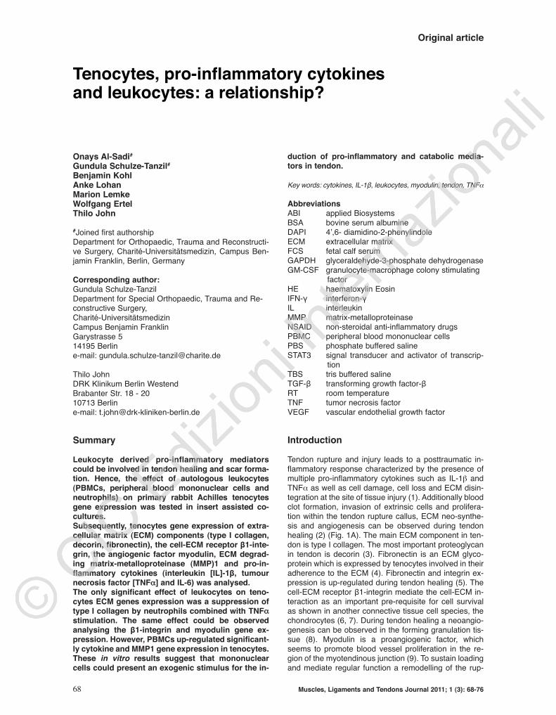
Original article
68 Muscles, Ligaments and Tendons Journal 2011; 1 (3): 68-76
Onays Al-Sadi#
Gundula Schulze-Tanzil#
Benjamin KohlAnke LohanMarion LemkeWolfgang ErtelThilo John
#Joined first authorshipDepartment for Orthopaedic, Trauma and Reconstructi-ve Surgery, Charité-Universitätsmedizin, Campus Ben-jamin Franklin, Berlin, Germany
Corresponding author:Gundula Schulze-TanzilDepartment for Special Orthopaedic, Trauma and Re-constructive Surgery, Charité-UniversitätsmedizinCampus Benjamin FranklinGarystrasse 514195 Berline-mail: [email protected]
Thilo JohnDRK Klinikum Berlin WestendBrabanter Str. 18 - 2010713 Berline-mail: [email protected]
Summary
Leukocyte derived pro-inflammatory mediatorscould be involved in tendon healing and scar forma-tion. Hence, the effect of autologous leukocytes(PBMCs, peripheral blood mononuclear cells andneutrophils) on primary rabbit Achilles tenocytesgene expression was tested in insert assisted co-cultures. Subsequently, tenocytes gene expression of extra-cellular matrix (ECM) components (type I collagen,decorin, fibronectin), the cell-ECM receptor β1-inte-grin, the angiogenic factor myodulin, ECM degrad-ing matrix-metalloproteinase (MMP)1 and pro-in-flammatory cytokines (interleukin [IL]-1β, tumournecrosis factor [TNFα] and IL-6) was analysed. The only significant effect of leukocytes on teno-cytes ECM genes expression was a suppression oftype I collagen by neutrophils combined with TNFα
stimulation. The same effect could be observedanalysing the β1-integrin and myodulin gene ex-pression. However, PBMCs up-regulated significant-ly cytokine and MMP1 gene expression in tenocytes. These in vitro results suggest that mononuclearcells could present an exogenic stimulus for the in-
duction of pro-inflammatory and catabolic media-tors in tendon.
Key words: cytokines, IL-1β, leukocytes, myodulin, tendon, TNFα
AbbreviationsABI applied BiosystemsBSA bovine serum albumineDAPI 4’,6- diamidino-2-phenylindoleECM extracellular matrixFCS fetal calf serumGAPDH glyceraldehyde-3-phosphate dehydrogenaseGM-CSF granulocyte-macrophage colony stimulating
factorHE haematoxylin EosinIFN-γ interferon-γIL interleukinMMP matrix-metalloproteinaseNSAID non-steroidal anti-inflammatory drugsPBMC peripheral blood mononuclear cellsPBS phosphate buffered salineSTAT3 signal transducer and activator of transcrip-
tionTBS tris buffered salineTGF-β transforming growth factor-βRT room temperatureTNF tumor necrosis factorVEGF vascular endothelial growth factor
Introduction
Tendon rupture and injury leads to a posttraumatic in-flammatory response characterized by the presence ofmultiple pro-inflammatory cytokines such as IL-1b andTNFα as well as cell damage, cell loss and ECM disin-tegration at the site of tissue injury (1). Additionally bloodclot formation, invasion of extrinsic cells and prolifera-tion within the tendon rupture callus, ECM neo-synthe-sis and angiogenesis can be observed during tendonhealing (2) (Fig. 1A). The main ECM component in ten-don is type I collagen. The most important proteoglycanin tendon is decorin (3). Fibronectin is an ECM glyco-protein which is expressed by tenocytes involved in theiradherence to the ECM (4). Fibronectin and integrin ex-pression is up-regulated during tendon healing (5). Thecell-ECM receptor β1-integrin mediate the cell-ECM in-teraction as an important pre-requisite for cell survivalas shown in another connective tissue cell species, thechondrocytes (6, 7). During tendon healing a neoangio-genesis can be observed in the forming granulation tis-sue (8). Myodulin is a proangiogenic factor, whichseems to promote blood vessel proliferation in the re-gion of the myotendinous junction (9). To sustain loadingand mediate regular function a remodelling of the rup-
Tenocytes, pro-inflammatory cytokines and leukocytes: a relationship?

Muscles, Ligaments and Tendons Journal 2011; 1 (3) 68-76 69
Tenocytes, cytokines and leukocytes
ture callus is necessary (2). Callus remodelling in injuredtendons requires more than three months in the rabbitmodel (8). Remodelling is mediated by ECM degradingenzymes such as MMP1. Particularly MMP1 which iswell known to be able to cleave type I collagen has beenshown in tendon and cultured tenocytes (10).The impact of the inflammatory response on the tendonhealing outcome is still unclear. TNFα and IL-1β stimulatetenocytes in vitro to produce further pro- and anti-inflam-matory cytokines e.g. IL-1β, TNFα, IL-6 and IL-10 andECM degrading enzymes such as MMPs (11,12). Thissuggests an autoactivation of tenocytes by exogenic cy-tokines. However, apart from IL-1β and TNFαmultiple oth-er cytokines are released by leukocytes. Krause et al., (13)reported recently that e.g. IL-2, IL-8, IL-10, IL-17, TNFα,interferon (IFN)-γ, and granulocyte-macrophage colony
stimulating factor (GM-CSF) are produced by PBMCs.Some of them might probably be involved in tendon heal-ing. The cytokine concentration produced by leukocytesmight differ from that usually used in in vitro experimentswith isolated recombinant cytokines. PBMCs consist ofmonocytes and several lymphocyte subpopulations. Despite the stimulatory effect of platelets and their ex-tracts on tendon healing is meanwhile intensely dis-cussed (14-16), the effect of exogenic pro-inflammatorymediators su ch as cytokines released by leukocytes ontenocytes has not yet been studied in tendon tissue andtenocytes at all. Hen ce, this study was undertaken to as-sess the direct interactive response of Achilles teno-cytes to leuko cyte-derived soluble mediators in an indi-rect co-culture setting.
Materials and Methods
Tenocytes isolation and cul-tureRabbit primary Achilles tenocyteswere isolated as described previ-ously (17,18) from the midsub-stance of seven Achilles tendonsof healthy one year-aged adultfemale donors post mortem. Thisstudy was approved by the Berlinreview board for experimentswith animals. The identity of theisolated tenocytes was confirmedby immunolabelling of tenomod-ulin and tenascin C. Cultureswere grown at 37°C in a humidi-fied atmosphere with 5% CO
2
and the growth medium waschanged every three days.Growth medium for tenocyte cul-ture consisted of Ham’s F-12/Dulbecco’s modified Eagle’smedium (50/50, Biochrom-Se ro -med, Munich, Germany) contain-ing 10% fetal calf serum (FCS,Biochrom-Seromed), 25 μg/mLascorbic acid (Sigma-Aldrich,Munich, Germany), 50 IU/mLstreptomycin, 50 IU/mL penicillin,2.5 μg/mL amphotericin B, es-sential amino acids, L-glutamine(all: Biochrom-Seromed).
Immunofluorescence labellingof tenocytesCover slips seeded with teno-cytes were fixed in 4%paraformaldehyde, then rinsedin Tris buffered saline (TBS:0.05 M Tris, 0.015 M NaCl, pH7.6) before blocked with pro-tease-free donkey serum (5%diluted in TBS) for 30 min atroom temperature (RT), rinsedand incubated with the poly-clonal goat tenomodulin (SantaCruz Biotechnology, Inc., USA),
Figure 1A-B. Simplified scheme depicturing aspects of early tendon healing and teno-cytes/ leukocytes co-culturing using insert culture.

monoclonal mouse tenascin C antibody BC-24 (Gene-Tex Inc, Biozol, Eching, Germany), polyclonal rabbittype I collagen antibody (27.5 µg/mL, Acris Antibodies,Herford, Germany) and polyclonal rabbit decorin (0,5mg/mL, 1:40, Acris, Hiddenhausen, Germany) antibod-ies in a humidifier chamber overnight at 4°C. Sectionswere subsequently washed with TBS before incubationwith donkey-anti-rabbit-Alexa-Fluor®488 (10 mg/mL,Invitrogen) secondary antibody for 30 min at RT. Neg-ative controls included omitting the primary antibodyduring the staining procedure. Cell nuclei were coun-terstained using 4’,6- diamidino-2-phenylindole (DAPI)(0.1 µg/mL, Roche). Cover slips were rinsed severaltimes with TBS, embedded with Fluoromount G(Southern Biotech, Diagnostica, Birmingham, USA)and examined using fluorescence microscopy (Ax-ioskop 40, soft imaging solution, Muenster Carl Zeiss,Jena, Germany). Images were taken using a XC30camera system (Olympus, Germany).
Isolation of peripheral blood mononuclear cells andgranulocytes7 mL heparinized rabbit whole blood was centrifugedfor 10 min at 400 g. 3 mL sedimented blood cells weremixed with 12,5 mL solution B (2% FCS in phosphatebuffered saline [PBS], 100 U/mL polymixin B). Resus-pended blood cells were added to 7.5 mL Biocoll(Biochrom-Seromed) and sedimented for 30 min at400 g. The interphase was separated and mixed with12.5 mL solution B. The cell pellet containing the redblood cells and granulocytes (predominately neu-trophils) was used for isolation of the neutrophils (seebelow). The PBMCs were washed in solution B. To iso-late the neutrophils, the pellet was mixed with 40 mLsolution B. 6.6 mL dextran was added and the suspen-sion left to form sediment for 1 h. The supernatant wasthen separated and mixed with solution B, before be-ing centrifuged at 600 g for 10 min. Leukocytes werecounted and immediately cryo-preserved until use.
HE staining of tendon, tenocytes and leukocytesHaematoxylin Eosin (HE) staining were performed usingparaffin-sections of tendon, cover slips seeded withtenocytes or cytospines of leukocytes as described pre-viously (8).
Two-chamber co-culture of te -nocytes and leukocytesA two-chamber system was usedfor co-culturing consisting of 6-well plates and inserts (pore size0.4 µm) (Beckton Dickinson,Franklin Lakes, USA) (Fig. 1B).For the experiments tenocyteswere cultured at 15000/cm2 in 6-well plates for 24 h, subsequentlyserum-starved (1% FCS contain-ing growth medium) for 2 h. Thecryo-preserved autologous leuko-cytes were gently thawed, pelletedin serum-starved medium andthen 4 mio leukocytes, suspendedin 1% FCS growth medium perwell were added to the insert serv-ing as upper chamber. Some of
the co-cultures were stimulated with 10 ng/mL humanTNFα (peproTech EC Um kirch, Germany) under similarcon ditions.
RNA isolation from tenocytes The insert containing the leukocytes was removed fromthe 6 well plates. Tenocytes were rinsed with PBS and in-cubated 5 min with RLT buffer (Qiagen, Hilden, Ger-many) + 10% mercaptoethanol. Total RNA was isolatedusing Qiagen RNA isolation mini kit (Qiagen) and theRNA quantity and quality was evaluated using the RNA6000 Nano assay (Agilent Technologies, Waldbronn,Germany). Reverse transcription was performed usingthe Quanti Tect Reverse Transcription Kit according tothe manufacturer’s instructions (Qiagen).
Messenger RNA analysis by real time detectionpolymerase chain reaction Real time detection polymerase chain reaction (RTD-PCR) analyses were performed to obtain quantitativegene expression data. To assess the type I collagengene expression specific primers for the type I collagenand the house-keeping gene β-actin both obtained fromthe company Qiagen were used. Specific primers for my-odulin, decorin, fibronectin, β1-integrin, IL-1β, TNFα, IL-6, MMP1 and the house-keeping gene glyceraldehyde-3-phosphate dehydrogenase (GAPDH) (3) gene se-quences (Tab. 1) in the rab bit were used which derivedfrom Applied Biosystems (ABI), Foster City USA. Type Icollagen gene expression was normalized versus β-actin, expression of the other genes versus GAPDH. ForRTD PCR, 1 µL aliquots of the cDNA were amplified us-ing RTD-PCR in a 20 µL reaction mixture using theQuantitec Probe RTD-PCR kit (Qiagen) or TaqMan GeneExpression Assay. All assays were performed in an Opti-con 1 – Real-Time-Cycler (OpticonTM RTD-PCR, Biorad).The conditions for TaqMan analyses were: 10 min at90°C, and then for 41 cycles 15 s at 95°C, 30 s at 56°Cand followed by 6°C cooling. The lack of primer dimersand the specifity of amplification were further confirmedby efficacy testing using rabbit Achilles tendon cDNA andagarose gel electrophoresis of PCR products all showinga single band of expected size. Relative gene expressionlevels were normalized versus the housekeeping genesand calculated with the 2-deltaCT method (19).
O. Al-Sadi et al.
70 Muscles, Ligaments and Tendons Journal 2011; 1 (3): 68-76
Table I. Oligonucleotides used for RTD PCR analysis with rabbit tenocytes cDNA.
Muscles, Ligaments and Tendons Journal 2011; 1 (3) 68-76 71
Western blot analysisWestern blotting was used to determine tenocytes β1-integrin and MMP1 protein synthesis when co-culturedwith leukocytes for 24 h, stimulated with TNFα or re-mained untreated as a control. Tenocyte monolayerswere washed with PBS solution, whole cell proteinswere extracted by incubation with lysis buffer (25 mMHEPES, pH 7.5, 1% Triton X-100, 5 mM CaCl
2, 2 mM
DTT, 1 mM EGTA [all: Carl-Roth, Karlsruhe, Germany]and proteinase inhibitors [proteinase complete mini,Roche]) on ice for 30 min. Cell debris was removed bycentrifugation. Supernatants were stored at –80°C un-til use. Total protein concentration of whole cell ex-tracts was normalized using Bradford protein assay(Roti-Nanquant, Carl-Roth) and bovine serum albumin(BSA) as a standard. Samples were separated by Tris-glycine SDS-PAGE (12% acrylamide) under reducingconditions before being transferred to a nitrocellulosemembrane (Carl-Roth), using a transblot apparatus(Bio-Rad). Equal protein loading was controlled by theuse of Ponceau S staining (Sigma-Aldrich) and b-actinhouse-keeping protein expression. Membranes wereblocked using blocking buffer (3% BSA/ PBS/ 0.05%Tween20) for 1 h at RT and incubated overnight at 4°Cwith primary antibodies (mouse anti-β1-integrin [Milli-pore Corporation/Chemicon International, Billerica,USA] monoclonal mouse anti-MMP1 [25 µg/ml, R&Dsystems, USA] or monoclonal mouse anti-β-actin [Sig-ma-Aldrich]) diluted 1:1000 in blocking buffer. Mem-branes were washed with PBS/ 0.05% Tween20 andincubated with horseradish peroxidase conjugated withsecondary goat-anti-mouse IgG antibodies (1:5000,Dako, Glostrup, Denmark) for 2 h at RT. Specific bind-ing was detected by chemiluminescence using RotiLu-min™ (Carl-Roth) and X-ray films. The developed filmswere scanned and protein bands were semi quantifiedby densitometric scanning (AlphaDigiDoc, Cell Bio-sciences).
Statistical analysisNormalized data were expressed as the mean and errorof mean (mean ± SEM). Differences between experi-mental groups were considered significant at p <0.05 asdetermined by student`s paired two-tailed t test (Graph-Pad Prism 5, GraphPad software inc, San Diego, Cali-fornia, USA).
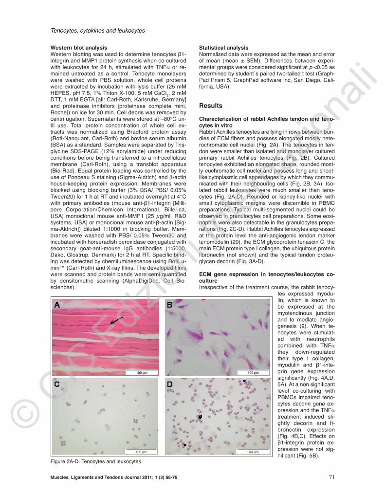
Results
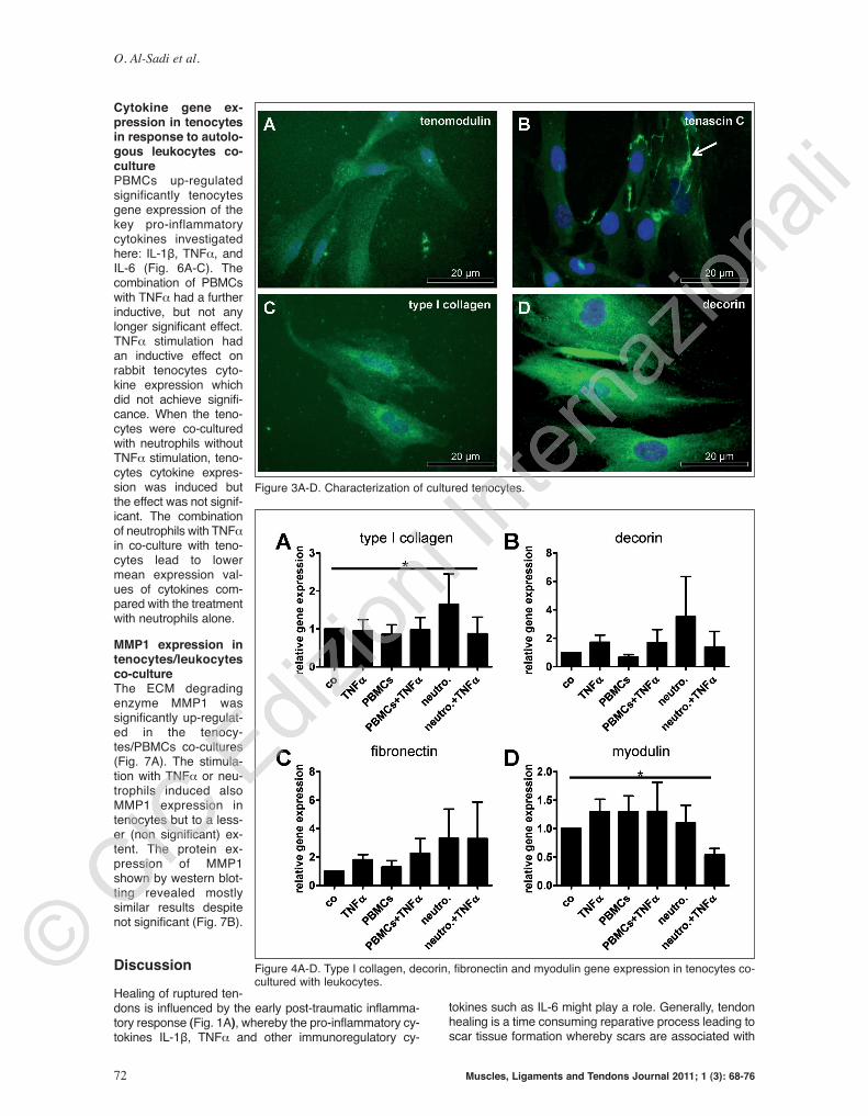
Characterization of rabbit Achilles tendon and teno-cytes in vitro Rabbit Achilles tenocytes are lying in rows between bun-dles of ECM fibers and possess elongated mostly hete-rochromatic cell nuclei (Fig. 2A). The tenocytes in ten-don were smaller than isolated and monolayer culturedprimary rabbit Achilles tenocytes (Fig. 2B). Culturedtenocytes exhibited an elongated shape, roun ded most-ly euchromatic cell nuclei and possess long and sheet-like cytoplasmic cell appendages by which they commu-nicated with their neighbouring cells (Fig. 2B, 3A). Iso-lated rabbit leukocytes were much smaller than teno-cytes (Fig. 2A-D). Rounded or kidney-like nuclei withsmall cytoplasmic margins were discernible in PBMCpre parations. Typical multi-segmented nuclei could beobserved in granulocytes cell pre parations. So me eosi -nophils were also detectable in the granulocytes prepa-rations (Fig. 2C-D). Rabbit Achilles tenocytes expressedat the protein level the anti-angiogenic tendon markertenomodulin (20), the ECM glycoprotein te nascin C, themain ECM protein type I collagen, the ubiquitous proteinfibronectin (not shown) and the typical tendon proteo-glycan decorin (Fig. 3A-D).
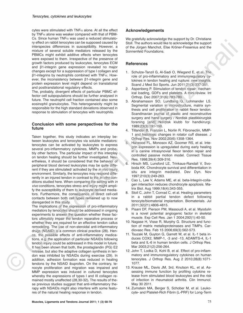
ECM gene expression in tenocytes/leukocy tes co-cultureIrrespective of the treat ment course, the rabbit tenocy-
tes expressed myodu-lin, whi ch is known tobe expressed at themyotendinous junctionand to mediate angio-genesis (9). When te -nocytes we re stimulat-ed with neutrophilscombined with TNFαthey down-regulatedtheir type I collagen,myodulin and β1-inte-grin gene expressionsignificantly (Fig. 4A,D,5A). At a non significantlevel co-culturing withPBMCs impaired teno-cytes de corin gene ex-pression and the TNFαtreatment induced sli -ghtly decorin and fi-bronectin expression(Fig. 4B,C). Effects onβ1-integrin protein ex-pression we re not sig-nificant (Fig. 5B).
Tenocytes, cytokines and leukocytes
Figure 2A-D. Tenocytes and leukocytes.
O. Al-Sadi et al.
72 Muscles, Ligaments and Tendons Journal 2011; 1 (3): 68-76
Cytokine gene ex-pression in tenocytesin response to autolo-gous leukocytes co-culturePBMCs up-regulatedsignificantly tenocytesgene expression of thekey pro-inflammatorycytokines investigatedhere: IL-1β, TNFα, andIL-6 (Fig. 6A-C). Thecombination of PBMCswith TNFα had a furtherinductive, but not anylonger significant effect.TNFα stimulation hadan inductive effect onrabbit tenocytes cyto -kine expression whichdid not achieve signifi-cance. When the teno-cytes were co-culturedwith neutrophils withoutTNFα stimulation, teno-cytes cytokine expres-sion was induced butthe effect was not signif-icant. The combinationof neutrophils with TNFαin co-culture with teno-cytes lead to lowermean expression val-ues of cytokines com-pared with the treatmentwith neutrophils alone.
MMP1 expression intenocytes/leukocytesco-cultureThe ECM degradingenzyme MMP1 wassignificantly up-regulat-ed in the teno cy -tes/PBMCs co-cultures(Fig. 7A). The stimula-tion with TNFα or neu-trophils induced alsoMMP1 expression intenocytes but to a less-er (non significant) ex-tent. The protein ex-pression of MMP1shown by western blot-ting revealed mostlysimilar results despitenot significant (Fig. 7B).
Discussion
Healing of ruptured ten-dons is influenced by the early post-traumatic inflamma-tory response (Fig. 1A), whereby the pro-inflammatory cy-tokines IL-1β, TNFα and other immunoregulatory cy-
tokines such as IL-6 might play a role. Generally, tendonhealing is a time consuming reparative process leading toscar tissue formation whereby scars are associated with
Figure 3A-D. Characterization of cultured tenocytes.
Figure 4A-D. Type I collagen, decorin, fibronectin and myodulin gene expression in tenocytes co-cultured with leukocytes.

Tenocytes, cytokines and leukocytes
Muscles, Ligaments and Tendons Journal 2011; 1 (3) 68-76 73
Figure 5A-B. β1-integrin gene and protein expression in tenocytes co-cultured with leukocytes.
Figure 6A-C. Cytokine gene expression in tenocytes co-cultured with leukocytes.

O. Al-Sadi et al.
74 Muscles, Ligaments and Tendons Journal 2011; 1 (3): 68-76
an altered function of tendon. IL-1β and TNFα are able tostrongly activate tenocytes to up-regulate their gene ex-pression for multiple cytokines such as IL-1β, TNFα, IL-6and IL-10 via auto- and paracrinic amplification loops. Ad-ditionally, these cytokines induce ECM degrading en-zymes such as MMPs and suppress their type I collagensynthesis initiating a catabolic cascade (11,12). Immigrating leukocytes are an important exogenic sour-ce for pro-inflammatory cytokines in healing tendons.They are directly released during post-traumatic bleedingor recruited from blood vessels by chemotactic signalsdue to tissue damage (Fig. 1A). Hence, in this study rab-bit tenocytes were co-cultured with autologous leukocy-tes to study the effect of leukocytes on tenocytes inducedby soluble mediators. The rabbit was used as a cellsource since it is a common experimental surgery modelto study tendon repair. Additionally, rabbit tenocytes arealso well characterized in vitro (21). Tenocytes werecombined with autologous leukocytes in the presentstudy to exclude effects of an immunogenic responseagainst allogenic cells influencing the results of co-cul-turing. However, when as a confirmation experiment hu-man hamstring tenocytes were combined with allogenichuman blood-derived leukocytes similar effects in regardto tenocytes cytokine induction by PBMCs were de-tectable (not shown). PBMCs induced the gene expres-sion of the ECM degradative enzyme MMP1, the pro-in-flammatory cytokines IL-1β, TNFα and IL-6 in tenocytes.MMP1, once activated, can cleave type I collagen.Hence, it is important for callus remodelling in the ad-vanced tendon healing phase. However, the activation ofMMP1 was not assessed in the present study: the non-glycosylated 52 kDa pro-enzyme was detected by west-ern blotting. IL-1β is a typical pro-inflammatory cytokine,up-regulated in injured tendons (22). A blockade of TNFαimproved early tendon healing in a rotator cuff healingmodel in the rat (23). In contrast, IL-6 has been implicat-ed to play essential roles in the wound healing process(24). Tendon healing was reduced in IL-6 knock out mice
(25). Nakama et al. (26) showed that IL-6 promotesblood vessel proliferation induced by vascular endothe-lial growth factor (VEGF)-dependent angiogenesis viathe Signal Transducer and Activator of Transcription(STAT3) signalling pathway. STAT3 activation may there-fore play a critical role in tendon adaptation to ischaemicstress by maintaining the intratendinous vasculature af-ter tendon injury. Since IL-6 can increase vascular per-meability, leading to oedema, this may explain the oc-currence of swelling which is a typically feature in the firsthours and days after a tendon injury and maybe neces-sary for the wound healing processes. Myodulin is a pro-angiogenic factor and could be de -monstrated in the present study in cultured rabbit teno-cytes. Myodulin expression was not restricted to the my-otendinous junction as suggested by (9) since in thepresent study the midsubstance of tendon was used fortenocyte isolation. However, tenocytes myodulin geneexpression was neither induced by PBMCs nor by neu-trophils in a significant manner but rather suppressed byneutrophils combined with TNFα.Peripheral blood-derived leukocytes were used in thepresent study, but one can assess that leukocytes immi-grating in response to chemotactic signals and hence,activated, might present a different cytokine expressionprofile and release further growth factors and mediatorsinfluencing tendon healing. Leukocytes cytokine expres-sion pattern might also be influenced by direct cell-cellcontacts between tenocytes and leukocytes and hereby,lead to a modified/enhanced response of tenocytes. Soa direct co-culture system should be applied in futurestudies. Additionally, the soluble mediators released bythe autologous leukocytes into the culture supernatantsshould be further characterised to identify key effectorcytokines and chemokines. Not only typical pro-inflam-matory cytokines but also growth factors are releasedby leukocytes such as transforming growth factor(TGF)β1 or vascular endothelial growth factor (VEGF)(14,27). In comparison to the effect of leukocytes, teno-
Figure 7A-B. MMP1 gene and protein expression in tenocytes co-cultured with leukocytes.
Tenocytes, cytokines and leukocytes
Muscles, Ligaments and Tendons Journal 2011; 1 (3) 68-76 75
cytes were stimulated with TNFα alone. At all the effectby TNFα alone was weaker compared with that of PBM-Cs. Since human TNFα was used a reduced stimulato-ry effect on rabbit tenocytes can be supposed caused byinterspecies differences in susceptibility. However, amixture of several soluble mediators released by thePBMCs might exhibit additive effects when tenocyteswere exposed to them. Irrespective of the presence ofgrowth factors produced by leukocytes, tenocytes ECMand β1-integrin gene expression revealed no majorchanges except for a suppression of type I collagen andβ1-integrins by neutrophils combined with TNFα. How-ever, the inconsistency between β1-integrin gene andprotein expression level might depend on translationaland posttranslational regulatory effects.The, probably, divergent effects of particular PBMC ef-fector cell subpopulations should be further analysed infuture. The neutrophil cell fraction contained also someeosinophil granulocytes. This heterogeneicity might beresponsible for the high standard deviations observed inresponse to stimulation of tenocytes with neutrophils.
Conclusion with some perspectives for thefuture
Taken together, this study indicates an interplay be-tween leukocytes and tenocytes via soluble mediators:tenocytes can be activated by leukocytes to expressseveral pro-inflammatory cytokines, MMPs and proba-bly other factors. The particular impact of this interplayon tendon healing should be further investigated. Nev-ertheless, it should be considered that the behavior ofperipheral blood derived leukocytes may be quite diffe-rent if they are stimulated/ activated by the in vivo injuryenvironment. Similarly, the tenocytes may respond diffe-rently in an injured tendon in contrast to the in vitro con-ditions studied here. When comparing the setting with invivo conditions, tenocytes stress and injury might ampli-fy the susceptibility of them to leukocyte derived media-tors. Furthermore, the consequence of direct cell-cellcontacts between both cell types remained up to nowdisregarded in this study.The implications of the expression of pro-inflammatorymediators by tenocytes should be addressed in ongoingexperiments to answer the question whether these fac-tors ultimately impair the tendon reparative process orwhether they are required for an effective tendon callusremodeling. The use of non-steroidal anti-inflammatorydrugs (NSAID) is a common clinical practice (28). Hen-ce, the possible effects of anti-inflammatory medica-tions, e.g. the application of particular NSAIDs followingtendon injury could be addressed in this model in future.It has been shown that both, the prostaglandin (PG) E2release, but also the adaptive collagen synthesis in ten-don was inhibited by NSAIDs during exercise (29). Inaddition, adhesion formation was reduced in healingtendons by the NSAID ibuprofen. On the contrary, te-nocyte proliferation and migration was impaired andMMP expression was induced in cultured tenocyteswhereby the expressions of types I and III collagen re-mained mostly unaffected (28,30-32). The results of the-se previous studies suggest that anti-inflammatory the-rapy with NSAIDs might also interfere with some featu-res of the natural healing response in tendon.
Acknowledgements
We gratefully acknowledge the support by Dr. Christiane
Stoll. The authors would like to acknowledge the support
of the Jürgen Manchot, Else Kröner-Fresenius and the
Sonnenfeld Foundations.
References
1. Schulze-Tanzil G, Al-Sadi O, Wiegand E, et al. The
role of pro-inflammatory and immunoregulatory cy-
tokines in tendon healing and rupture: new insights.
Scand J Med Sci Sports. Jun 2011;21(3):337-351.
2. Aspenberg P. Stimulation of tendon repair: mechan-
ical loading, GDFs and platelets. A mini-review. Int
Orthop. Dec 2007;31(6):783-789.
3. Abrahamsson SO, Lundborg G, Lohmander LS.
Segmental variation in microstructure, matrix syn-
thesis and cell proliferation in rabbit flexor tendon.
Scandinavian journal of plastic and reconstructive
surgery and hand surgery / Nordisk plastikkirurgisk
forening [and] Nordisk klubb for handkirurgi.
1989;23(3):191-198.
4. Tillander B, Franzen L, Norlin R. Fibronectin, MMP-
1 and histologic changes in rotator cuff disease. J
Orthop Res. Nov 2002;20(6):1358-1364.
5. Harwood FL, Monosov AZ, Goomer RS, et al. Inte-
grin expression is upregulated during early healing
in a canine intrasynovial flexor tendon repair and
controlled passive motion model. Connect Tissue
Res. 1998;39(4):309-316.
6. Hirsch MS, Lunsford LE, Trinkaus-Randall V, Svo-
boda KK. Chondrocyte survival and differentiation in
situ are integrin mediated. Dev Dyn. Nov
1997;210(3):249-263.
7. Cao L, Lee V, Adams ME, et al. beta-Integrin-colla-
gen interaction reduces chondrocyte apoptosis. Ma-
trix Biol. Aug 1999;18(4):343-355.
8. Stoll C, John T, Conrad C, et al. Healing parameters
in a rabbit partial tendon defect following
tenocyte/biomaterial implantation. Biomaterials. Jul
2011;32(21):4806-4815.
9. Pisani DF, Pierson PM, Massoudi A, et al. Myodulin
is a novel potential angiogenic factor in skeletal
muscle. Exp Cell Res. Jan 1 2004;292(1):40-50.
10. Nagase H, Visse R, Murphy G. Structure and func-
tion of matrix metalloproteinases and TIMPs. Car-
diovasc Res. Feb 15 2006;69(3):562-573.
11. Tsuzaki M, Guyton G, Garrett W, et al. IL-1 beta in-
duces COX2, MMP-1, -3 and -13, ADAMTS-4, IL-1
beta and IL-6 in human tendon cells. J Orthop Res.
Mar 2003;21(2):256-264.
12. John T, Lodka D, Kohl B, et al. Effect of pro-inflam-
matory and immunoregulatory cytokines on human
tenocytes. J Orthop Res. Aug 2 2010;28(8):1071-
1077.
13. Krause ML, Davis JM, 3rd, Knutson KL, et al. As-
sessing immune function by profiling cytokine re-
lease from stimulated blood leukocytes and the risk
of infection in rheumatoid arthritis. Clin Immunol.
May 30 2011.
14. Zumstein MA, Berger S, Schober M, et al. Leuko-
cyte- and Platelet-Rich Fibrin (L-PRF) for Long-Term
O. Al-Sadi et al.
76 Muscles, Ligaments and Tendons Journal 2011; 1 (3): 68-76
Delivery of Growth Factor in Rotator Cuff Repair:Review, Preliminary Results and Future Directions.Curr Pharm Biotechnol. Jul 8 2011.
15. de Mos M, van der Windt AE, Jahr H, et al. Canplatelet-rich plasma enhance tendon repair? A cellculture study. Am J Sports Med. Jun 2008;36(6):1171-1178.
16. Kajikawa Y, Morihara T, Sakamoto H, et al. Platelet-rich plasma enhances the initial mobilization of cir-culation-derived cells for tendon healing. J CellPhysiol. Jun 2008;215(3):837-845.
17. Schulze-Tanzil G, Mobasheri A, Clegg PD, SendzikJ, John T, Shakibaei M. Cultivation of human teno-cytes in high-density culture. Histochem Cell Biol.Sep 2004;122(3):219-228.
18. Stoll C, John T, Endres M, et al. Extracellular matrixexpression of human tenocytes in three-dimension-al air-liquid and PLGA cultures compared with ten-don tissue: Implications for tendon tissue engineer-ing. J Orthop Res. Sep 24 2010;28(9)1170-1177.
19. Livak KJ, Schmittgen TD. Analysis of relative geneexpression data using real-time quantitative PCRand the 2(-Delta Delta C(T)) Method. Methods (SanDiego, Calif. Dec 2001;25(4):402-408.
20. Docheva D, Hunziker EB, Fassler R, Brandau O.Tenomodulin is necessary for tenocyte proliferationand tendon maturation. Mol Cell Biol. Jan2005;25(2):699-705.
21. Bernard-Beaubois K, Hecquet C, Houcine O,Hayem G, Adolphe M. Culture and characterizationof juvenile rabbit tenocytes. Cell Biol Toxicol. Feb1997;13(2):103-113.
22. Berglund M, Hart DA, Wiig M. The inflammatory re-sponse and hyaluronan synthases in the rabbit flex-or tendon and tendon sheath following injury. J HandSurg. Oct 2007;32(5):581-587.
23. Gulotta LV, Kovacevic D, Cordasco F, Rodeo SA.Evaluation of Tumor Necrosis Factor alpha Block-ade on Early Tendon-to-Bone Healing in a Rat Rota-tor Cuff Repair Model. Arthroscopy. Jun 25 2011.
24. Gallucci RM, Simeonova PP, Matheson JM, et al.Impaired cutaneous wound healing in interleukin-6-deficient and immunosuppressed mice. Faseb J.Dec 2000;14(15):2525-2531.
25. Lin TW, Cardenas L, Glaser DL, Soslowsky LJ. Ten-don healing in interleukin-4 and interleukin-6 knock-out mice. J. Biomech. 2006;39(1):61-69.
26. Nakama K, Gotoh M, Yamada T, et al. Interleukin-6-induced activation of signal transducer and activatorof transcription-3 in ruptured rotator cuff tendon. J IntMed Res. Nov-Dec 2006;34(6):624-631.
27. Dohan Ehrenfest DM, Bielecki T, Jimbo R, et al. Dothe Fibrin Architecture and Leukocyte Content Influ-ence the Growth Factor Release of Platelet Con-centrates? An Evidence-Based Answer Comparinga Pure Platelet-Rich Plasma (P-PRP) Gel and aLeukocyte- and Platelet-Rich Fibrin (L-PRF). CurrPharm Biotechnol. Jul 8 2011.
28. Tsai WC, Hsu CC, Chang HN, Lin YC, Lin MS, PangJH. Ibuprofen upregulates expressions of matrixmetalloproteinase-1, -8, -9, and -13 without affectingexpressions of types I and III collagen in tendoncells. J Orthop Res. Apr 2010;28(4):487-491.
29. Christensen B, Dandanell S, Kjaer M, Langberg H.Effect of anti-inflammatory medication on the run-ning-induced rise in patella tendon collagen synthe-sis in humans. J Appl Physiol. Jan 2011;110(1):137-141.
30. Tan V, Nourbakhsh A, Capo J, Cottrell JA, Meyen-hofer M, O’Connor JP. Effects of nonsteroidal anti-inflammatory drugs on flexor tendon adhesion. JHand Surg. Jun 2010;35(6):941-947.
31. Tsai WC, Tang FT, Hsu CC, Hsu YH, Pang JH, Shi-ue CC. Ibuprofen inhibition of tendon cell prolifera-tion and upregulation of the cyclin kinase inhibitorp21CIP1. J Orthop Res. May 2004;22(3):586-591.
32. Tsai WC, Hsu CC, Chen CP, Chen MJ, Lin MS, PangJH. Ibuprofen inhibition of tendon cell migration anddown-regulation of paxillin expression. J OrthopRes. Mar 2006;24(3):551-558.