Embed Size (px)
Citation preview
Cancer Cell
Previews
et al., 2002). No mutations in p38α have been reported in primary tumors; the presence of three other p38 MAPK genes could compensate for any par-tial loss of function of p38α.
The successful creation of a tumor requires that the apoptotic and antip-roliferative effects of p38 MAPK must be suppressed. Dolado et al. propose that one mechanism employed by tumor cells to overcome the tumor-suppressive function of p38 MAPK is to uncouple the production of ROS from p38 MAPK activation. The increased expression of GSTm proteins (Gstm1 and GSTm2) observed in tumor cells may serve this function. Dolado et al. show that reduced expression of GSTm2 in MCF7 breast cancer cells increased p38 MAPK activity and apoptosis, whereas forced overex-pression of GSTm2 further potentiated the transformed phenotype. Since
GSTm proteins can inhibit the ASK1/p38 pathway, these data are compel-ling. However, it is unclear if the over-expression of GSTm proteins in these cancer cells is a cause of or a result of transformation or if the increased levels reflect responses to tumor treatment or passage in tissue culture. Neverthe-less, proteins that serve as sensors for ROS levels (e.g., GSTm1/2) and other proteins that attenuate the p38 MAPK pathway (e.g., WIP1) represent candi-date drug targets for the design of new therapies for cancer.
RefeRences
Brancho, D., Tanaka, N., Jaeschke, A., Ven-tura, J.J., Kelkar, N., Tanaka, Y., Kyuuma, M., Takeshita, T., Flavell, R.A., and Davis, R.J. (2003). Genes Dev. 17, 1969–1978.
Bulavin, D.V., Demidov, O.N., Saito, S., Kaura-niemi, P., Phillips, C., Amundson, S.A., Ambro-sino, C., Sauter, G., Nebreda, A.R., Anderson,
C.W., et al. (2002). Nat. Genet. 31, 210–215.
Bulavin, D.V., and Fornace, A.J., Jr. (2004). Adv. Cancer Res. 92, 95–118.
Bulavin, D.V., Phillips, C., Nannenga, B., Timofeev, O., Donehower, L.A., Anderson, C.W., Appella, E., and Fornace, A.J., Jr. (2004). Nat. Genet. 36, 343–350.
Chen, G., Hitomi, M., Han, J., and Stacey, D.W. (2000). J. Biol. Chem. 275, 38973–38980.
Dolado, I., Swat, A., Ajenjo, N., De Vita, G., Cuadrado, A., and Nebreda, A.R. (2007). Can-cer Cell, this issue.
Irani, K., Xia, Y., Zweier, J.L., Sollott, S.J., Der, C.J., Fearon, E.R., Sundaresan, M., Finkel, T., and Goldschmidt-Clermont, P.J. (1997). Sci-ence 275, 1649–1652.
Kennedy, N.J., and Davis, R.J. (2003). Cell Cy-cle 2, 199–201.
Lavoie, J.N., L’Allemain, G., Brunet, A., Muller, R., and Pouyssegur, J. (1996). J. Biol. Chem. 271, 20608–20616.
Manke, I.A., Nguyen, A., Lim, D., Stewart, M.Q., Elia, A.E., and Yaffe, M.B. (2005). Mol. Cell 17, 37–48.
Ten Genes for Inherited Breast cancerTom Walsh1 and Mary-Claire King1,*1Departments of Medicine and Genome Sciences, University of Washington, Seattle, Washington 98195, USA*Correspondence: [email protected], [email protected] 10.1016/j.ccr.2007.01.010
Inherited breast cancer is associated with germline mutations in ten different genes in pathways critical to genomic integrity. BRCA1 and BRCA2 mutations confer very high risks of breast and ovarian cancer. p53 and PTEN mutations lead to very high breast cancer risks associated with rare cancer syndromes. Mutations in CHEK2, ATM, NBS1, RAD50, BRIP1, and PALB2 are associated with doubling of breast cancer risks. In addition, biallelic mutations in BRCA2, BRIP1, and PALB2 cause Fanconi anemia. The convergence of these genes in a shared role reveals underlying biology of these illnesses and suggests still other breast cancer genes.
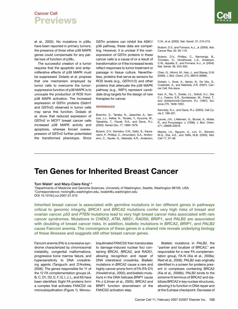
Fanconi anemia (FA) is a recessive syn-drome characterized by chromosomal instability, congenital malformations, progressive bone marrow failure, and hypersensitivity to DNA crosslink-ing agents (Taniguchi and D’Andrea, 2006). The genes responsible for 11 of the 12 FA complementation groups (A, B, C, D1, D2, E, F, G, I, J, L, and M) have been identified. Eight FA proteins form a complex that activates FANCD2 via monoubiquitination (Figure 1). Monou-
biquitinated FANCD2 then translocates to damage-induced nuclear foci con-taining BRCA1, BRCA2, and RAD51, allowing recognition and repair of DNA interstrand crosslinks. Biallelic mutations in BRCA2 cause a rare and highly cancer-prone form of FA (FA-D1) (Howlett et al., 2002), and biallelic muta-tions in the DNA helicase BRIP1 cause FA-J (Litman et al., 2005). BRCA2 and BRIP1 function downstream of the FANCD2 activation step.
Cancer Cell 11
Biallelic mutations in PALB2, the “partner and localizer of BRCA2,” are responsible for a new FA complemen-tation group, FA-N (Xia et al., 2006a; Reid et al., 2006). PALB2 was originally identified in a screen for proteins pres-ent in complexes containing BRCA2 (Xia et al., 2006b). PALB2 binds to the extreme N terminus of BRCA2 and sta-bilizes BRCA2 in key nuclear structures, allowing it to function in DNA repair and at the S phase checkpoint. Decrease of
, February 2007 ©2007 Elsevier Inc. 103
Cancer Cell
Previews
figure 1. Interactions of Proteins Associated with Inherited Breast cancer and with fanconi AnemiaA complex of eight Fanconi proteins (A, B, C, E, F, G, L, and M) activates FANCD2 via monoubiq-uitination, allowing FANCD2 to translocate to damage-induced nuclear foci that contain BRCA1, BRCA2, and RAD51. DNA damage activates ATM and CHEK2, which in turn activate BRCA1 by phosphorylation. PTEN binds to the Rad51 promoter and may regulate its transcription (Shen et al., 2007). Proteins indicated in red carry germline mutations that predispose to breast cancer. The numbers indicate different breast-cancer-associated mutations identified thus far in each gene. A germline variant in the promoter of Rad51 (blue) may modify breast cancer risk in BRCA2 mutation carriers (Levy-Lahad et al., 2001).
PALB2 expression in HeLa cells by siR-NAs leads to mitomycin C sensitivity, which causes interstrand crosslinking and eventually double-strand breaks. Mitomycin C sensitivity is a hallmark of Fanconi anemia.
Reid and colleagues identified bial-lelic protein-truncating mutations in PALB2 in 7 of 82 individuals with FA not due to known genes (Reid et al., 2006). In a lymphoblastoid line from one patient with two such muta-tions, transfection of wild-type PALB2 reversed sensitivity to mitomycin C. All seven individuals with FA-N developed cancer as children; six of the seven patients died before age 4.
Xia and colleagues evaluated one FA patient with normal monoubiquitination of FANCD2 and wild-type sequences of BRCA2 and BRIP1 (Xia et al., 2006a). This patient carried a premature trun-cation in exon 4 of PALB2 inherited from the mother and a complete dele-tion of PALB2 inherited from the father. A subline of the patient’s lymphoblasts developed MMC resistance, that is, became a phenotypic revertant. Genomic analysis of this line revealed an Alu-mediated deletion of exon 4 that contains the premature truncation,
104 Cancer Cell 11, February 2007 ©200
creating an in-frame mRNA with exon 3 spliced to exon 5.
The cellular and cancer pheno-types of patients with biallelic BRCA2 mutations and of patients with biallelic PALB2 mutations are quite similar. Het-erozygosity for mutations in BRCA2 are associated with very high risk of breast cancer. Is it possible that heterozygos-ity for mutations in PALB2 also increase risk of breast cancer?
Rahman and colleagues sequenced the 13 coding exons of PALB2 in DNA from 923 familial breast cancer patients with wild-type sequences at BRCA1 and BRCA2 (Rahman et al., 2006). Five different truncating mutations of PALB2 were identified in ten patients (1% of the series; two of the five muta-tions were also observed in the FA-N families described above). No truncat-ing mutations of PALB2 appeared in 1084 control individuals. The relative risk of breast cancer associated with PALB2 was estimated at 2.3 (3.0 for women younger than 50 years and 1.9 for women older than 50 years).
Erkko and colleagues sequenced PALB2 in DNA from 113 families from northern Finland with breast and ovar-ian cancer, identifying a premature
7 Elsevier Inc.
truncation in three families (Erkko et al., 2007). Further genotyping revealed the allele in 18 of 1918 (0.9%) breast cancer cases not selected for family history from the northern Finland pop-ulation and 6 of 2501 (0.2%) controls, consistent with a 2- to 4-fold increased risk to mutation carriers.
PALB2 is a new addition to the growing list of genes associated with approximately 2-fold increased risk of breast cancer. CHEK2 was the first gene of this type described. Truncating allele 1100delC of CHEK2 was identi-fied through a combined linkage and candidate gene approach in a single severely affected breast cancer fam-ily (Meijers-Heijboer et al., 2002). This allele has a frequency of 0.002–0.005 in northern Europe populations and con-fers an approximately 2-fold increased risk of breast cancer. Other CHEK2 mutations have been subsequently associated with similarly increased risk of breast cancer (Walsh et al., 2006). In NBS1, a rare protein-truncating allele first identified in Polish breast cancer patients is associated with approxi-mately 2-fold increased risk. Rare mutations of BRIP1 (FA-J) were also identified in breast cancer families (Seal et al., 2006). Heterozygous car-riers of ATM mutations have long been suspected of having increased risk of breast cancer. Recent comprehen-sive sequencing of ATM in 434 familial breast cancer patients revealed seven protein-truncating alleles, two splice mutations, and two missense alleles (one of which appeared in two families) experimentally verified to affect ATM function. In contrast, only two ATM sequence variants were found in 521 controls (Renwick et al., 2006). Finally, a protein-truncating allele of RAD50 identified in northern Finland conferred an approximate 4-fold increased risk of breast cancer (Heikkinen et al., 2006). In each of these genes, more mutations are likely to be found: more populations will be analyzed, genomic rearrange-ments will be evaluated, and a larger subset of missense alterations will be validated by functional assays.
These ten genes share two impor-tant features in their impact on breast cancer. (1) A single deleterious muta-tion in any one of them is sufficient to
Cancer Cell
Previews
significantly increase breast cancer risk. (2) There are many deleterious mutations, and each mutation is indi-vidually rare. That is, for none of these genes (individually or in combination) does increased risk of breast cancer result from additive effects of mul-tiple common alleles, each of small influence. Inherited breast cancer is highly genetically heterogeneous with respect to both loci and alleles involved. All evidence to date is that the model that best reflects this het-erogeneity is not a “common disease-common allele” model, but instead a “common disease-multiple rare alleles” model.
The ten known genes for inherited breast cancer function in a pathway whose role is to preserve genomic integrity. Roughly 50% of familial breast cancer remains unresolved by any of these genes. Clearly other genes in this pathway are worthy of in-depth genomic analysis in unre-solved families. Furthermore, in thus far unrecognized members of this pathway, mutations may also be asso-ciated with inherited breast cancer.
Wilms tumor (WT) is a childhood embryonal cancer of the kidney. Unlike most tumors, Wilms tumors gener-ally exhibit few, if any, chromosomal
Wilms Tumor GeTwist to the storVicki Huff1,*1Department of Cancer Genetics, Unit 101Houston, TX 77030, USA*Correspondence: [email protected] 10.1016/j.ccr.2007.01.011
The study of the genetics of Wilmlishing discoveries. Ironically, howgene mutations have been identgene, WTX, that is mutated somexplain the genetic etiology of a that X chromosome genes can p
RefeRences
Erkko, H., Xia, B., Nikkila, J., Schleutker, J., Syrjakoski, K., Mannermaa, A., Kallioniemi, A, Pylkas, K., Karppinen, S-M., Rapakko, K., et al. (2007). Nature. Published online February 7, 2007. 10.1038/nature05609.
Heikkinen, K., Rapakko, K., Karppinen, S.M., Erkko, H., Knuutila, S., Lundan, T., Mannermaa, A., Borresen-Dale, A.L., Borg, A., Barkardottir, R.B., et al. (2006). Carcinogenesis 27, 1593–1599.
Howlett, N.G., Taniguchi, T., Olson, S., Cox, B., Waisfisz, Q., De Die-Smulders, C., Persky, N., Grompe, M., Joenje, H., Pals, G., et al. (2002). Science 297, 606–609.
Levy-Lahad, E., Lahad, A., Eisenberg, S., Da-gan, E., Paperna, T., Kasinetz, L., Catane, R., Kaufman, B., Beller, U., Renbaum, P., et al. (2001). Proc. Natl. Acad. Sci. USA 98, 3232–3236.
Litman, R., Peng, M., Jin, Z., Zhang, F., Zhang, J., Powell, S., Andreassen, P.R., and Cantor, S.B. (2005). Cancer Cell 8, 255–265.
Meijers-Heijboer, H., van den Ouweland, A., Kli-jn, J., Wasielewski, M., de Snoo, A., Oldenburg, R., Hollestelle, A., Houben, M., Crepin, E., van Veghel-Plandsoen, M., et al. (2002). Nat. Genet. 31, 55–59.
Rahman, N., Seal, S., Thompson, D., Kelly, P., Renwick, A., Elliott, A., Reid, S., Spanova, K., Barfoot, R., Chagtai, T., et al. (2006). Nat. Genet. Published online December 31, 2006. 10.1038/
Cancer Cell 1
abnormalities, and therefore WT was originally thought to represent a sim-ple model for studying the genetic etiology of cancer. WT genetics, how-
netics: A new, Uy
0, University of Texas M.D. Anderson Canc
s tumor has led to several highly uever, the identification of “WT ge
ified in only ?25% of tumors. Theatically in ?30% of Wilms tumo
substantial proportion of tumors alay in cancer genetics.
ng1959.
Reid, S., Schindler, D., Hanenberg, H., Barker, K., Hanks, S., Kalb, R., Neveling, K., Kelly, P., Seal, S., Freund, M., et al. (2006). Nat. Genet. Published online December 31, 2006. 10.1038/ng1947.
Renwick, A., Thompson, D., Seal, S., Kelly, P., Chagtai, T., Ahmed, M., North, B., Jayatilake, H., Barfoot, R., Spanova, K., et al. (2006). Nat. Genet. 38, 873–875.
Seal, S., Thompson, D., Renwick, A., Elliott, A., Kelly, P., Barfoot, R., Chagtai, T., Jayatilake, H., Ahmed, M., Spanova, K., et al. (2006). Nat. Genet. 38, 1239–1241.
Shen, W.H., Balajee, A.S., Wang, J., Wu, H., Eng, C., Pandolfi, P.P., and Yin, Y. (2007). Cell 128, 157–170.
Taniguchi, T., and D’Andrea, A.D. (2006). Blood 107, 4223–4233.
Walsh, T., Casadei, S., Coats, K.H., Swisher, E., Stray, S.M., Higgins, J., Roach, K.C., Mandell, J., Lee, M.K., Ciernikova, S., et al. (2006). JAMA 295, 1379–1388.
Xia, B., Dorsman, J.C., Ameziane, N., de Vries, Y., Rooimans, M.A., Sheng, Q., Pals, G., Errami, A., Gluckman, E., Llera, J., et al. (2006a). Nat. Genet. Published online December 31, 2006. 10.1038/ng1942.
Xia, B., Sheng, Q., Nakanishi, K., Ohashi, A., Wu, J., Christ, N., Liu, X., Jasin, M., Couch, F.J., and Livingston, D.M. (2006b). Mol. Cell 22, 719–729.
1, February 2007 ©2007 Elsevier Inc. 105
ever, has turned out to be anything but simple; the road to identifying and understanding “WT genes” has been littered with false leads and dashed
nX-pected
er Center, 1515 Holcombe Boulevard,
nexpected and precedent-estab-nes” has been painfully slow, and discovery of an X chromosome
rs is notable both for helping to nd also for underscoring the role


![Fundamentals of genetics A.ppt [Read-Only]...inherited as multiple genes that alsoinherited as multiple genes that also illustrate incomplete dominance. What would be the possible](https://img.pdfslide.us/doc/110x75/5ecbfad337999c04ec2d38b8/fundamentals-of-genetics-appt-read-only-inherited-as-multiple-genes-that.jpg)