Embed Size (px)
Citation preview
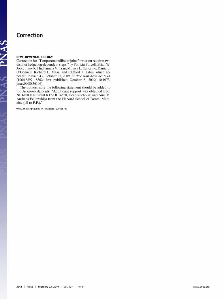
Correction
DEVELOPMENTAL BIOLOGYCorrection for “Temporomandibular joint formation requires twodistinct hedgehog-dependent steps,” by Patricia Purcell, BrianW.Joo, JimmyK.Hu, PamelaV. Tran,Monica L. Calicchio,Daniel J.O’Connell, Richard L. Maas, and Clifford J. Tabin, which ap-peared in issue 43, October 27, 2009, of Proc Natl Acad Sci USA(106:18297–18302; first published October 8, 2009; 10.1073/pnas.0908836106).The authors note the following statement should be added to
the Acknowledgments: “Additional support was obtained fromNIH/NIDCR Grant K12-DE14528, Dean’s Scholar, and Aina M.Auskaps Fellowships from the Harvard School of Dental Medi-cine (all to P.P.).”
www.pnas.org/cgi/doi/10.1073/pnas.1000188107
3942 | PNAS | February 23, 2010 | vol. 107 | no. 8 www.pnas.org
Temporomandibular joint formation requires twodistinct hedgehog-dependent stepsPatricia Purcella,b, Brian W. Jooa,b, Jimmy K. Hua, Pamela V. Tranb, Monica L. Calicchioc, Daniel J. O’Connellb,Richard L. Maasb, and Clifford J. Tabina,1
aDepartment of Genetics, Harvard Medical School, Boston, MA 02115; bDivision of Genetics, Department of Medicine, Brigham and Women’s Hospital andHarvard Medical School, Boston, MA 02115; and cDepartment of Pathology, Children’s Hospital Boston, Boston, MA 02115
Contributed by Clifford J. Tabin, August 10, 2009 (sent for review July 17, 2009)
We conducted a genetic analysis of the developing temporo-man-dibular or temporomandi-bular joint (TMJ), a highly specialized sy-novial joint that permits movement and function of the mammalianjaw. First, we used laser capture microdissection to perform a ge-nome-wide expression analysis of each of its developing components.The expression patterns of genes identified in this screen wereexamined in the TMJ and compared with those of other synovialjoints, including the shoulder and the hip joints. Striking differenceswere noted, indicating that the TMJ forms via a distinct molecularprogram. Several components of the hedgehog (Hh) signaling path-way are among the genes identified in the screen, including Gli2,which is expressed specifically in the condyle and in the disk of thedeveloping TMJ. We found that mice deficient in Gli2 display aberrantTMJ development such that the condyle loses its growth-plate-likecellular organization and no disk is formed. In addition, we used aconditional strategy to remove Smo, a positive effector of the Hhsignaling pathway, from chondrocyte progenitors. This cell autono-mous loss of Hh signaling allows for disk formation, but the resultingstructure fails to separate from the condyle. Thus, these experimentsestablish that Hh signaling acts at two distinct steps in disk morpho-genesis, condyle initiation, and disk–condyle separation and providea molecular framework for future studies of the TMJ.
Indian hedgehog � Gli2 � synovial joints � microarray
The temporomandibular joint (TMJ) is a complex structure thatis essential for jaw movement and found only in mammals. Its
major components include the glenoid fossa of the temporal bone,the condylar head of the mandible, and a fibrocartilaginous disk thatis located between these bones, dividing the joint cavity into twocompartments. Both the condyle and the glenoid fossa are endo-chondral in origin. The first evidence of TMJ formation duringdevelopment is the appearance of distinct mesenchymal conden-sations, the temporal and condylar blastemas. The condylar blast-ema rapidly grows toward the temporal blastema, closing the gapbetween them while a distinct articular disk forms within the jointas a separate condensation (1).
The TMJ differs from most synovial joints in several ways. First,the TMJ forms by appositional growth, as opposed to segmentationof a continuous skeletal condensation. Second, in the TMJ, thearticular surfaces of the condyle and glenoid fossa are covered bya layer of fibrous rather than hyaline cartilage. Last, the two bonesare in contact with an intervening fibrocartilaginous disk ratherthan articulating with each other directly. The development of theTMJ during prenatal life also lags behind other joints in both thetime of its initiation and its development. In the mouse, all of themajor anatomical features of the TMJ, including the disk, arepresent by E16.5, although the condyle and glenoid fossa continueto increase in size and density into adulthood.
Although the structural features of the TMJ are well docu-mented, little information is available with respect to the genetic,cellular, and molecular mechanisms involved in TMJ morphogen-esis. In contrast, studies of other skeletal elements, most notably ofthe developing limb, have provided a wealth of information aboutsignals involved in synovial joint formation. Most synovial joints
develop by the cleavage or segmentation of a continuous skeletalcondensation (2–4). The first morphological sign of joint formationis the appearance of a transverse stripe of cells, the interzone, athree-layered region with reduced cell density in the center thatmarks the area destined to become the joint space (5, 6). Thismorphological change is presaged by molecular events, includingthe down-regulation of several genes expressed in the remainder ofthe developing cartilage, such as Sox9, a member of the Sox familyof transcription factors present in all chondroprogenitor cells (7, 8).Conversely, a large number of genes are induced specifically in thelocation of the future joint. Prominent among these are Wnt9a(formerly called Wnt14), a canonical Wnt ligand (9–11), anddepending on the specific joint, Gdf5, Gdf6, or Gdf7, members ofthe BMP/TGF� superfamily (12–15). Strikingly, these genes areexpressed during and act in the formation of joints that formbetween the long bones by segmentation and in other classes ofjoints such as those between vertebrae and those between calvarialmembranous bones. Different members of the Gdf family areexpressed in diverse joints, and the loss of Gdf activity results in thefailure of joint formation (16). In addition, �-catenin activity, a keyeffector of the canonical Wnt pathway, is required for joint forma-tion, and ectopic Wnt9a is sufficient to initiate the formation of ajoint interzone (9, 11, 17).
The condyle is an important growth site in the mandible withsimilarities to the growth plate of the long bones, and it displays fourdistinct zones: a fibrous cell layer, a progenitor cell layer, a zone offlattened chondrocytes, and a zone of hypertrophic chondrocytes(Fig. 1, upper right) (18, 19). One key gene previously noted to beexpressed during and function within the growth plate of thecondylar cartilage is Indian hedgehog (Ihh) (20–22). Ihh has beenstudied extensively during endochondral ossification of the longbones, where it plays several distinct roles. Secreted by prehyper-trophic condrocytes that are just entering the differentiation path-way, Ihh is critical for maintaining the growth of adjacent prolif-erating chondrocytes. In addition, Ihh plays an indirect role inregulating the rate of chondrocyte differentiation by acting in anegative feedback loop with a second secreted protein, parathyroid-hormone-related protein (PTHrP), in the periarticular perichon-drium. Chondrocytes within the range of PTHrP signaling are inturn blocked from entering the differentiation pathway. Thus, Ihh,in conjunction with PTHrP, plays a crucial role in organizing thegrowth plate (23–25). In potentially analogous fashion, in theabsence of Ihh, the organization of the growth-plate-like zone in theTMJ condyle is disrupted, and the TMJ disk does not form (21). Ihhsignals through its receptor Ptc1, itself a transcriptional target ofIhh. Acting through a second transmembrane protein, Smo, Ihhactivity serves to regulate the processing and activity of the Glifamily of transcription factors (26). Gli1 itself is transcriptionallyup-regulated by Ihh signaling and is a transcriptional activator of
Author contributions: P.P. and C.J.T. designed research; P.P., B.W.J., and M.L.C. performedresearch; J.K.H. and P.V.T. contributed new reagents/analytic tools; P.P., D.J.O., R.L.M., andC.J.T. analyzed data; and P.P. and C.J.T. wrote the paper.
The authors declare no conflict of interest.
1To whom correspondence should be addressed. E-mail: [email protected].
www.pnas.org�cgi�doi�10.1073�pnas.0908836106 PNAS � October 27, 2009 � vol. 106 � no. 43 � 18297–18302
DEV
ELO
PMEN
TAL
BIO
LOG
Y
Ihh targets. Gli3 acts predominantly as a transcriptional repressorand is down-regulated transcriptionally by Ihh signaling. Moreover,Ihh activity alters the processing of full-length Gli3 protein such thatthe repressor form is not produced. Finally, Gli2 functions mostlyas a transcriptional activator, although in the absence of Ihh activityit retains some repressor function (27, 28). Functional Gli2 isnecessary for normal endochondral bone development, as indicatedin mice lacking the Gli2 gene (29, 30), whereas in early limbdevelopment Gli3 is the only significant mediator of hedgehog (Hh)signaling (downstream of the Ihh-relative, Sonic hedgehog) (31).During skeletogenesis, the three Gli family members are active andexhibit some degree of functional redundancy in mediating Ihhsignaling (27, 29).
In the present study, we sought to identify and further investigategenes that participate in TMJ development. Our observationsindicate that the TMJ is unique not only structurally but also withrespect to the expression of several key regulatory molecules.Furthermore, we analyzed the role of Hh signaling and found thatit acts during at least two distinct steps in TMJ disk morphogenesis.
Results and DiscussionIdentification of Genes Expressed in the TMJ. To identify genesexpressed during the development of the major structures of theTMJ, we focused on tissues in the E16.5 mouse. At this stage, all ofthe distinct components of the TMJ have formed, but the joint is stillin the process of maturation. The articular condyle and fossa wereisolated separately by laser capture microdissection (LCM). TheTMJ disk was included with the condyle sample. The RNA wasextracted from the isolated tissues in triplicate and was subjected tomicroarray analysis. The E16.5 whole mouse was used as a refer-ence sample. We found 3,035 probes enriched more than two-foldin the condyle sample with respect to the whole mouse referenceand 1,465 probes enriched more than two-fold in the fossa. Geneswith the highest signal intensities and fold enrichment were vali-dated by in situ hybridization and selected for further analysis, witha particular focus on genes involved in known signaling pathways.
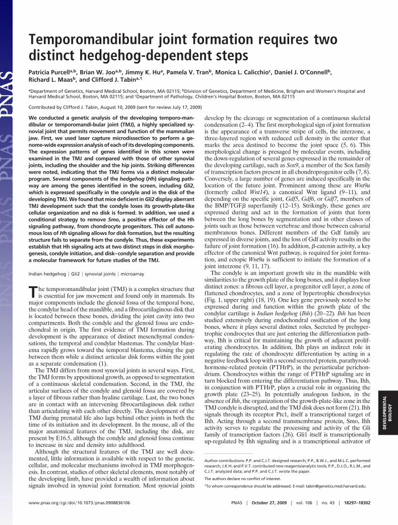
Consistent with the fact that both the condyle and fossa form byendochondral ossification, our array data showed the expression ofmany genes known to be involved in this process. For example, FGFand FGF receptor (Fgfr) gene families encode essential signalingmolecules that function throughout all stages of bone development.Fgfr1, Fgfr2, and Fgfr3 were highly expressed in our microarrays andwere found by in situ analysis to be expressed in time-dependent
and spatially restricted patterns. Fgfr1 was expressed in the perios-teum of the condyle and fossa, Fgfr2 in the perichondrium of thecondyle and fossa, and Fgfr3 in the immature chondrocytes of thecondyle [Fig. 1 A–C (32)]. The transcription factor Sox9 was highlyexpressed in proliferating chondrocytes in the condyle at E16.5 andwas down-regulated in hypertrophic chondrocytes (Fig. 1F). Thisfinding is in agreement with the fact that Sox9 is expressed in allchondroprogenitor cells and all chondrocytes and has an essentialrole in cartilage development (7, 33, 34). Expression of Sox5 andSox6, two transcription factors that act downstream of Sox9 in othertissues, was seen in chondroprogenitor cells in a similar pattern asthat of Sox9, indicating that the downstream role of Sox9 isconserved in the TMJ. However, their expression was weaker andmore restricted, being localized toward the tip of the condyle (Fig.1 D and E). A member of the SCAN (SRE-ZBP, Ctfin51, AW-1,and Number 18) family of zinc finger proteins of unknown function,Zfp445, also showed a very similar expression pattern to that ofSox9 in chondroprogenitor cells. In addition, it was also expressedin disk and fossa at E16.5 (Fig. 1G). Wnt6 and BMP7 were detectedin the microarray. Both genes have been shown previously to playimportant roles in limb joint development. Wnt6 is particularlyenriched in the perichondrium of the condyle, although it is alsoexpressed in the chondroprogenitors (Fig. 1H). BMP7 is foundin the chondroprogenitor cells of the condyle (Fig. 1I).
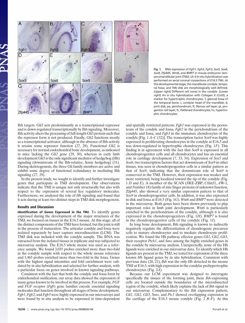
In long bone development, Ihh signaling is known to indirectlynegatively regulate the differentiation of chondrogenic precursorcells to mature chondrocytes and to mediate chondrocyte prolif-eration. We found the Hh pathway effector genes Gli1, Gli2, Gli3,their receptor Ptch1, and Smo among the highly enriched genes inthe condyle by microarray analysis. Unexpectedly, none of the Hhligands were enriched in our microarray data. To identify which Hhligands are present in the TMJ, we tested for expression of the threeknown Hh ligand genes by in situ hybridization. Consistent withprevious data (20, 21), Ihh was the only Hh detected in the mouseTMJ at E16.5, with high expression in the condylar prehypertrophicchondrocytes (Fig. 2A).
Because our LCM experiment was designed to interrogatespecifically the tissues of the forming joint, these Ihh-expressingcells are located outside the boundaries of the microdissectedregion of the condyle, which likely explains the lack of Ihh signal inour microarray. Complementary to the Ihh expression pattern,Gli1, Gli2, Gli3, Smo, and Ptc1 showed overlapping expression inthe cartilage of the E16.5 mouse condyle (Fig. 2 B–F). At this
Fig. 1. RNA expression of Fgfr1, Fgfr2, Fgfr3, Sox5, Sox6,Sox9, Zfp445, Wnt6, and BMP7 in mouse embryonic tem-poromandibular joint (TMJ). (A–I) In situ hybridization wasperformed on serial coronal cryosections of E16.5 TMJ. Atthis developmental stage, the mandibular condyle, tempo-ral fossa, and TMJ disk are morphologically well defined.(Upper right) Different cell zones in the condyle. (Lowerright) An in situ hybridization with Collagen X (ColX), amarker for hypertrophic chondrocytes. f, glenoid fossa ofthe temporal bone; c, condylar head of the mandible; d,joint disk; pe, perichondrium; fl, fibrous cell layer; pl, pro-genitor cell layer; fc, flattened chondrocytes; hc, hypertro-phic chondrocytes.
18298 � www.pnas.org�cgi�doi�10.1073�pnas.0908836106 Purcell et al.
embryonic stage, the Gli2 expression was the strongest among theGli genes, which together with Smo were the only genes in the Hhsignaling pathway that were expressed at readily detectable levels inthe joint disk (Fig. 2 C and E).
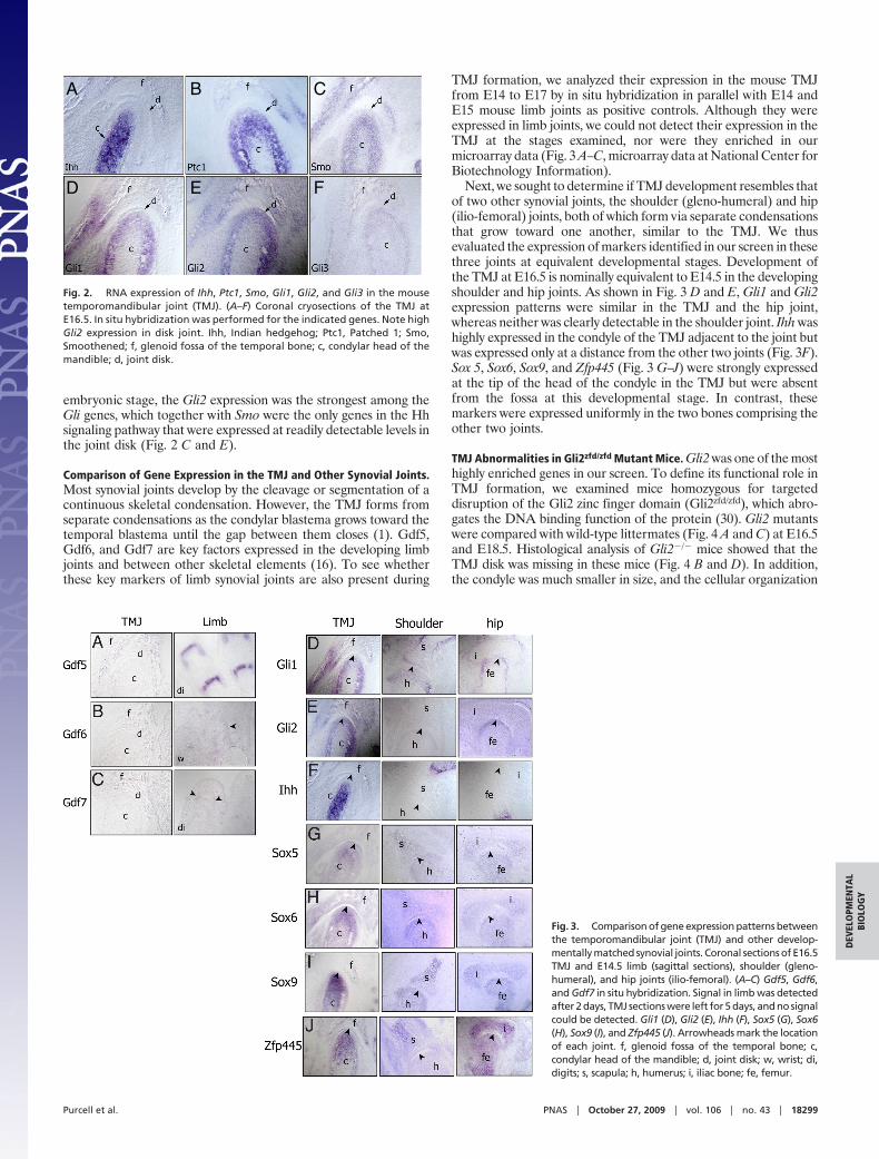
Comparison of Gene Expression in the TMJ and Other Synovial Joints.Most synovial joints develop by the cleavage or segmentation of acontinuous skeletal condensation. However, the TMJ forms fromseparate condensations as the condylar blastema grows toward thetemporal blastema until the gap between them closes (1). Gdf5,Gdf6, and Gdf7 are key factors expressed in the developing limbjoints and between other skeletal elements (16). To see whetherthese key markers of limb synovial joints are also present during
TMJ formation, we analyzed their expression in the mouse TMJfrom E14 to E17 by in situ hybridization in parallel with E14 andE15 mouse limb joints as positive controls. Although they wereexpressed in limb joints, we could not detect their expression in theTMJ at the stages examined, nor were they enriched in ourmicroarray data (Fig. 3 A–C, microarray data at National Center forBiotechnology Information).
Next, we sought to determine if TMJ development resembles thatof two other synovial joints, the shoulder (gleno-humeral) and hip(ilio-femoral) joints, both of which form via separate condensationsthat grow toward one another, similar to the TMJ. We thusevaluated the expression of markers identified in our screen in thesethree joints at equivalent developmental stages. Development ofthe TMJ at E16.5 is nominally equivalent to E14.5 in the developingshoulder and hip joints. As shown in Fig. 3 D and E, Gli1 and Gli2expression patterns were similar in the TMJ and the hip joint,whereas neither was clearly detectable in the shoulder joint. Ihh washighly expressed in the condyle of the TMJ adjacent to the joint butwas expressed only at a distance from the other two joints (Fig. 3F).Sox 5, Sox6, Sox9, and Zfp445 (Fig. 3 G–J) were strongly expressedat the tip of the head of the condyle in the TMJ but were absentfrom the fossa at this developmental stage. In contrast, thesemarkers were expressed uniformly in the two bones comprising theother two joints.
TMJ Abnormalities in Gli2zfd/zfd Mutant Mice. Gli2 was one of the mosthighly enriched genes in our screen. To define its functional role inTMJ formation, we examined mice homozygous for targeteddisruption of the Gli2 zinc finger domain (Gli2zfd/zfd), which abro-gates the DNA binding function of the protein (30). Gli2 mutantswere compared with wild-type littermates (Fig. 4 A and C) at E16.5and E18.5. Histological analysis of Gli2�/� mice showed that theTMJ disk was missing in these mice (Fig. 4 B and D). In addition,the condyle was much smaller in size, and the cellular organization
A B C
D E F
Fig. 2. RNA expression of Ihh, Ptc1, Smo, Gli1, Gli2, and Gli3 in the mousetemporomandibular joint (TMJ). (A–F) Coronal cryosections of the TMJ atE16.5. In situ hybridization was performed for the indicated genes. Note highGli2 expression in disk joint. Ihh, Indian hedgehog; Ptc1, Patched 1; Smo,Smoothened; f, glenoid fossa of the temporal bone; c, condylar head of themandible; d, joint disk.
Fig. 3. Comparison of gene expression patterns betweenthe temporomandibular joint (TMJ) and other develop-mentally matched synovial joints. Coronal sections of E16.5TMJ and E14.5 limb (sagittal sections), shoulder (gleno-humeral), and hip joints (ilio-femoral). (A–C) Gdf5, Gdf6,and Gdf7 in situ hybridization. Signal in limb was detectedafter 2 days, TMJ sections were left for 5 days, and no signalcould be detected. Gli1 (D), Gli2 (E), Ihh (F), Sox5 (G), Sox6(H), Sox9 (I), and Zfp445 (J). Arrowheads mark the locationof each joint. f, glenoid fossa of the temporal bone; c,condylar head of the mandible; d, joint disk; w, wrist; di,digits; s, scapula; h, humerus; i, iliac bone; fe, femur.
Purcell et al. PNAS � October 27, 2009 � vol. 106 � no. 43 � 18299
DEV
ELO
PMEN
TAL
BIO
LOG
Y
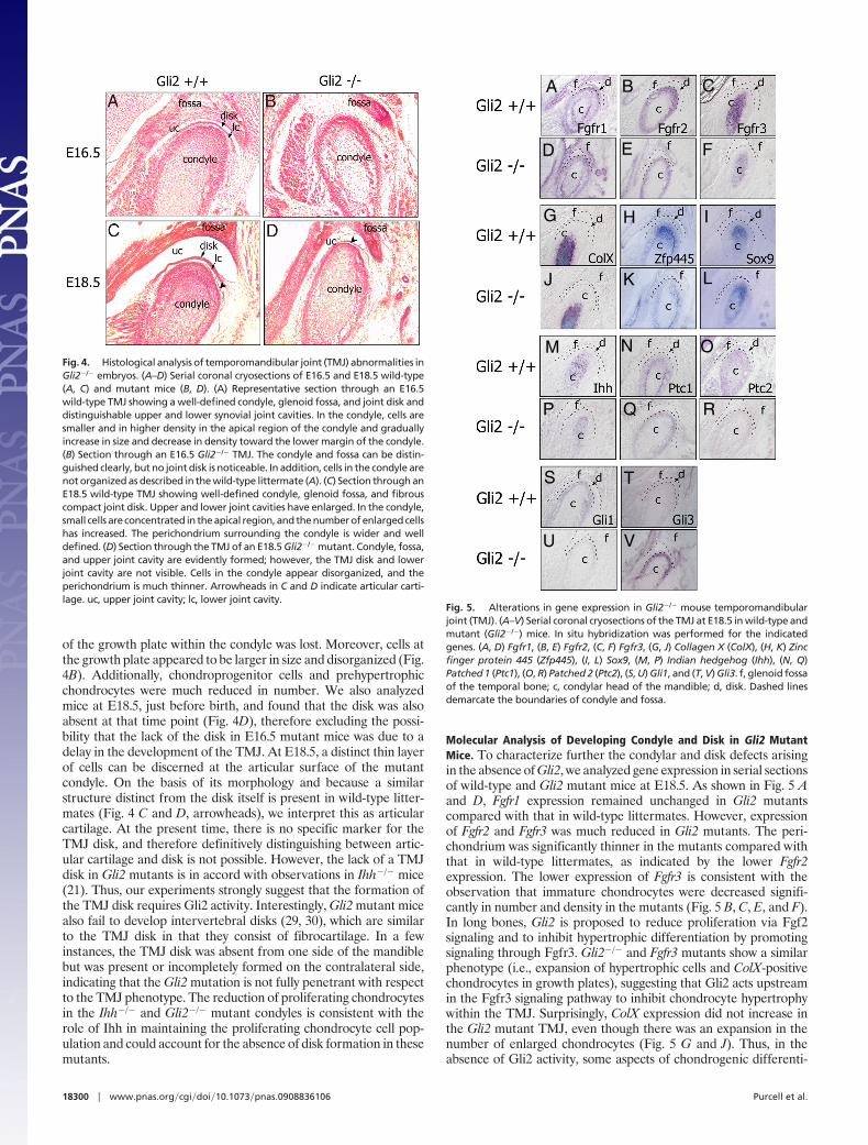
of the growth plate within the condyle was lost. Moreover, cells atthe growth plate appeared to be larger in size and disorganized (Fig.4B). Additionally, chondroprogenitor cells and prehypertrophicchondrocytes were much reduced in number. We also analyzedmice at E18.5, just before birth, and found that the disk was alsoabsent at that time point (Fig. 4D), therefore excluding the possi-bility that the lack of the disk in E16.5 mutant mice was due to adelay in the development of the TMJ. At E18.5, a distinct thin layerof cells can be discerned at the articular surface of the mutantcondyle. On the basis of its morphology and because a similarstructure distinct from the disk itself is present in wild-type litter-mates (Fig. 4 C and D, arrowheads), we interpret this as articularcartilage. At the present time, there is no specific marker for theTMJ disk, and therefore definitively distinguishing between artic-ular cartilage and disk is not possible. However, the lack of a TMJdisk in Gli2 mutants is in accord with observations in Ihh�/� mice(21). Thus, our experiments strongly suggest that the formation ofthe TMJ disk requires Gli2 activity. Interestingly, Gli2 mutant micealso fail to develop intervertebral disks (29, 30), which are similarto the TMJ disk in that they consist of fibrocartilage. In a fewinstances, the TMJ disk was absent from one side of the mandiblebut was present or incompletely formed on the contralateral side,indicating that the Gli2 mutation is not fully penetrant with respectto the TMJ phenotype. The reduction of proliferating chondrocytesin the Ihh�/� and Gli2�/� mutant condyles is consistent with therole of Ihh in maintaining the proliferating chondrocyte cell pop-ulation and could account for the absence of disk formation in thesemutants.
Molecular Analysis of Developing Condyle and Disk in Gli2 MutantMice. To characterize further the condylar and disk defects arisingin the absence of Gli2, we analyzed gene expression in serial sectionsof wild-type and Gli2 mutant mice at E18.5. As shown in Fig. 5 Aand D, Fgfr1 expression remained unchanged in Gli2 mutantscompared with that in wild-type littermates. However, expressionof Fgfr2 and Fgfr3 was much reduced in Gli2 mutants. The peri-chondrium was significantly thinner in the mutants compared withthat in wild-type littermates, as indicated by the lower Fgfr2expression. The lower expression of Fgfr3 is consistent with theobservation that immature chondrocytes were decreased signifi-cantly in number and density in the mutants (Fig. 5 B, C, E, and F).In long bones, Gli2 is proposed to reduce proliferation via Fgf2signaling and to inhibit hypertrophic differentiation by promotingsignaling through Fgfr3. Gli2�/� and Fgfr3 mutants show a similarphenotype (i.e., expansion of hypertrophic cells and ColX-positivechondrocytes in growth plates), suggesting that Gli2 acts upstreamin the Fgfr3 signaling pathway to inhibit chondrocyte hypertrophywithin the TMJ. Surprisingly, ColX expression did not increase inthe Gli2 mutant TMJ, even though there was an expansion in thenumber of enlarged chondrocytes (Fig. 5 G and J). Thus, in theabsence of Gli2 activity, some aspects of chondrogenic differenti-
Fig. 4. Histological analysis of temporomandibular joint (TMJ) abnormalities inGli2�/� embryos. (A–D) Serial coronal cryosections of E16.5 and E18.5 wild-type(A, C) and mutant mice (B, D). (A) Representative section through an E16.5wild-type TMJ showing a well-defined condyle, glenoid fossa, and joint disk anddistinguishable upper and lower synovial joint cavities. In the condyle, cells aresmaller and in higher density in the apical region of the condyle and graduallyincrease in size and decrease in density toward the lower margin of the condyle.(B) Section through an E16.5 Gli2�/� TMJ. The condyle and fossa can be distin-guished clearly, but no joint disk is noticeable. In addition, cells in the condyle arenot organized as described in the wild-type littermate (A). (C) Section through anE18.5 wild-type TMJ showing well-defined condyle, glenoid fossa, and fibrouscompact joint disk. Upper and lower joint cavities have enlarged. In the condyle,small cells are concentrated in the apical region, and the number of enlarged cellshas increased. The perichondrium surrounding the condyle is wider and welldefined. (D) Section through the TMJ of an E18.5 Gli2�/� mutant. Condyle, fossa,and upper joint cavity are evidently formed; however, the TMJ disk and lowerjoint cavity are not visible. Cells in the condyle appear disorganized, and theperichondrium is much thinner. Arrowheads in C and D indicate articular carti-lage. uc, upper joint cavity; lc, lower joint cavity.
A B C
D E F
G H I
J K L
M N O
P Q R
S T
U V
Fig. 5. Alterations in gene expression in Gli2�/� mouse temporomandibularjoint (TMJ). (A–V) Serial coronal cryosections of the TMJ at E18.5 in wild-type andmutant (Gli2�/�) mice. In situ hybridization was performed for the indicatedgenes. (A, D) Fgfr1, (B, E) Fgfr2, (C, F) Fgfr3, (G, J) Collagen X (ColX), (H, K) Zincfinger protein 445 (Zfp445), (I, L) Sox9, (M, P) Indian hedgehog (Ihh), (N, Q)Patched 1 (Ptc1), (O, R) Patched 2 (Ptc2), (S, U) Gli1, and (T, V) Gli3. f, glenoid fossaof the temporal bone; c, condylar head of the mandible; d, disk. Dashed linesdemarcate the boundaries of condyle and fossa.
18300 � www.pnas.org�cgi�doi�10.1073�pnas.0908836106 Purcell et al.
ation are aberrant in the forming TMJ. Similarly, Sox9 expressionalso was unchanged in the condyle despite a reduction in cells in thechondroprogenitor layer (Fig. 5 I and L). In contrast, Zfp445, whichshows similar expression to Sox9 in wild-type chondroprogenitorcells, was reduced dramatically in the Gli2 mutant (Fig. 5 H and K).Ihh, Ptc1, Ptc2, and Gli1 expression was reduced in the Gli2 mutant;in contrast, Gli3 was considerably up-regulated (Fig. 5 M–V). Gli3is well known to act as a repressor of Sonic hedgehog (Shh) and arepressor of osteoblastic bone formation. Gli3 overexpression in thecondyle of Gli2 mutants possibly is contributing to the absence ofTMJ disk formation. Gli proteins show partial functional redun-dancy in other tissues where they are coexpressed. Although Gli1
and Gli3 are expressed within the mutant TMJ, they are largelyabsent in the forming disk and are thus apparently unable tocompensate for the lack of Gli2 in TMJ disk development.
Hh Signaling Is Required at Two Distinct Steps in TMJ Disk Formation.In the absence of either Gli2 (this work) or Ihh activity (20), theTMJ disk fails to form, and in the latter case, the process has beenshown to be Gli3-independent (20). Interestingly, Ihh is expressedduring early chondrogenesis within condensing mesenchymal cells,and in a potentially analogous fashion, both our observations andpublished data indicate that Ihh is expressed in the TMJ condyleduring early mesenchymal condensation before disk formation[approximately E13.5 (22)]. These observations suggest that Hhsignaling through Gli2 could act very early in inducing disk differ-entiation. Alternatively, because Gli2 continues to be expressedwithin the disk after it has undergone chondrogenic differentiationand initiated Sox5, Sox6, and Sox9 expression (Fig. 1), Hh could berequired at a later stage to maintain the disk.
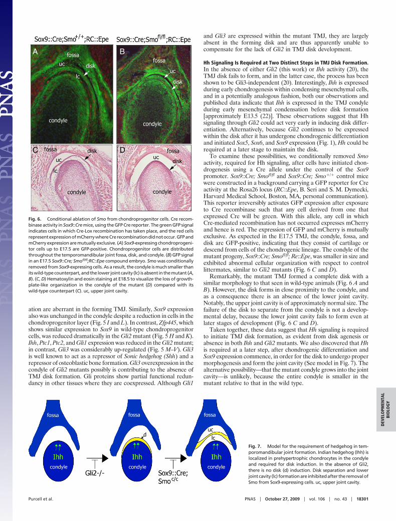
To examine these possibilities, we conditionally removed Smoactivity, required for Hh signaling, after cells have initiated chon-drogenesis using a Cre allele under the control of the Sox9promoter. Sox9::Cre; Smofl/fl and Sox9::Cre; Smo�/� control micewere constructed in a background carrying a GFP reporter for Creactivity at the Rosa26 locus (RC::Epe, B. Seri and S. M. Dymecki,Harvard Medical School, Boston, MA, personal communication).This reporter irreversibly activates GFP expression after exposureto Cre recombinase such that any cell derived from one thatexpressed Cre will be green. With this allele, any cell in whichCre-mediated recombination has not occurred expresses mCherryand hence is red. The expression of GFP and mCherry is mutuallyexclusive. As expected in the E17.5 TMJ, the condyle, fossa, anddisk are GFP-positive, indicating that they consist of cartilage ordescend from cells of the chondrogenic lineage. The condyle of themutant progeny, Sox9::Cre; Smofl/fl; Re::Epe, was smaller in size andexhibited abnormal cellular organization with respect to controllittermates, similar to Gli2 mutants (Fig. 6 C and D).
Remarkably, the mutant TMJ formed a complete disk with asimilar morphology to that seen in wild-type animals (Fig. 6 A andB). However, the disk forms in close proximity to the condyle, andas a consequence there is an absence of the lower joint cavity.Notably, the upper joint cavity is of approximately normal size. Thefailure of the disk to separate from the condyle is not a develop-mental delay, because the lower joint cavity fails to form even atlater stages of development (Fig. 6 C and D).
Taken together, these data suggest that Hh signaling is requiredto initiate TMJ disk formation, as evident from disk agenesis orabsence in both Ihh and Gli2 mutants. We also discovered that Hhis required at a later step, after chondrogenic differentiation andSox9 expression commence, in order for the disk to undergo propermorphogenesis and form the joint cavity (See model in Fig. 7). Thealternative possibility—that the mutant condyle grows into the jointcavity—is unlikely, because the entire condyle is smaller in themutant relative to that in the wild type.
A B
C D
Fig. 6. Conditional ablation of Smo from chondroprogenitor cells. Cre recom-binase activity in Sox9::Cre mice, using the GFP Cre reporter. The green GFP signalindicates cells in which Cre-Lox recombination has taken place, and the red cellsrepresentexpressionofmCherrywhereCrerecombinationdidnotoccur.GFPandmCherry expression are mutually exclusive. (A) Sox9-expressing chondroprogeni-tor cells up to E17.5 are GFP-positive. Chondroprogenitor cells are distributedthroughout the temporomandibular joint fossa, disk, and condyle. (B) GFP signalin an E17.5 Sox9::Cre; Smofl/fl;RC::Epe compound embryo. Smo was conditionallyremoved from Sox9-expressing cells. As a result, the condyle is much smaller thanits wild-type counterpart, and the lower joint cavity (lc) is absent in the mutant (A,B). (C, D) Hematoxylin and eosin staining at E18.5 to visualize the loss of growth-plate-like organization in the condyle of the mutant (D) compared with itswild-type counterpart (C). uc, upper joint cavity.
Fig. 7. Model for the requirement of hedgehog in tem-poromandibular joint formation. Indian hedgehog (Ihh) islocalized in prehypertrophic chondrocytes in the condyleand required for disk induction. In the absence of Gli2,there is no disk (d) induction. Disk separation and lowerjoint cavity (lc) formation are inhibited after the removal ofSmo from Sox9-expressing cells. uc, upper joint cavity.
Purcell et al. PNAS � October 27, 2009 � vol. 106 � no. 43 � 18301
DEV
ELO
PMEN
TAL
BIO
LOG
Y
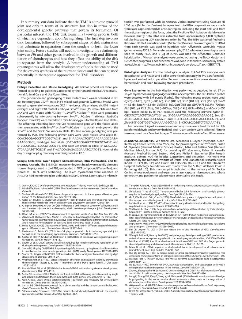
In summary, our data indicate that the TMJ is a unique synovialjoint not only in terms of its structure but also in terms of thedevelopmental genetic pathways that govern its formation. Ofparticular interest, the TMJ disk forms in a two-step process, bothof which are dependent upon Hh signaling. The first step involvesdisk formation, followed by subsequent stages of disk maturationthat culminate in separation from the condyle to form the lowerjoint cavity. Future studies will need to investigate the relationshipbetween Hh and other genes involved in the growth and differen-tiation of chondrocytes and how they affect the ability of the diskto separate from the condyle. A better understanding of TMJorganogenesis will allow the development of tools that can be usedfor the ex vivo synthesis of the relevant tissues and that can be usedpotentially in therapeutic approaches for TMJ disorders.
MethodsEmbryo Collection and Mouse Genotyping. All animal procedures were per-formed according to guidelines approved by the Harvard Medical Area Institu-tional Animal Care and Use Committee.
The generation and genotyping of Gli2 mutant mice was as described in ref.29. Heterozygous Gli2�/� mice in F1 mixed backgrounds (C3H/HeJ: Fvb/N) weremated to generate homozygous Gli2�/� embryos. We analyzed six E16 mutantembryos and eight E18 mutant embryos. Smofl/fl mice (35) were obtained fromJAX and were mated with RC::Epe�/�. Smofl/fl; RC::Epe�/� mice were generatedsubsequently by intercrossing between Smofl/�; RC::Epe�/� siblings. Sox9::Creknock-in mice (36) were mated with mice homozygous for the floxed Smo alleles.The offspring inheriting both the Cre-recombinase gene and the floxed allelethen were mated with Smofl/fl or Smofl/fl; RC::Epe�/� to obtain embryos with theSmofl/fl and the Sox9::Cre knock-in allele. Routine mouse genotyping was per-formed by PCR. The following primer pairs were used: floxed Smo allele (5�-CCACTGCGAGCCTTTGCGCTAC-3� and 5�-AAGAACTCGTCAAGAAGGCGATA-GAAGGCG-3�), wild-type Smo allele (5�-CCACTGCGAGCCTTTGCGCTAC-3� and5�-CCCATCACCTCCGCGTCGCA-3�), and Sox9-Cre knock-in allele (5�-GCAGAAC-CTGAAGATGTTCGC-3� and 5�-ACACCAGAGACGGAAATCCATC-3�). Noon of theday of vaginal plug discovery was designated as E0.5.
Sample Collection, Laser Capture Microdissection, RNA Purification, and Mi-croarray Analysis. The E16.5 CD1 mouse embryonic heads were rapidly dissectedfrom embryos, rinsed in cold PBS, embedded in OCT compound (Tissue-Tek), andstored at �80 °C until sectioning. The 8-�m cryosections were collected onArcturus PEN membrane glass slides (Molecular Devices). Laser capture microdis-
section was performed with an Arcturus Veritas instrument using CapSure HSLCM caps (Molecular Devices). Independent total RNA preparations were madefrom laser-captured condyle articular region, including the joint disk, and fromthe articular region of the fossa, using the PicoPure RNA isolation kit (MolecularDevices). Briefly, total RNA was extracted from approximately 1,000 capturedcells by incubating LCM caps in extraction buffer. The RNA was amplified usingRiboAmp HS RNA amplification kit (Molecular Devices). Five micrograms of cDNAfrom each sample was used to hybridize with Affymetrix GeneChip mousegenome array 430 2.0. For a reference sample, E16.5 whole mouse embryos wereused to purify RNA, and 5 �g of cDNA was used for Affymetrix GeneChiphybridization. Microarray analyses were carried out using the Bioconductor andGeneSifter programs. Each experiment was done in triplicate. Microarray data isaccessible at http://www.ncbi.nlm.nih.gov/geo/query/acc.cgi?acc�GSE17473.
Histological Analyses. For the histological analysis, E14.5–E19.5 embryos weredecapitated, and heads and bodies were fixed separately in 4% paraformalde-hyde and embedded in paraffin. Ten-micrometer sections were stained withhematoxylin and eosin following standard procedures.
Gene Expression. In situ hybridization was performed as described in ref. 37 on16-�m cryosections using digoxigenin (DIG)-labeled probes. The DIG-labeled probeswere detected with BM purple (Roche). The following mouse probes were used:Fgfr1(�3.6 kb), Fgfr2 (�900 bp), Sox5 (488 bp), Sox6 (481 bp), Sox9 (255 bp), Wnt6(�1.6kb),Bmp7 (�2.1kb),Gdf5 (551bp),Gdf6 (481bp),GDF7 (478bp), Ihh (700bp),Ptc1 (406bp),Ptc2 (2kb),Gli1 (�800bp),Gli2 (�1kb),Gli3 (�400bp),andColX (�650bp). Probes made by RT-PCR product used the following primers: Fgfr3 (5�-CGCATCCTCACTGTGACATC-3� and 3�-GGAAATGAGAGGGCCAGAAC-5�), Smo (5�-AGAGCAAGATGATCGCCAAG-3� and 3�-ATCCAAGATCTCAGCCTCCA-5�), andZfp445 (5�-GCGTGGGTAGAAAAAGGCTA-3�, 3�-CTCATCCCGGTCTGTCAAAT-5�).
For fluorescent signal visualization on sections, tissues were fixed for 2–4 h in 4%paraformaldehyde and cryoembedded, and 10-�m sections were collected. Pictureswere captured on a Zeiss AxioImager Z1 microscope with an AxioCam HRm camera.
ACKNOWLEDGMENTS. We thank Drs. Alexandra L. Joyner (Memorial Sloan-Kettering Cancer Center, New York, NY) for providing the Gli2zfd/zfd mice, SusanM. Dymecki (Harvard Medical School, Boston, MA) and Bettina Seri (HarvardMedical School, Boston, MA) for providing RC:: Epe mice, Rahul N. Kanadia(Harvard Medical School, Boston, MA) and Philip P. Stashenko (The ForsythInstitute, Boston, MA) for helpful suggestions and discussion. This work wassupported by the National Institute of Dental and Craniofacial Research AwardK22DE-016309 (to P.P.) and Grant P01 DK56246 (to C.J.T.) and the Children’sHospital Pathology Foundation with funds from the S. Burt Wolbach Chair inPathology (M.L.C.). This manuscript is dedicated to the memory of Dr. TuckerCollins, whose equipment and expertise in laser capture microscopy, and whosegenerosity and passion for science were essential to this study.
1. Avery JK (2001) Oral Development and Histology (Thieme, New York) 3rd Ed, p 435.2. Hinchliffe JR and Johnson DR (1980) The Development of the Vertebrate Limb (Clarendon,
Oxford).3. Shubin NH, Alberch P (1986) A morphogenetic approach to the origin and basic organi-
zation of the tetrapod limb. Evol Biol 20:319–387.4. Oster GF, Shubin N, Murray JD, Alberch P (1988) Evolution and morphogenic rules: The
shape of the vertebrate limb in ontogeny and phylogeny. Evolution 42:862–884.5. Craig FM, Bentley G, Archer CW (1987) The spatial and temporal pattern of collagens I and II
and keratan sulphate in the developing chick metatarsophalangeal joint. Development99:383–391.
6. Khan IM, et al. (2007) The development of synovial joints. Curr Top Dev Biol 79:1–36.7. Akiyama H, Chaboissier MC, Martin JF, Schedl A, de Crombrugghe B (2002) The transcription
factor Sox9 has essential roles in successive steps of the chondrocyte differentiation pathwayand is required for expression of Sox5 and Sox6. Genes Dev 16:2813–2828.
8. Ikeda T, et al. (2005) Distinct roles of Sox5, Sox6, and Sox9 in different stages of chondro-genic differentiation. J Bone Miner Metab 23:337–340.
9. Hartmann C, Tabin CJ (2001) Wnt-14 plays a pivotal role in inducing synovial jointformation in the developing appendicular skeleton. Cell 104:341–351.
10. Spater D, Hill TP, Gruber M, Hartmann C (2006) Role of canonical Wnt-signalling in jointformation. Eur Cell Mater 12:71–80.
11. Spater D, et al. (2006) Wnt9a signaling is required for joint integrity and regulation of Ihhduring chondrogenesis. Development 133:3039–3049.
12. StormEE,KingsleyDM(1996) Jointpatterningdefects causedbysingleanddoublemutationsin members of the bone morphogenetic protein (BMP) family. Development 122:3969–3979.
13. Storm EE, Kingsley DM (1999) GDF5 coordinates bone and joint formation during digitdevelopment. Dev Biol 209:11–27.
14. Wolfman NM, et al. (1997) Ectopic induction of tendon and ligament in rats by growth anddifferentiation factors 5, 6, and 7, members of the TGF-� gene family. J Clin Invest100:321–330.
15. Francis-West PH, et al. (1999) Mechanisms of GDF-5 action during skeletal development.Development 126:1305–1315.
16. Settle SH, Jr, et al. (2003) Multiple joint and skeletal patterning defects caused by singleand double mutations in the mouse Gdf6 and Gdf5 genes. Dev Biol 254:116–130.
17. Guo X, et al. (2004) Wnt/�-catenin signaling is sufficient and necessary for synovial jointformation. Genes Dev 18(19):2404–2417.
18. Sarnat BG (1966) Developmental facial abnormalities and the temporomandibular joint.Dent Clin North Am Nov:587–600.
19. Silbermann M, Frommer J (1972) The nature of endochondral ossification in the mandib-ular condyle of the mouse. Anat Rec 172:659–667.
20. Tang GH, Rabie AB, Hagg U (2004) Indian hedgehog: A mechanotransduction mediator incondylar cartilage. J Dent Res 83:434–438.
21. Shibukawa Y, et al. (2007) Temporomandibular joint formation and condyle growthrequire Indian hedgehog signaling. Dev Dyn 236:426–434.
22. Gu S, Wei N, Yu L, Fei J, Chen Y (2008) Shox2-deficiency leads to dysplasia and ankylosis ofthe temporomandibular joint in mice. Mech Dev 125:729–742.
23. Lanske B, et al. (1996) PTH/PTHrP receptor in early development and Indian hedgehog-regulated bone growth. Science 273:663–666.
24. Vortkamp A, et al. (1996) Regulation of rate of cartilage differentiation by Indian hedge-hog and PTH-related protein. Science 273:613–622.
25. St-Jacques B, Hammerschmidt M, McMahon AP (1999) Indian hedgehog signaling regu-latesproliferationanddifferentiationofchondrocytesand isessential forboneformation.Genes Dev 13:2072–2086.
26. Ingham PW, McMahon AP (2001) Hedgehog signaling in animal development: Paradigmsand principles. Genes Dev 15:3059–3087.
27. Bai CB, Joyner AL (2001) Gli1 can rescue the in vivo function of Gli2. Development128:5161–5172.
28. Wang B, Fallon JF, Beachy PA (2000) Hedgehog-regulated processing of Gli3 produces ananterior/posterior repressor gradient in the developing vertebrate limb. Cell 100:423–434.
29. Mo R, et al. (1997) Specific and redundant functions of Gli2 and Gli3 zinc finger genes inskeletal patterning and development. Development 124(1):113–123.
30. Miao D, et al. (2004) Impaired endochondral bone development and osteopenia inGli2-deficient mice. Exp Cell Res 294:210–222.
31. Hui CC, Joyner AL (1993) A mouse model of greig cephalopolysyndactyly syndrome: Theextra-toesJ mutation contains an intragenic deletion of the Gli3 gene. Nat Genet 3:241–246.
32. Rice DP, Rice R, Thesleff I (2003) Fgfr mRNA isoforms in craniofacial bone development.Bone 33:14–27.
33. Ng LJ, et al. (1997) SOX9 binds DNA, activates transcription, and coexpresses with type IIcollagen during chondrogenesis in the mouse. Dev Biol 183:108–121.
34. Zhao Q, Eberspaecher H, Lefebvre V, De Crombrugghe B (1997) Parallel expression of Sox9and Col2a1 in cells undergoing chondrogenesis. Dev Dyn 209:377–386.
35. Long F, Zhang XM, Karp S, Yang Y, McMahon AP (2001) Genetic manipulation of hedge-hog signaling in the endochondral skeleton reveals a direct role in the regulation ofchondrocyte proliferation. Development 128:5099–5108.
36. Akiyama H, et al. (2005) Osteo-chondroprogenitor cells are derived from Sox9 expressingprecursors. Proc Natl Acad Sci USA 102:14665–14670.
37. BrentAE,SchweitzerR,TabinCJ (2003)Asomitic compartmentof tendonprogenitors.Cell113:235–248.
18302 � www.pnas.org�cgi�doi�10.1073�pnas.0908836106 Purcell et al.