Embed Size (px)
Citation preview

D. K. Kido1
E. D. Caine2
M. LeMay3
S. Ekholm1
H. Booth2
R. Panzer4
Received May 27, 1988; accepted after revision October 25, 1988.
Presented at the annual meeting of the American Society of Neuroradiology, New York, May 1987.
This work was supported in part by grants AG/ NS 03644; MH00473; and MH40381 to the University of Rochester Clinical Research Center to study psychopathology of the elderly.
1 Department of Radiology, University of Rochester Medical Center, P.O. Box 648, 601 Elmwood Ave ., Rochester, NY 14642. Address reprint requests to D. K. Kido.
2 Department of Psychiatry, University of Rochester Medical Center, Rochester NY 14642.
3 Department of Radiology, Brigham and Wornens Hospital, Boston MA 02115.
' Department of Medicine. University of Rochester Medical Center, Rochester NY 14642.
AJNR 10:551-555, May/June 1989 0195-6108/89/1003-0551 © American Society of Neuroradiology
551
Temporal Lobe Atrophy in Patients with Alzheimer Disease: A CT Study
CT was used to document temporal lobe atrophy in 39 patients who were diagnosed clinically as having Alzheimer disease; the results were compared with those from 29 healthy elderly control subjects who were matched for age and education. The diagnosis of Alzheimer disease was made according to clinical criteria consistent with those specified by an NINCDS workshop. These included detailed medical and neuropsychological assessments.
Temporal lobe atrophy was assessed by evaluating the temporal horns and sylvian cisterns. Temporal horn measurements greater than 3 mm occurred only in patients with Alzheimer disease while measurements less than or equal to this occurred in both Alzheimer patients and control subjects. Subjective evaluation of the sylvian cistern indicated that 24/29 controls had normal-appearing cisterns while only 5/39 Alzheimer patients had similar findings. In contrast to temporal horns, sylvian cisterns were more sensitive but less specific as discriminators between Alzheimer patients and normal controls.
The cerebral atrophy that occurs in patients with Alzheimer disease overlaps that which occurs in normal individuals during their sixth and seventh decades [1 , 2]. Most attempts to differentiate morphologically these two groups with CT have focused on enlargement of the lateral ventricles and cerebral sulci. Several investigators have reported that linear and planar CT measurements of the lateral ventricles or ratios between the ventricles and cranium can differentiate Alzheimer disease from normal aging [3-5]. Others have disagreed [6 , 7]. More recently , it has been suggested that volumetric measurements of the lateral ventricles can separate these two groups [8, 9]. Sulcal enlargement [1 0, 11], gray/white differentiation, and brain density have also been measured in an attempt to separate Alzheimer patients from control subjects, but these parameters have produced negative or controversial results [12-16].
Atrophy of the temporal lobe has been largely ignored as a CT indicator of Alzheimer disease despite pathologic studies that indicate that atrophy and neurofibrillary tangles frequently occur in that location [17 -20]. The reason for this may be that atrophy in the frontal and parietal lobes, and subsequent sulcal and ventricular enlargement, is more prominent than similar changes in the temporal lobes. To determine whether temporal lobe atrophy can be used to differentiate individuals in the earlier stages of Alzheimer disease from normal control subjects, we reviewed CT scans obtained from Alzheimer patients and normal controls. Temporal lobe atrophy was determined by separately analyzing sylvian cistern and temporal horn enlargement. We related temporal lobe atrophy to cognitive and functional changes in Alzheimer patients to determine whether the CT indexes we used could detect patients in the earliest stages of their disease.
Materials and Methods
Twenty-nine normal control subjects and 39 patients with Alzheimer disease participated in this study. Informed consent was obtained from all individuals who were scanned for

552 KIDO ET AL. AJNR:10, May/June 1989
research purposes. Patients were diagnosed as having "probable" Alzheimer disease on the basis of NINCDS-ADRDA criteria: that is, all patients had histories, clinical and physical examinations, and laboratory evaluations that did not indicate treatable medical and psychiatric illnesses or other conditions associated with dementia (21] . Patients underwent comprehensive neuropsychological testing as part of their diagnostic assessment. Subsequently, Alzheimer patients were followed for 12-24 months to confirm their progressive course. Normal controls were recruited from the community; potential subjects were excluded who had a history of alcoholism, psychiatric illness, head trauma, significant neurologic disease, or medical illness that affected mentation, or who were using medications with central actions or cognitive side effects. Thus, we recruited a physically healthy, functionally active group of control subjects. All normal control subjects and 36/39 Alzheimer patients were tested with the Mini-Mental State (MMS) examination (normal range = 24-30) (22] , and all patients were also evaluated with a functional rating scale (maximum score= 13), modified from Shoulson and Fahn (23] , that has been shown to correlate highly with CT changes in Huntington disease (24]. Functional ratings were not conducted for controls, since the selection criteria we used placed the sample at the "ceiling" of our test instrument.
Women outnumbered men in both the Alzheimer group (24/39) and the control group (18/29). In the 55-69 age group, there were 18 Alzheimer patients and 20 control subjects. The mean age of Alzheimer patients in this group was 63.6 ± 3.5 years as compared with controls, whose mean age was 61 .9 ± 3.9 years. The controls in this group were better educated than the Alzheimer patients (15.3 ± 1.9 years of education versus 11 .7 ± 4.7 years). In the 70+ age group, Alzheimer patients outnumbered controls (21 to nine). The mean age of Alzheimer patients in this older group was 75.5 ± 4.3 years; controls averaged 76.0 ± 6.3 years. Education for the Alzheimer group was 12.9 ± 4.0 years; controls averaged 13.7 ± 3.0 years.
The CT scans were performed on a GE 8800 scanner. The scans were taken 15° to Reid's baseline by utilizing the scout view. The scans were usually obtained with 5-mm contiguous sections, but a few scans were taken with 1 0-mm sections and a 3-mm overlap. The scanner was operated at 120 kVp, 120 mA, and 2 sec.
The CT scans were subjectively examined for temporal lobe atrophy by separately evaluating the size of the temporal horns and
1 2
sylvian cisterns. Temporal horn size was subjectively evaluated and assigned a number on a scale of 1 to 4 according to whether the horns appeared normal or slightly, moderately, or markedly enlarged. In addition, the symmetry of the temporal horns was noted. The anteroposterior diameter of the tip of the temporal horn, the width of the body of the temporal horn (anterior third), and the border between the body and tip of the temporal horns were measured in mm by using the em scale on the corresponding CT image (Fig . 1). When the temporal horns were asymmetric, the larger side was measured.
The sylvian cisterns were subjectively evaluated and assigned a number on a scale of 1 to 4 according to whether they appeared to be normal or slightly, moderately, or markedly enlarged. The anteroposterior distance between the posterior inferior border of the frontal lobe and the anterior tip of the temporal lobe was measured in mm. In addition, the width of the sylvian cistern was measured 1 em behind the tip of the temporal lobe (Fig. 2). Measurements were accurate to ± 1.0 mm. All CT measurements were conducted by neuroradiologists blinded to diagnosis, MMS score, and functional rating . The subject's age was available to CT raters.
The ability of subjective ratings and objective measurements to discriminate at various cut-off levels between Alzheimer patients and controls was assessed through receiver operating characteristic (ROC) analysis (25]. The area under the ROC curves was used as an indicator of the overall discriminating ability of each measurement and to compare the different measures (26]. In this method, a perfect test has an area of 1.0 and a useless test has an area of 0.5.
To assess interrater reliability, subjective and objective measurements were obtained from two independent neuroradiologists on a representative sample of 18 Alzheimer patients and 24 controls. Their measurements were compared through Pearson correlation coefficients and discrimination through comparison of ROC curve areas for paired data (27].
Results
The temporal horns appeared subjectively enlarged in 26/ 39 Alzheimer patients while similar changes were present in only one control (Fig. 3). Differentiating normal from mild temporal horn enlargement was occasionally difficult, but differentiating mild from moderate temporal horn enlargement was not difficult (Fig. 4). Temporal horn asymmetry was
Fig. 1.-0bjective measurements of right temporal horn. Anteroposterior diameter of tip of right temporal horn (curved arrow), width of temporal horn body (arrowhead), and border between lip and body of temporal horn (arrows) are marked.
Fig. 2.-0bjective measurements of lett sylvian cistern. Anteroposterior distance between posterior interior border of frontal lobe and anterior tip of temporal lobe (straight arrow) and width of sylvian cistern (curved arrow) are marked.
AJNR:10, May{June 1989 CT OF TEMPORAL LOBE ATROPHY 553
en .... c Q)
...... 0
80
60
40
20
0
~Co ntr o l .AD
None Mild Moderate Severe
T emporal Horn Enlargement
Fig. 3.-Distribution of subjective temporal horn measurements in normal controls (n = 29) and patients with Alzheimer disease (n = 39).
detected in 21 /39 Alzheimer patients while similar changes were present in only 5/29 controls (Fig . 5).
Measurements of the tip , body, and border between the tip and body of the temporal horn in both Alzheimer patients and controls are given in Table 1. Linear measurements >3 mm in any of these three parameters, indicating the presence of temporal lobe atrophy, occurred only in Alzheimer patients. Measurements :53 mm occurred in both Alzheimer patients and control subjects. The one control who was subjectively judged to have mild temporal horn enlargement had an anteroposterior tip measurement of 3 mm, a body width of 2 mm, and an oblique border of 3 mm.
A 8
The Alzheimer patients with no subjective temporal horn enlargement demonstrated cognitive changes on their MMS tests; their mean score was 19.9 ± 5.8 (n = 12). Alzheimer patients with mild temporal horn enlargement (n = 14) had a mean MMS score of 14.5 ± 6.7; those with moderate enlargement (n = 7) had a mean score of 16.4 ± 6.4 (both MMS scores consistent with moderate dementia); and those with severe enlargement (n = 3) had a mean score of 8.0 ± 6.0 (severe dementia). The normal subjects had a mean MMS score of 29.2 ± 1.0 (n = 29). Functional ratings in Alzheimer patients were also diminished (normal-appearing temporal horns= 7.9 ± 2.5 [mild/moderate functional impairment], mild = 6.6 ± 3.4 [moderate functional impairment], moderate = 7.1 ± 2.8, and severe= 4.0 ± 3.2 [moderatejseverefunctional impairment]). It was notable that in this cross-sectional study functional and cognitive scores tended to show a decline in
TABLE 1: Temporal Horn Measurements
Enlargement Tip Oblique Border Body
(mm) AD (Controls) AD (Contro ls) AD (Controls)
0 3 (4) 3 (6) 6 (18) 1 5 (16) 7 (11) 3 (9) 2 7 (7) 3 (11) 7 (2) 3 7 (2) 4 (1) 3 (0) 4 7 (0) 2 (0) 5 (0) 5 6 (0) 7 (0) 6 (0) 6 1 (0) 5 (0) 4 (0) 7 1 (0) 0 (0) 1 (0) 8 2 (0) 5 (0) 3 (0) 9 and above 0 (0) 3 (0) 1 (0)
Note.-AD =Alzheimer disease patients.
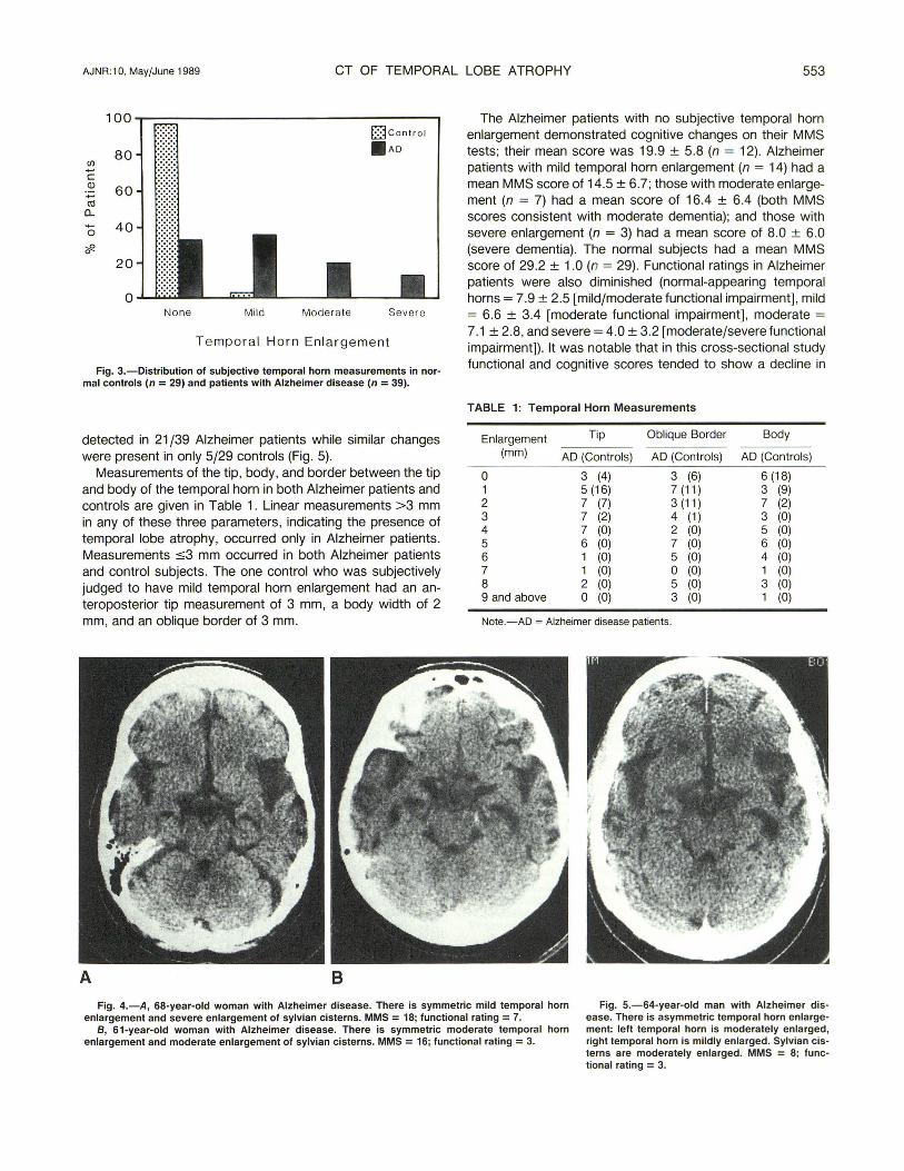
Fig. 4.-A , 68-year-old woman with Alzheimer disease. There is symmetric mild temporal horn enlargement and severe enlargement of sylvian cisterns. MMS = 18; functional rating = 7.
Fig. 5.-64-year-old man with Alzheimer disease. There is asymmetric temporal horn enlargement: left temporal horn is moderately enlarged, right temporal horn is mildly enlarged. Sylvian cisterns are moderately enlarged. MMS = 8; functional rating = 3.
B, 61-year-old woman with Alzheimer disease. There is symmetric moderate temporal horn enlargement and moderate enlargement of sylvian cisterns. MMS = 16; functional rating = 3.

554 KIDO ET AL. AJNR :10, MayfJune 1989
relation to the full spectrum of CT changes as independent variables but proved insensitive to apparent differences between patients with mild and moderate temporal horn enlargement.
Subjective ratings showed that individuals with Alzheimer disease had larger-appearing sylvian cisterns than controls did (Fig . 6). These findings were corroborated objectively by two measurements (Table 2). First, the anteroposterior distance of the right anterior sylvian cistern was 2.7 mm greater in Alzheimer patients than in controls (p < .001 ). Second, the width of the right anterior sylvian cistern was 2.9 mm greater in Alzheimer patients than in controls (p < .0001 ). The measurements of the left sylvian cistern were similar to those on the right. Although the sylvian cisterns of 35/39 Alzheimer patients and 29/29 controls appeared grossly symmetric, minor asymmetries were frequently present (Fig. 7).
A comparison of the accuracy of classifying individuals as Alzheimer patients or control subjects by using the areas under the ROC curves showed no significant differences among the subjective and objective measurements. The areas varied from .80 ± .05 to .87 ± .04 (p = NS). Although analysis based on a single cut-off level showed a trend for the sylvian cistern measure to be more sensitive and the temporal horn measure to be more specific, there were no differences in overall accuracy.
100 Q control
80 .AD (J) ..... c Q) 60 ..... co
a.. ..... 0
40
;,R 0
20
0 None Mil d Moder ale Severe
Sy lv ian Fi ssure Enlargement
Fig. 6.-Distribution of subjective sylvian cistern measurements in normal controls (n = 29) and patients with Alzheimer disease (n = 39).
TABLE 2: Sylvian Cistern Measurements
Space between frontal and temporal lobes (mm)
Right Left
Width of sylvian cistern (mm)
Right Left
Controls (n = 29)
AD Significance (n = 39) oft-test
4.9 ± 3.3 7.6 ± 3.3 p < .001 5.0 ± 3.8 7.5 ± 3.3 p < .006
5.0 ± 2.0 7.9 ± 2 .2 p < .0001 5.0 ± 1.9 7.7 ± 2.4 p < .0001
Note.- AD = Alzheimer disease patients.
Fig. 7.-68-year-old woman, normal control, with moderate sylvian cistern enlargement. Tip of right temporal lobe is pointed, causing sylvian cistern to appear triangular. Tip of left temporal lobe is flatter than right, causing left sylvian cistern to appear square. MMS = 30.
lnterrater reliability was good, with Pearson correlation coefficients ranging from .87 to .93 for the subjective and objective estimates. There was a trend for one reader to record higher subjective and objective measurements of atrophy, with a difference averaging 0.9 mm higher across all objective measurements. However, there were no significant differences in the two readers ' ability to discriminate between Alzheimer disease as measured by ROC curve areas .
Discussion
The temporal horn measurements were quite specific but insensitive for identifying Alzheimer disease in our population . Temporal horns larger than 3 mm occurred only in Alzheimer patients. In contrast, the sylvian cistern measurements tended to be sensitive but not specific. This combination of test operating characteristics suggests that use of both measurements might be worthwhile when it is important to discriminate between Alzheimer disease and normal aging. Thus, given its high sensitivity, a normal sylvian cistern might help rule out Alzheimer disease. Highly abnormal temporal horn measurements, given their high specificity, might help rule in disease. Our observations confirm an earlier study that indicated that multiple measurements of the temporal lobe and its surrounding structures can be used to separate Alzheimer patients from normal controls [20]. However, it remains to be determined whether measurements of the temporal lobe can be used to separate Alzheimer disease from the other diseases that cause dementia.
The CT representation of temporal lobe atrophy in Alzheimer patients results primarily from degenerative changes in the hippocampus, amygdala, and the adjacent white matter [2 , 18, 28-30]. The amygdala and hippocampus are associated with memory, learning, and motivational changes, which are characteristic findings in patients with Alzheimer disease

AJNR :10, MayfJune 1989 CT OF TEMPORAL LOBE ATROPHY 555
[29-31]. The CT findings we measured in the temporal lobe are indirect measurements of these structures, and it is not surprising that the MMS and functional scores in our crosssectional study sample indicate that clinical changes occur in advance of detectable indirect morphologic CT changes. Therefore, CT scanning will continue to be used primarily to diagnose the 2-1 0% of patients with dementia who have treatable disorders [32 , 33] .
MR may be able to detect atrophy in the temporal lobes earlier than CT, because the former can image the hippocampus and amygdala directly, possibly allowing detection of more subtle structural changes. However, MR detection of hippocampal and amygdaloid atrophy must still be correlated with symptomatic changes. It remains to be determined whether grossly detectable pathology precedes clinically definable cognitive and functional declines. PET and SPECT appear to be more promising than either CT or MR, as they are able to measure abnormal metabolism and cerebral blood flow [34-36] . These two techniques may separate dementia patients with Alzheimer disease from those with multiinfarct dementia [37-39] . Unfortunately, PET continues to be an expensive research tool. SPECT, on the other hand, should provide a clinically useful tool when compounds such as 1231-iodoamphetamine are used [39-40] .
REFERENCES
1. Tomlinson BE, Blessed G, Roth M. Observations on the brains of nondemented old people. J Neural Sci 1968;7:331-356
2. Tomlinson BE, Blessed G, Roth M. Observations on the brains of demented old people. J Neural Sci 1970;11 :205-242
3. Ford CV, Winter J. Computerized axial tomograms, and dementia in elderly patients. J Gerontal 1981 ;36:164-169
4. de Leon MJ, Ferris SH, George AE, Reisberg B, Kricheff II , Gershon S Computed tomography evaluations of brain-behavior relationships in senile dementia of the Alzheimer's type. Neurobiol Aging 1980;1 :69 -79.
5. Jacoby RJ, Levy R. Computed tomography in the elderly. 2. Senile dementia: diagnosis and functional impairment. Br J Psychiatry 1980;136:256- 269
6. Hughes CP, Gada M. Computed tomography and aging of the brain. Radiology 1981 ;139:391-396
7. Brinkman SO, Sarwar M, Levin HS, Morris HH Ill . Quantitative indexes of computed tomography in dementia and normal aging. Radiology 1981;138:89-92
8. Gada M, Hughes CP, Danziger W, Chi 0 , Jost G, Berg L. Volumetric measurements of the cerebrospinal fluid spaces in demented subjects and controls. Radiology 1982; 144:535-538
9. George AE, de Leon MJ, Rosenbloom S, et al. Ventricular volume and cognitive deficit: a computed tomographic study. Radiology 1983;149: 493-498
10. Arai H, Kobayashi K, Ikeda K, Nagao Y, Ogihara R, Kosaka K. A computed tomography study of Alzheimer's disease. J Neurol1983;229:69-77
11. Roberts MD, Caird Fl. Computerized tomography and intellectual impairment in the elderly . J Neural Neurosurg Psychiatry 1976;39:986-989
12. George AE, de Leon MJ , Ferris SH, Kricheff II. Parenchymal CT correlates of senile dementia (Alzheimer's disease): Loss of grey-white discriminality. AJNR 1981 ;2:205-21 3
13. Naser MA, Gebhardt C, Levine HL. Decrease computerized tomography numbers in patients with presenile dementia. Arch Neural 1980;37: 401-409
14. Bondareff W, Baldy R, Levy R. Quantitative computed tomography in
dementia. Arch Gen Psychiatry 1981 ;38:1365-1368 15. Wilson RS, Fox JH , Huckman MS, Bacon LD , Lobick JJ . Computed
tomography in dementia. Neurology 1982;32 :1 054- 1057 16. Gada M, Danziger WL, Chi 0 , Hughes CP, Goben LA. Brain parenchymal
density measurements by CT in demented subjects and normal controls. Radiology 1983;147:703- 71 0
17. Corsellis JAN. The limbic areas in Alzheimer's disease and in other conditions associated with dementia. In : Wolstenholme GEW, O'Connor M, eds. Alzheimer 's disease and related conditions. London: Churchill , 1970: 37-45
18. Kemper T. Neuroanatomical and neuropathological changes in normal aging and in dementia. In: Albert M, ed. Clinical neurology of aging. New York : Oxford University Press, 1984:9-52
19. LeMay M. CT changes in dementing diseases: a review. AJNR 1986;7: 841-853
20. LeMay M, Stafford JL, Sandor T, Albert M, Haykal H, Zamani. Statistical assessment of perceptual CT scan ratings in patients with Alzheimer's type dementia. J Comput Assist Tomogr 1986;10:802-809
21. McKhann G, Drachman D, Folstein M, Katzman R, Price D, Stadlan G. Clinical diagnosis of Alzheimer's disease. Neurology 1984;34:939- 944
22. Folstein MF, Folstein SE, McHugh PR . "Mini-Mental State": A practical method for grading the cognitive state of patients for the clinician . J Psychiatr Res 1975;12:189-198
23. Shoulson I, Fahn S. Huntington's disease: clinical care and evaluation . Neurology 1979;29:1-3
24. Shoulson I, Plassche W. OdoroH C. Huntington 's disease: caudate atrophy parallels functional impairment. Neurology 1982;32:143
25. Metz CE. Basic principles of ROC analysis. Semin Nucl Med 1978;8: 283-298
26. Hanley JA, McNeil BJ . The meaning and use of the area under a receiver operating characteristic (ROC) curve. Radiology 1982;143:29-36
27 . Hanley JA, McNeil BJ . A method of comparing the areas under receiver operating characteristic curves derived from the same cases. Radiology 1983;148:839-843
28. Ball MJ , Lo P. Granulovascular degeneration in the aging brain and in dementia. J Neuropathol Exp Neurol1977;36:474- 478
29. Herzog AG, Kemper TL. Amygdaloid changes in aging and dementia. Arch
Neurol1980;37:625-629 30. Hyman BT, Van Huesen GW, Damasio AR, Barnes CL. Alzheimer's dis
ease: cell-specific pathology isolates the hippocampal formation . Science
1984;225: 11 68-1 170 31. Van Buren JM, Barke RD. The mesial temporal substratum of memory.
Brain 1972;95:599-632 32. Bradshaw JR, Thompson JLG, Cambell MJ . Computed tomography in the
investigation of dementia. Br Med J 1983;286:277-280 33. Larson EB, Reifler BV, Featherstone HJ , English DR. Dementia in elderly
outpatients: a prospective study. Ann Intern Med 1984;100:417-423 34. Friedlord RP, Budinger TF, Brant-Zawadzki M, Jagust WJ . The diagnosis
of Alzheimer-type dementia. JAMA 1984;252:2750-2752 35. de Leon MJ , Ferris SH, George AE, et al. Positron emission tomographic
studies of aging and Alzheimer's disease. AJNR 1983;4:568- 571 36. McGeer PL, Kama H, Harrop R, et al. Positron emission tomography in
patients with clinically diagnosed Alzheimer' s disease. Can Med Assoc J 1986;134:597-607
37 . Benson OF, Kuhl DE, Hawkins RA, Phelps ME, Cummings JL, Trai SY. The fluorodeoxyglucose 18F scan in Alzheimer's disease and multi-infarct dementia. Arch Neurol1983 ;40:711 - 714
38. Phelps ME, Mezziotta JC, Huang SC. Study of cerebral function with positron computed tomography . J Cereb Blood Flow Metab 1982;2: 113-162
39 . Cohen MB, Graham LS, Lake R, et al. Diagnosis of Alzheimer's disease and multi-infarct dementia by tomographic imaging of iodine-123 IMP. J
Nucl Med 1986;27 :769- 774 40. Johnson KA, Mueller ST, Walske TM , English RJ , Holman BL. Cerebral
perfusion imaging in Alzheimer's disease. Use of single photon emission computed tomography and iofetamine hydrochloride 1' 23
. Arch Neural
1987;44:165- 168