Embed Size (px)
Citation preview

http://www.diva-portal.org
Postprint
This is the accepted version of a paper published in Journal of the American ChemicalSociety. This paper has been peer-reviewed but does not include the final publisher proof-corrections or journal pagination.
Citation for the original published paper (version of record):
Yuan, N., Pascanu, V., Huang, Z., Valiente, A., Heidenreich, N. et al. (2018)Probing the Evolution of Palladium Species in Pd@MOF Catalysts during the HeckCoupling Reaction: An Operando X-ray Absorption Spectroscopy StudyJournal of the American Chemical Society, 140(26): 8206-8217https://doi.org/10.1021/jacs.8b03505
Access to the published version may require subscription.
N.B. When citing this work, cite the original published paper.
Permanent link to this version:http://urn.kb.se/resolve?urn=urn:nbn:se:su:diva-159117

Probing the evolution of palladium species in Pd@MOF cata-lysts during the Heck coupling reaction: An operando X-ray ab-sorption spectroscopy study
Ning Yuan,†,‡,§,$ Vlad Pascanu,†,¤,$ Zhehao Huang,†,‡ Alejandro Valiente, †,¤ Niclas Heidenreich, Sebas-tian Leubner, A. Ken Inge,†,‡ Jakob Gaar,¤ Norbert Stock, Ingmar Persson,§* Belén Martín-Matute,†,¤,* Xiaodong Zou†,‡,*
† Berzelii Center EXSELENT on Porous Materials, Stockholm University, SE-106 91 Stockholm, Sweden
‡ Department of Materials and Environmental Chemistry, Stockholm University, SE-106 91 Stockholm, Sweden
§ Department of Molecular Sciences, Swedish University of Agricultural Sciences, P.O. Box 7015, SE-750 07 Uppsala, Sweden
¤ Department of Organic Chemistry, Stockholm University, SE-106 91 Stockholm, Sweden
Institut für Anorganische Chemie, Christian-Albrechts-Universität zu Kiel, DE-24118 Kiel, Germany
ABSTRACT: The mechanism of the Heck C–C coupling reaction catalyzed by Pd@MOFs has been investigated using operando X-ray absorption spectroscopy (XAS) and powder X-ray diffraction (PXRD) combined with transmission electron microscopy (TEM) analysis and nuclear magnetic resonance (1H NMR) kinetic studies. A custom-made reaction cell was used allowing operando PXRD and XAS data collection using high-energy synchrotron radiation. By analyzing the XAS data in combination with ex situ studies, the evolution of the palladium species is followed from the as-synthesized to its deactivated form. An adaptive reaction mechanism is proposed. Mononuclear Pd(II) complexes are found to be the dominant active species at the beginning of the reaction, which then gradually transform into Pd nanoclusters with 13-20 Pd atoms on average in later cat-alytic turnovers. Consumption of available reagent and substrate leads to coordination of Cl– ions to their surfaces, which causes the poisoning of the active sites. By understanding the deactivation process, it was possible to tune the reaction con-ditions and prolong the lifetime of the catalyst.
INTRODUCTION
Sustainable catalysis by transition metals1 is essential for further advances in the large-scale production of specialty chemicals for pharmaceuticals, advanced materials or agro-chemicals. In this context, C–C coupling reactions remain in-dispensable for creating the backbone of organic molecules. To minimize, recycle or replace the heavy metals involved in these processes, for economic and environmental rea-sons, a thorough understanding of reaction mechanisms is required. The Heck coupling is one such process of utmost importance, forging new C–C bonds between aryl halides and olefins, in the presence of a metal catalyst, via a two-electron redox cycle.2
Detailed investigations into the catalyst activation, nature of active species, factors that govern reactivity and possible deactivation pathways are more difficult to perform in the case of emerging heterogeneous systems such as metal-or-ganic framework (MOF)-supported catalysts,3 for which so-lution-specific characterization methods are not suitable. Heterogeneous mechanisms are notoriously difficult to monitor4,5 and adapted operando methods are highly de-manding to achieve a deeper understanding.6
X-ray absorption spectroscopy (XAS) is a powerful ele-ment-specific technique for investigating the oxidation
state and coordination environment of particular atoms in a material, regardless of the aggregation state of the sample (solid, liquid or gas). The concentration of the investigated element can be as low as a few mM. These properties make XAS an ideal method to obtain detailed information about catalytically active centers supported in solid matrices such as MOFs. Moreover, operando XAS methods provide great opportunities to monitor in real time the structural changes of the catalytic center while avoiding interference from the solid support.7
Constant improvements in the time resolution of XAS methods led to various reactors being designed and adapted to various reaction conditions. In situ/operando XAS measurements became possible in both gas-solid8 and liquid-solid9 reaction systems. In the latter case, Pd nano-particles supported on active carbon, Al2O3 or polymers
were studied by operando XAS for oxidation10 and C–C cou-pling11 reactions, revealing mechanistic aspects that were inaccessible with other techniques. Surprisingly, catalytic reactions starting from atomic Pd pre-catalysts are less ex-plored, although they could shed light in the debate over the heterogeneity of Pd-catalyzed processes.12
Meanwhile, MOFs evolved to become the most prolific supports in heterogeneous catalysis today, owing to their record-breaking porosity and their unprecedented degree

of tunability,13,14 which allow more complex catalysts to be embedded in a controlled manner.15 Our groups have previ-ously developed MOF-supported Pd catalysts that exhibited excellent reactivity and recyclability for C–C cross-coupling reactions,16 and for the functionalization of aromatic C–H bonds,17 which are essential tools for creating new organic molecules. However, preliminary ex situ investigations re-vealed that despite the apparent recyclability, the catalyst compositions showed obvious differences before and after the reaction.18 In addition, the same MOF-supported cata-lysts suffered from an unexplainable rapid deactivation in other reactions (e.g. the Heck coupling). These peculiar ob-servations highlighted the necessity to identify better tech-niques for studying the behavior of MOF-supported cata-lysts.
A custom reactor was earlier developed by us19 for the simultaneous acquisition of operando XAS and powder X-ray diffraction (PXRD) data. This set-up enables the correla-tion of information about the active catalytic species with the changes in the structural stability of the crystalline solid support. The reactor was designed for operation at synchro-tron beamlines. It includes an in-built miniaturized stirring plate and parameters like temperature, pressure as well as the addition of reagents can be controlled remotely. Dupli-cate experiments under identical conditions were followed by ex situ NMR spectroscopy, transmission electron micros-copy (TEM), scanning transmission electron microscopy (STEM), and energy dispersive spectroscopy (EDS). Com-bining the information extracted from these methods we have unambiguously probed the entire “lifetime” evolution of the Pd species in Pd(II)@MOF pre-catalysts during the Heck coupling reaction. This “lifetime” includes activation of the pre-catalyst, catalysis driven by different types of ac-tive species and deactivation of the catalysts. This infor-mation further enabled us to manipulate and prolong the activity of our catalyst.
The method described herein is widely applicable to the study of complex catalytic systems promoted by the major-ity of supported transition metal complexes, where tradi-tional spectroscopic techniques are not sufficient.
EXPERIMENTAL SECTION
Materials
Pd(II)@MIL-101-NH2, Pd(0)@MIL-101-NH2, Pd(II)@MIL-88B-NH2 and Pd(0)@MIL-88B-NH2 (Pd@MOF, all with ca. 7–8 wt% Pd loading) were synthesized according to proce-dures previously reported.16a,20 Details of synthesis and characterization are given in Section S1 of the Supporting Information (SI). All reagents and solvents were used as ob-tained from commercial suppliers without further purifica-tion.
Reactor for operando experiments
Reactions were carried out in a custom-built reactor (Figure 1) developed at the Christian-Albrechts University (Kiel, Germany) in cooperation with the beamline staff at the P08 beamline, PETRA III, DESY (Hamburg, Germany).19 The re-actor allows the analysis of chemical reactions and crystal-lographic transformations under solvothermal conditions using synchrotron-based characterization techniques. It
consists of an aluminum casing that holds Duran© glass vials with a maximum volume of 6 mL. The inner diameter of the vials used was 10 mm and the thickness of the glass wall was 1.0 mm. The whole reactor was aligned on the beamline in transmission geometry. The aluminum casing was sur-rounded by a heating mantle made of copper wires. Typi-cally, the reaction mixture was brought to target tempera-ture in less than 1 min. The actual temperature of the reac-tion mixture was constantly monitored using a PTFE-coated thermocouple and kept close to the target temperature through a combination of resistive heating and direct cool-ing of the heating mantle with compressed air. To initiate very fast reactions or alter chemical parameters during the reaction, the injection of reagents was triggered remotely via two tubes embedded into the reactor’s cap. The tubes were connected to a neMESYS syringe pump fitted with 5 mL glass syringes. A magnetic stirrer was placed under the base of the aluminum casing to ensure homogeneous distri-bution of particles in the pathway of the X-ray beam.
Figure 1. Custom-made reactor for combined operando XAS and PXRD measurements.
Operando XAS and PXRD experiments
Operando XAS and PXRD data were collected on the beam-line BM01B at the ESRF, Grenoble, France. Detailed descrip-tions of data collection and analysis are given in Section S2. PXRD data were collected on a Dexela 2D detector using X-rays with an energy of 24.55 keV (λ = 0.505 Å).21 XAS meas-urements were performed at the Pd K-edge (24.35 keV) in transmission mode with an energy range from 24.00 to 25.25 keV. During the catalytic reactions XAS and PXRD data were collected in bundles of 5 scans each, alternating be-tween the two modes of the beamline. The acquisition of five XAS spectra required ca. 17 min (3.4 min/scan). The time necessary to switch from XAS to PXRD, collect five dif-fraction patterns and switch back to XAS was ca. 2.4 min. This sequence was repeated for the entire duration of the experiment, as described in Figure 2.
For PXRD data, minimal changes were observed between the five consecutive frames. Therefore, intensities from the

five frames were summed up in order to improve the inten-sity statistics. For XAS data, all scans were treated individu-ally as the sample composition changed with time. The data treatment, including pre-edge subtraction, spline removal, normalization and Fourier transformation, was performed with the EXAFSPAK package.22 The experimental k3-weighted EXAFS oscillations were analyzed by non-linear least-squares fits of the data to the EXAFS equation, refining the model parameters, number of backscattering atoms (N), mean interatomic distances (R), Debye-Waller factor coeffi-cients (σ2), and threshold energy (Eo). The spectrum of a pal-ladium metal foil was recorded simultaneously in transmis-sion mode as reference with the first inflection point of the absorption edge defined as 24.350 keV.23 The theoretical phases and amplitudes used in the refinements were calcu-lated using the FEFF7 program.24 The standard deviations reported for the refined parameters were obtained from k3
weighted least-squares refinements of the EXAFS function (k), and without including systematic errors. These statis-tical error values allow reasonable comparisons, e.g. of the significance of comparing relative shifts in the distances. However, the variations in the refined parameters, includ-ing the shift in the Eo value (with k = 0), using different mod-els and data ranges, indicate that the accuracy of the dis-tances given for an individual complex is between ±0.005 and ±0.02 Å for well-defined interactions. The “standard de-viations” have been increased accordingly to include esti-mated additional systematic errors.
NMR and TEM experiments
The experiments at the beamline were repeated in the la-boratory under identical conditions in order to acquire NMR and TEM data and correlate it with that obtained by XAS. Small aliquots (<50 µL) were taken from the hot reac-tion mixture at regular intervals, extracted with cold CDCl3 and analyzed by 1H NMR spectroscopy to measure conver-sion. The 1H NMR spectra were recorded at 400 MHz on a Bruker Advance spectrometer. Alternatively, a droplet of the aliquot was transferred to a Cu grid without further treatment. The grid was immediately placed into the vac-uum chamber of the microscope for TEM and EDS analysis.
TEM observations and EDS mapping were performed on a JEOL JEM-2100F microscope equipped with a Schottky field emission gun, and operated at 200 kV. Images were rec-orded with a Gatan Ultrascan 1000 CCD camera.
Catalytic reactions under standard conditions
Aryl iodide (0.1 mmol), olefin (1.3 equiv.), sodium acetate (2 equiv.), and Pd@MOF (0.003 mmol) were stirred in a mixture of deionized water (0.5 mL) and dimethoxyethane (DME, 1.5 mL) in a vial sealed with a septum-fitted cap. The reactions were performed in air. Minimum reactivity was observed at 60 °C and optimal reactivity was observed at 90 °C. Under these conditions, the catalyst was completely de-activated after one run and could not be recycled. Details about the optimization of reaction parameters and the sub-strate scope are given in Sections S3 and S4. p-Iodobenzo-nitrile and tert-butyl acrylate were chosen as model sub-strates for the operando experiments (Scheme 1).
Scheme 1. Model Heck coupling for operando experiments.
Catalytic reactions for operando experiments
To achieve XAS data of sufficient quality and a rapid com-pletion of the organic reaction, the total concentration of re-agents was doubled and the catalyst loading was further in-creased to ca. 18 mol%. p-Iodobenzonitrile (0.4 mmol), tert-butyl acrylate (1.5 equiv.), NaOAc (2 equiv.), and Pd@MOF catalyst (0.075 mmol) were mixed in H2O (1 mL) and DME (3 mL) in 6 mL vials. Full conversion was typically achieved within 1 h. At higher overall concentrations, an increased viscosity of the reaction medium impeded stirring. Control experiments confirmed that besides an increased reaction rate, no further differences were observed compared to those under standard conditions. Therefore, the conclu-sions derived from operando studies can be considered rel-evant for general catalytic experiments.
The following sequence of operations was used as illus-trated in Figure 2: 1. (t < 0 min): p-Iodobenzonitrile (0.4 mmol, 91 mg), sodium acetate (2 equiv., 66 mg) and Pd@MOF catalyst (0.075 mmol, ca. 100 mg for 8 wt% Pd loading in MOF) together with H2O (1 mL) and DME (2 mL) were added to the reaction cell. XAS and PXRD data acquisi-tion was already initiated while the mixture was allowed to stir to homogenize for 1020 min. 2. (t = 0 min): tert-butyl acrylate (1.5 equiv.) was injected with the remaining sol-vent (DME, 1 mL) and 1 mL headspace gas was simultane-ously extracted to avoid overpressure. 3. (t = 010 min): the temperature was increased to 60 °C in less than 1 min and
held constant for 10 min. 4. (t = 1030 min): the tempera-ture was further increased by 10 °C every 10 min up to 90 °C. 5. (t = 3090 min): the temperature was maintained con-stant at 90 °C for 60 min to achieve completion of the or-ganic reaction. 6. (t > 90min): the mixture was cooled down gradually to room temperature (RT) during which the XAS and PXRD data acquisition was continued.
Results and discussion
1. Catalytic activity (NMR Spectroscopy)
Kinetic profiles of the Heck reaction using Pd(II)@MIL-101-NH2 as pre-catalyst are presented in Figure 3a, where both conversion and temperature are plotted as a function of time. Intriguingly, after an initial short period of rapid con-version at 60 °C, a plateau was reached at ca. 70–80 °C (20–30 min after olefin addition) with a clearly diminished ac-tivity (Figure 3a, solid blue line). Upon further heating, the rate increased again and the reaction reached completion in ca. 60–70 min. This unusual behavior is indicative of differ-ent mechanisms being dominant under different reaction conditions. At the attempt to isolate a single operating mode, we performed additional experiments maintaining the temperature constant at 60 °C. To our surprise, the re-action stopped after only one turnover (Figure 3a, dashed blue line).

Figure 2. Timeline of the operando XAS and PXRD experiments.
Figure 3. Reaction profile for the Heck coupling catalyzed by Pd(II)@MIL-101-NH2 (a) and Pd(II)@MIL-88B-NH2 (b).
We hypothesized that during one of the catalytic steps, after oxidative addition in the first catalytic turn-over, Pd lost its coordination to the MOF linker, due to the poorer coordinating ability of anilines in the MOF compared to ancillary ligands such as phosphines.25 This also agrees with our result from XAS showing that the Pd–N mean distance decreased from 2.114(8) Å (Pd coordinated to Ar–NH2 in the MOF) to 1.993(9) Å (Pd coordinated to R–CN and/or C ligands) at 47 min (vide infra, Table 1).26 Without the nitrogen donor, Pd species did not have the necessary electron density to perform
a second oxidative addition. Furthermore, these Pd spe-cies became mobile and could agglomerate into clusters and further into nanoparticles. Therefore, the plateau observed during the stepwise heating process (ca. 20–30 min, Figure 3a, solid blue line) is likely due to the de-creased amount of mononuclear Pd complexes coordi-nated to the MOF linker while the mobile Pd species are unreactive at low temperature (below 90 °C). We antic-ipated that in order to catalyze the reaction at 60 °C, Pd might need to coordinate to a more electron rich ligand (such as Ar-NH2). This was confirmed by adding aniline in stoichiometric amounts to Pd into the reaction mix-ture. Aniline represents a small homogeneous model for the MOF linker that can simulate the same coordination mode. Under these conditions, oxidative addition is faster than aggregation, and full conversion was achieved at 60 °C (Figure 3a, dotted blue line). The shape of the reaction profile suggests a completely ho-mogeneous reaction. This is also confirmed by TEM and EDS studies at various stages of the catalytic reaction; no Pd nanoparticles were observed before the comple-tion of the catalytic reaction (Figures S1-2, Section S5).
To establish the generality of this behavior, we also tested Pd(II)@MIL-88B-NH2, and found that it operates through a related mechanism, with subtle but im-portant differences (Figure 3b, Figure S3, Section S6). When the catalyst was tested at 60 °C, the first turnover occurred at a high rate, as in the case of Pd(II)@MIL-101-NH2. Afterwards the reaction continued, although at a slower rate, instead of being completely suppressed as for Pd(II)@MIL-101-NH2 (dashed blue line). When using the stepwise temperature ramp to 90 °C, a similar behavior as for Pd(II)@MIL-101-NH2 was observed (solid green line), but the reactivity plateau was less pronounced. We have previously shown that Pd(II) in MIL-88B-NH2 is coordinated by two amino ligands in a chelating fashion, not only one as in the case of MIL-101-NH2.20 In this way, there is a higher probability that Pd remains partly coordinated to amino donors in the MOF, even after the first turnover and does not agglom-erate as fast as in MIL-101-NH2. Thus, although the ac-tivity was diminished, this effect was not as drastic as for Pd(II)@MIL-101-NH2. Furthermore, a sustained ac-tivity due to coordination to the second MOF linker is
20 °C
60 °C
70 °C
80 °C
90 °C
Reaction timeline [min]
Reaction t
em
pera
ture
[°C]
Reagents are mixedand the suspensionis homogenized.
t = 0 min; T = 20 °C Olefin is added and Pd is reduced.
t = 0.5 min; T = 60 °C Heck reaction starts now!
t = 30 min; T = 90 °COptimal temp. for Heck reaction.
t = 20 min; T = 80 °C
t = 10 min; T = 70 °C
Data acquisition is started.
- 20 - 10 0 30 60 90 12010 20
t = 90 min; T = 90 °C:Heck reaction is completed.Reaction mixture is cooled.
Data acquisition is continued.
t = 110 min; T = 30 °C

also in agreement with literature reports showing that bipyridine-type and other chelating ligands could cata-lyze the Heck coupling reaction more efficiently.27 It is worth mentioning that the kinetic profiles for Pd(II)@MIL-101-NH2 and Pd(II)@MIL-88-NH2 at the early stage (up to 15 min) of the reaction have almost perfect overlap (Figure S4, Section S6). This indicates that the reaction mechanism was the same for both ma-terials at this early stage, and the first catalytic turnover occurred inside the pores.
Importantly, when the reaction mixture was heated to 90 °C, the reactivity was restored for both catalysts (Fig-ures 3a-3b). This indicates that as-synthesized Pd cen-ters may have changed their coordination environment to form mobile mononuclear Pd complexes and Pd clus-ters or nanoparticles under the reaction conditions, which altered the reactivity of the catalysts at 90 °C. We anticipate that such drastic changes in the Pd coordina-tion environment should be reflected in the XAS spec-tra. Indeed, the operando XAS monitoring of the Heck reaction revealed previously unseen details, which sup-port a dynamic, adaptive behavior of Pd centers upon exposure to physical and chemical stimuli in the reac-tion mixture. The XAS data is presented in the following section.
2. Nature of the active species and catalyst de-activation (X-ray absorption spectroscopy)
X-ray absorption spectroscopy (XAS) revealed that clear changes at the Pd centers occurred as soon as the catalyst was added to the reaction mixture. Both XANES and Fourier transformed (FT) EXAFS of as-synthesized Pd(II)@MIL-101-NH2, with the formula [Pd(Ar-NH2)(CH3CN)Cl2] are markedly different from those rec-orded in the first operando XAS scan (Figures 4 and 5, Figures S5a-b, Section S7). Even though the Heck reac-tion had not yet started, anionic Cl– ligands were already replaced by neutral N-ligands, presumably p-iodoben-zonitrile, which form more stable complexes by several orders of magnitude with Pd(II) in aqueous media (de-tails can be found in Section S2).28
We found that the Pd(II) in the resulting cationic com-plex was rapidly reduced upon olefin addition, in a Wacker-type process.29 This was proved by carrying out the oxidation of the olefin using Pd(II)@MIL-101-NH2 (30 mol% Pd) under the same XAS reaction conditions.
Despite the low stability of the resulting α-formylacetate derivative in basic media and high tem-perature, we were able to observe characteristic alde-hyde signals (9.9-9.6 ppm) in the 1H NMR spectrum (Figure S6, Section S8). This process required both ole-fin and water (Figure S7, Section S8). In the absence of H2O, Pd remained in oxidation state +II, even upon heat-ing to 60 °C, and was homogeneously distributed in the MOF crystals as indicated from the TEM images (Figure
S7a, Section S8). The in situ generated atomic Pd(0) trig-gers the Heck coupling, being re-oxidized by the aryl io-dide substrate in the initial oxidative addition step of the catalytic cycle (vide infra, Figure 8).
The XANES features of as-synthesized Pd(II)@MIL-101-NH2 and their evolution as a function of time under Heck conditions is presented in Figure 4. The overview of all XANES spectra recorded during one operando ex-periment (as described in Figure 2) is shown in Figure 4a. The XANES spectra have the same edge features ap-peared as one smooth peak at the beginning of the measurement. Then the smooth peak gradually split into two smaller peaks. This overview indicates an ini-tial stable state and thereafter a transformation stage of Pd species during the measurement.
To follow the changes in detail, representative XANES spectra were selected and compared with as-synthe-sized Pd(II)@MIL-101-NH2 and a metallic Pd foil refer-ence (Figures 4b and 4c). Figure 4b focuses on the initial stages of the reaction. It is noted that the first XANES spectrum recorded under operando measurement con-dition shows significant changes compared with the XANES spectrum of the as-synthesized Pd(II)@MIL-101-NH2 (Figure 4b, green line). This reveals the changes of the coordination environment of Pd atoms when the as-synthesized catalyst was added into the re-action mixture. From the start of the operando data ac-quisition until the reaction temperature reached 80 °C, the XANES spectra overlap and show almost identical edge positions at 24354 eV and similar profiles after the edge. This indicates that the observed Pd species have a similar coordination environment from the start of the operando measurement at RT to 80 °C. During this pe-riod, edge positions are located close to the as-synthe-sized Pd(II)@MIL-101-NH2 and about 4 eV above the Pd metal foil reference, which has the oxidation state zero and K-edge position at 24350 eV. Considering that a one-step change in oxidation number commonly intro-duces a K-edge shift to a higher energy by 2.0 to 2.5 eV, these results confirm that Pd mainly maintained an ox-idation state of +II during this reaction period.30 These observations are in agreement with the NMR data (vide supra). We can conclude that the freshly reduced Pd(0) was short-lived and reacted immediately upon heating to 60 °C. It is thus reasonable that at sufficiently high concentration of aryl iodide, Pd is re-oxidized much faster than the time resolution of the present XAS exper-iments (ca. 3.4 min/scan). A cationic resting state as a Pd(II) complex during the catalytic cycle is likely to be the one observed by XAS, thus not showing major dif-ferences from the pre-catalyst. Figure 4c includes se-lected XANES spectra of Pd(II)@MIL-101-NH2 covering the entire experiment. The spectrum of the recycled cat-alyst and that of the Pd foil are also included. When the spectra were observed compared to those recorded at

Figure 4. Normalized Pd K-edge XANES spectra of Pd(II)@MIL-101-NH2 catalyst as a function of measurement time. (a) All XANES spectra displayed in three-dimensions. (b) Selected XANES spectra showing the as-synthesized Pd(II)@MIL-101-NH2, the heating steps and Pd reference. (c) Selected XANES spectra at RT, 90 °C, cooling back to RT, recycled and Pd reference.
Figure 5. Fourier transformed k3-weighted EXAFS data as a function of time showing the coordination environment of Pd in Pd(II)@MIL-101-NH2. The spectra are not phase cor-rected. * The peak at ca. 1.9 Å comprises the signal of Pd–Cl single scattering and the side peak of Pd–Pd single scatter-ing.
system was heated to 90 °C, changes in the XANES shifted towards a lower energy. These changes became even more pronounced for the recycled catalyst. Its edge position fur-ther shifted to 24352 eV indicating the presence of Pd in both metallic Pd(0) and oxidized Pd(+II) forms. These ob-servations suggest a gradual increase in the degree of trans-formation of mononuclear Pd(II) complexes into Pd(0) nanoclusters, and part of the Pd atoms in recycled catalyst remained in an oxidized form.
The composition of Pd species during this transformation and their corresponding ratios were studied by linear com-bination fit (LCF) of the XANES spectra. Detailed LCF fit and analysis are described in Figure S8 and Section S9. The fairly good fit results confirm that the catalysis system consisted of mononuclear Pd(II) complexes and Pd(0) nanoclusters. The fractions of different Pd components during the oper-ando experiment are displayed in Figure 6. At the start of the operando measurement, the catalyst consisted of 100% mononuclear Pd(II) complexes and 0% nanoclusters. The complexes started transforming into nanoclusters from 31 min when the temperature reached 90 °C. Generally, the rate of transformation was high at the beginning, appearing as steep slopes and then decreased. The transformation reached completion in the recycled catalyst where only Pd(0) nanoclusters were present.
Figure 6. Fractions of mononuclear Pd(II) complexes and Pd(0) nanoclusters in the catalyst derived from the linear combination fit of the XANES spectra.
The specific coordination environment of Pd atoms in the MOF was further investigated by analyzing the correspond-ing FT EXAFS spectra from the same operando experiment
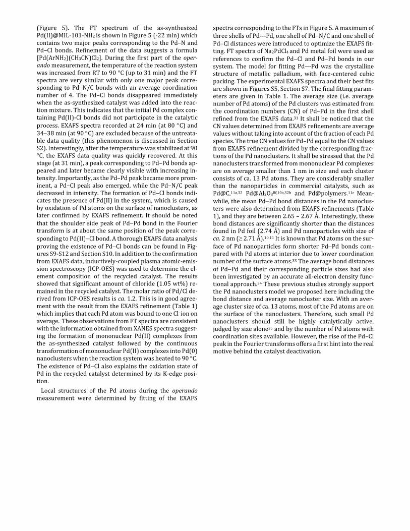
(Figure 5). The FT spectrum of the as-synthesized Pd(II)@MIL-101-NH2 is shown in Figure 5 (-22 min) which contains two major peaks corresponding to the Pd–N and Pd–Cl bonds. Refinement of the data suggests a formula [Pd(ArNH2)(CH3CN)Cl2]. During the first part of the oper-ando measurement, the temperature of the reaction system was increased from RT to 90 °C (up to 31 min) and the FT spectra are very similar with only one major peak corre-sponding to Pd–N/C bonds with an average coordination number of 4. The Pd–Cl bonds disappeared immediately when the as-synthesized catalyst was added into the reac-tion mixture. This indicates that the initial Pd complex con-taining Pd(II)-Cl bonds did not participate in the catalytic process. EXAFS spectra recorded at 24 min (at 80 C) and 34–38 min (at 90 C) are excluded because of the untreata-ble data quality (this phenomenon is discussed in Section S2). Interestingly, after the temperature was stabilized at 90 °C, the EXAFS data quality was quickly recovered. At this stage (at 31 min), a peak corresponding to Pd–Pd bonds ap-peared and later became clearly visible with increasing in-tensity. Importantly, as the Pd–Pd peak became more prom-inent, a Pd–Cl peak also emerged, while the Pd–N/C peak decreased in intensity. The formation of Pd–Cl bonds indi-cates the presence of Pd(II) in the system, which is caused by oxidation of Pd atoms on the surface of nanoclusters, as later confirmed by EXAFS refinement. It should be noted that the shoulder side peak of PdPd bond in the Fourier transform is at about the same position of the peak corre-sponding to Pd(II)Cl bond. A thorough EXAFS data analysis proving the existence of PdCl bonds can be found in Fig-ures S9-S12 and Section S10. In addition to the confirmation from EXAFS data, inductively-coupled plasma atomic-emis-sion spectroscopy (ICP-OES) was used to determine the el-ement composition of the recycled catalyst. The results showed that significant amount of chloride (1.05 wt%) re-mained in the recycled catalyst. The molar ratio of Pd/Cl de-rived from ICP-OES results is ca. 1.2. This is in good agree-ment with the result from the EXAFS refinement (Table 1) which implies that each Pd atom was bound to one Cl- ion on average. These observations from FT spectra are consistent with the information obtained from XANES spectra suggest-ing the formation of mononuclear Pd(II) complexes from the as-synthesized catalyst followed by the continuous transformation of mononuclear Pd(II) complexes into Pd(0) nanoclusters when the reaction system was heated to 90 °C. The existence of PdCl also explains the oxidation state of Pd in the recycled catalyst determined by its K-edge posi-tion.
Local structures of the Pd atoms during the operando measurement were determined by fitting of the EXAFS
spectra corresponding to the FTs in Figure 5. A maximum of three shells of Pd---Pd, one shell of PdN/C and one shell of PdCl distances were introduced to optimize the EXAFS fit-ting. FT spectra of Na2PdCl4 and Pd metal foil were used as references to confirm the PdCl and PdPd bonds in our system. The model for fitting Pd---Pd was the crystalline structure of metallic palladium, with face-centered cubic packing. The experimental EXAFS spectra and their best fits are shown in Figures S5, Section S7. The final fitting param-eters are given in Table 1. The average size (i.e. average number of Pd atoms) of the Pd clusters was estimated from the coordination numbers (CN) of Pd–Pd in the first shell refined from the EXAFS data.31 It shall be noticed that the CN values determined from EXAFS refinements are average values without taking into account of the fraction of each Pd species. The true CN values for Pd–Pd equal to the CN values from EXAFS refinement divided by the corresponding frac-tions of the Pd nanoclusters. It shall be stressed that the Pd nanoclusters transformed from mononuclear Pd complexes are on average smaller than 1 nm in size and each cluster consists of ca. 13 Pd atoms. They are considerably smaller than the nanoparticles in commercial catalysts, such as Pd@C,11a,32 Pd@Al2O38f,10a,32b and [email protected] Mean-while, the mean PdPd bond distances in the Pd nanoclus-ters were also determined from EXAFS refinements (Table 1), and they are between 2.65 – 2.67 Å. Interestingly, these bond distances are significantly shorter than the distances found in Pd foil (2.74 Å) and Pd nanoparticles with size of ca. 2 nm (≥ 2.71 Å).10,11 It is known that Pd atoms on the sur-
face of Pd nanoparticles form shorter PdPd bonds com-pared with Pd atoms at interior due to lower coordination number of the surface atoms.33 The average bond distances of PdPd and their corresponding particle sizes had also been investigated by an accurate all-electron density func-tional approach.34 These previous studies strongly support the Pd nanoclusters model we proposed here including the bond distance and average nanocluster size. With an aver-age cluster size of ca. 13 atoms, most of the Pd atoms are on the surface of the nanoclusters. Therefore, such small Pd nanoclusters should still be highly catalytically active, judged by size alone35 and by the number of Pd atoms with coordination sites available. However, the rise of the Pd–Cl peak in the Fourier transforms offers a first hint into the real motive behind the catalyst deactivation.

Table 1. Refined distances (d/Å), and mean number of distances (N) in selected scans using the Pd(II)@MIL-101-NH2 catalyst.
a The standard deviations in parentheses were obtained from k3-weighted least square refinements of the EXAFS function χ(k) and do not include systematic errors of the measurement. b The estimated error of the coordination is ca. 25% of the given value when refined. Underscored parameters have been optimized and fixed in the refinements. c The CN values in parentheses are the true coordination numbers of PdPd 1st shell when the fractions of Pd nanoclusters were taken into account. These values were used to determine the average size of Pd nanoclusters. d The bond distance and CN value of PdPd 1st shell at 31 min contain relatively big errors because of the weak EXAFS signal when Pd nanoclusters just started to form.
Figure 7. Proposed Pd species at different stages during the Heck coupling reaction using Pd(II)@MIL-101-NH2 as a catalyst. The red trace shows the temperature and the blue trace shows conversion to the desired product. The value of CN in the figure refers to the coordination number of Pd–Pd in the first shell.
Catalyst d(Pd-
N/C) (Å)a CNb
d(Pd-Cl) (Å)a
CNb d(Pd···
Pd) 1st shell (Å)a
CNb,c d(Pd···
Pd) 2nd shell (Å)a
CNb d(Pd···
Pd) 3rd shell (Å)a
CNb
As synth. 2.114(8) 2.0 2.298(1) 2.0 - - - - - - First scan 2.083(6) 4.0 - - - - - - - -
31 min 2.056(4) 3.8 2.710(7)d 0.4(8.0)d
c
44 min 2.037(6) 3.4 - - 2.674(5) 0.8(4.0) - - - - 47 min 1.993(9) 2.9 - - 2.666(7) 1.8(4.5) 3.68(2) 1.0 - - 50 min 2.026(8) 2.8 - - 2.655(4) 2.7(6.0) 3.82(2) 1.0 - - 57 min 1.980(6) 1.3 2.37(1) 0.4 2.654(4) 2.8(5.6) 3.76(2) 1.0 4.68(2) 2.0 63 min 1.956(5) 1.0 2.35(1) 0.7 2.653(4) 3.1(5.6) 3.74(4) 1.0 4.73(2) 2.0 73 min 1.979(7) 0.8 2.38(2) 1.0 2.662(3) 3.2(4.9) 3.76(1) 1.0 4.73(1) 2.0 85 min 1.959(7) 0.9 2.38(1) 1.0 2.662(3) 3.7(4.9) 3.78(2) 1.0 4.75(1) 2.0
115 min 1.978(7) 0.8 2.384(7) 1.0 2.662(2) 4.2(5.3) 3.76(2) 2.0 4.85(1) 3.0 Recycled 2.415(3) 1.0 2.689(1) 6.5 3.815(6) 3.0 4.747(3) 6.0
Pd foil 2.741(1) 12.0 3.904(8) 6.0 4.767(2) 24.0

Figure 8. Proposed mechanism for the Heck coupling reaction catalyzed by Pd(II)@MIL-101-NH2.
The average coordination number of PdN/C maintained at 4.0 when there was no transformation of Pd(II) species. As the mononuclear Pd(II) started to reconstruct into Pd(0) nanoclusters, the average CN of PdN/C decreased to ca. 3.4 at 44 min and to ca. 0.8 at 115 min indicating a continuous transformation process. The first PdCl bonds were ob-served after 57 min with an average CN of ca. 0.4. The reac-tion conversion had at this time already reached ca. 95% (Figure 3a). The average CN then further increased and ended up at ca. 1.0. It shall be noted that the bond distances of the reformed PdCl bonds were 2.35-2.42 Å, which is sig-nificantly longer than those in Pd(II) complexes20. This indi-cates that the Pd atoms coordinated to Cl- were surface at-oms of the Pd nanoclusters, where each Pd atom bound to several Pd atoms and the PdCl bonds became longer in comparison with Pd(II) complexes. This also indicates that when Cl- ions are coordinated to the relatively bulky and less mobile Pd nanoclusters, they could not easily be re-placed by the amino groups of the MOF linkers. In addition, no EXAFS signals corresponding to PdClCl and PdClPdCl multiple scattering paths were observed, which excludes the presence of PdCl2. Furthermore, no EXAFS signal corresponding to PdO single scattering was observed in the recycled catalyst, which suggests that sur-face Pd of the nanoclusters were coordinated to Cl- ions. Otherwise, they would have been oxidized in air to form PdO bonds.
The recycled Pd(II)@MIL-101-NH2 was measured by ex situ EXAFS (Figure S5f, Section S7). No EXAFS signal from PdN/C distances was found from the refinement and aver-age coordination numbers of PdPd and PdCl bonds were determined to be ca. 6.5 and ca. 1.0, respectively. The PdPd
bond distance is longer (ca. 2.69 Å) than PdPd bond dis-tances (2.65 – 2.67 Å) during operando measurement (44 – 115 min) indicating larger clusters, but still shorter than the PdPd distance of a 2 nm Pd nanoparticle. It is reasonable to propose that at this stage of the process, Pd nanoclusters became the absolute dominating species with an average size of ca. 20 atoms,34 and the surfaces of these Pd nanoclus-ters were almost covered by Cl- ions leading to the deactiva-tion of the catalyst. It shall be noted that the coordination numbers and the estimated sizes of Pd clusters obtained from the EXAFS fitting are an average of various Pd species. In reality, it is likely that both larger Pd nanoparticles with higher coordination numbers and smaller Pd clusters with lower coordination numbers coexist in the sample.
Based on the operando XANES and EXAFS analyses, the evolution of Pd species was followed and the catalytically active species under Heck conditions were identified. The kinetic profile (vide supra) of the identified Pd species are illustrated in Figure 7. The active species were mononuclear Pd(II) complexes coordinating to the linkers of the MOF at the beginning of the reaction below 90 C. The mononuclear Pd(II) complexes then dissociated from the linkers after one run and their activity was lost when the temperature was lower than 90 C. As the temperature was raised and main-tained at 90 C, these mobile mononuclear Pd(II) complexes gradually converted to Pd nanoclusters of ca. 13 Pd atoms on average. A mixture of the mobile mononuclear Pd(II) complexes and Pd nanoclusters co-existed and became the active species to catalyze the reaction. The activity of the mobile mononuclear Pd complexes at 90 C was confirmed by adding a second load of reagents at 70 min, when the sys-tem consisted of ca. 40% mononuclear Pd complexes and 60% Pd nanoclusters covered and poisoned by coordinated
Cl– ions (Figure 7). A full conversion of the new reagents was achieved in another 70 min at 90 C. This experimental re-sult proved that the mobile Pd mononuclear complexes are catalytically active after the reaction system was heated to 90 C during the operando measurement. At the beginning of this stage, mononuclear Pd complexes appeared also as the major Pd species compared to the Pd nanocluster. The mononuclear Pd complexes have even better activity than the corresponding Pd nanoclusters considering the size and the number of coordination sites available. This continuous conversion of the Pd species also indicated that mononu-clear Pd complexes had a relatively high activity because it could compete with the aggregation process under the re-ducing conditions. After Cl– ions were bound to the surface of the Pd nanoclusters, the remaining mononuclear Pd(II) complexes catalyzed the reaction to its completion. When the substrates were completely consumed, mononuclear Pd complexes could not react and they self-aggregated into Pd nanoclusters, which were quickly poisoned by remaining Cl- ions in the system. A similar evolution of Pd species was also observed from Pd(II)@MIL-88B-NH2 catalyzed reaction. The corresponding XANES and EXAFS spectra can be found in Figure S13, Section S11.
A mechanism derived from the above-described investi-gations is proposed and shown in Figure 8. The freshly pro-duced Pd(0) complex (I) represents the active species that initiate the catalytic cycle by oxidative addition in the C–I bond, leading to the Pd(L2)ArI intermediate (II). The ab-sence of any Pd–I distances in the EXAFS data is not surpris-ing. The oxidative addition is rapidly followed by a ligand exchange reforming the favored cationic species stabilized by a neutral N-ligand, following a similar preference as ob-served in the early stages of the experiment. This leads to an intermediate complex (III), to which the olefin coordinates to form a -complex (IV). Intermediates III and IV contain solely PdC/N bonds, in agreement with the XAS data. Based on this analysis, it is reasonable to conclude that the resting state of Pd species observed by XAS in the early stages of the measurement corresponds to intermediate IV, in which Pd has an oxidation state of +II. This is also in agreement with numerous reports that pointed out the mi-gratory insertion step to be turnover-limiting. In the follow-ing step, a -hydride elimination yields the final organic product and a palladium hydride species (VI) that upon re-ductive deprotonation returns to Pd(0), completing this cy-cle.
3. Framework stability and catalyst distribution
inside the MOFs (TEM, EDS, PXRD)
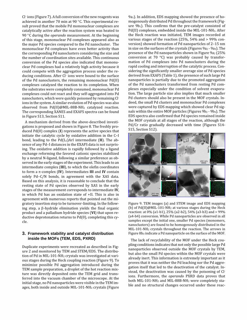
Duplicate experiments were recreated as described in Fig-ure 2 and monitored by TEM and STEM/EDS. The distribu-tion of Pd in MIL-101-NH2 crystals was investigated at vari-ous stages during the Heck coupling reaction (Figure 9). To minimize possible Pd aggregation introduced during the TEM sample preparation, a droplet of the hot reaction mix-ture was directly deposited onto the TEM grid and trans-ferred into the vacuum chamber of the microscope. At the initial stage, no Pd nanoparticles were visible in the TEM im-ages, both inside and outside MIL-101-NH2 crystals (Figure
9a1). In addition, EDS mapping showed the presence of ho-mogenously distributed Pd throughout the framework (Fig-ure 9b1). This confirms that the pre-catalyst consisted of Pd(II) complexes, embedded inside the MIL-101-NH2. After the Heck reaction was initiated, TEM images recorded at various stages of the reaction (25%, 54% and > 99% con-version) showed formation of Pd nanoparticles of 2–15 nm in size on the surfaces of the crystals (Figures 9a2 - 9a4). The presence of the Pd nanoparticles shown in Figure 9a2 (25% conversion at 70 °C) was probably caused by transfor-mation of Pd complexes into Pd nanoclusters during the rapid cooling and interruption of the catalytic process. Con-sidering the significantly smaller average size of Pd species derived from EXAFS (Table 1), the presence of such large Pd nanoparticles is partially due to the promoted aggregation of the Pd nanoclusters transformed from resting Pd com-plexes especially under the condition of solvent evapora-tion. The large particle size also implies that much smaller Pd clusters should also be present in the MOF crystals. In-deed, the small Pd clusters and mononuclear Pd complexes were captured by EDS mapping which showed clear Pd sig-nals within the entire MOF particles (Figures 9b2 - 9b4). The EDS spectra also confirmed that Pd species remained inside the MOF crystals at all stages of the reaction, although the Pd/Cr ratio gradually decreased with time (Figures S14-S15, Section S12).
Figure 9. TEM images (a) and STEM image and EDS mapping (b) of Pd(II)@MIL-101-NH2 at various stages during the Heck reaction: at 0% (a1-b1), 25% (a2-b2), 54% (a3-b3) and > 99% (a4-b4) conversion. While Pd nanoparticles are observed in all samples except the initial one, smaller Pd species (monomers, nanoclusters) are found to be homogenously distributed in the MIL-101-NH2 crystals throughout the reaction. The arrows in Figure 8b3 indicate a Pd nanoparticle on the surface of the MOF.
The lack of recyclability of the MOF under the Heck cou-pling conditions indicates that not only the possible large Pd nanoparticles observed outside the MOF crystals by TEM, but also the small Pd species within the MOF crystals were already inert. This information is extremely important as it proves that it was neither the Pd leaching nor the Pd aggre-gation itself that led to the deactivation of the catalyst. In-stead, the deactivation was caused by the poisoning of Cl- ions. Furthermore, the operando PXRD data proves that both MIL-101-NH2 and MIL-88B-NH2 were completely sta-ble and no structural changes occurred under these reac-

tion conditions (Figure S16, Section S13). Based on all infor-mation acquired, a clear picture emerges. The deactivation of the catalyst is not of a physical nature. Different from the structural degradation factors commonly suggested in the literature such as Pd leaching and agglomeration or MOF decomposition, the predominant deactivation pathway here is of a chemical nature. The irreversible capping of small Pd clusters with chloride ligands makes the reactive sites inaccessible to the reaction partners, long before Pd leaching or aggregation becomes a major issue.
4. Recyclability
With the above information in hand, we revisited the issue of recyclability. Initial studies showed a dramatic loss of ac-tivity in the second cycle, after the catalyst was recovered, washed and dried. However, when fresh reagents were added during the reaction phase of high activity, before a complete inhibition by Cl– ions was achieved, a high TOF could be maintained for at least 3 cycles. Despite the dis-turbance introduced by fresh reagents (temperature, solu-bility, reagent ratios) the reaction was faster than what would have been necessary to convert three batches of starting materials by fresh catalysts (Section S14). This ex-periment was purely illustrative and does not represent a practical experimental procedure. However, it suggests that operation under continuous flow would be a suitable way to improve the total turnover of the catalyst. Cl– ions could be washed away at the beginning of the experiment and a con-stant feed of fresh reagents would postpone the deactiva-tion process.
Even though the recyclability of the material did not work as expected, we observed remarkable differences when comparing the activity of the Pd(II)@MOF with its homoge-neous counterparts. When the reaction was performed us-ing the same palladium precursors (i.e. Na2PdCl4 and PdCl2(MeCN)2) as catalysts without being impregnated into the MOF support, the yields dropped to 42-43% for both Pd(II) salts. This clearly shows the beneficial effect of the MOF support in this catalysis.
5. Discussion
The correlation of data from multiple characterization tech-niques portrays Pd@MIL-101-NH2 and Pd@MIL-88B-NH2 as heterotopic multifaceted catalysts with more than one mode of action. In their as-synthesized dry form, both Pd(II)@MOF pre-catalysts contain Pd centers in a +II oxida-tion state and square planar coordination geometry, bear-ing two anionic Cl– ligands. When the catalyst was mixed with the other reagents except for olefin, the chloride lig-ands were immediately released and replaced by neutral ni-trogen ligands, forming a cationic complex favored by the polar aqueous environment. Upon olefin addition in the presence of water, Pd(II) was rapidly reduced to Pd(0) even at room temperature. The freshly reduced Pd underwent a fast oxidative addition, initiating the catalytic cycle. The Heck coupling product was already detectable immediately after reaching 60 °C, proving that mononuclear Pd(II) com-plexes attaching to the linkers of the MOFs were competent
active species to catalyze this transformation at relatively low temperatures.
The first catalytic turnovers have taken place inside the MOF pores, and proceeded with comparable rate for both Pd@MIL-101-NH2 and Pd@MIL-88B-NH2 catalysts. During the first cycle, the coordination to the amino-terephthalate linker was lost in the case of MIL-101-NH2, leading to the formation of an even more electron-deficient Pd species, which could travel through the pores. A second oxidative addition could not be initiated by this form of electrophilic Pd, which instead gradually agglomerated into small nanoclusters. The resulting mixture of the mobile mononu-clear Pd complexes and the Pd nanoclusters was inert at 60 °C, and became active in the catalytic cycle at 90 °C. How-ever, upon depletion of the starting materials and further upon cooling the reaction mixture, the surface of the Pd clusters was covered by Cl– ions and their catalytic proper-ties were finally lost. Similar conclusions can be derived for Pd(II)@MIL-88B-NH2 although the chelating coordination mode of the MOF and the more confined pore space attenu-ated the deactivation process.
This study fits into a larger effort to bring a better under-standing to the complex behavior of palladium under reac-tion conditions specific for C–C and C–heteroatom bond for-mation.36 In recent years, it has become evident that the ac-tual landscape of active species is more diverse and dy-namic than previously believed. Starting from mononuclear Pd catalysts, formation of Pd clusters is often unavoidable. However, these particulate species not only represent an al-ternative resting state for Pd but can also get involved di-rectly in the second stage of catalysis. The presence of rap-idly inter-convertible species that possess different levels of reactivity makes it challenging to quantify precisely their contribution to the total observed catalytic effect.37 Moreo-ver, commonly employed heterogeneity tests may not al-ways provide reliable results.38 Without a better under-standing of these phenomena, a rational design of superior catalysts would not be achievable.
Towards this goal, the method presented herein can re-veal fine details of the reaction mechanism by monitoring more precisely the evolution of different Pd species and the effects of Pd phase transitions on its catalytic activity. More-over, we are continuously working to expand the applicabil-ity of the method. Further development of the in situ reactor will make the acquisition of fluorescence XAS data in oper-ando mode feasible. This would be especially beneficial to measure highly diluted samples or catalysts with relatively low absorption edge energies. Furthermore, the new gener-ation of synchrotron radiation sources will provide great opportunities to improve the time resolution and investi-gate reactions with significantly shorter half-lifetimes.
Conclusions
The mechanism of a Pd@MOF-catalyzed Heck C–C coupling reaction was investigated in detail using a newly developed reaction cell suitable for operando studies of heterogeneous catalysts. For the first time we probed the entire lifetime of the catalyst and revealed fine details of the reaction mecha-nism that would otherwise remain hidden. Different active species operating at different reaction stages and under dif-ferent reaction conditions have been identified, together

with their activation and deactivation pathways. The irre-versible deactivation of the catalyst was provoked neither by leaching of the active species, nor by decomposition of the crystalline framework. Instead a chemical deactivation mechanism was identified, in which Cl- ions cover the sur-face of transformed Pd clusters and nanoparticles, blocking the access of starting materials to the active sites. This in-formation can be used to prolong the lifetime of the catalyst and to design improved catalysts and processes in the fu-ture. Importantly, the method described is widely applica-ble to study diverse chemical reactions catalyzed by transi-tion metals, including both homogeneous and heterogene-ous systems, and has the potential to provide unprece-dented insight into the fine details of the mechanism. Other complex mechanisms of MOF-catalyzed processes are cur-rently under investigation in our laboratories.
ASSOCIATED CONTENT
Supporting Information Catalysts syntheses; optimization of reaction conditions, substrate scope and recycling experi-ments; additional studies of kinetic profiles by 1H NMR; details on the XAS and PXRD data collection; XAS data treatment and refinement results; additional TEM and EDS results. This mate-rial is available free of charge via the Internet at http://pubs.acs.org.
AUTHOR INFORMATION
Corresponding Authors
*[email protected] *[email protected]
(1) Egorova, K. S.; Ananikov V. P. Angew. Chem. Int. Ed. 2016, 55, 12150–12162.
(2) Jutand, A. Mechanisms of the Mizoroki–Heck Reaction (Chapter 1), in The Mizoroki-Heck Reaction (ed M. Oestreich), 2009, John Wiley & Sons, Ltd, Chichester, UK. (3) For some of the most recent reviews discussing catalysis with MOFs, see: a) Cohen, S. M.; Zhang, Z.; Boissonnault, J. A. Inorg. Chem. 2016, 55, 7281–7290; b) Dhakshinamoorthy, A.; Asiri, A. M.; Gar-cia, H. Catal. Sci. Technol. 2016, 6, 5238–5261; c) Dhakshinamoor-thy, A.; Asiri, A. M.; Garcia, H. Chem. Soc. Rev. 2015, 44, 1922–1947; d) Chughtai, A. H.; Ahmad, N.; Younus, H. A.; Laypkov, A.; Verpoort, F. Chem. Soc. Rev. 2015, 44, 6804–6849; e) Evans, J. D.; Sumby C. J.; Doonan, C. J. Chem. Soc. Rev. 2014, 43, 5933–5951; f) Gascon, J.; Corma, A.; Kapteijn, F.; Llabres i Xamena, F. X. ACS. Catal. 2014, 4, 361–378; g) Janssen, K. P. F.; De Cremer, G.; Neely, R. K.; Kubarev, A. V.; Van Loon, J.; Martens, J. A.; De Vos, D. E.; Roeffaers, M. B. J.; Hofkens, J. Chem. Soc. Rev. 2014, 43, 990–1006; h) Valvekens, P.; Vermoortele, F.; De Vos, D. Catal. Sci. Technol. 2013, 3, 1435–1445; i) Rogge, S. M. J.; Bavykina, A.; Hajek, J.; Garcia, H.; Olivos-Suarez, A. I.; Sepúlveda-Escribano, A.; Vimont, A.; Clet, G.; Bazin, P.; Kapteijn, F.; Daturi, M.; Ramos-Fernandez, E. V.; Llabrés i Xamena, F. X.; Van Speybroeck, V.; Gascon, J. Chem. Soc. Rev. 2017, 46, 3134–3184; j) Leus, K.; Liu, Y. Y; Voort P. V. D. Cat. Rev. Sci. Eng. 2014, 56, 1–56.
(4) For recent reviews, discussing the complexity of heterogene-ous catalytic mechanisms, see: a) Eremin, D. B.; Ananikov, V. P. Coord. Chem. Rev. 2017, 346, 2–19; b) Schauermann, S.; Freund, H.-J. Acc. Chem. Res. 2015, 48, 2775–2782; c) Schauermann, S.; Nilius N.; Shalkhutdinov, S.; Freund, H.-J. Acc. Chem. Res. 2013, 46, 1673–1681; d) Yang, X.-F.; Wang, A.; Qiao, B.; Li, J.; Liu, J.; Zhang, T. Acc. Chem. Res. 2013, 46, 1740–1748.
Author Contributions
$These authors contributed equally to this work.
Notes The authors declare no competing financial interest.
ACKNOWLEDGMENT
This work has been supported by the MATsynCELL project through Röntgen-Ångström Cluster, supported by the Swedish Research Council (VR) and the German Federal Ministry of Ed-ucation and Research (BMBF). We are also thankful to the Ber-zelii Center EXSELENT, the Swedish Research Council and the Project Management Organization at DESY (Deutsches Electro-nen-Synchrotron). The allocation of beamtime at BM01B (SNBL), European Synchrotron Radiation Facility (ESRF) is gratefully acknowledged. A.K.I. is supported by the Knut and Alice Wallenberg Foundation (KAW) through the MAX IV post-doctoral scholarship. B.M.-M. was supported by VINNOVA through a VINNMER grant. We thank KAW for the project grant 3DEM-NATUR and a grant for purchasing the TEMs. We thank Prof. Lynne McCukser for assistance during the beamtime ap-plications, and the staff of beamline BM01B (SNBL), particu-larly Dr. Michela Brunelli and Dr. Hermann Emerich at the ESRF for the assistance during data collection. We also thank Dr A. Bermejo Gómez for helpful discussions.
REFERENCES
(5) Metzger, E. D.; Comito, R. J.; Hendon, C. H.; Dinca, M. J. Am. Chem. Soc. 2017, 139, 757–762.
(6) Morris, R. J.; Russell Jr., J. N.; Karwacki C. J. J. Phys. Chem. Lett. 2015, 6, 4923–4926.
(7) a) Bordiga, S.; Groppo, E.; Agostini, G.; van Bokhoven, J. A.; Lamberti, C. Chem. Rev. 2013, 113, 1736–1850; b) Singh, J.; Lamberti, C.; van Bokhoven, J. A. Chem. Soc. Rev. 2010, 39, 4754–4766; c) Bordiga, S.; Bonino, F.; Lillerud, K. P.; Lamberti, C. Chem. Soc. Rev 2010, 39, 4885–4927.
(8) a) Jung, U.; Elsen, A.; Li, Y.; Smith, J. G.; Small, M. W.; Stach E. A.; Frenkel, A. I.; Nuzzo, R. G. ACS Catal. 2015, 5, 1539–1551; b) Smit, E. De; Cinquini, F.; Beale, A. M.; Safonova, O. V; Beek, W. Van; Sautet, P.; Weckhuysen, B. M.; Supe, N.; Horowitz, R. J.; Cedex, F.-G. J. Am. Chem. Soc. 2010, 132, 14928–14921; c) Rønning, M.; Tsakoumis, N. E.; Voronov, A.; Johnsen, R. E.; Norby, P.; Van Beek, W.; Borg, Ø; Rytter, E.; Holmen, A. Catal. Today 2010, 155, 289–295; d) van Beek, W.; Safonova, O. V.; Wiker, G.; Emerich, H. Phase Transitions 2011, 84, 726–732; e) Grunwaldt, J.-D.; Clausen, B. S. Topics Catal. 2002, 18, 37–43; f) Matam, S. K.; Aguirre, M. H.; Weidenkaff, A.; Ferri, D. J. Phys. Chem. C 2010, 114, 9439–9443; g) Reina, T. R.; Xu, W.; Ivanova, S.; Centeno, M. Á.; Hanson, J.; Rodriguez, J. A.; Odriozola, J. A. Catal. Today 2013, 205, 41–48; h) Cassinelli, W. H.; Martins, L.; Passos, A. R.; Pulcinelli, S. H.; Santilli, C. V.; Rochet, A.; Briois, V. Catal. Today 2014, 229, 114–122; i) Braglia, L.; Borfecchia, E.; Martini, A.; Bugaev, A. L.; Soldatov, A. V; Øien-Ødegaard, S.; Bleken, B. T. L.; Olsbye, U.; Lillerud, K. P.; Lomachenko, K. A.; Agostini, G.; Manzoli, M.; Lamberti, C. Phys. Chem. Chem. Phys. 2017, 19, 27489–27507; j) Andersen, C. W.; Borfecchia, E.; Bremholm, M.; Jørgensen, M. R. V.; Vennestrøm, P. N. R.; Lamberti, C.; Lundegaard, L. F.; Iversen, B. B. Angew. Chemie Int. Ed. 2017, 56, 10367–10372; k) Barzan, C.; Piovano, A.; Braglia, L.;

Martino, G. A.; Lamberti, C.; Bordiga, S.; Groppo, E. J. Am. Chem. Soc. 2017, 139, 17064–17073; l) Øien, S.; Agostini, G.; Svelle, S.; Borfecchia, E.; Lomachenko, K. A.; Mino, L.; Gallo, E.; Bordiga, S.; Olsbye, U.; Lillerud, K. P.; Lamberti, C. Chem. Mater. 2015, 27, 1042–1056; m) Zhou, Y.; Doronkin, D. E.; Chen, M.; Wei, S.; Grunwaldt, J.-D. ACS Catal. 2016, 6, 7799–7809; n) Stötzel, J.; Frahm, R.; Kimmerle, B.; Nachtegaal, M.; Grunwaldt, J.-D. J. Phys. Chem. C 2012, 116, 599–609; o) Doronkin, D. E.; Casapu, M.; Günter, T.; Müller, O.; Frahm, R.; Grunwaldt, J.-D. J. Phys. Chem. C 2014, 118, 10204–10212; p) Wezendonk, T. A.; Santos, V. P.; Nasalevich, M. A.; Warringa, Q. S. E.; Dugulan, A. I.; Chojecki, A.; Koeken, A. C. J.; Ruitenbeek, M.; Meima, G.; Islam, H. U.; Sankar, G.; Makkee, M.; Kapteijn, F.; Gascon, J. ACS Catal. 2016, 6, 3236–3247. (9) a) Gorlin, Y.; Lassalle-Kaiser, B.; Benck, J. D.; Gul, S.; Webb, S. M.; Yachandra, V. K.; Yano, J.; Jaramillo, T. F. J. Am. Chem. Soc. 2013, 135, 8525–8534; b) Newton, M. A.; Brazier, J. B.; Barreiro, E. M.; Parry, S.; Emmerich, H.; Adrio, L. A.; Mulligan, C. J.; Hellgardt, K.; Hii, K. K. (Mimi). Green Chem. 2016, 18, 406–411.
(10) a) Grunwaldt, J.-D.; Caravati, M.; Baiker, A. J. Phys. Chem. B 2006, 110, 9916–9922; b) Lee, A. F.; Ellis, C. V.; Naughton, J. N.; Newton, M. A.; Parlett, C. M. A.; Wilson, K. J. Am. Chem. Soc. 2011, 133, 5724–5727; c) Parlett, C. M. A.; Gaskell, C. V.; Naughton, J. N.; Newton, M. A.; Wilson, K.; Lee, A. F. Catal. Today 2013, 205, 76–85. (11) a) Reimann, S.; Stötzel, J.; Frahm, R.; Kleist, W.; Grunwaldt, J. D.; Baiker, A. J. Am. Chem. Soc. 2011, 133, 3921–3930; b) Brazier, J. B.; Nguyen, B. N.; Adrio, L. A.; Barreiro, E. M.; Leong, W. P.; Newton, M. A.; Figueroa, S. J. A.; Hellgardt, K.; Hii, K. K. M. Catal. Today 2014, 229, 95–103; c) Ellis, P. J.; Fairlamb, I. J. S.; Hackett, S. F. J.; Wilson, K.; Lee, A. F. Angew. Chem., Int. Ed. 2010, 49, 1820–1824; d) Fiddy, S. G.; Evans, J.; Neisius, T.; Newton, M. A.; Tsoureas, N.; Tulloch, A. A. D.; Danopoulos, A. A. Chem. Eur. J. 2007, 13, 3652–3659.
(12) Zalesskiy, S. S.; Ananikov, V. P. Organometallics 2012, 31, 2302–2309.
(13) For general aspects of MOF chemistry, see: a) Howarth, A. J.; Peters, A. W.; Vermeulen, N. A.; Wang, T. C.; Hupp, J. T.; Farha O. K. Chem. Mater. 2017, 29, 26–39; b) Howarth, A. J.; Liu, Y.; Li, P.; Li, Z.; Wang, T. C.; Hupp, J. T.; Farha O. K. Nat. Rev. Mater. 2016, 15018; c) Ferguson, A.; Liu, L.; Tapperwijn, S. J.; Perl, D.; Coudert, F.-X.; Van Cleuvenbergen, S.; Verbiest, T.; van der Veen, M. A.; Telfer, S. G. Nat. Chem. 2016, 8, 250–257; d) Furukawa, H.; Cordova, K. E.; O’
Keeffe, M.; Yaghi, O. M. Science 2013, 341, 1230444; e) Ferey, G.; Chem. Soc. Rev. 2008, 37, 191–214.
(14) For detailed discussions on the complexity and tunability of MOF supports, see: a) Fracaroli, A. M.; Siman, P.; Nagib, D. A.; Su-zuki, M.; Furukawa, H.; Toste, F. D.; Yaghi, O. M. J. Am. Chem. Soc. 2016, 138, 8352–8355; b) Furukawa, H.; Muller, U.; Yaghi, O. M. An-gew. Chem. Int. Ed. 2015, 54, 3417–3430; c) Sue, A. C.-H.; Manninge, R. V.; Deng, H.; Cao, D.; Wang, C.; Gandara, F.; Stoddart, J. F.; White-lam, S.; Yaghi, O. M. Proc. Natl. Acad. Sci. 2015, 137, 7810–7816.
(15) For some interesting recent examples of MOFs in catalysis, see: a) Li, J.; Yu, X.; Xu, M.; Liu, W.; Sandraz, E.; Lan, H.; Wang, J.; Cohen, S. M. J. Am. Chem. Soc. 2017, 139, 611–614; b) Lv, X.-L.; Wang, K.; Wang, B.; Su, J.; Zou, X.; Xie, Y.; Li, J.-R.; Zhou, H.-C. J. Am. Chem. Soc. 2017, 139, 211–217; c) Yu, X.; Cohen, S. M. J. Am. Chem. Soc. 2016, 138, 12320–12323; d) Noh, H.; Cui, Y.; Peters, A. W.; Pahls, D. R.; Ortuno, M. A.; Vermeulen, N. A.; Cramer, C. J.; Gagliardi L.; Hupp, J. T.; Farha, O. K. J. Am. Chem. Soc. 2016, 138, 14720–14 726; e) Rimoldi, M.; Nakamura, A.; Vermeulen, N. A.; Henkelis, J. J.; Blackburn, A. K.; Hupp, J. T.; Stoddart J. F.; Farha, O. K. Chem. Sci. 2016, 7, 4980–4984; f) Mon, M.; Ferrando-Soria, J.; Grancha, T.; Fortea-Perez, F. R.; Gascon, J.; Leyva-Perez, A.; Armentano, D.; Pardo, E. J. Am. Chem. Soc. 2016, 138, 7864–8767; g) Abednatanzi, S.; Derakhshandeh, P. G.; Abbasi, A.; Voort, P. V. D.; Leus, K. Chem-CatChem 2016, 8, 3672–3679.
(16) a) Pascanu, V.; Yao, Q.; Bermejo Gómez, A.; Gustafsson, M.; Yun, Y., Wan, W.; Samain, L.; Zou, X.; Martín-Matute, B. Chem. Eur. J. 2013, 19, 17483–17493; b) Pascanu, V.; Hansen, P.; Bermejo Gómez, A.; Ayats, C.; Platero-Prats, A. E.; Johansson, M. J.; Pericàs, M. À.; Martín-Matute, B. ChemSusChem 2015, 8, 123–130.
(17) Pascanu, V.; Carson, F.; Vico Solano, M.; Su, J.; Zou, X.; Johans-son, M. J.; Martin-Matute. B. Chem. Eur. J. 2016, 22, 3729–3737.
(18) Carson, F.; Pascanu, V.; Bermejo Gómez, A.; Zhang, Y.; Plat-ero-Prats, A. E.; Zou, X.; Martín-Matute B. Chem. Eur. J. 2015, 21, 10896–10902.
(19) Heidenreich, N.; Rütt, U.; Köppen, M.; Inge, A. K.; Beier, S.; Dippel, A.-C.; Suren, R.; Stock, N. Rev. Sci. Instrum. 2017, 88, 104102.
(20) Pascanu, V.; Bermejo Gómez, A.; Ayats, C.; Platero-Prats, A. E.; Carson, F.; Su, J.; Yao, Q.; Pericàs, M. A.; Zou, X.; Martín-Matute B. ACS Catal. 2015, 5, 472–479.
(21) Abdala, P.M.; Mauroy, H.; van Beek, W. J. Appl. Cryst. 2014, 47, 449–457.
(22) George, G. N.; Pickering, I. J. EXAFSPAK – A Suite of Computer Programs for Analysis of X-ray Absorption Spectra, SSRL, Stanford, CA. 1993.
(23) Thompson, A.; Attwood, D.; Gullikson, E.; Howells, M.; Kim, K.-J.; Kirz, J.; Kortright, J.; Lindau, I.; Liu, Y.; Pianetta, P.; Robinson, A.; Scofield, J.; Underwood, J.; Williams, G.; Winick, H. X-ray data booklet, Lawrence Berkeley National Laboratory, Berkeley, 3rd Ed., 2009.
(24) Zabinsky, S. I.; Rehr, J. J.; Ankudinov, A.; Albers, R. C.; Eller, M. J. Phys. Rev. B 1995, 52, 2995–3009.
(25) Sigeev, A. S.; Peregudov, A. S.; Cheprakov, A. V.; Beletskaya I. P. Adv. Synth. Catal. 2015, 357, 417–429. (26) a) Ahsen, B. V.; Bley, B.; Proemmel, S.; Wartchow, R.; Willner, H. Z. anorg. Allg. Chem. 1998, 624, 1225–1234; b) Gebauer, T.; Frenzen, G.; Dehnicke, K. Z. Naturforsch. 1992, 47, 1505–1512; c) Massa, W.; Wocadlo, S.; Dehnicke, K.; Gebauer, T. Z. Kristallogr. 1996, 211, 120–121.
(27) a) Kong, G.-Q.; Ou, S.; Zou, C.; Wu, C.-D. J. Am. Chem. Soc., 2012, 134, 19851–19857; b) Chen, L.; Rangan, S.; Li, J.; Jiang, H.; Li, Y. Green Chem. 2014, 16, 3978–3985. (28) a) IUPAC Stability Constants, Academic Sofware, Otley, UK; b) Hellquist, B.; Elding, L. I.; Ducommun, Y. Inorg. Chem. 1988, 27, 3620–3623; c) Vargaftik, M. N.; German, E. D.; Dogonadze, R. R.; Syrkin, Y. K. Dokl. Akad. Nauk SSSR 1972, 206, 370–376; d) Boily, J.-F.; Seward, T. M. Geochim. Cosmochim. Acta 2005, 69, 3773–3789; e) Kragten J. Talanta 1980, 27, 375–377; f) Elding, L. I. Inorg. Chim. Acta 1972, 6, 647–651; g) Rittner, W.; Gulko, A.; Schmuckler, G. Talanta 1970, 17, 807–816.
(29) Henry, P. M.; Keith, J. A. Angew. Chem., Int. Ed., 2009, 48, 9038–9049.
(30) Jalilehvand, F. Structural of Hydrated Ions and Cyano Com-plexes by X-Ray Absorption Spectroscopy. Ph.D. Dissertation. Royal Institute of Technology, Stockholm, 2000. (31) Jentys, A. Phys. Chem. Chem. Phys. 1999, 1, 4059–4063. (32) a) Pikna, L.; Milkovič, O.; Saksl, K.; Heželová, M.; Smrčová, M.; Puliš, P.; Michalik, S.; Gamcová, J. J. Solid State Chem. 2014, 212, 197–204; b) Agostini, G.; Lamberti, C.; Pellegrini, R.; Leofanti, G.; Giannici, F.; Longo, A.; Groppo, E. ACS Catal. 2014, 4, 187–194; c) Shimizu, K.; Kubo, T.; Satsuma, A.; Kamachi, T.; Yoshizawa, K. ACS Catal. 2012, 2, 2467–2474. (33) Qi, W.; Huang, B.; Wang, M. Nanoscale Res. Lett. 2009, 4, 269–273. (34) Krüger, S.; Vent, S.; Nörtemann, F.; Staufer, M.; Rösch, N. J. Chem. Phys. 2001, 115, 2082–2087. (35) a) Phan, N. T. S.; Van Der Sluys, M.; Jones, C. W. Adv. Synth. Catal. 2006, 348, 609–679; b) Yu, L.; Huang, Y.; Wei, Z.; Ding, Y.; Su, C.; Xu, Q. J. Org. Chem. 2015, 80, 8677–8683; c) Martins, D. de L.; Alvarez, H. M.; Aguiar, L. C. S.; Antunes, O. A. C. Appl. Catal. A Gen. 2011, 408, 47–53; d) Thathagar, M. B.; ten Elshof, J. E.; Rothenberg, G. Angew. Chemie Int. Ed. 2006, 45, 2886–2890.
(36) a) Ananikov, V. P.; Beletskaya I. P. Organometallics 2012, 31, 1296–1604; b) Selivanova, A. V.; Tyurin, V. S.; Beletskaya, I. P. ChemPlusChem, 2014, 79, 1278–1283; b) Kashin A. N.; Ganina O. G.; Cheprakov A. V.; Beletskaya I. P. ChemCatChem 2015, 7, 2113–2121.

(37) Pentsak, E. O.; Kashin, A. S.; Polynsky, M. V.; Kvashnina, K. O.; Glatzel, P.; Ananikov, V. P. Chem. Sci. 2015, 6, 3302–3313.
(38) Crabtree, R. H. Chem. Rev. 2012, 112, 1536–1554.