Embed Size (px)
Citation preview
J. exp. Bio!. 156, 267-285 (1991) 2 6 7Printed in Great Britain © 7711" Company of Biologists Limited 1991
TEMPERATURE SENSITIVITY OF GRADED SYNAPTICTRANSMISSION IN THE LOBSTER STOMATOGASTRIC
GANGLION
BY BRUCE R. JOHNSON1, JACK H. PECK2
AND RONALD M. HARRIS-WARRICK1
1 Section of Neurobiology and Behavior, Seeley G. Mudd Hall, CornellUniversity, Ithaca, NY 14853, USA, 2Department of Psychology, Ithaca College,
Ithaca, NY 14850, USA
Accepted 19 October 1990
Summary
We examined the temperature sensitivity of graded chemical synaptic strengthwithin the pyloric circuit of the spiny lobster stomatogastric ganglion. Coolingfrom 20.4°C to 11.3°C reduced the gTaded synaptic potential (GSP) amplitude atall six pyloric synapses tested. Cooling appeared to reduce the slope of the linearpart of the input-output curve at three of these synapses, and did not significantlyalter the threshold for transmitter release at any synapses. Pairs of neurons with apresynaptic pyloric dilator (PD) cell showed reductions in graded synapticstrength at 16.5 °C but those with presynaptic lateral pyloric (LP) or ventral dilator(VD) cells did not. A generalized decrease in input resistance is not responsiblefor the reduced GSP amplitude upon cooling, as determined by input resistance,action potential amplitude and electrical coupling measurements. We concludethat cooling reduces graded chemical strength by a direct synaptic action. Sincethe PD and VD cells use the same transmitter and act on some of the samepostsynaptic cells, their differential sensitivity to cooling further suggests apresynaptic site of action. The temperature range used in our experimentsencompasses the range that the animal normally encounters in nature. Thus, therelative importance of graded synaptic interactions in generating the pyloric motorrhythm may vary with transient changes in temperature.
Introduction
Temperature is an important environmental modulator of the behavior ofectothermic animals. Consequently, the effects of altered temperature on neur-onal properties and interactions have been extensively studied in ectotherms(reviewed by Florey, 1978; Prosser and Nelson, 1981; Stephens, 1985, 1990).Synaptic junctions are, of course, important targets where changes in temperatureact to modify behavior. Most studies of temperature effects on chemical synaptic
Key words: graded synaptic transmission, temperature, stomatogastric ganglion, lobster,Panulirus interruptus.
268 B. R. JOHNSON, J. H. PECK AND R. M. HARRIS-WARRICK
transmission have focused on synapses where action potentials normally triggerthe release of transmitter. Many synapses, however, release transmitter as acontinuously graded function of presynaptic membrane potential. In invert-ebrates, for example, graded chemical synaptic transmission is used for trans-mission of sensory information (reviewed by Bush, 1981; Shaw, 1981; see alsoWilkens, 1988), in the production of motor patterns (reviewed by Simmers, 1981;Wilson and Phillips, 1983; Siegler, 1985; Hartline et al. 1988; see also Spencer,1988; DiCaprio, 1989; Toga et al. 1990) and for neuromuscular transmission (Davisand Stretton, 1989). The effects of changing temperature on these graded synapseshave not been studied.
We have examined the temperature sensitivity of graded chemical synaptictransmission in a neuronal circuit that appears to depend on graded interactionsfor its normal function. We chose the pyloric motor circuit of the stomatogastricganglion (STG) which generates rhythmic foregut activity in decapod crustaceans(Claiborne and Ayers, 1987). In the spiny lobster Panulirus interruptus, this circuitis a well-defined central pattern generator (CPG) network composed of 14neurons in six major classes whose synaptic interconnectivity is known in detail(Mulloney, 1987; see Fig. 1). All 14 neurons use action potentials to send signals todistant targets (muscles or neurons in other ganglia) but, within the STG, thesespiking neurons depend primarily on graded transmission to generate motorpatterns (Raper, 1979; Anderson and Barker, 1981; Graubard et al. 1983; Russelland Graubard, 1987; Hartline etal. 1988). We report here that the strength ofgraded chemical synaptic transmission between pyloric circuit neurons is tempera-ture-sensitive over a range that the animal normally encounters in nature. Inaddition, we provide evidence that temperature effects occur directly at thesynaptic junction and cannot be explained by a generalized input resistance changein the pre- and/or postsynaptic cell.
Materials and methods
Pacific spiny lobsters (Panulirus interruptus Randall) were purchased fromMarinus Inc. (Long Beach, CA) and maintained in marine aquaria at 15°C. Thestomatogastric nervous system (Selverston et al. 1976) was dissected and placed ina preparation dish filled with Panulirus saline of the following composition (inmmol P 1 ) : NaCl, 479; KC1,12.8; CaCl2,13.7; Na2SO4, 3.9; MgSO4,10.0; glucose,2.0; Tris base, 11.1; maleic acid, 5.1; pH7.35 (Mulloney and Selverston, 1974).The STG was desheathed, enclosed in a small (1 ml) pool of saline surrounded byVaseline and constantly perfused at 5 ml min"1 with oxygenated saline. The salinetemperature was controlled with a peltier device and monitored by a smallthermistor positioned within a few millimeters of the STG.
Standard intracellular techniques were used for current injection and voltagerecordings using KCl-filled (3moll"1, 10-20MQ) microelectrodes. The cellbodies of the pyloric neurons (anterior burster, AB; inferior cardiac, IC; lateralpyloric, LP; pyloric dilator, PD; pyloric, PY; ventral dilator, VD) were identified

Temperature sensitivity of graded synoptic transmission 269
Fig. 1. Summary of the synaptic connections in the pyloric central pattern generator ofPanulirus interruptus (modified from Mulloney, 1987, and Johnson and Harris-Warrick, 1990). There are eight PY and two PD neurons and one of each other celltype. Resistor symbols indicate electrotonic connections between and among celltypes; thick-lined symbols indicate strong connections, thin-lined symbols indicateweaker connections and dashed symbols indicate weak connections. Filled circlesindicate inhibitory chemical synapses. PY, pyloric; AB, anterior burster; IC, inferiorcardiac; LP, lateral pyloric; VD, ventral dilator.
during rhythmic pyloric activity at 16.5°C by: (1) matching action potentialsrecorded extracellularly from an appropriate motor nerve root and intracellularlyfrom the soma; (2) the timing of spike activity within the pyloric rhythm; (3) thecharacteristic shape of membrane potential oscillations and action potentialamplitudes; and (4) the synaptic connectivity (Fig. 1). The PY cell population inthese experiments was a mixture of early- and late-firing PYs (Hartline et al. 1987).
Following cell identification, we replaced the saline in a second Vaseline-walledpool surrounding the input nerve to the STG with 10 moll tetrodotoxin(TTX). This procedure eliminated all descending modulatory inputs to the STGand thus stopped rhythmic pyloric activity (Russell, 1979; Nagy and Miller, 1987).Possible ascending modulatory inputs from muscle stretch receptors (Katz et al.1989) were eliminated in the initial dissection by removing the appropriate nerves.In some experiments, we isolated pairs of neurons from the rest of the pyloriccircuit to ensure that changes in synaptic efficacy were not occurring indirectly(Johnson and Harris-Warrick, 1990). This isolation involved 6-carboxyfluoresceinphotoinactivation (Miller and Selverston, 1979; Flamm and Harris-Warrick, 1986)and pharmacological blockade (Bidaut, 1980; Eisen and Marder, 1982; Marderand Eisen, 1984).
All experiments were conducted over a temperature range of 20.4-11.3°C,which approximates the natural temperature range (21-12°C) for these animals(G. Martin, Marinus, Inc., unpublished observations). Cooling and heating ratesranged from 0.5 to 1.0°min~1 and measurements were taken after at least 5minequilibration time at each temperature. Each series of measurements was made atthree temperatures: 20.4°C (which we will refer to as the high temperature),
270 B. R. JOHNSON, J. H . PECK AND R. M. HARRIS-WARRICK
16.5°C (mid temperature) and 11.3°C (low temperature). In most experiments,initial measurements were made at the high temperature; some experiments werebegun at either the middle or low temperature and showed no qualitativedifference in the results. A final measurement was always repeated at the initialtest temperature to ensure reversibility. Electrodes were removed from the cellbefore each temperature change and the cell was re-penetrated after a newtemperature had been reached. This procedure was necessary because there wassufficient movement of the STG during temperature changes to dislodge theelectrodes and/or damage the cell.
Measurements of graded synaptic strength
The effect of cooling on graded chemical transmission between pyloric circuitneurons was examined after the STG had been superfused with 10~ molP 1 TTXin saline to block spiking synaptic transmission. Graded synaptic strength betweena pyloric cell pair at different temperatures was determined from input-output(I/O) curves measured with two presynaptic electrodes (for current injection andvoltage recording) and one postsynaptic electrode to record the gTaded synapticpotential (GSP, Johnson and Harris-Warrick, 1990). I/O curves were constructedfrom 1 s presynaptic peak polarizations of varying amplitude and sign, plottedagainst the peak amplitude of the postsynaptic polarization. The stimulation ratewas 0.2 Hz; there was no obvious decrement in postsynaptic responses withsquare-wave presynaptic depolarizations at this frequency. Peak GSP amplitudeswere compared at the same levels of presynaptic membrane potential at eachsynapse (ranging from —35 to — 25 mV at different synapses). These levels variedamong synapses because the peak GSP amplitude sometimes declined with verylarge presynaptic depolarizations (not shown in the I/O curves of Fig. 4);amplitude comparisons were made before this decline in GSP amplitude. We donot understand the cause of this decline in GSP amplitude, but weak electricalcoupling, which is generally present between pyloric cells (Mulloney, 1987), maycontribute (Johnson and Harris-Warrick, 1990). The slope of the I/O curve wasobtained from a simple regression line through the data points with measurableGSPs, excluding data points after the peak GSP amplitude had been reached. Thethreshold for a detectable postsynaptic response was calculated as the x-interceptpoint of this regression line. Because the sites of the synaptic contacts areelectrically distant from our recording site in the cell body, our I/O curve slopeand threshold measurements are estimates only; however, they serve here asuseful points of comparison among the different temperature conditions.
The input resistance of the cell body was measured with two electrodes, one topass current and one to record the resulting voltage changes. The input resistancewas taken as the slope of the I/V relationship over the linear part of the I/V curve(i.e. hyperpolarized from rest). Antidromic action potentials were elicited bystimulation of the respective peripheral motor nerves with 0.5 ms suprathresholdstimuli at 0.33 Hz. Action potential amplitudes were measured from the restingpotential to the peak depolarization. I/O curves for electrical coupling between
Temperature sensitivity of graded synaptic transmission 271
pairs of pyloric cells were gathered using the same stimulation and recordingprotocol as that used for gTaded chemical transmission. Picrotoxin (PTX)(5xlO~6moir1) was added to the TTX saline for measurements of PY cellcoupling. This appeared to increase PY cell input resistance slightly above that ofother pyloric cells (see Fig. 6) and served to make their weak electrical couplingmore apparent. Coupling coefficients were determined from the slopes of theseI/O curves. To ensure that weak capacitative coupling between the electrodes wasnot mistaken for weak electrical transmission, we compared weak cellularelectrotonic responses with electrode responses after the postsynaptic electrodehad been withdrawn to the bath. Statistical comparisions were made withrepeated-measures analysis of variance (ANOVA) and subsequent protectedMests.
Results
Effects of temperature on graded chemical synaptic strength
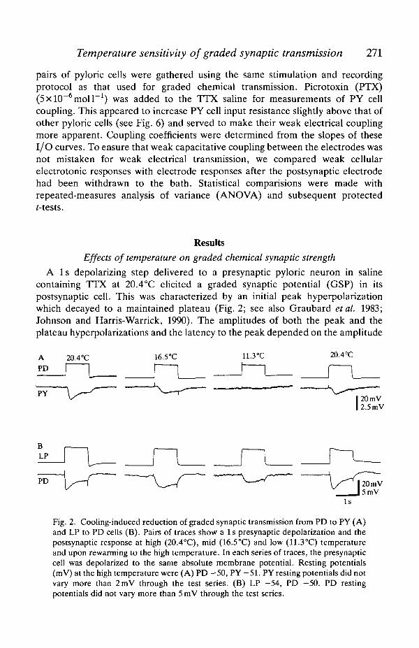
A 1 s depolarizing step delivered to a presynaptic pyloric neuron in salinecontaining TTX at 20.4°C elicited a graded synaptic potential (GSP) in itspostsynaptic cell. This was characterized by an initial peak hyperpolarizationwhich decayed to a maintained plateau (Fig. 2; see also Graubard et al. 1983;Johnson and Harris-Warrick, 1990). The amplitudes of both the peak and theplateau hyperpolarizations and the latency to the peak depended on the amplitude
20.4°C
20 mV2.5 raV
PD 20mV5mV
Fig. 2. Cooling-induced reduction of graded synaptic transmission from PD to PY (A)and LP to PD cells (B). Pairs of traces show a Is presynaptic depolarization and thepostsynaptic response at high (20.4°C), mid (16.5°C) and low (11.3°C) temperatureand upon rewarming to the high temperature. In each series of traces, the presynapticcell was depolarized to the same absolute membrane potential. Resting potentials(mV) at the high temperature were (A) PD -50, PY -51 . PY resting potentials did notvary more than 2mV through the test series. (B) LP -54, PD -50. PD restingpotentials did not vary more than 5mV through the test series.
272 B. R. JOHNSON, J. H. PECK AND R. M. HARRIS-WARRICK
of the presynaptic depolarization (Graubard etal. 1983). With large presynapticdepolarizations (approximately 30mV from rest), the GSP reached a maximumamplitude; larger presynaptic depolarization often elicited smaller GSPs but couldstill shorten the GSP rise time. In our experiments, we did not detect transmitterrelease at rest, as observed in earlier studies (Graubard etal. 1983; Johnson andHarris-Warrick, 1990), probably because of the relatively hyperpolarized restingpotentials of the presynaptic cells (see Table 2).
Cooling from 20.4°C (high temperature) to 11.3°C (low temperature) caused aprogressive reversible reduction in the peak GSP amplitude at all the synapses weexamined in the pyloric circuit. The different synaptic pairs did not, however, allexhibit the same temperature sensitivity. For example, Fig. 2A shows that, inresponse to an identical PD depolarization, the peak amplitude of the GSP in a PYcell was reduced to 52% of the high-temperature value at 16.5 °C (the midtemperature) and was abolished at the low temperature. A second synapse, fromLP to PD, (Fig. 2B) was less affected: cooling from high to mid temperaturereduced the GSP amplitude to 73 % and this was further reduced to 45 % at thelow temperature. In both examples, the reduction in graded synaptic strength wasat least partially reversed upon re-warming to the high temperature.
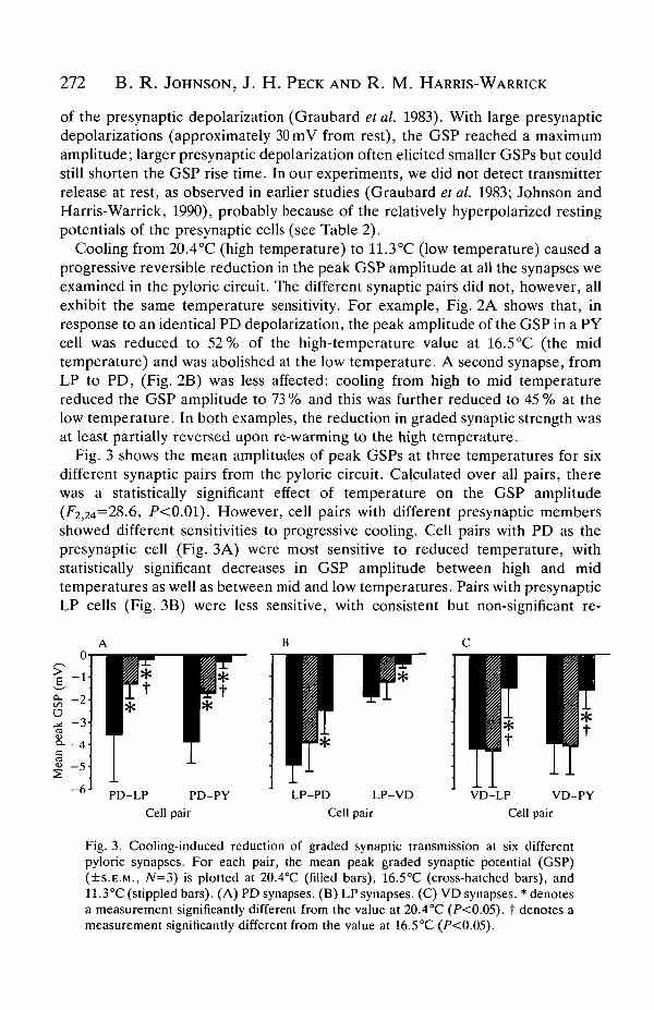
Fig. 3 shows the mean amplitudes of peak GSPs at three temperatures for sixdifferent synaptic pairs from the pyloric circuit. Calculated over all pairs, therewas a statistically significant effect of temperature on the GSP amplitude(^2,24=28.6, P<0.01). However, cell pairs with different presynaptic membersshowed different sensitivities to progressive cooling. Cell pairs with PD as thepresynaptic cell (Fig. 3A) were most sensitive to reduced temperature, withstatistically significant decreases in GSP amplitude between high and midtemperatures as well as between mid and low temperatures. Pairs with presynapticLP cells (Fig. 3B) were less sensitive, with consistent but non-significant re-
- 6 J
PD-LP PD-PYCell pair
LP-PD LP-VDCell pair
VD-LP VD-PYCell pair
Fig. 3. Cooling-induced reduction of graded synaptic transmission at six differentpyloric synapses. For each pair, the mean peak graded synaptic potential (GSP)(+S.E.M., N=3) is plotted at 20.4°C (filled bars), 16.5°C (cross-hatched bars), and11.3°C (stippled bars). (A) PD synapses. (B) LP synapses. (C) VD synapses. * denotesa measurement significantly different from the value at 20.4°C (P<0.05). t denotes ameasurement significantly different from the value at 16.5°C (P<0.05).
Temperature sensitivity of graded synoptic transmission 273
ductions in GSP amplitude at the mid temperature and significant reductions at thelow temperature. Pairs with presynaptic VD cells (Fig. 3C) showed no reductionin GSP amplitude at the mid temperature, but a significant decrease at the lowtemperature. These differences between presynaptic cells suggest that loweredtemperature selectively affects presynaptic function (see Discussion).
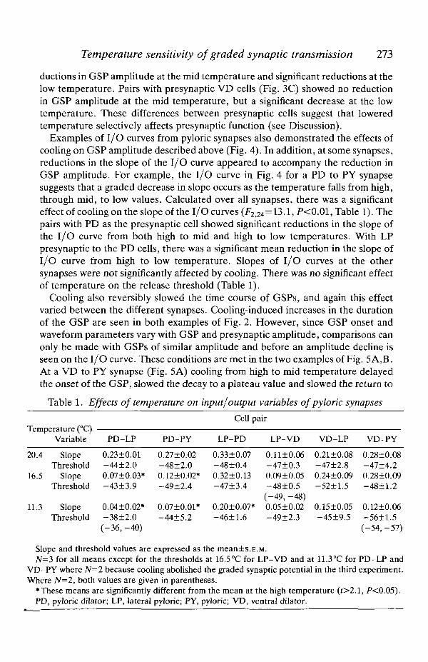
Examples of I/O curves from pyloric synapses also demonstrated the effects ofcooling on GSP amplitude described above (Fig. 4). In addition, at some synapses,reductions in the slope of the I/O curve appeared to accompany the reduction inGSP amplitude. For example, the I/O curve in Fig. 4 for a PD to PY synapsesuggests that a graded decrease in slope occurs as the temperature falls from high,through mid, to low values. Calculated over all synapses, there was a significanteffect of cooling on the slope of the I/O curves (F2,24=13.1, P<0.01, Table 1). Thepairs with PD as the presynaptic cell showed significant reductions in the slope ofthe I/O curve from both high to mid and high to low temperatures. With LPpresynaptic to the PD cells, there was a significant mean reduction in the slope ofI/O curve from high to low temperature. Slopes of I/O curves at the othersynapses were not significantly affected by cooling. There was no significant effectof temperature on the release threshold (Table 1).
Cooling also reversibly slowed the time course of GSPs, and again this effectvaried between the different synapses. Cooling-induced increases in the durationof the GSP are seen in both examples of Fig. 2. However, since GSP onset andwaveform parameters vary with GSP and presynaptic amplitude, comparisons canonly be made with GSPs of similar amplitude and before an amplitude decline isseen on the I/O curve. These conditions are met in the two examples of Fig. 5A,B.At a VD to PY synapse (Fig. 5A) cooling from high to mid temperature delayedthe onset of the GSP, slowed the decay to a plateau value and slowed the return to
Table 1. Effects of temperature on input/output variables of pyloric synapses
TprnrA C111L
20.4
16.5
11.3
>erature (°C)Variable
SlopeThreshold
SlopeThreshold
SlopeThreshold
PD-LP
0.23±0.01-44±2.00.07±0.03*-43±3.9
0.0410.02*-3812.0
PD-PY
0.27±0.02-48±2.00.12±0.02*-49±2.4
0.07±0.01*-44+5.2
Cell
LP-PD
0.33±0.07-48±0.40.32±0.13-47±3.4
0.20±0.07-46±1.6
pair
LP-VD
0.11±0.06-47±0.30.09±0.05-48±0.5(-49, -48)
* 0.05±0.02-49+2.3
VD-LP
0.21±0.08-47±2.80.24±0.09-52±1.5
0.15±0.05-45±9.5
VD-PY
0.28±0.08-47±4.20.2810.09-48±1.2
0.12±0.06-5611.5
(-36,-40) (-54,-57)
Slope and threshold values are expressed as the meanis.E.M.N=3 for all means except for the thresholds at 16.5°C for LP-VD and at 11.3°C for PD-LP and
VD-PY where N=2 because cooling abolished the graded synaptic potential in the third experiment.Where N=2, both values are given in parentheses.
•These means are significantly different from the mean at the high temperature (t>2.1, P<0.05).PD, pyloric dilator; LP, lateral pyloric; PY, pyloric; VD, ventral dilator.
274 B. R. JOHNSON, J. H. PECK AND R. M. HARRIS-WARRICK
_2c
ote
D -U
CL03C>-»
1-
o-
- 1 -
- 2 -
°0
ii- 2
- 3 4- 4
- 4 0 - 3 0 - 2 0 - 7 0 -60 -50 - 4 0 - 3 0
PD potential (mV)
CM O * O *
- 7 0 - 6 0 - 5 0 - 4 0 - 3 0
1,
0- 1
- 2
- 3
- 4
- 5
- 6- 7 0 -60 - 5 0 - 4 0 - 3 0
LP potential (mV)
2
0
- 2
- 4
- 6 1 J * «
-60 - 5 0 - 4 0 -30 - 2 0 -"70 - 6 0 - 5 0 - 4 0 - 3 0
VD potential (mV)
Fig. 4. Effects of temperature on input-output curves (peak graded synaptic poten-tial, GSP, amplitude in postsynaptic cell plotted against presynaptic membranepotential) for graded chemical transmission at six different pyloric synapses. Hyper-polarizations from the presynaptic resting potential are not shown because there wasno resting transmitter release at any of these synapses. Open circles indicatemeasurements at 20.4°C, filled triangles are measurements at 16.5°C and filled squaresare measurements at 11.3°C. The AB cell was killed for the PD to PY experiment butnot for the PD to LP experiment. However, in other PD to LP experiments where theAB cell was killed, similar reductions in the GSP were seen with cooling. The AB andPD cells were killed for the VD to LP and VD to PY experiments.
Temperature sensitivity of graded synoptic transmission 275
A
20.4°C
20.4°C
5mV
200 ms
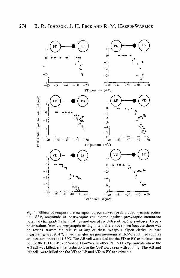
Fig. 5. Effects of cooling on the time course of graded synaptic potentials (GSPs) andelectrotonic potentials. Digitized postsynaptic responses to presynaptic stimulation atdifferent temperatures are overlaid. Presynaptic stimulation time for all traces isindicated by the bottom bar. (A) VD-PY GSP at 20.4 and 16.5°C. The AB and PDcells were killed for this experiment. (B) LP-PD GSP at 20.4 and 11.3°C. (C) PD-PDelectrotonic postsynaptic potential at 20.4 and 11.3°C.
the baseline after the end of the presynaptic stimulation. At an LP to PD synapse(Fig. 5B), with a faster rise time to peak, cooling from high to low temperature didnot change the GSP onset but did increase the time to peak and the decay toplateau. These changes in GSP time course are probably not the result of generalchanges in membrane properties, since electrotonic potentials between electricallycoupled PY or PD cells at high and low temperatures did not show different timecourses (Fig. 5C).
Effects of temperature on pyloric cell input resistance
Cell body input resistance
Cooling-induced reductions in GSP amplitude could arise from direct effects atthe synapse itself, or through a general reduction in cell input resistance. Since thesynapses are located in the neuropil of the STG (King, 1976), a temperature-jnduced change in the passive spread of current would affect both neuronal
276 B. R. JOHNSON, J. H. PECK AND R. M. HARRIS-WARRICK
16 -i
VD LPCell type
PY
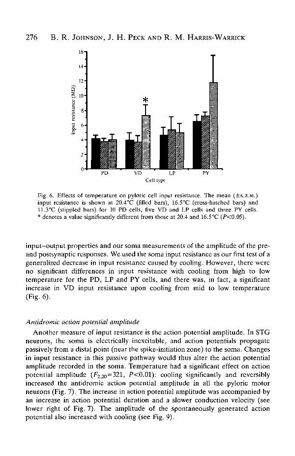
Fig. 6. Effects of temperature on pyloric cell input resistance. The mean (±S.E.M.)input resistance is shown at 20.4°C (filled bars), 16.5°C (cross-hatched bars) and11.3°C (stippled bars) for 10 PD cells, five VD and LP cells and three PY cells.* denotes a value significantly different from'those at 20.4 and 16.5 °C (P<0.05).
input-output properties and our soma measurements of the amplitude of the pre-and postsynaptic responses. We used the soma input resistance as our first test of ageneralized decrease in input resistance caused by cooling. However, there wereno significant differences in input resistance with cooling from high to lowtemperature for the PD, LP and PY cells, and there was, in fact, a significantincrease in VD input resistance upon cooling from mid to low temperature(Fig. 6).
Antidromic action potential amplitude
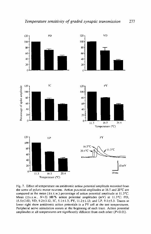
Another measure of input resistance is the action potential amplitude. In STGneurons, the soma is electrically inexcitable, and action potentials propagatepassively from a distal point (near the spike-initiation zone) to the soma. Changesin input resistance in this passive pathway would thus alter the action potentialamplitude recorded in the soma. Temperature had a significant effect on actionpotential amplitude (F2,2o=321, P<0.01): cooling significantly and reversiblyincreased the antidromic action potential amplitude in all the pyloric motorneurons (Fig. 7). The increase in action potential amplitude was accompanied byan increase in action potential duration and a slower conduction velocity (seelower right of Fig. 7). The amplitude of the spontaneously generated actionpotential also increased with cooling (see Fig. 9).
Temperature sensitivity of graded synoptic transmission 277
120 -i
100-
80
120
100-
80
60
40
20
VD
120
= 100
80
60
40
20
1C
11.3 16.5 20.4Temperature (°C)
120 l
100
LP PY
20 10 mV
11.3 16 5 20.4Temperature (°C)
10 ms
Fig. 7. Effect of temperature on antidromic action potential amplitude recorded fromthe soma of pyloric motor neurons. Action potential amplitudes at 16.5 and 20°C arecompared as the mean (±S.E.M.) percentage of action potential amplitude at 11.3°C.Mean (±S.E.M., JV=3) 100% action potential amplitudes (mV) at 11.3°C: PD,15.5±2.02; VD, 9.2±3.12; IC, 5.1 + 1.3; PY, 11.2+1.13; and LP, 9.1±5.3. Traces atlower right show antidromic action potentials in a PY cell at the test temperatures.Peripheral nerve stimulation occurs at the beginning of each trace. Action potentialamplitudes at all temperatures are significantly different from each other (P<0.01).
278 B. R. JOHNSON, J. H. PECK AND R. M. HARRIS-WARRICK
PD-PDCell pair
PD-VDCell pair
PY-PYCell pair
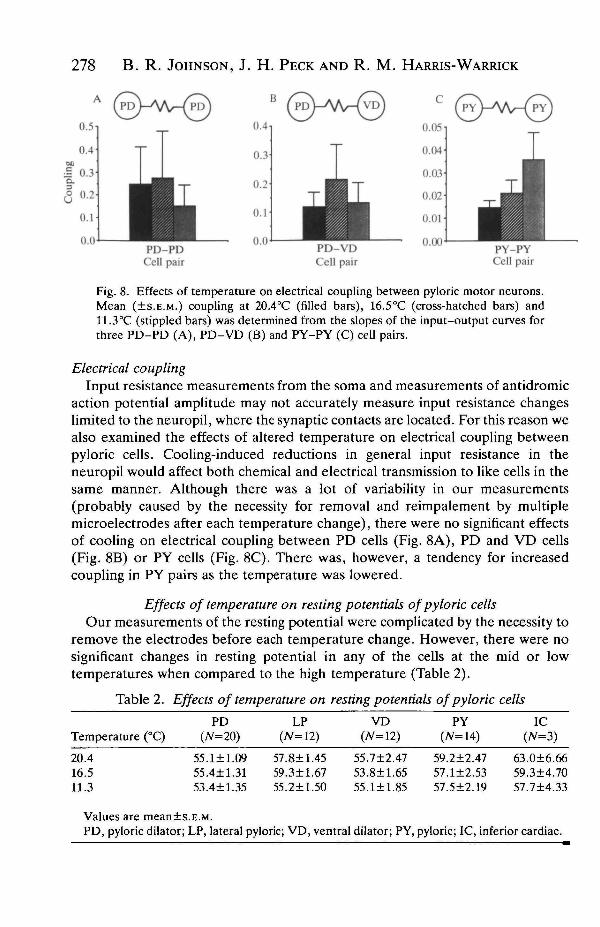
Fig. 8. Effects of temperature on electrical coupling between pyloric motor neurons.Mean (±S.E.M.) coupling at 20.4°C (filled bars), 16.5°C (cross-hatched bars) and11.3°C (stippled bars) was determined from the slopes of the input-output curves forthree PD-PD (A), PD-VD (B) and PY-PY (C) cell pairs.
Electrical couplingInput resistance measurements from the soma and measurements of antidromic
action potential amplitude may not accurately measure input resistance changeslimited to the neuropil, where the synaptic contacts are located. For this reason wealso examined the effects of altered temperature on electrical coupling betweenpyloric cells. Cooling-induced reductions in general input resistance in theneuropil would affect both chemical and electrical transmission to like cells in thesame manner. Although there was a lot of variability in our measurements(probably caused by the necessity for removal and reimpalement by multiplemicroelectrodes after each temperature change), there were no significant effectsof cooling on electrical coupling between PD cells (Fig. 8A), PD and VD cells(Fig. 8B) or PY cells (Fig. 8C). There was, however, a tendency for increasedcoupling in PY pairs as the temperature was lowered.
Effects of temperature on resting potentials of pyloric cellsOur measurements of the resting potential were complicated by the necessity to
remove the electrodes before each temperature change. However, there were nosignificant changes in resting potential in any of the cells at the mid or lowtemperatures when compared to the high temperature (Table 2).
Table 2. Effects of temperature on resting potentials of pyloric cells
PD LP VD(JV=20)Temperature (°C)
LP(7V=12)
PY(7V=14)
IC(N=3)
20.416.511.3
Values are mean±sPD, pyloric dilator;
55.55.53.
.E.M
LP,
1±14±1.4±1
.09
.31
.35
575955
lateral pyloric;
.8±1
.3±1
.2±1
VD,
.45
.67
.50
ventral
55.7±253.8±155.1±1
dilator;
.47
.65
.85
PY,
59.2±2.4757.1±2.5357.5±2.19
635957
.0±6.66
.3±4.70
.7±4.33
pyloric; IC, inferior cardiac.
Temperature sensitivity of graded synaptic transmission 279
20.4°C 11.3°C
PD
t
J100ms
10 mV5mV
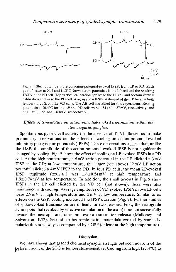
Fig. 9. Effect of temperature on action-potential-evoked IPSPs from LP to PD. Eachpair of traces at 20.4 and 11.3°C shows action potentials in the LP cell and the resultingIPSPs in the PD cell. Top vertical calibration applies to the LP cell and bottom verticalcalibration applies to the PD cell. Arrows show IPSPs at the end of the LP burst at bothtemperatures (from the VD cell). The AB cell was killed for this experiment. Restingpotentials at 20.4°C for the LP and PD cells were —54 and — 57mV, respectively, andat 11.3°C, -55 and -60mV, respectively.
Effects of temperature on action-potential-evoked transmission within thestomatogastic ganglion
Spontaneous pyloric cell activity (in the absence of TTX) allowed us to makepreliminary observations on the effects of cooling on action-potential-evokedinhibitory postsynaptic potentials (IPSPs). These observations suggest that, unlikethe GSP, the amplitude of the action-potential-evoked IPSP is not significantlychanged by cooling. Fig. 9 shows the effect of cooling on LP-evoked IPSPs in a PDcell. At the high temperature, a 6mV action potential in the LP elicited a 3mVIPSP in the PD; at low temperature, the larger (see above) 12mV LP actionpotential elicited a 4mV IPSP in the PD. In four PD cells, the mean LP-evokedIPSP amplitude (±S.E.M.) was 1.6±0.54mV at high temperature and1.9±0.74mV at low temperature. In addition, the small arrows in Fig. 9 showIPSPs in the LP cell elicited by the VD cell (not shown); these were alsomaintained with cooling. Average amplitudes of VD-evoked IPSPs in two LP cellswere 2.9mV at high temperature and 3mV at low temperature. Similar to itseffects on the GSP, cooling increased the IPSP duration (Fig. 9). Further studiesof spike-evoked transmission are difficult for two reasons. First, the retrogradeaction potential (evoked by selective stimulation of the axon) does not successfullyinvade the neuropil and does not evoke transmitter release (Mulloney andSelverston, 1972). Second, orthodromic action potentials evoked by soma de-polarization are always accompanied by a GSP (at least at the high temperature).
Discussion
We have shown that graded chemical synaptic strength between neurons of thepyloric circuit of the STG is temperature-sensitive. Cooling from high (20.4°C) to
280 B. R. JOHNSON, J. H . PECK AND R. M. HARRIS-WARRICK
low temperature (11.3°C) reduced the GSP amplitude at all pyloric synapsestested. These synapses did, however, display differential sensitivity to coolingfrom high to mid (16.5 °C) temperature: graded synaptic strength was reduced atsome synapses but not at others.
Mechanisms by which temperature changes graded synaptic strength
Cooling could reduce graded transmission by a reduction in the synaptic drivingforce, by a generalized reduction in membrane resistance and/or by directlyaltering the synaptic transmission process itself. Since the resting potential of thepyloric cells did not significantly change from high to low temperature, reducedsynaptic driving force is unlikely to account for graded synaptic strengthreductions with cooling.
A cooling-induced reduction in membrane input resistance could reduce gradedchemical transmission by reducing the passive spread of current from input sites tooutput sites in the neuropil (King, 1976). This would change graded synaptic I/Oproperties by decreasing the peak GSP amplitude and the slope of the I/O curve,and by decreasing our soma measurements of pre- and postsynaptic voltageresponses. Three lines of evidence indicate that a generalized decrease in inputresistance is not responsible for the reduced GSP amplitude upon cooling. First, astemperature is lowered, both pre- and postsynaptic cells show either an increase orno significant change in soma input resistance. Second, the amplitudes of bothantidromic and spontaneous action potentials progressively increase with cooling.This suggests that an input resistance increase occurs somewhere along thepathway that the passively propagated spike follows to the soma. Cooling is knownto increase input resistance in a variety of neurons and muscle cells fromectotherms (Prosser and Nelson, 1981; White, 1983; Klein and Prosser, 1985;Adams, 1987), including axons of the spiny lobster (Grossman and Kendig, 1984).An alternative explanation for the increased spike amplitude upon cooling couldbe that it propagates further towards the soma before failing. Third, there are nosignificant changes in electrical coupling between like or unlike cells with cooling.In fact, there is a tendency for electrical coupling to increase (PY to PY) ratherthan to decrease. Using electrical coupling measurements as indicators of inputresistance changes is complicated because they reflect a summation of effects onthe electrical junctional membranes and on non-junctional membranes. In severalspecies (including crustaceans), electrical junctional conductance is not tempera-ture-sensitive if the cooling is slow (Spray and Bennett, 1985), as it was in theseexperiments. Thus, junctional conductance changes with cooling probably do notcontribute to any coupling changes we observed between pyloric cells. Our resultssuggest that the input resistance, monitored by measuring soma input resistance,antidromic spike height or electrical coupling, may be increasing at some sites inthe cell, but is certainly not decreasing with cooling and therefore cannot accountfor the reduction in GSP strength.
We conclude that, in the pyloric motor circuit, the strength of the gradedsynaptic interaction itself is reduced at lower temperatures. We did not directly
Temperature sensitivity of graded synaptic transmission 281
address the question of whether the temperature-sensitive steps in transmissionare pre- or postsynaptic, but cooling does not appear specifically to affect thepostsynaptic response to transmitter in pyloric cells. Graded synaptic strength atPD to LP and PD to PY synapses was reduced at 16.5°C, and sometimescompletely abolished at 11.3°C. In contrast, graded synaptic strength at VD to LPand VD to PY synapses changed little at the mid temperature. Only at the lowtemperature was there a reduction in the GSP at synapses with VD as thepresynaptic cell. Since both the PD and VD cells use the same transmitter,acetylcholine (Marder, 1987), one would expect that, if postsynaptic responsive-ness was altered by cooling, both VD and PD synapses onto LP and PY cells wouldbe similarly affected. One direct test of this would be to compare neuronalresponsiveness to extrinsically applied neurotransmitter. Unfortunately, we couldnot directly determine the postsynaptic response to applied acetylcholine becausemuscarinic agonists can also induce bursting potentials in pyloric cells (Nagy et al.1985). Based on the differential sensitivity of the PD and VD synapses to cooling,we suggest that cooling may reduce the graded synaptic strength between pyloricneurons by a presynaptic action. The differential sensitivity to cooling of gradedtransmission between pyloric cells could be due to a differential sensitivity ofcalcium entry into the terminals and/or to a differential sensitivity of the releasemechanism itself.
Comparison of the effects of temperature on graded and spike-evokedtransmission
Similar to our results with graded synaptic transmission in the pyloric circuit,lowered temperature reduces spike-evoked transmission at many synapses inother ectotherms (see reviews by Florey, 1978; Prosser and Nelson, 1981;Stephens, 1985, 1990), including the squid giant synapse (Weight and Erulkar,1976), motor neuron synapses in insect ganglia (Burrows, 1989), frog (Takeuchi,1958; Jensen, 1972) and lizard (Adams, 1989), and crustacean neuromuscularjunctions (Stephens, 1985). Since the input resistance generally increases withlowered temperature in these preparations, a change in input resistance cannotexplain the reduction in synaptic potential amplitude.
As we suggested for the cooling-induced reduction in GSP, decreases in spike-evoked transmission with cooling are also largely explained by presynaptictemperature effects. For example, low temperature reduces quantal content atseveral neuromuscular junctions (Takeuchi, 1958; White, 1983; Adams, 1989) anddecreases spontaneous transmitter release at frog (Fatt and Katz, 1952; Barrettet al. 1978) and crab muscle fibers (Parnas et al. 1975). Cooling may decreasesynaptic potentials by reducing calcium entry into the presynaptic terminal at thesquid giant synapse (Charlton and Atwood, 1979; Llinas et al. 1987). Like thethreshold for transmitter release in our preparation, the threshold depolarizationfor measuring calcium current at the squid giant synapse is not affected by cooling(Llinas et al. 1987). Recently, Delaney and Zucker (1990) found that coolingf educed neurotransmitter release elicited by flash-evoked release of caged calcium
282 B . R. JOHNSON, J. H. PECK AND R. M. HARRIS-WARRICK
injected into the squid giant synapse. This suggests that, in addition to reducingcalcium influx into the squid presynaptic terminal, lowered temperature affects therelease processes that follow calcium influx.
In our experiments, cooling increased the latency of the GSP and prolonged itsduration, although these effects varied from cell pair to cell pair. These effects ofcooling are not due to general changes in the membrane properties of pyloric cells,since cooling did not change the latency and time course of electrical couplingpotentials (Fig. 5C). Similar results are found in spike-activated synapses, wherethe evidence suggests that this arises primarily from an effect of temperature onthe release process itself (Katz and Miledi, 1965; Barrett and Stevens, 1972; Llinasetal. 1987; Delaney and Zucker, 1990).
Importance of graded synoptic transmission in organizing the pyloric motorpattern
Graded synaptic interactions between neurons of the pyloric circuit are thoughtto organize the motor patterns produced by this CPG, while the same cells useaction potentials to send signals to distant targets (Raper, 1979; Anderson andBarker, 1981; Russell and Graubard, 1987; Hartline etal. 1988). The reduction inthe graded synaptic strength at temperatures near the lower natural range of12-21 °C for the spiny lobster implies, however, that the role of graded trans-mission may vary with temperature. The pyloric motor pattern remains robust at11.3°C, although, compared to the rhythm at 20.4°C, the frequency is slower andfiring phase relationships between the pyloric cells are modified (B. R. Johnson,J. H. Peck and R. M. Harris-Warrick, unpublished observations). Perhaps at lowtemperature, the increase in action potential amplitude and duration maintainsspike-evoked synaptic interactions, thus compensating for weaker graded interac-tions between the pyloric cells. The importance of graded and spike-evokedtransmission for pyloric motor pattern generation at different temperatures cannotbe determined at present because temperature may affect other neuronalproperties that are important for pattern generation. We must also examine theeffects of temperature on such properties as endogenous bursting (Johnson et al.1990), plateau generation, firing threshold and post-inhibitory rebound (Getting,1989) to understand fully the constellation of temperature effects that mayproduce variable pyloric motor patterns.
We must emphasize that we have only examined the effects of acute tempera-ture changes on graded synaptic transmission. We have not yet studied thecompensatory changes that could occur in lobsters maintained for long periods atlow temperature. It is clear that many ectotherms, including crustaceans, canacclimate a variety of neural functions to match a wide range of thermal conditions(Langerspetz, 1974; Florey, 1978; Prosser and Nelson, 1981; Stephens, 1985); thisincludes acclimatory shifts in neuromuscular transmission (Harri and Florey, 1979;Stephens and Atwood, 1982; Blundon, 1989). Seasonal acclimatory changescertainly may occur in graded synaptic transmission such that its functional
Temperature sensitivity of graded synaptic transmission 283
significance in organizing the pyloric motor pattern at lowered temperatures wouldbe maintained.
Approximately 99% of animal species are poikilotherms, and must contendwith a body temperature at or near environmental levels (Florey, 1978). Despitethe importance of temperature for most animals, the neural mechanisms oftemperature modification of behavior remain poorly understood. The well-defined pyloric network of the crustacean STG could be a model system to studyhow temperature acutely affects motor pattern production at different neuronallevels (cellular, synaptic and network) and at what neuronal level(s) compensatoryacclimation may occur.
This work was supported by National Research Service Award NS07859 to BRJand N1H grant NS17323 and Hatch Act grant NYC-191410 to RMH-W.
ReferencesADAMS, B. A. (1987). Thermal dependence of passive electrical properties of lizard muscle
fibers. J, exp. Biol. 133, 169-182.ADAMS, B. A. (1989). Temperature and synaptic efficacy in frog skeletal muscle. J. Physiol,
Lond. 408, 443-455.ANDERSON, W. W. AND BARKER, D. L. (1981). Synaptic mechanisms that generate network
oscillations in the absence of discrete postsynaptic potentials. /. exp. Zool. 216, 187-191.BARRETT, E. F., BARRETT, J. N., BOTZ, D., CHANG, D. B. AND MAHAFFEY, D. (1978).
Temperature-sensitive aspects of evoked and spontaneous transmitter release at the frogneuromuscular junction. /. Physiol., Lond. 279, 253-273.
BARRETT, E. F. AND STEVENS, C. F. (1972). The kinetics of transmitter release at the frogneuromuscular junction. /. Physiol., Lond. 121, 691-708.
BIDAUT, M. (1980). Pharmacological dissection of the pyloric network of the lobsterstomatogastric ganglion using picrotoxin. J. Neurophysiol. 44, 1089-1101.
BLUNDON, J. A. (1989). Effects of temperature and thermal history on neuromuscularproperties of two crustacean species. /. comp. Physiol. B 158, 689-696.
BURROWS, M. (1989). Effects of temperature on a central synapse between identified motorneurons in the locust. J. comp. Physiol. A 165, 687-695.
BUSH, B. M. H. (1981). Non-impulsive stretch receptors in crustaceans. In Neurones WithoutImpulses (ed. A. Roberts and B. M. H. Bush), pp. 147-176. Cambridge: CambridgeUniversity Press.
CHARLTON, M. P. AND ATWOOD, H. L. (1979). Synaptic transmission: temperature sensitivity ofcalcium entry in presynaptic terminals. Brain Res. 170, 543-546.
CLAIBORNE, B. J. AND AYERS, J. (1987). Functional anatomy and behavior. In The CrustaceanStomatogastric System (ed. A. I. Selverston, and M. Moulins), pp. 9-29. Berlin: Springer-Verlag.
DAVIS, R. E. AND STRETTON, A. O. W. (1989). Signaling properties of Ascaris motorneurons:graded active responses, graded synaptic transmission, and tonic transmitter release.Neuroscience 9, 415-425.
DELANEY, K. R. AND ZUCKER, R. S. (1990). Calcium released by photolysis of DM-nitrophenstimulates transmitter release at squid giant synapse. J. Physiol., Lond. 426, 473-498.
DICAPRIO, R. A. (1989). Nonspiking interneurons in the ventilatory central pattern generator ofthe shore crab, Carcinus maenas. J. comp. Neurol. 285, 83-106.
EISEN, J. S. AND MARDER, E. (1982). Mechanisms underlying pattern generation in lobsterstomatogastric ganglion as determined by selective inactivation of identified neurons.III. Synaptic connections of electrically coupled pyloric neurons. J. Neurophysiol. 48,1392-1415.
284 B. R. JOHNSON, J. H. PECK AND R. M. HARRIS-WARRICK
FATT, P. AND KATZ, B. (1952). Spontaneous subthreshold activity at motor nerve endings. /.Physiol., Lend. 117, 109-128.
FLAMM, R. E. AND HARRIS-WARRICK, R. M. (1986). Aminergic modulation in lobsterstomatogastric ganglion. II. Target neurons of dopamine, octopamine, and serotonin withinthe pyloric circuit. J. Neurophysiol. 55, 866-881.
FLOREY, E. (1978). Comparative aspects of the temperature dependence of transmitter and drugaction. Adv. Pharmac. Ther. 8, 309-322.
GETTING, P. A. (1989). Emerging principles governing the operation of neural networks.A. Rev. Neurosci. 12, 185-205.
GRAUBARD, K., RAPER, J. A. AND HARTLINE, D. K. (1983). Graded synaptic transmissionbetween identified spiking neurons. /. Neurophysiol. 50, 508-520.
GROSSMAN, Y. AND KENDIG, J. J. (1984). Pressure and temperature: time dependent modulationof membrane properties in a bifurcating axon. /. Neurophysiol. 52, 692-708.
HARRI, M. AND FLOREY, E. (1979). The effects of acclimation temperature on a neuromuscularsystem of the crayfish, Astacus leptodactylus. J. exp. Biol. 78, 281-293.
HARTLINE, D. K., GASSIE, D. V. AND SIRCHIA, C. D. (1987). PY cell types in the stomatogastricganglion of Panulirus. In The Crustacean Stomatogastric System (ed. A. I. Selverston, and M.Moulins), pp. 75-77. Berlin, Springer-Verlag.
HARTLINE, D. K., RUSSELL, D. F., RAPER, J. A. AND GRAUBARD, K. (1988). Special cellular andsynaptic mechanisms in motor pattern generation. Comp. Biochem. Physiol. 91C, 115-131.
JENSEN, D. W. (1972). The effect of temperature on transmission at the neuromuscular junctionof the sartorius muscle of Rarta pipiens. Comp. Biochem. Physiol. 41A, 685-695.
JOHNSON, B. R. AND HARRIS-WARRICK, R. M. (1990). Aminergic modulation of graded synaptictransmission in the lobster stomatogastric ganglion. J. Neurosci. 10, 2066-2076.
JOHNSON, B. R., PECK, J. H. AND HARRIS-WARRICK, R. M. (1990). Elevated temperature altersthe ionic dependence of amine-induced oscillations in a conditional burster neuron. Soc.Neurosci. Abstr. 16, 855.
KATZ, B. AND MILEDI, R. (1965). The effect of temperature on the synaptic delay at theneuromuscular junction. J. Physiol., Lond. 181, 656-670.
KATZ, P. S., EIGG, M. H. AND HARRIS-WARRICK, R. M. (1989). Serotonergic/cholinergic musclereceptor cells in the crab stomatogastric nervous system. I. Identification and characterizationof the gastro-pyloric receptor cells. J. Neurophysiol. 62, 558-570.
KING, D. G. (1976). Organization of crustacean neuropil. II. Distribution of synaptic contactson identified motor neurons in lobster stomatogastric ganglion. J. Neurocytol. 5, 239-266.
KLEIN, M. G. AND PROSSER, C. L. (1985). The effects of temperature acclimation on the restingmembrane of skeletal muscle fibres from green sunfish. /. exp. Biol. 114, 563-579.
LANGERSPETZ, K. Y. H. (1974). Temperature acclimation and the nervous system. Biol. Rev. 49,477-514.
LUNAS, R., SUGIMORI, M. AND WALTON, K. (1987). Further studies on depolarization releasecoupling in squid giant synapse. In Molecular Mechanisms of Neuronal Responsiveness (ed. Y.H. Ehrlich, R. H. Lenox, E. Kornecki and W. O. Berry), pp. 1-17. New York: Plenum Press.
MARDER, E. (1987). Neurotransmitters and neuromodulators. In The Crustacean StomatogastricSystem (ed. A. I. Selverston and M. Moulins), pp. 263-300. Berlin: Springer-Verlag.
MARDER, E. AND EISEN, J. S. (1984). Transmitter identification of pyloric neurons: electricallycoupled neurons use different transmitters. J. Neurophysiol. 51, 1345-1361.
MILLER, J. P. AND SELVERSTON, A. I. (1979). Rapid killing of single neurons by irradiation ofintracellular injected dye. Science 206, 702-704.
MULLONEY, B. (1987). Neural circuits. In The Crustacean Stomatogastric System (ed. A. I.Selverston and M. Moulins), pp. 57-77. Berlin: Springer-Verlag.
MULLONEY, B. AND SELVERSTON, A. I. (1972). Antidromic action potentials fail to demonstrateknown interactions between neurons. Science Yll, 69-72.
MULLONEY, B. AND SELVERSTON, A. I. (1974). Organization of the stomatogastric ganglion of thespiny lobster. I. Neurons driving the lateral teeth. J. comp. Physiol. 91,1-32.
NAGY, F., BENSON, J. A. AND MOULINS, M. (1985). Cholinergic inputs reduce a steady outwardK+ cun-ent allowing activation of a Ca++ conductance which underlies the burst generatingoscillations in lobster pyloric neurons. Soc. Neurosci. Abstr. 11, 1022.
NAGY, F. AND MILLER, J. P. (1987). Pyloric pattern generation in Panulirus interruptus i |
Temperature sensitivity of graded synoptic transmission 285
terminated by blockade of activity through the stomatogastric nerve. In The CrustaceanStomatogastric System (ed. A. I. Selverston and M. Moulins), pp. 136-139. Berlin: Springer-Verlag.
PARNAS, I., RAHAMIMOFF, R. AND SARNE, Y. (1975). Tonic release of transmitter at theneuromuscular junction of the crab. /. Physiol., Lond. 250, 275-286.
PROSSER, C. L. AND NELSON, D. O. (1981). The role of nervous systems in temperatureadaptation of poikilotherms. A. Rev. Physiol. 43, 281-300.
RAPER, J. A. (1979). Non-impulse mediated synaptic transmission during the generation of acyclic motor program. Science 205, 304-306.
RUSSELL, D. F. (1979). CNS control of pattern generators in the lobster stomatogastricganglion. Brain Res. 177, 598-602.
RUSSELL, D. F. AND GRAUBARD, K. (1987). Cellular and synaptic properties. In The CrustaceanStomatogastric System (ed. A. I. Selverston and M. Moulins), pp. 79-100. Berlin: Springer-Verlag.
SELVERSTON, A. I., RUSSELL, D. F., MILLER, J. P. AND KING, D. G. (1976). The stomatogastricnervous system: structure and function of a small neural network. Prog. Neurobiol. 7,215-289.
SHAW, S. R. (1981). Anatomy and physiology of identified non-spiking cells in thephotoreceptor-lamina complex of the compound eye of insects, especially Diptera. InNeurones Without Impulses (ed. A. Roberts and B. M. H. Bush), pp. 61-116. Cambridge:Cambridge University Press.
SIEGLER, M. V. S. (1985). Nonspiking interneurons and motor control in insects. Adv. InsectPhysiol. 18, 249-304.
SIMMERS, A. J. (1981). Non-spiking interactions in crustacean rhythmic motor systems. InNeurones Without Impulses (ed. A. Roberts and B. M. H. Bush), pp. 61-116. Cambridge:Cambridge University Press.
SPENCER, A. N. (1988). Non-spiking interneurones in the pedal ganglia of a swimming mollusc./. exp. Biol. 134, 443-450.
SPRAY, D. C. AND BENNETT, M. V. L. (1985). Physiology and pharmacology of gap junctions.A. Rev. Physiol. 47, 281-303.
STEPHENS, P. J. (1985). The effects of temperature and acclimation on crustacean nerve-musclephysiology. Biol. Bull. mar. biol. Lab., Woods Hole 169, 92-105.
STEPHENS, P. J. (1990). The effects of temperature on the physiology of crustacean nerves andmuscles. J. therm. Biol. 15, 15-24.
STEPHENS, P. J. AND ATWOOD, H. L. (1982). Thermal acclimation in a crustacean neuromuscularsystem. J. exp. Biol. 98, 39-47.
TAKEUCHI, N. (1958). The effect of temperature on the neuromuscular junction of the frog. Jap.J. Physiol. 8, 391-404.
TOGA, T., TAKAHATA, M. AND HISADA, M. (1990). An identified set of local nonspikinginterneurons which control the activity of abdominal postural motoneurons in crayfish. /. exp.Biol. 148, 477-482.
WEIGHT, F. F. AND ERULKAR, S. D. (1976). Synaptic transmission and effects of temperature atthe squid giant synapse. Nature 261, 720-722.
WHITE, R. E. (1983). Effects of temperature change and acclimation temperature onneuromuscular function and lethality in crayfish. Physiol. Zool. 56, 174-194.
WILKENS, L. A. (1988). Hyperpolarizing photoreceptors in the eyes of the giant clam Tridacna:physiological evidence for both spiking and nonspiking cell types. J. comp. Physiol. A 163,73-84.
WILSON, J. A. AND PHILLIPS, C. E. (1983). Pre-motor non-spiking interneurons. Prog.Neurobiol. 20, 89-107.