Embed Size (px)
Citation preview
ORIGINAL PAPER
Temperature responses of growth, photosynthesis, fatty acidand nitrate reductase in Antarctic and temperate Stichococcus
Zhuo Chen • Chenliu He • Hanhua Hu
Received: 9 September 2011 / Accepted: 2 November 2011 / Published online: 15 November 2011
� Springer 2011
Abstract Stichococcus, a genus of green algae, distrib-
utes in ice-free areas throughout Antarctica. To understand
adaptive strategies of Stichococcus to permanently cold
environments, the physiological responses to temperature
of two psychrotolerants, S. bacillaris NJ-10 and S. minutus
NJ-17, isolated from rock surfaces in Antarctica were
compared with that of one temperate S. bacillaris
FACHB753. Two Antarctic Stichococcus strains grew at
temperature from 4 to 25�C, while the temperate strain
could grow above 30�C but could not survive at 4�C. The
photosynthetic activity of FACHB753 at lower than 10�C
was less than that of Antarctic algae. Nitrate reductase in
NJ-10 and NJ-17 had its optimal temperature at 20�C,
in comparison, the maximal activity of nitrate reductase in
FACHB753 was found at 25�C. When cultured at 4–15�C a
large portion of unsaturated fatty acids in the two Antarctic
species was detected and the regulation of the degree of
unsaturation of fatty acids by temperature was observed
only above 15�C, though the content of the major unsatu-
rated fatty acid aC18:3 in FACHB753 decreased with the
temperatures elevated from 10 to 25�C. Elevated nitrate
reductase activity and photosynthetic rates at low temper-
atures together with the high proportion of unsaturated
fatty acids contribute to the ability of the Antarctic
Stichococcus to thrive.
Keywords Antarctica �Nitrate reductase �Photosynthesis �Psychrotolerants � Stichococcus � Temperature response
Introduction
Antarctica is the coldest area on earth with an average
monthly temperature from -10 to -30�C in winter and
around 0�C in summer and it has the lowest temperature
record of -89.5�C. In general, the water temperature in
Antarctic ranges from -1.8 to 5.0�C no matter in winter or
in short summer (Wiencke and Dieck 1990). The fresh-
water Antarctic phytoplanktons are able to grow under the
constant low temperatures from 0 to 5.0�C (Priddle et al.
1986). Except psychrophiles, which could not survive
above 15�C, many Antarctic phytoplanktons exhibit the
ability to grow at temperatures above 20�C (Seaburg et al.
1981; Hu et al. 2008). These psychrotolerants are often
subjected to the ambient environment with temperatures
much lower than the optimal growth temperature. A long-
term exposure to the extremely low temperature may
induce corresponding adaptation of morphology, ultra-
structure, physiology, biochemical composition and gene
expression (Nagashima et al. 1995; Teoh et al. 2004; Stibal
and Elster 2005; Hu et al. 2008; Li et al. 2009; Lu et al.
2009). Many concerns have been concentrated on the polar
psychrophiles and few reports on the psychrotolerants can
be found. In comparison with mesophiles, the content of
the poly-unsaturated and short chain fatty acids in the
membrane lipid is much higher, the optimal enzyme
activity of nitrate reductase, glutathione reductase, argini-
nosuccinate lyase shifts toward lower temperatures and
rates of photosynthesis and respiration show a higher value
in polar psychrophiles (Vona et al. 2004; Di Martino
Rigano et al. 2006; Morgan-Kiss et al. 2006; Ding et al.
Communicated by S. Albers.
Z. Chen � C. He � H. Hu (&)
Institute of Hydrobiology, Chinese Academy of Sciences,
Wuhan 430072, People’s Republic of China
e-mail: [email protected]
Z. Chen
The Graduate University of Chinese Academy of Sciences,
Beijing 100049, People’s Republic of China
123
Extremophiles (2012) 16:127–133
DOI 10.1007/s00792-011-0412-1
2007). In contrast, Morgan-Kiss et al. (2008) found that the
composition of the membrane lipid in psychrophilic
Chlorella BI was similar to that in mesophilic one and no
short chain fatty acids were detected.
Stichococcus Nageli, a genus of green algae, which
includes about 14 species, dwells mostly in moist soil, on the
wall, the trunk and aquatic plants (Ettl and Gartner 1995;
Handa et al. 2003; Neustupa et al. 2007). Stichococcus
bacillaris, the model species of this genus, distributes all
over the world including Antarctica, showing an extensive
adaptability to changing temperatures, salinities and pH
values with short life cycles (Pollio et al. 1997). Moreover,
Stichococcus species have been identified to be a promising
alternative feedstock for biodiesel production owing to the
high lipid content together with high growth rate and bio-
mass (Olivieri et al. 2011). Many species of genus Sticho-
coccus have been isolated from Antarctica, which shows that
these show good adaptation to the extremely low tempera-
ture (Vinocur and Izaguirre 1994; Broady 1996; McKnight
et al. 2000; Massalski et al. 2001; Teoh et al. 2004; Hughes
2006). Study on the temperature responses of Stichococcus
will facilitate the understanding of its wide distribution in
Antarctica. In this study, we isolated two unicellular green
alga strains, NJ-10 and NJ-17, from the surface of Antarctic
rock. Based on the morphology, ultrastructure and 18S
rDNA sequence analysis, they belong to the genus Sticho-
coccus, one is S. bacillaris and the other is S. minutus. We
compared the temperature responses of these two Antarctic
Stichococcus with the temperate S. bacillaris FACHB753
and analyzed their cold tolerance.
Materials and methods
Isolation
Samples were scraped from the surface of wet rocks near
the Zhongshan Station (69�220S–76�220E) of Antarctica in
January 1999. In the laboratory, they were cultured in
BG11 liquid medium (Stanier et al. 1971) at 4�C and
50 lmol photons m-2 s-1 for approximately 2 weeks.
Then individual Stichococcus cells were isolated with
micropipettes after a series of dilutions of the culture. S.
bacillaris NJ-10 and S. minutus NJ-17 isolated were further
purified by repeated streaking on BG11 medium solidified
with 2% agar. S. bacillaris FACHB753 was purchased
from the FACHB-Collection (Freshwater Algae Collection
of the Institute of Hydrobiology, Chinese Academy of
Sciences, Wuhan, China). The algal strains were cultured
in BG11 medium at 4�C under continuous illumination
with an intensity of 50 lmol photons m-2 s-1 provided by
cool-white fluorescent lamps (Philips 40 W).
Light and electron microscopy
Cells in logarithmic growth phase (4�C) were observed and
photographed under the Olympus BX41 microscope
(Olympus, Tokyo, Japan) for light microscopy. For elec-
tron microscopy, cells were firstly fixed using glutaralde-
hyde (5%, v/v) in 0.2 M sodium cacodylate buffer (pH 7.2)
containing 6% (w/v) sucrose for 2 h, and then fixed in 2%
OsO4 for 1 h. After that, cells were dehydrated in a series
of gradient ethanol and embedded in Spurr’s resin. Ultra-
thin sections obtained by a diamond knife were double-
stained with 2% aqueous uranyl acetate and lead citrate.
Finally, specimens were examined by the Hitachi
H-7000FA transmission electron microscope (Hitachi,
Tokyo, Japan).
Molecular phylogenetics
The total genomic DNA of the algae was extracted
according to the manufacturer’s instructions of a glass
milk DNA isolation kit (Fermentas, Vilnius, Lithuania).
Polymerase chain reaction (PCR) was performed using
the general primers 18S-1 (50-tggttgatcctgccagtagtc-30)and 18S-2 (50-tgatccttctgcaggttcacc-30) to amplify 18S
rDNA gene as previously described (Hu et al. 2008). The
PCR products were gel purified and cloned into pMD18-
T (Takara, Dalian, China) for sequencing. Manipulations
of DNA were performed according to the standard
methods. Stichococcus species included in phylogenetic
analysis were selected as described by Neustupa et al.
(2007). Phylogenetic analyses were performed by
PAUP4.0b (Swofford 1998), with 1,000 bootstrap repli-
cates for neighbor-joining and parsimony analyses, 100
replicates for the maximum likelihood analysis using
Lobosphaera tirolensis (AB006051) and Coenocystis
inconstans (AB017435) as the outgroup. Genbank
number of S. bacillaris NJ-10, S. minutus NJ-17 and
S. bacillaris FACHB753 were JN400255, JN400256 and
EU045358, respectively.
Growth at various temperatures
Stichococcus cells were grown in 300-ml polycarbonate
flasks containing 100 ml BG11 liquid medium (the starting
optical density OD730 = 0.05) at six different tempera-
tures: 4, 10, 15, 20, 30 and 35�C under continuous illu-
mination with the intensity of 100 lmol photons m-2 s-1
in static culture, and each was performed in triplicate. Cell
density was monitored at 730 nm daily, and specific
growth rates (l) were calculated with the equation
l = (lnXt - lnX0)/t, in which X0 is the initial cell density
and Xt is the cell density after t days.
128 Extremophiles (2012) 16:127–133
123
Freeze tolerance characteristics
S. bacillaris NJ-10 and S. minutus NJ-17 grown at 4, 10, 15
and 25�C in logarithmic growth phase were harvested by
centrifugation and cell pellets were frozen at -20�C in the
dark for 8 days. The frozen-thawed cell pellets were
resuspended in fresh BG11 media, diluted to OD730 = 0.04
and cultivated in the test tube for 12 days at 4�C under
continuous illumination with the intensity of 100 lmol
photons m-2 s-1, and each was performed in triplicate.
Photosynthesis and respiration versus temperature
responses
Cells grown at 10�C in logarithmic growth phase were
collected by centrifugation, resuspended in a fresh BG11
medium (supplemented with 10 mM NaHCO3) and trans-
ferred to the Clark oxygen electrode (Hansatech Instru-
ments Ltd., Norfolk, UK) chamber. Photosynthesis under
the saturation intensity (about 700 lmol photons m-2 s-1)
and respiration versus temperature responses were mea-
sured at 5, 10, 15, 20, 25, 30 and 35�C, respectively.
Nitrate reductase assay
Algal cells grown at 10�C in logarithmic growth phase
were harvested by centrifugation (6,000g, 5 min). Cell
pellets were washed by 0.1 M Tris–HCl buffer (pH 7.5)
twice, placed in liquid nitrogen for a quick frozen and
grinded fully, then, protein extraction buffer (0.1 M Tris–
HCl, pH 7.5, 0.3 mM Na2EDTA, 2 mM DTT) was added.
Supernatants as the crude enzyme were collected after
centrifugation at 4�C, 12,000g for 20 min. Nitrate reduc-
tase activity was assayed in 2 ml reaction mixture con-
taining 1.2 ml 0.1 M KNO3, 0.4 ml 2 g l-1 NADH and
0.4 ml crude enzyme. The reaction was started by addition
of the electron donor NADH. The assays were carried out
at different temperatures for 30 min with triplicate reac-
tions and stopped by the addition of 0.5 ml of 1 M zinc
acetate solution plus 0.5 ml absolute ethanol, then the tubes
were quickly shaken, centrifuged at 4�C, 6,000g for 5 min
and the nitrite content in the supernatant was estimated at
540 nm with the addition of 1 ml 1% (w/v) sulfanilamide
and 1 ml 0.02% (w/v) N-(1-Naphthyl) ethylenediamine
dihydrochloride at 25�C after 30 min. Meanwhile, protein
concentration in crude enzyme was estimated by the
method of Bradford. Nitrate reductase activity was
expressed as nmol nitrite produced min-1 mg-1 protein.
Fatty acid analysis
Mid-log phase cultures of S. bacillaris NJ-10, S. minutus
NJ-17 and S. bacillaris FACHB753 grown at 4, 10, 15 and
25�C were harvested and freeze-dried. 0.3 g of freeze-dried
samples were used for the total lipids extraction with the
addition of petroleum ether–diethyl ether (1:1, v/v) for 3 h
and transesterified in 1 ml of 0.4 M KOH–MeOH for 3 h.
Then, ddH2O was added to the mixture for stratification
and the fatty acid methyl esters in the supernatants were
analyzed by gas chromatography (HP5890E, Hewlett-
Packard, Wilmington, USA), equipped with a glass column
(1.8 m 9 2 mm) packed with 5% (w/v) DEGS (Diethylene
Glycol Succinate) as stationary phase. The flow rate was
maintained at 30 ml min-1 with N2 as the carrier gas. The
column was held at 180�C and the injector and FID
detector were set at 220 and 240�C, respectively. Fatty
acids were identified according to retention time compar-
ison with the internal standard.
Results and discussion
Two Stichococcus species isolated from Antarctica
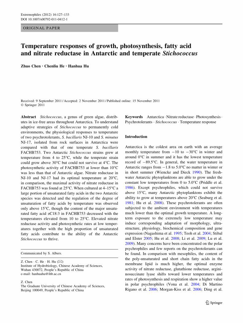
Based on the 18S rDNA sequences, two strains NJ-10 and
NJ-17 from Antarctica were identified to be Stichococcus
species. NJ-10 and NJ-17 were most closely related to
S. bacillaris s3 and S. mirabilis CCAP379/3, respectively,
with twenty-nine and eight different bases (Fig. 1).
Consistent with the previous studies (Handa et al. 2003;
Neustupa et al. 2007), different isolates attributed to
S. bacillaris do not branch together in the phylogenetic
tree, which shows a great difference in the 18S rDNA
sequences of this species.
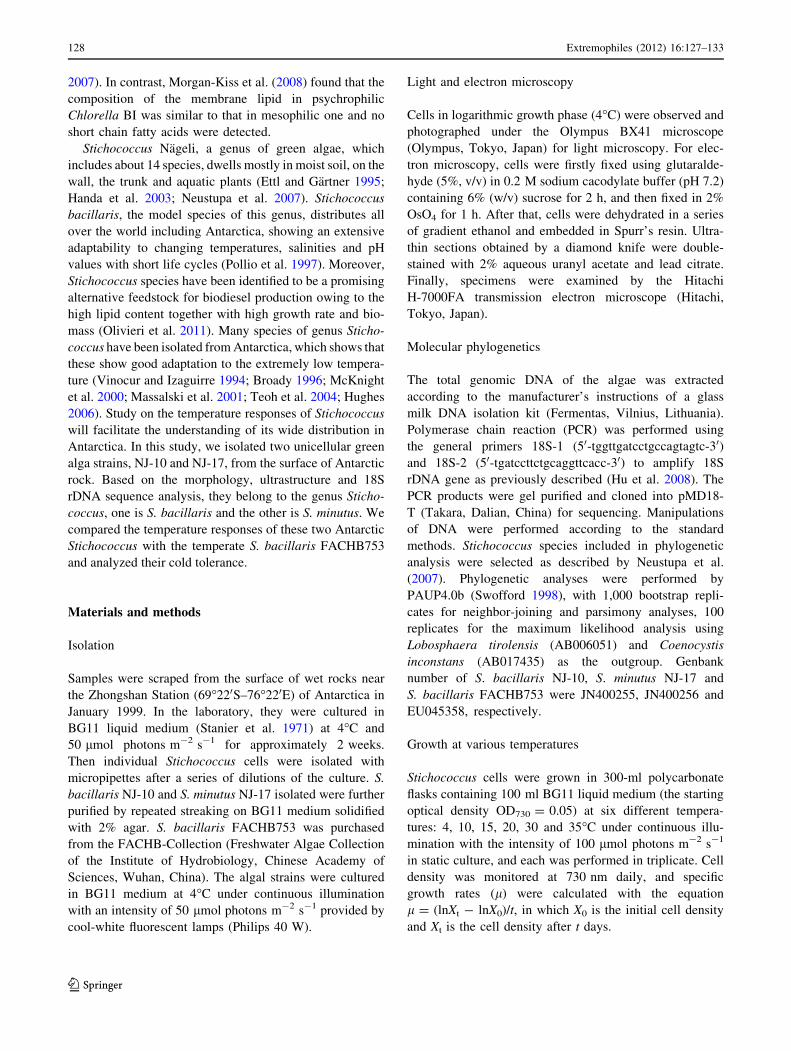
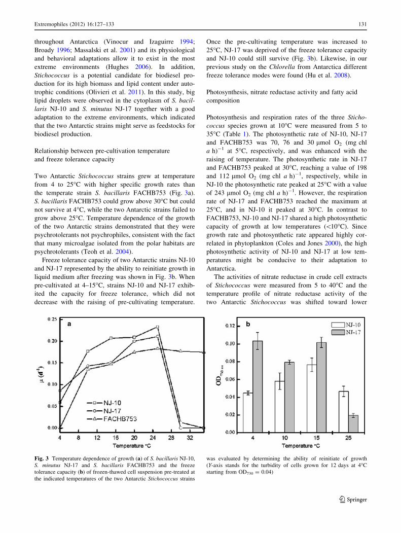
NJ-10 cells were cylindrical, 2.3–3.5 lm broad and
1.6–3.0 times as long and formed short filaments composed
of 2–6 cells, which very readily broke apart and slightly
constricted at cross walls. A lateral and plate-like chloro-
plast without pyrenoids covered only a small portion of the
cell (Fig. 2a). Cells of NJ-17 were small, short cylindrical,
with rounded ends, 1.7–2.6 lm broad and 3.5–5.6 lm long,
and no elliptic cells were observed (Fig. 2b). This alga
forms short filaments with a plate-like chloroplast occupy-
ing a slightly terminal lateral portion. Under transmission
electron microscopy, cells of the two Stichococcus strains
exhibited thick colloid out of cell walls and several big lipid
droplets within cytoplasm (Fig. 2c, d). In addition, multiple
starch grains were observed in the chloroplast of NJ-10
(Fig. 2c). However, Massalski et al. (2001) did not find
starch grains in S. bacillaris. On the basis of the morphol-
ogy, NJ-10 and NJ-17 was identified to be S. bacillaris and
S. minutus, respectively (Ettl and Gartner 1995).
Species of genus Stichococcus distribute widely and
have a good adaptation to the extremely low temperature
(Broady 1996). S. bacillaris is a ubiquitous unicellular
eukaryotic terrestrial alga found in ice-free areas
Extremophiles (2012) 16:127–133 129
123
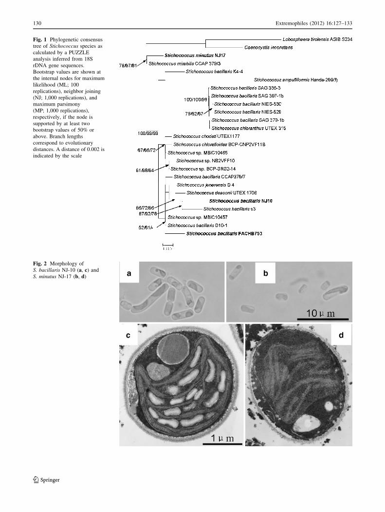
Fig. 1 Phylogenetic consensus
tree of Stichococcus species as
calculated by a PUZZLE
analysis inferred from 18S
rDNA gene sequences.
Bootstrap values are shown at
the internal nodes for maximum
likelihood (ML; 100
replications), neighbor joining
(NJ; 1,000 replications), and
maximum parsimony
(MP; 1,000 replications),
respectively, if the node is
supported by at least two
bootstrap values of 50% or
above. Branch lengths
correspond to evolutionary
distances. A distance of 0.002 is
indicated by the scale
Fig. 2 Morphology of
S. bacillaris NJ-10 (a, c) and
S. minutus NJ-17 (b, d)
130 Extremophiles (2012) 16:127–133
123
throughout Antarctica (Vinocur and Izaguirre 1994;
Broady 1996; Massalski et al. 2001) and its physiological
and behavioral adaptations allow it to exist in the most
extreme environments (Hughes 2006). In addition,
Stichococcus is a potential candidate for biodiesel pro-
duction for its high biomass and lipid content under auto-
trophic conditions (Olivieri et al. 2011). In this study, big
lipid droplets were observed in the cytoplasm of S. bacil-
laris NJ-10 and S. minutus NJ-17 together with a good
adaptation to the extreme environments, which indicated
that the two Antarctic strains might serve as feedstocks for
biodiesel production.
Relationship between pre-cultivation temperature
and freeze tolerance capacity
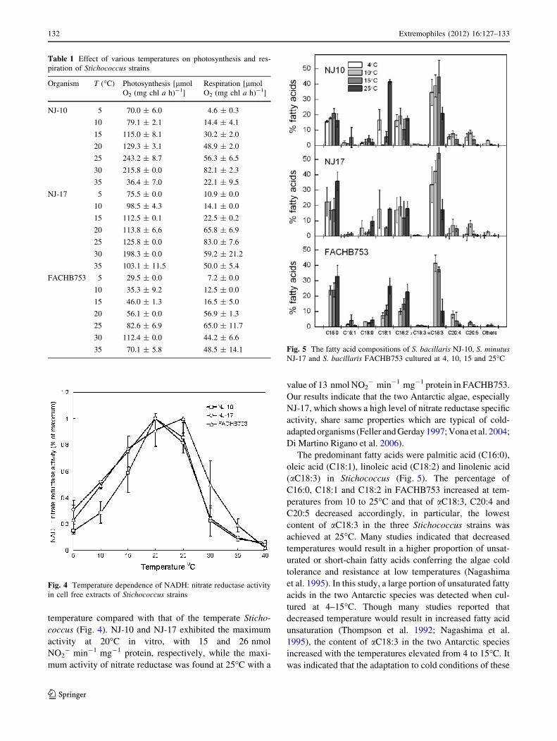
Two Antarctic Stichococcus strains grew at temperature
from 4 to 25�C with higher specific growth rates than
the temperate strain S. bacillaris FACHB753 (Fig. 3a).
S. bacillaris FACHB753 could grow above 30�C but could
not survive at 4�C, while the two Antarctic strains failed to
grow above 25�C. Temperature dependence of the growth
of the two Antarctic strains demonstrated that they were
psychrotolerants not psychrophiles, consistent with the fact
that many microalgae isolated from the polar habitats are
psychrotolerants (Teoh et al. 2004).
Freeze tolerance capacity of two Antarctic strains NJ-10
and NJ-17 represented by the ability to reinitiate growth in
liquid medium after freezing was shown in Fig. 3b. When
pre-cultivated at 4–15�C, strains NJ-10 and NJ-17 exhib-
ited the capacity for freeze tolerance, which did not
decrease with the raising of pre-cultivating temperature.
Once the pre-cultivating temperature was increased to
25�C, NJ-17 was deprived of the freeze tolerance capacity
and NJ-10 could still survive (Fig. 3b). Likewise, in our
previous study on the Chlorella from Antarctica different
freeze tolerance modes were found (Hu et al. 2008).
Photosynthesis, nitrate reductase activity and fatty acid
composition
Photosynthesis and respiration rates of the three Sticho-
coccus species grown at 10�C were measured from 5 to
35�C (Table 1). The photosynthetic rate of NJ-10, NJ-17
and FACHB753 was 70, 76 and 30 lmol O2 (mg chl
a h)-1 at 5�C, respectively, and was enhanced with the
raising of temperature. The photosynthetic rate in NJ-17
and FACHB753 peaked at 30�C, reaching a value of 198
and 112 lmol O2 (mg chl a h)-1, respectively, while in
NJ-10 the photosynthetic rate peaked at 25�C with a value
of 243 lmol O2 (mg chl a h)-1. However, the respiration
rate of NJ-17 and FACHB753 reached the maximum at
25�C, and in NJ-10 it peaked at 30�C. In contrast to
FACHB753, NJ-10 and NJ-17 shared a high photosynthetic
capacity of growth at low temperatures (\10�C). Since
growth rate and photosynthetic rate appeared highly cor-
related in phytoplankton (Coles and Jones 2000), the high
photosynthetic activity of NJ-10 and NJ-17 at low tem-
peratures might be conducive to their adaptation to
Antarctica.
The activities of nitrate reductase in crude cell extracts
of Stichococcus were measured from 5 to 40�C and the
temperature profile of nitrate reductase activity of the
two Antarctic Stichococcus was shifted toward lower
Fig. 3 Temperature dependence of growth (a) of S. bacillaris NJ-10,
S. minutus NJ-17 and S. bacillaris FACHB753 and the freeze
tolerance capacity (b) of frozen-thawed cell suspension pre-treated at
the indicated temperatures of the two Antarctic Stichococcus strains
was evaluated by determining the ability of reinitiate of growth
(Y-axis stands for the turbidity of cells grown for 12 days at 4�C
starting from OD730 = 0.04)
Extremophiles (2012) 16:127–133 131
123
temperature compared with that of the temperate Sticho-
coccus (Fig. 4). NJ-10 and NJ-17 exhibited the maximum
activity at 20�C in vitro, with 15 and 26 nmol
NO2- min-1 mg-1 protein, respectively, while the maxi-
mum activity of nitrate reductase was found at 25�C with a
value of 13 nmol NO2- min-1 mg-1 protein in FACHB753.
Our results indicate that the two Antarctic algae, especially
NJ-17, which shows a high level of nitrate reductase specific
activity, share same properties which are typical of cold-
adapted organisms (Feller and Gerday 1997; Vona et al. 2004;
Di Martino Rigano et al. 2006).
The predominant fatty acids were palmitic acid (C16:0),
oleic acid (C18:1), linoleic acid (C18:2) and linolenic acid
(aC18:3) in Stichococcus (Fig. 5). The percentage of
C16:0, C18:1 and C18:2 in FACHB753 increased at tem-
peratures from 10 to 25�C and that of aC18:3, C20:4 and
C20:5 decreased accordingly, in particular, the lowest
content of aC18:3 in the three Stichococcus strains was
achieved at 25�C. Many studies indicated that decreased
temperatures would result in a higher proportion of unsat-
urated or short-chain fatty acids conferring the algae cold
tolerance and resistance at low temperatures (Nagashima
et al. 1995). In this study, a large portion of unsaturated fatty
acids in the two Antarctic species was detected when cul-
tured at 4–15�C. Though many studies reported that
decreased temperature would result in increased fatty acid
unsaturation (Thompson et al. 1992; Nagashima et al.
1995), the content of aC18:3 in the two Antarctic species
increased with the temperatures elevated from 4 to 15�C. It
was indicated that the adaptation to cold conditions of these
Table 1 Effect of various temperatures on photosynthesis and res-
piration of Stichococcus strains
Organism T (�C) Photosynthesis [lmol
O2 (mg chl a h)-1]
Respiration [lmol
O2 (mg chl a h)-1]
NJ-10 5 70.0 ± 6.0 4.6 ± 0.3
10 79.1 ± 2.1 14.4 ± 4.1
15 115.0 ± 8.1 30.2 ± 2.0
20 129.3 ± 3.1 48.9 ± 2.0
25 243.2 ± 8.7 56.3 ± 6.5
30 215.8 ± 0.0 82.1 ± 2.3
35 36.4 ± 7.0 22.1 ± 9.5
NJ-17 5 75.5 ± 0.0 10.9 ± 0.0
10 98.5 ± 4.3 14.1 ± 0.0
15 112.5 ± 0.1 22.5 ± 0.2
20 113.8 ± 6.6 65.8 ± 6.9
25 125.8 ± 0.0 83.0 ± 7.6
30 198.3 ± 0.0 59.2 ± 21.2
35 103.1 ± 11.5 50.0 ± 5.4
FACHB753 5 29.5 ± 0.0 7.2 ± 0.0
10 35.3 ± 9.2 12.5 ± 0.0
15 46.0 ± 1.3 16.5 ± 5.0
20 56.1 ± 0.0 56.9 ± 1.3
25 82.6 ± 6.9 65.0 ± 11.7
30 112.4 ± 0.0 44.2 ± 6.6
35 70.1 ± 5.8 48.5 ± 14.1
Fig. 4 Temperature dependence of NADH: nitrate reductase activity
in cell free extracts of Stichococcus strains
Fig. 5 The fatty acid compositions of S. bacillaris NJ-10, S. minutusNJ-17 and S. bacillaris FACHB753 cultured at 4, 10, 15 and 25�C
132 Extremophiles (2012) 16:127–133
123
two Antarctic Stichococcus strains was not entirely depen-
dent on the content of unsaturated fatty acids.
The comparison of the different physiological responses
to temperature of one temperate and two Antarctic Sti-
chococcus strains would improve our understanding of
psychrotolerants’ adaptive strategies to permanently cold
environments. It seems that elevated nitrate reductase
activity and photosynthetic rates at low temperatures
together with the high proportion of unsaturated fatty acids
allow the two Stichococcus species to thrive in Antarctica.
Acknowledgments This research was supported by the National
Key Basic Research Project of China (2011CB200901) and National
Natural Science Foundation of China (No. 40606004).
References
Broady PA (1996) Diversity, distribution and dispersal of Antarctic
terrestrial algae. Biodivers Conserv 5:1307–1335
Coles JF, Jones RC (2000) Effect of temperature on photosynthesis-
light response and growth of four phytoplankton species isolated
from a tidal freshwater river. J Phycol 36:7–16
Di Martino Rigano V, Vona V, Lobosco O, Carillo P, Lunn JE,
Carfagna S, Esposito S, Caiazzo M, Rigano C (2006) Temper-
ature dependence of nitrate reductase in the psychrophilic
unicellular alga Koliella antarctica and the mesophilic alga
Chlorella sorokiniana. Plant Cell Environ 29:1400–1409
Ding Y, Miao JL, Wang QF, Zheng Z, Li GY, Jian JC, Wu ZH (2007)
Purification and characterization of a psychrophilic glutathione
reductase from Antarctic ice microalgae Chlamydomonas sp.
Strain ICE-L. Polar Biol 31:23–30
Ettl H, Gartner G (1995) Syllabus der Boden-, Luft-, und Flechte-
nalgen. Gustav Fischer Verlag, Stuttgart
Feller G, Gerday C (1997) Psychrophilic enzymes: molecular basis of
cold adaptation. Cell Mol Life Sci 53:830–841
Handa S, Nakahara M, Tsubota H, Deguchi H, Nakano T (2003) A
new aerial alga, Stichococcus ampulliformis sp. nov. (Trebouxi-
ophyceae, Chlorophyta) from Japan. Phycol Res 51:203–210
Hu H, Li H, Xu X (2008) Alternative cold response modes in
Chlorella (Chlorophyta, Trebouxiophyceae) from Antarctica.
Phycologia 47:28–34
Hughes KA (2006) Solar UV-B radiation, associated with ozone
depletion, inhibits the Antarctic terrestrial microalga, Stichococ-cus bacillaris. Polar Biol 29:327–336
Li H, Liu X, Wang Y, Hu H, Xu X (2009) Enhanced expression of
antifreeze protein genes drives the development of freeze
tolerance in an Antarctica isolate of Chlorella. Prog Nat Sci
19:1059–1062
Lu Y, Chi X, Yang Q, Li Z, Liu S, Gan Q, Qin S (2009) Molecular
cloning and stress-dependent expression of a gene encoding D12-
fatty acid desaturase in the Antarctic microalga Chlorellavulgaris NJ-7. Extremophiles 13:875–884
Massalski A, Mroziiiska T, Olech M (2001) Ultrastructural observa-
tions on five pioneer soil algae from ice denuded areas (King
George Island, West Antarctica). Polar Biosci 14:61–70
McKnight DM, Howes BL, Taylor CD, Goehringer DD (2000)
Phytoplankton dynamics in a stably stratified Antarctic lake
during winter darkness. J Phycol 36:852–861
Morgan-Kiss RM, Priscu JC, Pocock T, Gudynaite-Savitch L, Huner
NPA (2006) Adaptation and acclimation of photosynthetic
microorganisms to permanently cold environment. Microbiol
Mol Biol Rev 70:222–252
Morgan-Kiss RM, Ivanov AG, Modla S, Czymmek K, Huner NPA,
Priscu JC, Lisle JT, Hanson TE (2008) Identity and physiology
of a new psychrophilic eukaryotic green alga, Chlorella sp.,
strain BI, isolated from a transitory pond near Bratina Island,
Antarctica. Extremophiles 12:701–711
Nagashima H, Matsumoto GI, Ohtani S, Momose H (1995) Temper-
ature acclimation and the fatty acid composition of an Antarctic
green alga Chlorella. Proc NIPR Symp Polar Biol 8:194–199
Neustupa J, Elias M, Sejnohova L (2007) A taxonomic study of two
Stichococcus species (Trebouxiophyceae, Chlorophyta) with a
starch-enveloped pyrenoid. Nova Hedwigia 84:51–63
Olivieri G, Marzocchella A, Andreozzi R, Pinto G, Pollio A (2011)
Biodiesel production from Stichococcus strains at laboratory
scale. J Chem Technol Biotechnol 86:776–783
Pollio A, Aliotta G, Pinto G, Paterno M, Bevilacqua A (1997)
Ecophysiological characters and biochemical composition of
Stichococcus bacillaris Naegeli strains from low pH environ-
ments. Algol Stud/Arch Hydrobiol 84:129–143
Priddle J, Hawes I, Ellis-Evans JC (1986) Antarctic aquatic ecosys-
tems as habitats for phytoplankton. Biol Rev 61:199–238
Seaburg KG, Parker BC, Wharton RA, Simmons GM (1981)
Temperature–growth responses of algal isolates from Antarctic
oases. J Phycol 17:353–360
Stanier RY, Kunisawa R, Mandel M, Cohen-Bazire G (1971)
Purification and properties of unicellular blue-green algae (Order
Chroococcales). Bacteriol Rev 35:171–205
Stibal M, Elster J (2005) Growth and morphology variation as a
response to changing environmental factors in two Arctic species
of Raphidonema (Trebouxiophyceae) from snow and soil. Polar
Biol 28:558–567
Swofford DL (1998) PAUP* 4.0—phylogenetic analysis using parsi-
mony (*and other methods). Sinauer Associates, Sunderland
Teoh ML, Chu WL, Marchant H, Phang SM (2004) Influence of
culture temperature on the growth, biochemical composition and
fatty acid profiles of six Antarctic microalgae. J Appl Phycol
16:421–430
Thompson PA, Guo M, Harrisonp J, Whyte JNC (1992) Effects of
variation in temperature. II. On the fatty acid composition of
eight species of marine phytoplankton. J Phycol 28:488–497
Vinocur A, Izaguirre I (1994) Freshwater algae (excluding Cyano-
phyceae) from nine lakes and pools of Hope Bay, Antarctic
Peninsula. Antarct Sci 6:483–489
Vona V, Di Martino Rigano V, Lobosco O, Carfagna S, Esposito S,
Rigano C (2004) Temperature responses of growth, photosyn-
thesis, respiration and NADH: nitrate reductase in cryophilic and
mesophilic algae. New Phytol 163:325–331
Wiencke C, Dieck I (1990) Temperature requirements for growth and
survival of macroalgae from Antarctica and southern Chile. Mar
Ecol Progr Ser 59:157–170
Extremophiles (2012) 16:127–133 133
123