Embed Size (px)
Citation preview

08.06.2013 K.Frey, LMU – PTCOG 2013
K.Parodi, Ph.D.
Ludwig-Maximilian University (LMU) Munich, Germany Heidelberg University Hospital, Germany
Technologies Addressing the Range Uncertainty of Ion Therapy:
Positron-Emission-Tomography
08.06.2013 K.Frey, LMU – PTCOG 2013
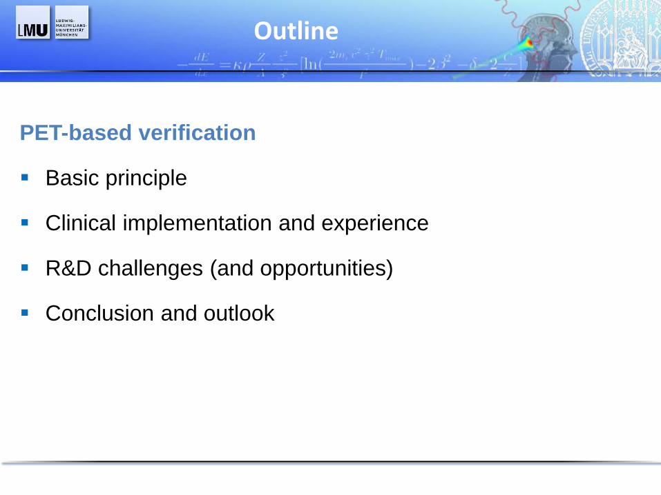
PET-based verification
Basic principle
Clinical implementation and experience
R&D challenges (and opportunities)
Conclusion and outlook
Outline

08.06.2013 K.Frey, LMU – PTCOG 2013
In-vivo visualization of treatment
• Nuclear reactions induce measurable emerging radiation
p or 12C
Target (e.g., 16O)
Projectile fragment (Z>1)
(vf ~ vp )
Target fragment (vt ~ 0 )
„Prompt“ p, d, t, n, g, …
„Delayed“ radioactive decay
• Primary ions are stopped somewhere within the patient, with dose
and range mainly dependent on Coulomb interaction
• Secondary radiation can be used as surrogate signal to infer
information on the beam range and treatment delivery
Only Positron-Emission-Tomography clinically investigated so far

08.06.2013 K.Frey, LMU – PTCOG 2013
p in PMMA
12C ions in PMMA
K. Parodi et al, IEEE TNS 2005
In-vivo PET-based verification
g-emission
b+-emitter yield (15O, 11C,..., with T1/2 ~ 2,
20,... min) as by-product of irradiation
A(r) D(r)
Tradeoff between better spatial
correlation (12C) and stronger signal (p)
Dose-guidance from comparison of
measured vs expected b+-activity
p or 12C
15O, 11C, ...
15O, 11C, ...
(projectile fragmentation only for Z>1)
11C, 10C

08.06.2013 K.Frey, LMU – PTCOG 2013
Clinical implementation (I): ibPET
g
g
Enghardt, … Parodi … Nucl Instrum Meth A 2004; Parodi et al Nucl Instrum Meth A 2005
In-beam PET
+ Patient in treatment position
+ Detection of short lived emitters (15O)
+ No prolongation of treatment session
o Morphological information from planning CT
- Limited-angle detection
- High integration costs
- Suitable for pulsed beam delivery
(measurement only in beam-off times)
Installation at GSI Darmstadt
used clinically for scanned 12C ions
GSI
Developed by HZDR
Dresden, Germany

08.06.2013 K.Frey, LMU – PTCOG 2013
Clinical implementation (I): ibPET
Enghardt, … Parodi … Nucl Instrum Meth A 2004; Parodi PhD Thesis 2004; Fiedler et al, PMB 2010
Experience from ibPET at GSI
+ Validation of TPS CT-range calibration
Initial CT-range cal.
Calc. PET
Meas. PET
Improved CT-range cal.
Calc. PET
Meas. PET
Calc. PET Meas. PET
Detection of over-range due to anatomy chage PET-based dose quantification
TPS dose Most-likely dose
+ Detection of mispositioning and anatomy
changes with indirect dose quantification
+ > 90% sensitivity / specificity in detecting
±6 mm range changes (in-silico trial)
o Minor degradation from washout
- Non-quantitative imaging, severe limited-
angle artifacts in extra-cranial sites
- Low counting statistics
08.06.2013 K.Frey, LMU – PTCOG 2013 Parodi et al, IJROBP 2007; Parodi et al, IEEE CR 2011; Bauer,.., Parodi, Radiother Oncol 2013
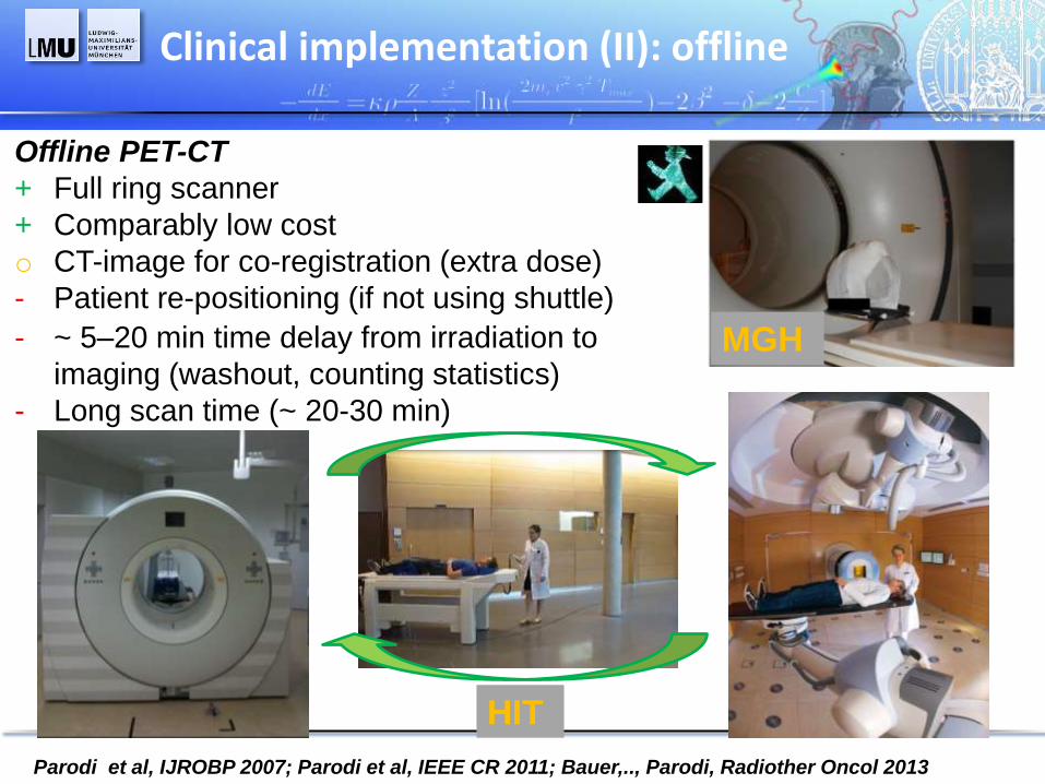
Offline PET-CT
+ Full ring scanner
+ Comparably low cost
o CT-image for co-registration (extra dose)
- Patient re-positioning (if not using shuttle)
MGH
HIT
Clinical implementation (II): offline
- ~ 5–20 min time delay from irradiation to
imaging (washout, counting statistics)
- Long scan time (~ 20-30 min)

08.06.2013 K.Frey, LMU – PTCOG 2013
Clinical implementation (II): offline PET/CT
Experience from offline PET/CT at MGH (p)
+ Activation detected in all subjects
Parodi et al, IJROBP 68, 2007; Knopf, Parodi et al, PMB 54, 2009; Knopf, Parodi et al, IJROBP 72, 2011
PET/CT Meas.
Field 1
Field 2
mGy
Scattered protons
Bq/ml
TPS Dose
- Low counting statistics
+ Feasibility of ± 3mm range monitoring in well
co-registered and low perfused tissues (H&N)
- Improper tissue classification from CT alone
- Limitations of universal washout modeling
- Co-registration and motion blurring in
extra-cranial sites
+ Washout modeling included in PET prediction
MC PET + washout
Bq/ml
Bq/ml
Phys. MC PET

08.06.2013 K.Frey, LMU – PTCOG 2013
Meas. PET Meas. PET
Scanned 12C ions
Clinical implementation (II): offline PET/CT
At HIT (p, 12C) similar findings as MGH, moreover
+ Feasibility and reproducibility of shuttle transport
+ Enhanced signal in distal part of the field due to 11C projectile fragments from 12C ion beam
Bauer et al, Radiother Oncol 2013, Kurz et al, Radiother Oncol 2012
Fraction 1 Fraction 2
+ Feasibility of range monitoring also in extracranial
sites, detection of mispositioning
+ Enhanced signal in necrotic areas („markers“)
- Even lower counting statistics for 12C ions than p
- Challenges of 4D gated imaging at low counts
Calc. PET on TP-CT
Meas. PET/CT
Scanned 12C ions
Calc. PET on PET-CT
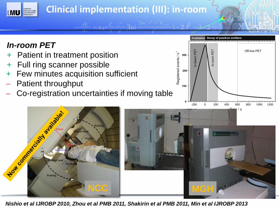
08.06.2013 K.Frey, LMU – PTCOG 2013 Nishio et al IJROBP 2010, Zhou et al PMB 2011, Shakirin et al PMB 2011, Min et al IJROBP 2013
+ Few minutes acquisition sufficient
Patient throughput
Co-registration uncertainties if moving table
NCC MGH
In-room PET
+ Patient in treatment position
+ Full ring scanner possible
Clinical implementation (III): in-room
08.06.2013 K.Frey, LMU – PTCOG 2013
Day 1
p beam
Clinical implementation (III): in-room PET
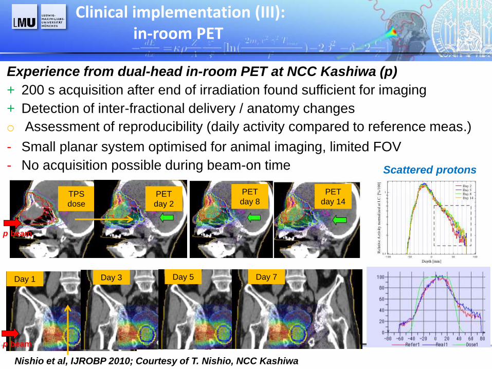
Experience from dual-head in-room PET at NCC Kashiwa (p)
+ 200 s acquisition after end of irradiation found sufficient for imaging
+ Detection of inter-fractional delivery / anatomy changes
Nishio et al, IJROBP 2010; Courtesy of T. Nishio, NCC Kashiwa
Day 3 Day 5 Day 7
TPS
dose
p beam
Scattered protons
PET
day 2
PET
day 14
PET
day 8
- Small planar system optimised for animal imaging, limited FOV
- No acquisition possible during beam-on time
o Assessment of reproducibility (daily activity compared to reference meas.)

08.06.2013 K.Frey, LMU – PTCOG 2013
Experience from full-ring in-room PET at MGH (p)
+ 5 min measurement started 2 min after irradiation end
similar to 20 min scan
+ Range agreement mostly within ±3 mm (4 - 11 mm rms)
TPS dose
Meas. PET
Calc. PET
Clinical implementation (III): in-room PET
Zhou et al PMB 2011, Min et al IJROBP 2013
- ~ 2 mm co-registration errors despite robotic couch and
radioactive markers
- Limited bore of scanner (only head and pediatric cases)
08.06.2013 K.Frey, LMU – PTCOG 2013
Remaining limitations of PET-based verification
Inaccurate prediction of activity distributions due to insufficient knowledge
of nuclear reaction cross sections and tissue composition
Degradation of activity distributions by washout and organ motion
Time-consuming evaluation requiring well trained staff
Imaging performances and integration costs for on-site implementations
R&D challenges

08.06.2013 K.Frey, LMU – PTCOG 2013
Ongoing efforts to
lmprove MC prediction via experimental
based adjustement of b+ activation
cross sections (only feasible for p)
Modeling of PET prediction
Bauer, .., Parodi, PMB 2013
TH-C-144-12
Frey et al, submitted to PMB
Landry, Parodi, et al, PMB 2013
TH-C-144-6 CT MR
Bauer et al,
In preparation
WE-G-500-6
Mitayake et al, MP 2011
Speed up calculation with analytical
approaches, ideally using same
pencil beam algorithms as TPS
Overcome limitations of CT-based
tissue classification by using MRI
information or Dual Energy CT
08.06.2013 K.Frey, LMU – PTCOG 2013
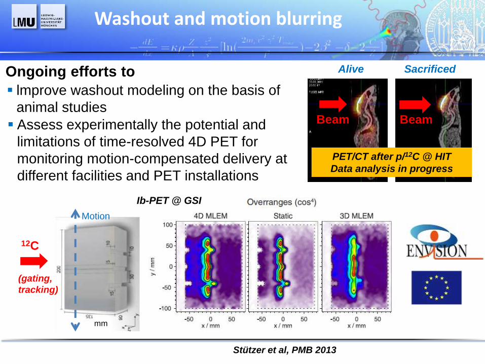
Washout and motion blurring
Ongoing efforts to
Stützer et al, PMB 2013
Assess experimentally the potential and
limitations of time-resolved 4D PET for
monitoring motion-compensated delivery at
different facilities and PET installations
Beam Beam
Alive Sacrificed
PET/CT after p/12C @ HIT
Data analysis in progress
lmprove washout modeling on the basis of
animal studies
mm
Ib-PET @ GSI
12C
(gating,
tracking)
Motion

08.06.2013 K.Frey, LMU – PTCOG 2013
Offline PET/CT @ HIT
Fr. # 4 Fr. # 3
Automated range assessment
Ongoing efforts to establish
Robust, automated range assessment from PET
distributions based on profile shift analysis or
% fall-off in BEV (meas. vs calc., meas. vs meas.)
Helmbrecht et al, PMB 2012
In-beam PET @ GSI
12C ions
Range Shift [mm]
in BEV
Decision support system for clinical workflow
Meas. PET Fr. # 3 Protons
Unholtz, …, Parodi,
IEEE MIC Conf. Rec. 2011
Range shift
in mm Similar approach for in-room PET @ MGH (Min et al, IJROBP 2013)
08.06.2013 K.Frey, LMU – PTCOG 2013
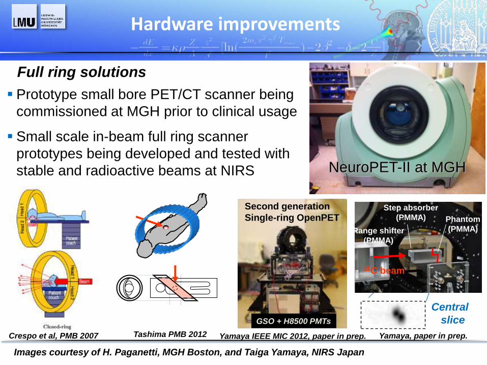
Hardware improvements
Crespo et al, PMB 2007
Full ring solutions
NeuroPET-II at MGH
Prototype small bore PET/CT scanner being
commissioned at MGH prior to clinical usage
Tashima PMB 2012
Central
slice
11C beam
Range shifter
(PMMA)
Phantom
(PMMA)
Step absorber
(PMMA)
Yamaya, paper in prep.
Yamaya IEEE MIC 2012, paper in prep.
Second generation
Single-ring OpenPET
GSO + H8500 PMTs
Small scale in-beam full ring scanner
prototypes being developed and tested with
stable and radioactive beams at NIRS
Images courtesy of H. Paganetti, MGH Boston, and Taiga Yamaya, NIRS Japan

08.06.2013 K.Frey, LMU – PTCOG 2013
Hardware improvements
Dual head solutions
New detector developments
towards ultra-fast Time-of-Flight
(TOF) in-beam PET
Crespo et al, PMB 2007
Beam OFF
Beam ON
11C 10C
Protons in PE
Cambraia Lopes et al, to be presented at IEEE MIC 2013
Philips dSiPM and LYSO crystals
Experimental set-up of TU-Delft group @ HIT
Small scale in-beam prototypes
being developed and tested
Images courtesy of D. Schaart and P. Cambraia Lopes, TU Delft
08.06.2013 K.Frey, LMU – PTCOG 2013
Conclusions and outlook
Clinical investigations of PET monitoring being reported for different
centers with different ions and delivery systems, as well as different
scanners (mostly adapted from nuclear medicine or small animal imaging)
Despite promising results (± 3mm range verification accuracy in
favorable H&N locations), several issues remain (counting statistics,
washout, co-registration and motion in extra-cranial sites, …)
Several groups are pursuing methodological improvements, but major
advancement being expected by next generation in-beam PET scanners
specifically optimized for this application
Although many promising new techniques are on the horizon, PET could
still play a role due to its intrinsic 3D, molecular imaging capabilities
when properly used to detect the major 15O contribution in the tumour

Thank you for your attention
The MC-modeling and in-vivo imaging research group at HIT:
J. Bauer, C. Kurz, A. Mairani*, I. Rinaldi, F. Sommerer*, D. Unholtz*, K. Frey,
M. Hildenbrandt (* alumni)
The colleagues at HIT, Universitätsklinikum Heidelberg and DKFZ:
J. Debus, S. Combs, T. Haberer, O. Jäkel, A. Abdollahi and teams
Collaborators / former colleagues:
A. Ferrari, F. Cerutti, CERN Geneva
C. Bert, N. Saito, N. Chaudhri, R. Kaderka, GSI Darmstadt
T. Bortfeld, H. Paganetti, G. El Fakhri, MGH Boston
W. Enghardt, F. Fiedler, K. Stützer, FZD (now HZDR) Dresden
Funding:
FP7 EU Project PARTNER
FP7 EU Project ENVISION
BMBF Project DOT-MOBI
BMBF Project SPARTA
Acknowledgements
Contributions from:
T. Nishio, NCC Kashiwa, Japan
T. Yamaya, NIRS Chiba, Japan
D.R. Schaart, P. Cambraia, TU Delft, The
Netherlands
P. Crespo, LIP Portugal

08.06.2013 K.Frey, LMU – PTCOG 2013
Automated range assessment
Unholtz, Bauer, …, Parodi, IEEE MIC CR 2011, Helmbrecht et al, PMB 2012
Profile shift analysis (*)
or % fall-off in BEV
(*) Knopf, Parodi et al, PMB 2008
PET difference
Shift [mm]
Distal Surface_Dose Distal Surface_PET
2 4 6 8 10
2
4
6
8
10
Dis
tan
ce
(cm
)
Distance (cm)
-20.00-18.00-16.00-14.00-12.00-10.00-8.000-6.000-4.000-2.00002.0004.0006.0008.00010.0012.0014.0016.0018.0020.00
Difference of distal surfaces
RMSD=8.8 mm
71% Coverage
PET – Dose difference
Courtesy of H. Paganetti and C.H.
Min MGH
20% of Max 50 Bq/ml
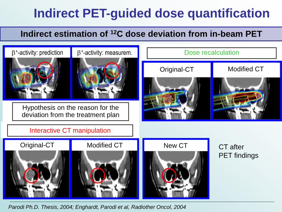
Original-CT Modified CT
Fast PET recalculation
b+-activity: prediction b+-activity: measurem.
Hypothesis on the reason for the deviation from the treatment plan
Dose recalculation
Modified CT Original-CT
Original-CT Modified CT
Interactive CT manipulation
New CT CT after
PET findings
Parodi Ph.D. Thesis, 2004; Enghardt, Parodi et al, Radiother Oncol, 2004
Indirect PET-guided dose quantification
Indirect estimation of 12C dose deviation from in-beam PET
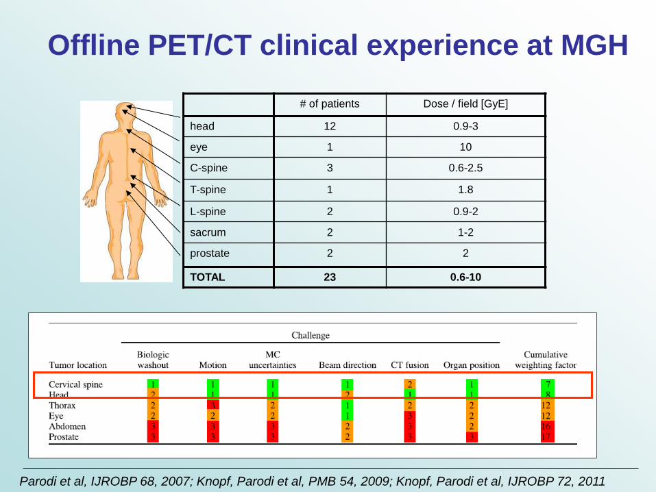
# of patients Dose / field [GyE]
head 12 0.9-3
eye 1 10
C-spine 3 0.6-2.5
T-spine 1 1.8
L-spine 2 0.9-2
sacrum 2 1-2
prostate 2 2
TOTAL 23 0.6-10
Offline PET/CT clinical experience at MGH
Parodi et al, IJROBP 68, 2007; Knopf, Parodi et al, PMB 54, 2009; Knopf, Parodi et al, IJROBP 72, 2011

g
g
In-beam PET for 12C ion therapy at GSI
Planned
dose
Once
Measured
b+-Activity
For every fraction
(typically 20 d @ 1Gy)
MC calculated
b+-Activity
Verification of
Beam range
Lateral position
In case of deviation
Timely reaction
Enghardt, … Parodi … Nucl Instrum Meth A 2004; Parodi et al Nucl Instrum Meth A 2005
> 400 patients
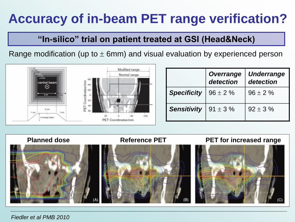
Planned dose Reference PET
Accuracy of in-beam PET range verification?
“In-silico” trial on patient treated at GSI (Head&Neck)
Range modification (up to 6mm) and visual evaluation by experienced person
Overrange
detection
Underrange
detection
Specificity 96 2 % 96 2 %
Sensitivity 91 3 % 92 3 %
Fiedler et al PMB 2010
PET for increased range