Embed Size (px)
Citation preview
s
TECHNIQUES AND TECHNOLOGIES
42 RETINA TODAY | JANUARY/FEBRUARY 2019
Macular hole surgery has evolved into an efficacious surgical technique since its description by Kelly and Wendell in 1991.1 Success
rates of greater than 90% are possible using just internal limiting membrane (ILM) peeling techniques in most eyes with small and recent macular holes. Nevertheless, larger holes, older holes, and those occurring secondary to high myopia or retinal detachment are associated with lower closure rates and reduced visual outcomes. These macu-lar hole pathologies pose a challenge, and a number of techniques have been devised to improve anatomic and visual outcomes in these eyes.
ILM FLAPS Michalewska et al described the
inverted flap technique in 2010 as an alternative to traditional ILM peel-ing in eyes with large macular holes or failed macular holes that have ILM present.2 Since that initial descrip-tion, a number of variations of the technique have been published. These include temporal flaps, circumferen-tial flaps, stuffing of the flap inside the macular hole, free flaps, and lens capsular flap transplantation.3-6 All of these techniques have anatomic closure rates of greater than 90%, even in eyes with large and failed macular
holes. However, visual outcomes with these techniques have been less than optimal, and each technique poses unique challenges.
OCT studies have identified bet-ter visual outcomes in cases that lack hyperreflectivity in the postop-erative period and in cases in which ellipsoid layer and outer retinal layer alterations are smaller after surgery.7 Pathophysiologically, larger and older macular holes tend to have elevated edges, leading to a larger inner hole and thus a larger scotoma. Bringing the edges in apposition with the retinal pigment epithelium (RPE) would reduce the size of the central scotoma. The inverted flap lying over
the surface of the retina in the area of the hole may act as scaffold on which fibroblasts can grow and help bring the edges of the hole closer together, thereby reducing the size of the sco-toma. Meanwhile, stuffing the hole or placing ILM or lens capsule inside the hole may create a larger scotoma and
Use of Autologous Blood in Macular Hole Surgery
Could blood be your friend during complex macular hole surgery? In some cases, maybe.
BY MARÍA H. BERROCAL, MD; and LUIS A. ACABA, BA
AT A GLANCE
s
Unsatisfactory visual and anatomic outcomes are sometimes seen after surgery for large macular holes.
s
Use of autologous blood during macular hole surgeries that employ internal limiting membrane flaps may be a promising technique.
s
A number of questions remain about the benefit of techniques that use autologous blood during macular hole surgeries, but good visual outcomes have been reported.
s WATCH IT NOW
BIT.LY/Berrocal0119
TECHNIQUES AND TECHNOLOGIES s
JANUARY/FEBRUARY 2019 | RETINA TODAY 43
could prevent approximation of the macular hole edge with the RPE, resulting in a suboptimal visual outcome despite anatomic closure.
Maintaining the flap in place is one of the challenges of inverted flap and free-flap techniques. For inverted flaps, leaving the flap attached temporally allows air to secure it as fluid-air exchange is performed because the gas moves from the temporal side first. Still, dislodgement of the flap occurs in a number of cases.
Maintaining attachment of free flaps in the foveal region is more challenging. Packing them inside a macular hole may prevent dislodging, but the ensuing scotoma can be larger. Securing free flaps under perfluorocarbon liquid and performing a direct oil exchange has been done, but this approach requires a second procedure for oil removal.
AUTOLOGOUS BLOOD In the 1990s, before ILM peeling became routine for
macular holes, distressing the edges of the hole to “stretch it” to cause bleeding and applying the resulting autologous blood was popularized as a means of improving anatomic and visual outcomes. The benefits of blood in the area of a macular hole remains controversial, but recent publications have shown some visual benefit.8,9
Lai et al described the technique of using an inverted flap with an autologous blood plug to secure the flap in place.9 This technique is promising, and it may warrant consider-ation when choosing one’s surgical approach to a macular hole (Table). (The technique can also be used with free flaps.) The blood clot that forms holds the flap in place while healing occurs. This approach can be performed without air or gas tamponade, which makes it particularly useful for patients who need to travel by plane or who cannot main-tain a prone position. The potential benefits of blood prod-ucts in the macular hole area merit further investigation.
TECHNIQUE FOR INVERTED FLAP WITH AUTOLOGOUS BLOOD A complete vitrectomy is performed on a 25- or 27-gauge
platform. The posterior hyaloid is detached and peeled with the vitrector, with or without triamcinolone staining. The ILM is stained with indocyanine green or brilliant blue dye. The ILM can then be peeled with the Finesse Flex Loop (Alcon), with forceps, or with the vitrectomy probe.
An edge is started and is then engaged with the vitrec-tomy probe and peeled circumferentially, leaving a segment
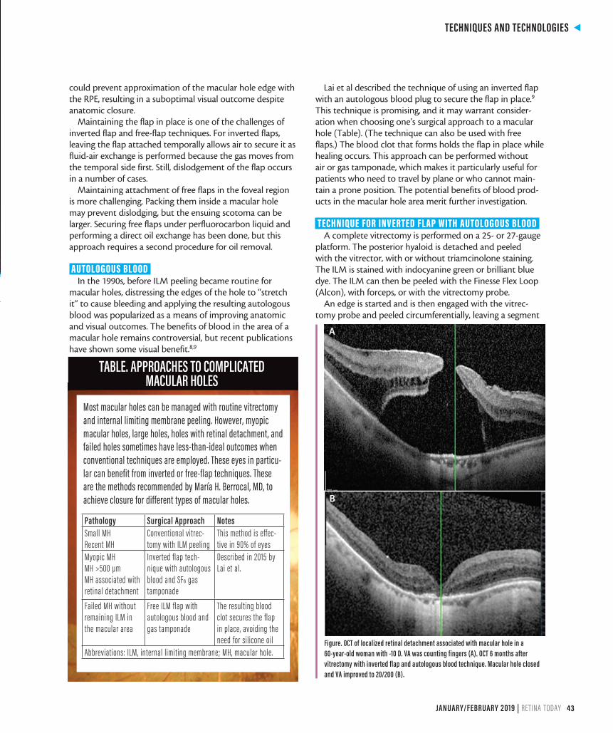
Figure. OCT of localized retinal detachment associated with macular hole in a 60-year-old woman with -10 D. VA was counting fingers (A). OCT 6 months after vitrectomy with inverted flap and autologous blood technique. Macular hole closed and VA improved to 20/200 (B).
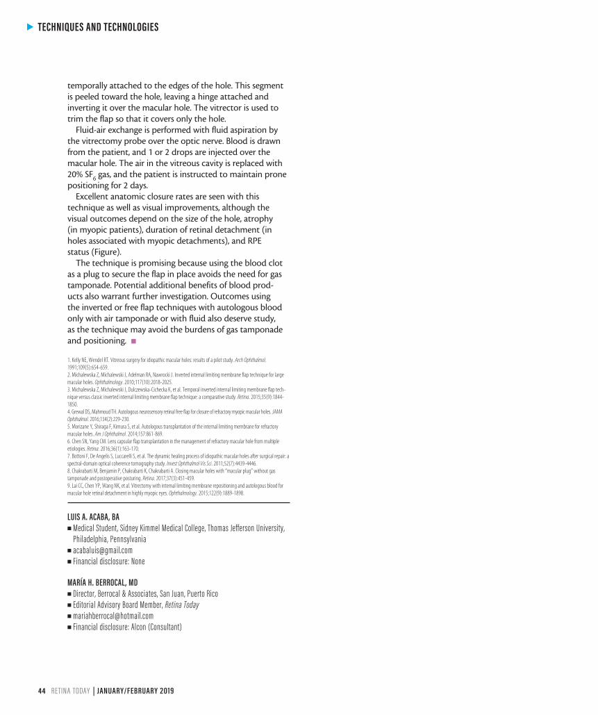
Most macular holes can be managed with routine vitrectomy and internal limiting membrane peeling. However, myopic macular holes, large holes, holes with retinal detachment, and failed holes sometimes have less-than-ideal outcomes when conventional techniques are employed. These eyes in particu-lar can benefit from inverted or free-flap techniques. These are the methods recommended by María H. Berrocal, MD, to achieve closure for different types of macular holes.
TABLE. APPROACHES TO COMPLICATED MACULAR HOLES
Pathology Surgical Approach NotesSmall MHRecent MH
Conventional vitrec-tomy with ILM peeling
This method is effec-tive in 90% of eyes
Myopic MHMH >500 µmMH associated with retinal detachment
Inverted flap tech-nique with autologous blood and SF6 gas tamponade
Described in 2015 by Lai et al.
Failed MH without remaining ILM in the macular area
Free ILM flap with autologous blood and gas tamponade
The resulting blood clot secures the flap in place, avoiding the need for silicone oil
Abbreviations: ILM, internal limiting membrane; MH, macular hole.
A
B
s
TECHNIQUES AND TECHNOLOGIES
44 RETINA TODAY | JANUARY/FEBRUARY 2019
temporally attached to the edges of the hole. This segment is peeled toward the hole, leaving a hinge attached and inverting it over the macular hole. The vitrector is used to trim the flap so that it covers only the hole.
Fluid-air exchange is performed with fluid aspiration by the vitrectomy probe over the optic nerve. Blood is drawn from the patient, and 1 or 2 drops are injected over the macular hole. The air in the vitreous cavity is replaced with 20% SF6 gas, and the patient is instructed to maintain prone positioning for 2 days.
Excellent anatomic closure rates are seen with this technique as well as visual improvements, although the visual outcomes depend on the size of the hole, atrophy (in myopic patients), duration of retinal detachment (in holes associated with myopic detachments), and RPE status (Figure).
The technique is promising because using the blood clot as a plug to secure the flap in place avoids the need for gas tamponade. Potential additional benefits of blood prod-ucts also warrant further investigation. Outcomes using the inverted or free flap techniques with autologous blood only with air tamponade or with fluid also deserve study, as the technique may avoid the burdens of gas tamponade and positioning. n
1. Kelly NE, Wendel RT. Vitreous surgery for idiopathic macular holes: results of a pilot study. Arch Ophthalmol. 1991;109(5):654-659.2. Michalewska Z, Michalewski J, Adelman RA, Nawrocki J. Inverted internal limiting membrane flap technique for large macular holes. Ophthalmology. 2010;117(10):2018-2025.3. Michalewska Z, Michalewski J, Dulczewska-Cichecka K, et al. Temporal inverted internal limiting membrane flap tech-nique versus classic inverted internal limiting membrane flap technique: a comparative study. Retina. 2015;35(9):1844-1850.4. Grewal DS, Mahmoud TH. Autologous neurosensory retinal free flap for closure of refractory myopic macular holes. JAMA Ophthalmol. 2016;134(2):229-230. 5. Morizane Y, Shiraga F, Kimura S, et al. Autologous transplantation of the internal limiting membrane for refractory macular holes. Am J Ophthalmol. 2014;157:861-869. 6. Chen SN, Yang CM. Lens capsular flap transplantation in the management of refractory macular hole from multiple etiologies. Retina. 2016;36(1):163-170.7. Bottoni F, De Angelis S, Luccarelli S, et al. The dynamic healing process of idiopathic macular holes after surgical repair: a spectral-domain optical coherence tomography study. Invest Ophthalmol Vis Sci. 2011;52(7):4439-4446.8. Chakrabarti M, Benjamin P, Chakrabarti K, Chakrabarti A. Closing macular holes with “macular plug” without gas tamponade and postoperative posturing. Retina. 2017;37(3):451-459.9. Lai CC, Chen YP, Wang NK, et al. Vitrectomy with internal limiting membrane repositioning and autologous blood for macular hole retinal detachment in highly myopic eyes. Ophthalmology. 2015;122(9):1889-1898.
LUIS A. ACABA, BAn Medical Student, Sidney Kimmel Medical College, Thomas Jefferson University,
Philadelphia, Pennsylvanian [email protected] Financial disclosure: None
MARÍA H. BERROCAL, MDn Director, Berrocal & Associates, San Juan, Puerto Rico n Editorial Advisory Board Member, Retina Todayn [email protected] Financial disclosure: Alcon (Consultant)