Embed Size (px)
Citation preview

Teachers’ Notes
Electromagnetic spectrum.................................................. 2 Radiowaves................................................................... 2 Infrared.......................................................................... 3 Thermography............................................................... 4 Visible light .................................................................... 5 X-rays............................................................................ 8 Radiotherapy................................................................. 9
Radioactivity ....................................................................... 11 Nuclear Medicine .......................................................... 13 PET ............................................................................... 16 Radiotherapy................................................................. 17
Ultrasound .......................................................................... 18 Doppler Ultrasound....................................................... 20
Worksheets
Electromagnetic spectrum.................................................. 22 Infrared ............................................................................... 23 X-Rays................................................................................ 24 Nuclear Medicine................................................................ 25 PET .................................................................................... 26 Ultrasound .......................................................................... 27
Answers........................................................................ 29
Acknowledgements ............................................... 32
www.teachingmedicalphysics.org.uk 1

Electromagnetic spectrum
National Curriculum requirements (KS4, Sc4 Physical Processes)
Students should be taught:
• That waves transfer energy without transferring matter (3d) • that the electromagnetic spectrum includes radio waves, microwaves,
infrared, visible light, ultraviolet waves, X-rays and gamma rays (3e) • some ways in which microwaves, infrared and ultraviolet waves are used
and the potential dangers of these (3f) • some uses of X-rays and gamma rays in medicine (3g) • how information can be transmitted along optical fibres (3h)
Radio waves
What happens when you use your mobile phone? These images show the effect of using a mobile phone on head temperature. An image of the head has been scanned into a computer. A computer program then worked out where the microwaves emitted by the mobile phone go, and the amount they heat the head by.
The maximum heating in this case was 0.2oC. The left hand image shows the surface of the head and the right hand image is a slice showing how the temperature changes deeper into the head. Q: Do you think this is dangerous?
oA: 0.2 C is about the same change as normal physiological changes, so is probably not significant but is of the same order of magnitude. The Government recommends minimum mobile phone use, especially for children.
www.teachingmedicalphysics.org.uk 2

Infrared
Pulse oximetry
A pulse oximeter showing the heart rate (81 beats per min) and oxygenation of the finger (99%). It uses red and near infrared light to determine the amount of blood and its colour. Blood absorbs light strongly so an increase in the amount of blood means that less light is measured. Blood carrying oxygen is also a brighter red than blood carrying deoxygenated blood. This means that we can measure both the amount of blood and the amount of oxygen it is carrying.
Near Infrared Spectroscopy (NIRS) Like pulse oximetry, near infrared spectroscopy (NIRS) uses near infrared light to determine the amount of blood and its colour. When the child observes a visual stimulus (alternating checkerboard pattern), the parts of the brain which respond to vision increase their electrical activity. This increased activity requires energy, so blood supply to the region increases. Blood is a strong absorber of near infrared light and so, by measuring the transmission of near infrared light across the head, we can get a measurement of blood volume and therefore brain activity. The black hat prevents light from the room being detected. The second picture shows NIRS being used on an adult volunteer. The third picture shows the measured blood flow response to flashing lights. The lights flash for 18 s. For the first 5 s, nothing happens, then there is a big increase in oxygenated blood and a smaller decrease in deoxygenated blood, so overall a small increase in blood volume. Even though the brain is using oxygen up, the body responds by sending more blood than is necessary, so the additional oxygenated blood exceeds the brain's requirements and the overall effect is an increase in oxygenated blood.
www.teachingmedicalphysics.org.uk 3

Thermography Thermograms are surface maps of temperature, created by measuring emitted infrared radiation. Hands
This picture shows a thermal picture of the temperature of the hand. On the left is a normal patient, where each finger is warm and has a similar temperature. The patient on the right has Raynaud's syndrome, which is a problem with the circulation in the fingers and toes. Reduced blood flow to fingers means he or she has some fingers which are much colder than the others.
Face This shows a thermogram of a child’s face. The lips, nose and eyes are warmer, and the head where it is insulated by hair is cooler and so is emitting less infrared radiation.
Back This shows a thermogram overlaid onto a normal photograph taken with visible light. The triangles are used to align the two images (though above the right shoulder you can see a slight mismatch). This person is healthy.
www.teachingmedicalphysics.org.uk 4

Visible Light
Endoscopy Endoscopy is a minimally invasive diagnostic medical procedure used to see inside the body by inserting a small scope through a natural body opening. It is often cited as a use of optical fibres, though modern endoscopes tend to use cameras to take the images.
The left hand picture shows an endoscope. The right hand picture shows an x-ray image of an endoscope passing down the oesophagus, into the stomach (far right), then round into the small intestine.
The left hand picture is taken in the stomach, with the endoscope bending round so you can see part of it coming out of the oesophagus. It shows the wall of the stomach lapsing back into the oesophagus, which is called a hernia. The right hand picture is again in the stomach and shows a parasitic worm. If treated with the right drugs, the worm will detach from the stomach wall, pass through the digestive system and leave the body in faeces.
www.teachingmedicalphysics.org.uk 5

Scanning laser ophthalmoscope This uses blue laser light to image the back of the eye (the retina). The mirrors scan the light across the retina allowing an image to be built up.
www.teachingmedicalphysics.org.uk 6

This image shows the optic disc (where the optic nerve leaves the eye) and has a large number of blood vessels supplying it. To the left of the image is the macula, the region of the eye with the most cones and with the fovea at the centre.
Photodynamic therapy (PDT) PDT can be used to treat Bowen’s disease (a kind of skin cancer). A cream called Metvix is rubbed onto the skin and it is preferentially absorbed by cancer cells, so the drug collects in the tumour. When the skin is then illuminated with visible light, the drug breaks down and destroys cells. The process is therefore doubly selective – first the drug collects in the tumour and second, only the tumour is illuminated. The pictures show the skin before and after treatment.
Blue light treatment of jaundice in babies Premature babies sometimes have jaundice. This makes them look yellow and is due to excess bilirubin, the yellow pigment in bruises. It is usually harmless but can be treated using blue light. The blue light breaks down the bilirubin so that it can be excreted in urine.
www.teachingmedicalphysics.org.uk 7

X-Rays
This is the first clinical x-ray taken by Wilhelm Roentgen on 22 Dec 1895. It shows his wife’s hand, with her wedding ring and the bones of her fingers clearly visible. She had been worried about the long hours he was spending in the lab, so Roentgen took this image to show hwhat he had been doing!
er
It was taken with a 15 minute exposure; a modern x-ray image has an expose of about10 ms
Click, and the picture fades into a modern x-ray
CT (computed tomography) takes images that are slices through the body. They can be reconstructed to make a 3D
age of the body. im This is a CT image which has been processed by computer so that only the bones and the muscles are shown. The second metatarsal bone (the bone that David Beckham broke in 2002) is shown.
www.gehealthcare.com
Similarly, this CT image of the pelvis which has been processed so that only the bones are shown.
www.gehealthcare.com
www.teachingmedicalphysics.org.uk 8

Radiotherapy Radiotherapy is the treatment of cancers using high energy radiation (x and gamma rays). Ionising radiation damages cells, and high enough doses can kill them. The cells in cancerous tissue are dividing very rapidly, making them more susceptible to damage by radiation. Healthy cells are also able to regenerate if they are only slightly damaged, cancerous cells cannot. Even so, care has to be taken to ensure that only the malignant cancer cells, and not the surrounding healthy tissue, receive a high dose.
This is a picture of a linear accelerator (linac). A linac generates high energy x-rays. It rotates around the body, with the tumour at the centre of rotation, irradiating the tumour from different directions. This means the tumour receives a large dose, but the dose to healthy tissue is minimal.
Demonstration: the linac moves around the ring irradiating the tumour from different directions. Q: Which part of the body has received the higher dose? A: The tumour
www.teachingmedicalphysics.org.uk 9
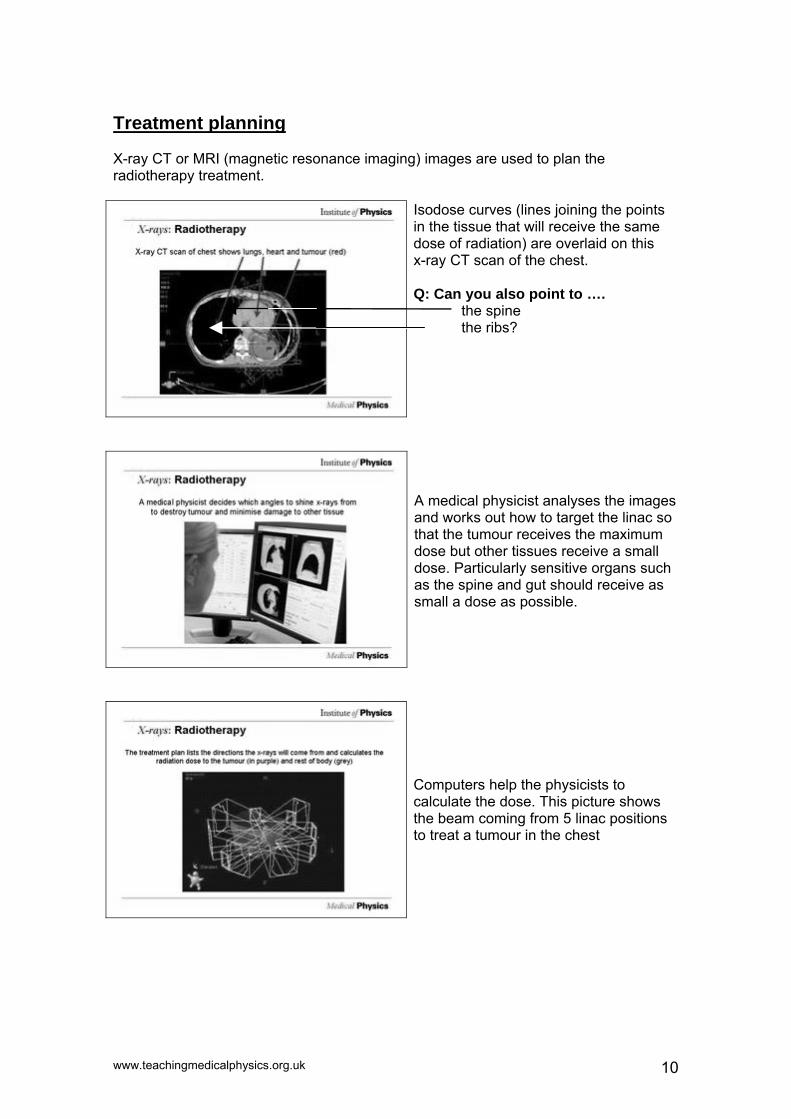
Treatment planning X-ray CT or MRI (magnetic resonance imaging) images are used to plan the radiotherapy treatment.
Isodose curves (lines joining the points in the tissue that will receive the same dose of radiation) are overlaid on this x-ray CT scan of the chest. Q: Can you also point to …. the spine the ribs?
A medical physicist analyses the images and works out how to target the linac so that the tumour receives the maximum dose but other tissues receive a small dose. Particularly sensitive organs such as the spine and gut should receive as small a dose as possible.
Computers help the physicists to calculate the dose. This picture shows the beam coming from 5 linac positions to treat a tumour in the chest
www.teachingmedicalphysics.org.uk 10
Radioactivity National Curriculum requirements (KS4, Physical Processes)
Students should be taught:
• that radioactivity arises from the breakdown of an unstable nucleus (6a) • about some sources of the ionising radiation found in all environments (6b) • the characteristics of alpha and beta particles and of gamma radiation (6c) • the meaning of the term ‘half-life’(6d) • the beneficial and harmful effects of ionising radiation on matter and living
organisms (6e) • some uses of radioactivity, including radioactive dating of rocks. (6f)
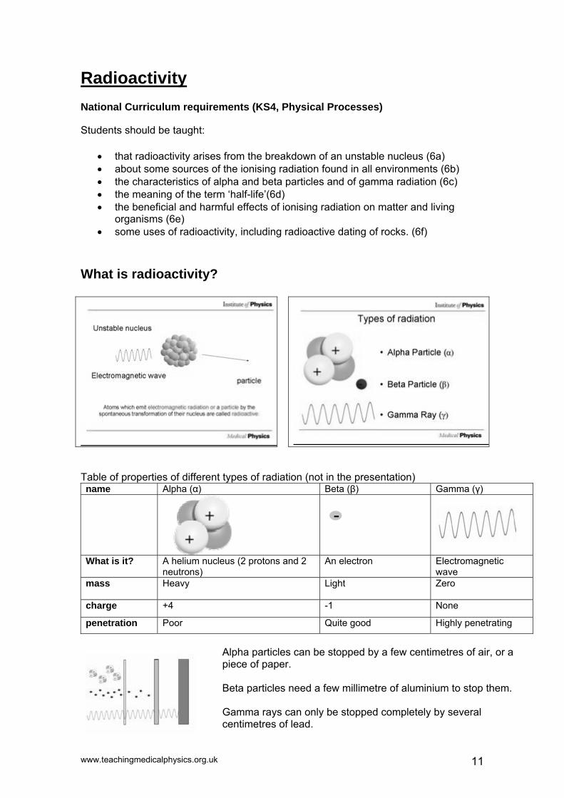
What is radioactivity?
Table of properties of different types of radiation (not in the presentation)
Alpha (α) Beta (β) Gamma (γ) name
What is it? A helium nucleus (2 protons and 2 neutrons)
An electron Electromagnetic wave
-
Heavy Light Zero mass
+4 -1 None charge
Poor Quite good Highly penetrating penetration
Alpha particles can be stopped by a few centimetres of air, or a piece of paper. Beta particles need a few millimetre of aluminium to stop them. Gamma rays can only be stopped completely by several centimetres of lead.
www.teachingmedicalphysics.org.uk 11

Uranite Uranite ore contains uranium, a radioactive element. It looks the same as a non-radioactive rock.
Radiation can be detected by a Geiger counter. These click every time either an alpha or beta particle, or a gamma ray, is detected.
www.teachingmedicalphysics.org.uk 12

Nuclear medicine
Nuclear medicine uses radioisotopes to investigate the function of the body. It works by following what happens to certain chemicals so the doctor can see if an organ is doing its job properly. The chemicals, called tracers, are 'labelled' with a radioactive isotope and their path followed through the body.
The radioisotopes are produced in generators where isotopes with long half-lives (e.g. molybdenum-99, half-life 67 hours) decay to isotopes with shorter lives (e.g. Technetium-99m, half life 6 hours). The Technetium-99m is drawn out of the generator in a solution and can be made into a range of different drugs (radiopharmaceuticals) that are absorbed by different parts of the body.
In the image you can see the lead glass used to shield the face of the medical physicist. Q: What material is this? A: Lead. Q: Why? A: Because lead can absorb all types of radiation, even gamma rays.
www.teachingmedicalphysics.org.uk 13

The radiopharmaceutical is drawn up into a syringe shielded with lead and its dose checked before it is injected into the patient.
The gamma rays given off by the radioisotope are detected by a gamma-camera (a detector that takes images with gamma rays) which is connected to a computer and gives an image of the distribution of the isotope in the patient.
The image shows where the drug is absorbed, and if several pictures are taken over a period of time it can also show how quickly the isotope is absorbed. This is a bone scan made using technetium-99m.
Q: Can you see where the patient was injected? A: By the elbow joint in the patient’s left arm. You can tell this as some of the radioactive technetium-99m has remained there.
www.teachingmedicalphysics.org.uk 14

The images show the build up of the tracer in the kidneys over time. The left kidney is healthy, but there is a blockage stopping the isotope from reaching the right kidney. In the PowerPoint presentation these images fade in after each other with only one mouse click.
Ventilation/perfusion scan. This involves two tests which may be done together or separately. The patient is injected with a radioactive drug which remains in the bloodstream around the lungs. An image of the distribution of this drug (on the right) shows the blood perfusion in the lungs. This should be uniform. A light area may show a blockage where the blood is not adequately perfusing the lung.
The second test is a ventilation scan, where the patient breathes radioactive xenon or krypton gas. This shows those parts of the lungs which are adequately ventilated. Combining both scans allows a doctor to work out whether the lung is functioning properly, and allows operations to be designed so as to reduce long-term damage.
Computer software enables us to look at the kidney from all directions. This image of a kidney rotates in the PowerPoint presentation.
www.teachingmedicalphysics.org.uk 15

PET Positron Emission Tomography (PET) scanning uses beta+ emitting isotopes. The isotope decays emitting a positron (which is a positive electron, also called a beta+ particle, and is a particle of antimatter). The positron can only travel about 1 mm before losing its energy and slowing down. When it slows down enough, it will meet a negative electron from a nearby atom, and they will 'annihilate', leaving no particles. Their energy is converted into two gamma rays which travel in opposite directions so that momentum is conserved.
This is used to make the radionuclides used in PET imaging by bombarding atoms with accelerated protons. It must be near to the PET scanner as the radionuclides used only have a short half-life (e.g. 2 mins for Oxygen-15).
The information from the PET scan (in colour on slide) can be superimposed on an x-ray CT image (grey/blue on slide). In this way, doctors get the benefit of high contrast from the PET scan and good spatial resolution from the CT image.
www.teachingmedicalphysics.org.uk 16

Radiotherapy For more information see radiotherapy in the electromagnetic spectrum section (pg 9-10).
Modern radiotherapy units use linear accelerators (linacs for short). These produce very high energy x-ray beams. The head rotates around the patient with the centre of the circle in the centre of the area to be treated. Older units worked in a similar way but used a single cobalt-60 source which emitted gamma rays. A more modern approach to radiotherapy using cobalt-60 is with a Gamma Knife.
Gamma knife The gamma knife is used for brain surgery, but is non-invasive (it is performed without cutting the skin or muscles and the skull does not need to be opened), though a frame needs to be attached to the skull using 4 screws (fitted under local anaesthetic). The gamma knife has 201 cobalt-60 sources, which emit gamma rays and have a half-life of 5.26 years. The sources are positioned in a hemisphere inside the unit. The patients head, held in the frame, is held inside a helmet with 201 holes to precisely target the radiation. When treatment starts, the patients head is moved inside the unit.
The treatment is planned using CT or MRI images, so that the sources are correctly targeted to irradiate the tumour and avoid healthy tissue, especially around the eye and cochlea. The gamma knife is used to treat benign and malignant tumours, blood vessel malformations, pain, and some movement and psychiatric disorders. There are currently three in the UK (two in London and one in Sheffield).
www.teachingmedicalphysics.org.uk 17

Ultrasound
National Curriculum requirements (KS4, Sc4 Physical Processes)
Students should be taught:
• about sound and ultrasound waves, and some medical and other uses of ultrasound (3l)
How does it work? The probe is coupled to the skin to make a good connection using a special coupling gel. The probe emits an ultrasound pulse. When the ultrasound wave reaches a boundary (such as the edge of an organ), some of it is reflected and some of it continues. The same thing happens at every boundary.
Many elements can be joined together to make a probe which can create an image.
www.teachingmedicalphysics.org.uk 18
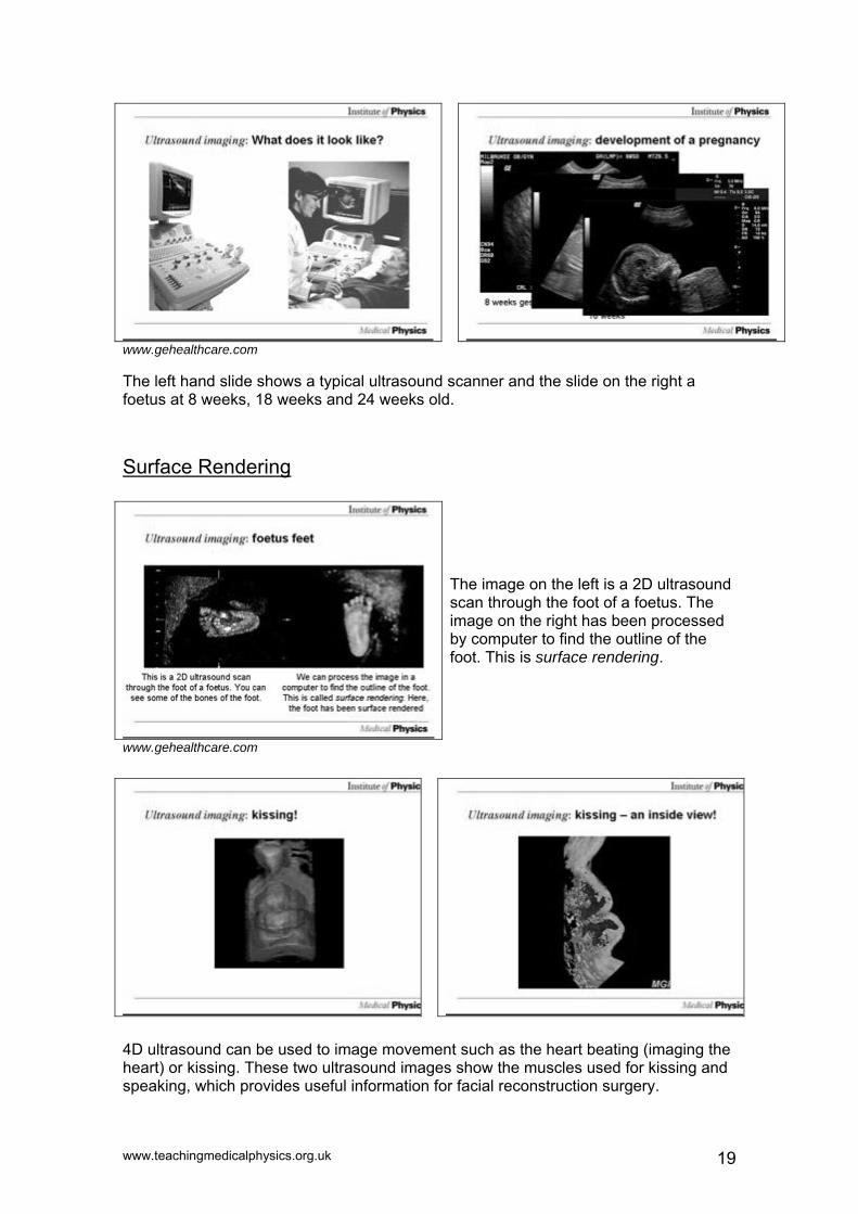
www.gehealthcare.com The left hand slide shows a typical ultrasound scanner and the slide on the right a foetus at 8 weeks, 18 weeks and 24 weeks old. Surface Rendering
The image on the left is a 2D ultrasound scan through the foot of a foetus. The image on the right has been processed by computer to find the outline of the foot. This is surface rendering.
www.gehealthcare.com
4D ultrasound can be used to image movement such as the heart beating (imaging the heart) or kissing. These two ultrasound images show the muscles used for kissing and speaking, which provides useful information for facial reconstruction surgery.
www.teachingmedicalphysics.org.uk 19

Doppler Ultrasound
The ultrasound probe emits an ultrasound wave. It travels through tissue and echoes off boundaries, as with standard ultrasound. In most cases, the echo has the same frequency as the emitted wave but if the target is moving, as in the case of a blood cell, the wavelength of the reflected wave will be modified by the Doppler effect. It is interesting to realise that there are two Doppler shifts occurring. The first occurs between the transmitted wave and the blood cell (with the probe acting as a source). But then, the beam echoes off the blood cell, so the moving blood cell behaves as a source and there is a second Doppler shift of the echoed wave, between the blood cell and the detector. Hence, the Doppler shifted frequency is twice what you would initially expect.
In this image, the Doppler ultrasound probe is measuring the blood flow in the radial artery, the same one you measure your pulse from. Doppler imaging can be combined with normal ultrasound imaging which can be used to image blood flow. Colour can be added to the image to make it easier to see.
Click on the loudspeaker icon to hear the sound. Q: Can you work out the heart rate?
www.teachingmedicalphysics.org.uk 20

Both the image and the Doppler data of the healthy carotid artery are clean and smooth as the smooth walled vessels lead to laminar flow.
The partially blocked carotid artery causes turbulent flow, as seen from the red and blue regions in the Doppler image (blood flow towards and away from the probe).
www.gehealthcare.com
This is a complicated image of the heart of a foetus. It shows the blood moving between the ventricles and the arteries. In the PowerPoint presentation you can see the motion of the heart as it beats.
Safety Ultrasound imaging has been used for 50 years, and is thought to be safe, though some energy is absorbed by the tissue, causing heating. This is very small in 2D ultrasound, and though new 4D imaging uses more energy it is thought to be safe.
Q: Should we use it to diagnose foetal illness? A: Yes, the benefit of being able to diagnose and treat the foetus is greater than the
risk of causing it harm. Q: Should we use it to make videos of healthy babies for parents? A: Some people say that because there is no medical benefit to the baby, the risk
outweighs the benefit. Others say that these scans help the parents to bond with the baby, so there is a benefit. What do you think?
[END OF PRESENTATIONS]
www.teachingmedicalphysics.org.uk 21

The Electromagnetic Spectrum
1) Name the parts of the Electromagnetic Spectrum
Low Energy High Energy
_ _ _ _ _ _ _ _ _ _ _ _ _ _ _ _ _ _ _ _ _ _ _ _ _ _ _ _ _ _ _ _ _ _ _ _ _-_ _ _ / _ _ _ _ _
2) Write a) what part of the body the image shows, b) the part of the
electromagnetic spectrum used and c) the name of the method used to take each image.
a)…………………… ……………………. ……………………… b)…………………... ……………………. ………………………. c)…………………… ……………………. ………………………. 3) Match the Medical Physics job with the part of the Electromagnetic
spectrum used (each word can be used more than once or not at all)
Imaging a fracture in a bone Gamma rays (γ) Thermography X-rays Treating cancer Infra Red (IR) CT scans Visible Endoscopy Ultraviolet (UV) Photodynamic therapy Bone scan
www.teachingmedicalphysics.org.uk 22

Infrared Radiation
1) a. Colour in your key. b. Colour in outside the face where it is coldest. c. Colour in the rest of the picture, working out which parts of your face give out the least and most infrared radiation.
30oC
32oC
34oC
2) Look at the images of the hands below
One person has healthy hands; the other person has a poor blood supply to their fingers (Raynaud's syndrome) a. Label one picture HEALTHY and POOR BLOOD SUPPLY
………………………………. …………………………… b. On each hand in the left hand image, label a blood vessel c. What are the names of the three types of blood vessel? …………………., ……………………., ………………………
www.teachingmedicalphysics.org.uk 23

X-Rays 1) a) What differences can you see between the two x-ray images?
………………………………………………… ……….…………………………………………………………….…………………………………………………………….……………………………………………………………………………………………………………………………………………………………………………
b) What year was x-ray 1 taken in? circle your answer 1795 1895 1995
c) Label a knuckle in each image d) What is the feature marked in the left image?
2) Radiotherapy- fill in the gaps in the paragraph below using the words in the box
healthy dose dividing tumour kill cancerous DNA X-rays irradiating
X-rays and other radiation can damage the _ _ _ in cells and kill them. Cells which are ________ rapidly are more likely to be killed, so we use x-rays to ____ the rapidly-dividing ______ cells. We must make sure that _______ tissue is undamaged. A linear accelerator generates ______. It rotates around the body, ___________the tumour from all directions.
This means the ______ receives a higher ____than the healthy tissue. 3) R A S I S O T I M I L E RadiotherapyD I V I D I N G C O U M Treatment, Plan A C N D O M E A A X E I Dose, LimitS I D X S R M H N I G R Cells, Dividing Y P A R E H T O I D A R Mitosis, Meiosis P S L C Y G A C L Y M A DNA, DamageC I N A A A E M V U A D Linac, X-rayE A J M N L R C O G D I Gamma, IrradiateC L M E L O T X E H I A CancerM A R S T R U O M U T T TumourG S I S O I E M E N R E
www.teachingmedicalphysics.org.uk 24

Nuclear Medicine
1) Sort these sentences into the correct order
• Patient injected with radiopharmaceutical
• Computer reconstructs image
• Drug labelled with radioisotope
• Patient imaged with Gamma camera
• Drug moves through bloodstream to organ
2) a) Which of these isotopes would be suitable for use as a tracer?
Circle your choice.
14C Carbon-14 half-life: 5730 years 57Co Cobalt- 57 half-life : 271 days 99mTc Technetium-99m half-life: 6hours 15O Oxygen-15 half-life : 2mins
b) Why did you choose that radioisotope? ……………………………………………………………………………………… ………………………………………………………………………………………………………………………………………………………………………………………………………………………………………………………………………
3) Sort the following statements into those that suggest radioactivity
is Beneficial those that suggest it is Harmful, and those that suggest it could be Either
Radiation can kill cells
Radiation most affects cells that are dividing
Radiation damages DNA and can cause mutations
Exposure to radiation can lead to sickness and burns
Gamma rays can only be
completely stopped by lead Radioactive
isotopes can be used as tracers in the body Radiation can cause cancer
but is also used to cure it
www.teachingmedicalphysics.org.uk 25

PET
1) A cross section of a PET scanner is shown below. The detectors ere detected at the same time.
shaded in show gamma rays which w
y drawing lines between the airs of detectors that detected
ere
) What does PET stand for? ) What type of radiation do the isotopes used in PET scans emit?
rticle meets an electron?
isotopes? topes are used in PET scans (circle the
Fluorine-17
8) Wh
Bpgamma rays, can you draw whyou think the tumour is?
234) What happens when this pa5) What is then produced? 6) What is the name of the machine used to produce the7) Which of the following iso
correct answers) Carbon-10 Oxygen-15 Fluorine-16 Carbon-11 Oxygen-16 Carbon-12 Oxygen-17 Fluorine-18 ich of these is a PET scanner?
9) What are PET scans often used to detect?
www.teachingmedicalphysics.org.uk 26

Ultrasound
1) Find the words listed, in the fastest time possible!
A Y S G N I G A M I UltrasoundL C O D W O H C E Q High, Frequency D N U O S A R T L U Sound, Wave O E N P V E S E H E ImagingL U D P S U O P P G FoetusP Q O L T W N H R I SonarH E T E A Y A L C N Dolphin, Bats I R O R B I R V S E Echo, Echoes N F H G I H O C E Y Doppler
2) Look at the images of each foetus below
a. Write the age of the foetus under each picture (8 weeks, 18 weeks)
8cm 14cm
Age……………………………… Age………………………………
b. On the 8 week old foetus, label the head, arms and legs c. On the 18 week old foetus, label the skull and spine d. How long is a full term pregnancy? ………………………….
3) How many uses of ultrasound can you list (medical and industrial)? ………………………………………………………………………………………………………………………………………………………………………………………………………………………………………………………………......................................................................................................................................................................................................................................
Images courtesy of GE Healthcare (www.gehealthcare.com)
www.teachingmedicalphysics.org.uk 27

a) Match the correct age of the foetus in each surface rendered ultrasound scan to the correct picture
b) Label the umbilical cord in each picture (hint, you can’t see it in the picture of the 32 week old foetus)
4) A dolphin hears an echo from a fish 0.02 seconds after it makes a
ed of sound in water is 1500m/s) noise (Hint, spe
a. How far has the ultrasound wave travelled? b. How far away is the fish? c. The dolphin swims closer to a distance of 9m from
the fish, How long will it take to hear the echo?
10 week old foetus
14cm
12 week old foetus18cm
21 week old foetus9cm
32 week old foetus11cm
Images courtesy of GE Healthcare (www.gehealthcare.com)
www.teachingmedicalphysics.org.uk 28

The Electromagnetic Spectrum - Answers
Low Energy High Energy
radiowave infrared visible ultraviolet x-ray/gamma ray
2. a) abdomen, lungs, oesophagus b) x-ray, gamma, visible c) x-ray CT (computed tomography), nuclear medicine (or gamma camera), endoscopy 3.
Gamma rays (γ) Imaging a fracture in a bone Thermography Treating Cancer CT scans Endoscopy Photodynamic therapy Bone scan
X-rays
Infra Red (IR)
Visible
Ultraviolet (UV)
Infrared - Answers
2 a) Left: healthy Right: Poor blood supply b) Artery, vein and capillary
www.teachingmedicalphysics.org.uk 29

X-Rays- Answers
1) a. One is a negative, one is positive, different noise levels in images, more detail
of bones seen in right hand image. b. 1895 d. wedding ring
2) X-rays and other radiation can damage the DNA in cells and kill them. Cells which are dividing rapidly are more likely to be killed, so we use x-rays to kill the rapidly-dividing cancerous cells. We must make sure that healthy tissue is undamaged. A Linear accelerator generates x-rays. It rotates around the body, irradiating the tumour from all directions. This means the tumour receives a higher dose than the healthy tissue.
Nuclear Medicine- Answers
1) Drug labelled with radioisotope Patient injected with radiopharmaceutical Drug moves through bloodstream to organ Patient imaged with Gamma camera Computer reconstructs image
2) a) Technetium-99m b) It has a suitable half-life, Carbon-14 and Cobalt-57 decay too slowly and Oxygen-15 decays too quickly, though O-15 is used in PET imaging.
3) Exposure to radiation causes sickness and burns – bad
Radioactive isotopes can be used as tracers in the body – good. Radiation can cause cancer, but is also used to treat it - either Others, arguments for both
PET- Answers
2) Positron emission tomography 3) Beta-+, positrons, positive electrons 4) They annihilate 5) Two gamma rays, travelling in opposite directions 6) A cyclotron 7) C-11, O-15, F-18 8) A)a cyclotron b) PET scanner c) gamma knife 9) tumour
www.teachingmedicalphysics.org.uk 30

Ultrasound - Answers
leg 2) skull
Age………8 weeks………………… Age…………18 weeks………….
d. 40 weeks 3) Pre natal scans, heart imaging, blood flow in arteries (Doppler ultrasound) SONAR, precision cleaning, detecting flaws (manufacturing)
2) a. 30m, b. 15m c. 0.012s
Images courtesy of GE Healthcare (www.gehealthcare.com)
10 week old foetus
12 week old foetus
21 week old foetus
32 week old foetus
headspine
arm
www.teachingmedicalphysics.org.uk 31

Acknowledgements This booklet and worksheets were written by Emily Cook. The lessons were developed by Adam Gibson, Jeff Jones, David Sang, Angela Newing, Nicola Hannam and Emily Cook. We are grateful to the following people and institutions for providing images:
Dr Gerard van Leeuwen Dr Clare Elwell Dr Kevin Howells Dr Sandy Mosse Dr Paul Campbell Dr Topun Austin Prof Alf Linney Dr Jing Deng Dr David Taylor Nature GE Healthcare Elekta Paul Burke Cochlear Europe Ltd The Nuclear Medicine and Radiotherapy Departments of the Royal Free Hospital, London UCL's Medical Illustration Unit The Cromwell Gamma Knife Centre, London Prof Jem Hebden
We have attempted to obtain permission and acknowledge the contributor of every image. If we have inadvertently used images in error, please contact us.
www.teachingmedicalphysics.org.uk 32