Embed Size (px)
Citation preview
07/12/2016
1
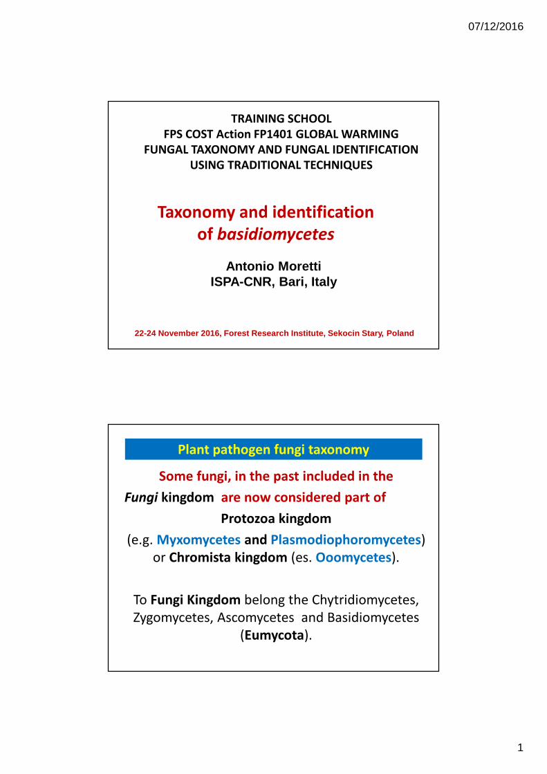
Taxonomy and identification
of basidiomycetes
TRAINING SCHOOL
FPS COST Action FP1401 GLOBAL WARMING
FUNGAL TAXONOMY AND FUNGAL IDENTIFICATION
USING TRADITIONAL TECHNIQUES
Antonio MorettiISPA-CNR, Bari, Italy
22-24 November 2016, Forest Research Institute, Sekocin Stary, Poland
Some fungi, in the past included in the
Fungi kingdom are now considered part of
Protozoa kingdom
(e.g. Myxomycetes and Plasmodiophoromycetes)
or Chromista kingdom (es. Ooomycetes).
To Fungi Kingdom belong the Chytridiomycetes,
Zygomycetes, Ascomycetes and Basidiomycetes
(Eumycota).
Plant pathogen fungi taxonomy
07/12/2016
2
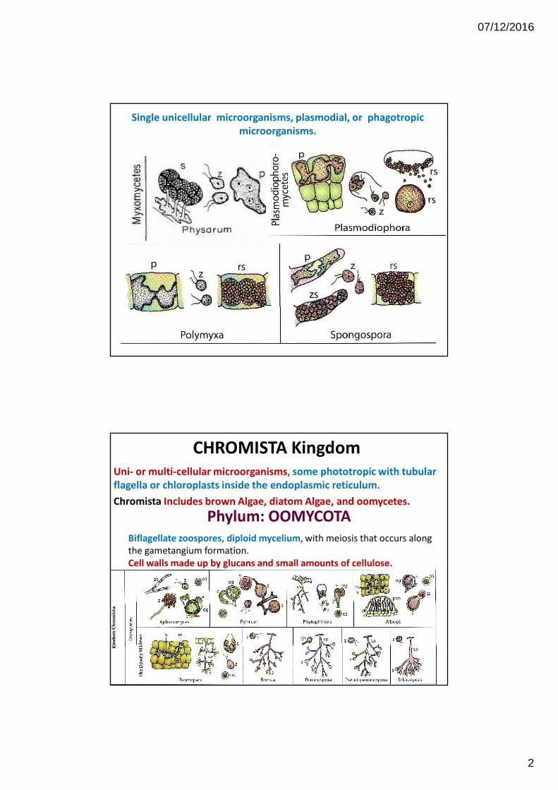
Single unicellular microorganisms, plasmodial, or phagotropic
microorganisms.
Uni- or multi-cellular microorganisms, some phototropic with tubular
flagella or chloroplasts inside the endoplasmic reticulum.
Chromista Includes brown Algae, diatom Algae, and oomycetes.
CHROMISTA Kingdom
Phylum: OOMYCOTA
Biflagellate zoospores, diploid mycelium, with meiosis that occurs along
the gametangium formation.
Cell walls made up by glucans and small amounts of cellulose.
07/12/2016
3
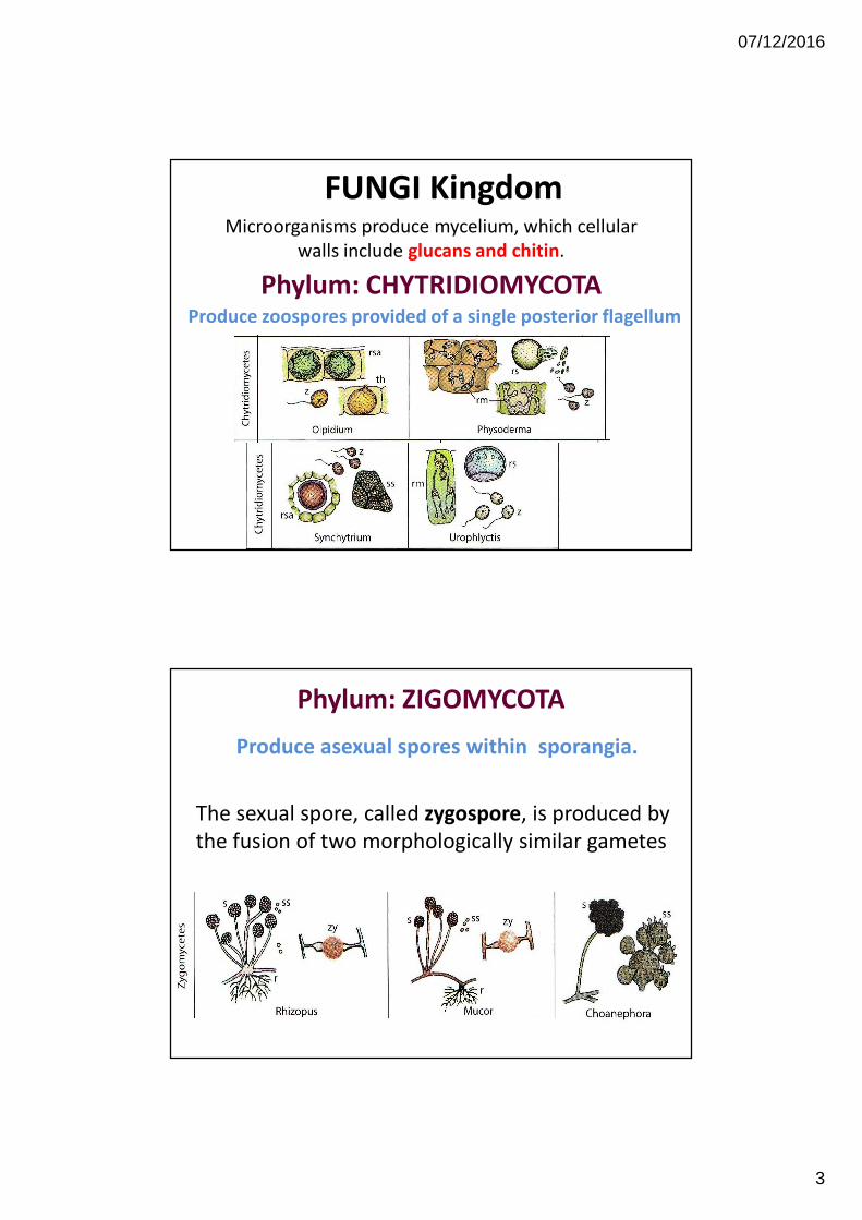
FUNGI KingdomMicroorganisms produce mycelium, which cellular
walls include glucans and chitin.
Phylum: CHYTRIDIOMYCOTAProduce zoospores provided of a single posterior flagellum
Produce asexual spores within sporangia.
The sexual spore, called zygospore, is produced by
the fusion of two morphologically similar gametes
Phylum: ZIGOMYCOTA
07/12/2016
4
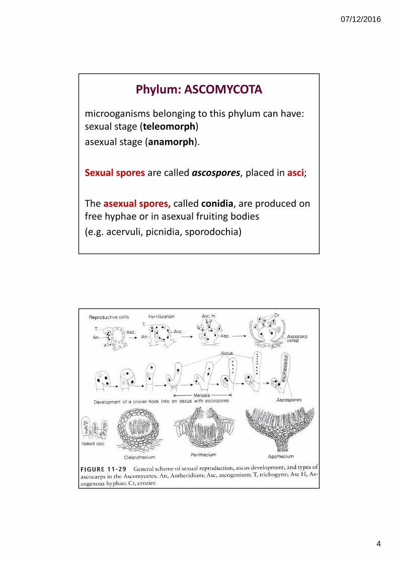
microoganisms belonging to this phylum can have:
sexual stage (teleomorph)
asexual stage (anamorph).
Sexual spores are called ascospores, placed in asci;
The asexual spores, called conidia, are produced on
free hyphae or in asexual fruiting bodies
(e.g. acervuli, picnidia, sporodochia)
Phylum: ASCOMYCOTA
07/12/2016
5
Main trait of Basidiomycetes is the production of
meiospores (= meiosis products), the basidiospores,
born in club-shaped structures called basidia.
Basidiomycetes have chitinose cell walls,
multilayers, and hyphae with septa provided of a
barrel-shaped swelling around their central pore
called dolipore.
Phylum: BASIDIOMYCOTA
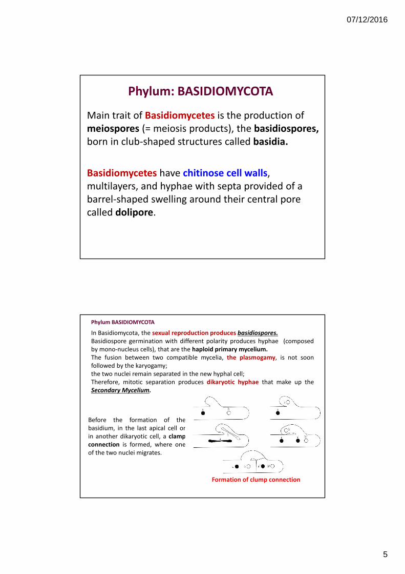
In Basidiomycota, the sexual reproduction produces basidiospores.
Basidiospore germination with different polarity produces hyphae (composed
by mono-nucleus cells), that are the haploid primary mycelium.
The fusion between two compatible mycelia, the plasmogamy, is not soon
followed by the karyogamy;
the two nuclei remain separated in the new hyphal cell;
Therefore, mitotic separation produces dikaryotic hyphae that make up the
Secondary Mycelium.
Phylum BASIDIOMYCOTA
Before the formation of the
basidium, in the last apical cell or
in another dikaryotic cell, a clamp
connection is formed, where one
of the two nuclei migrates.
Formation of clump connection
07/12/2016
6
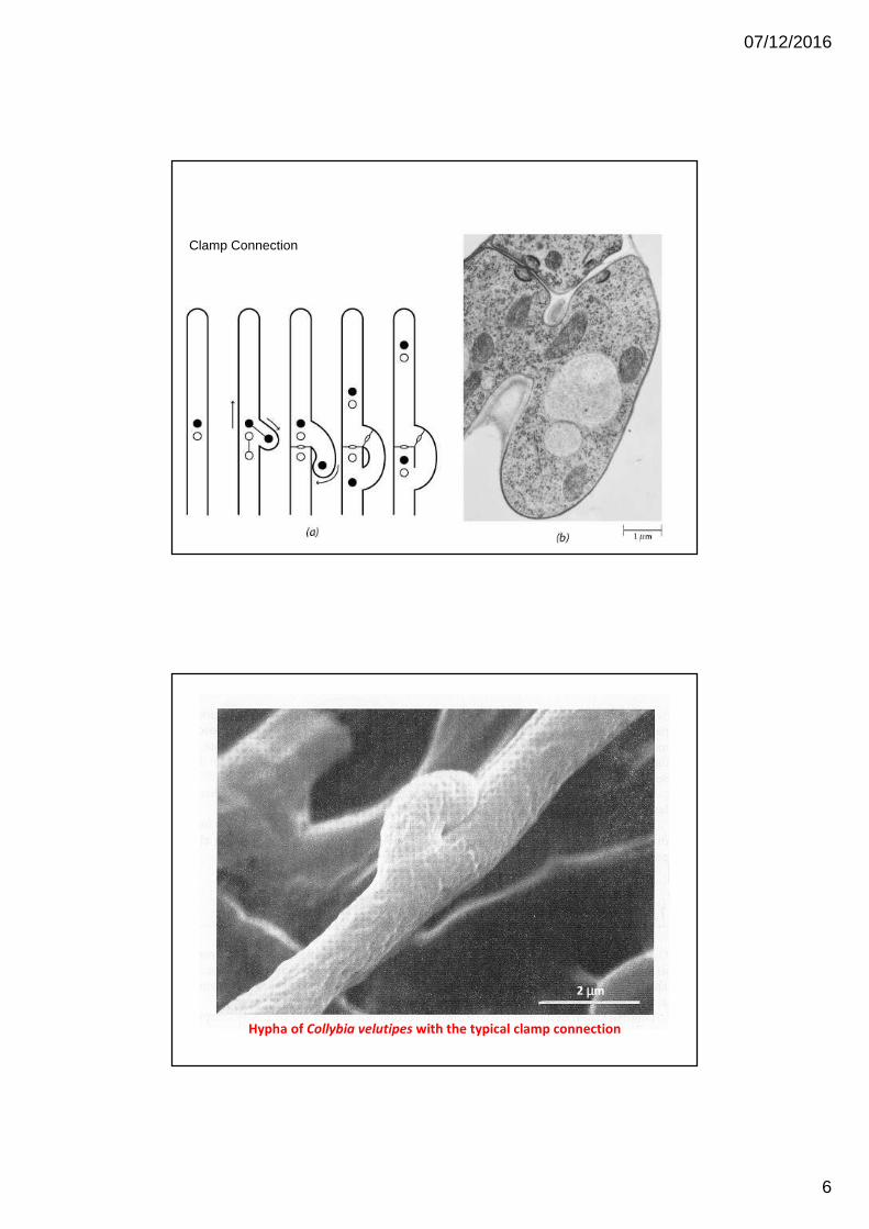
Clamp Connection
2 µm
Hypha of Collybia velutipes with the typical clamp connection
07/12/2016
7
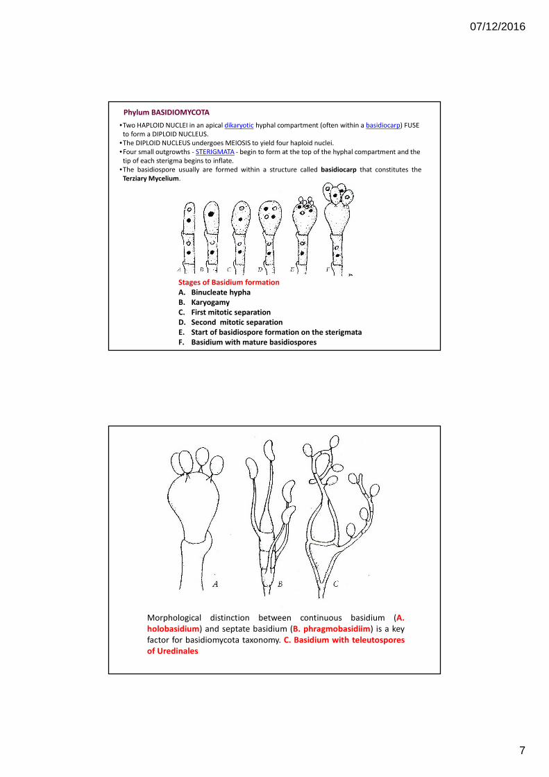
Phylum BASIDIOMYCOTA
Stages of Basidium formation
A. Binucleate hypha
B. Karyogamy
C. First mitotic separation
D. Second mitotic separation
E. Start of basidiospore formation on the sterigmata
F. Basidium with mature basidiospores
•Two HAPLOID NUCLEI in an apical dikaryotic hyphal compartment (often within a basidiocarp) FUSE
to form a DIPLOID NUCLEUS.
•The DIPLOID NUCLEUS undergoes MEIOSIS to yield four haploid nuclei.
•Four small outgrowths - STERIGMATA - begin to form at the top of the hyphal compartment and the
tip of each sterigma begins to inflate.
•The basidiospore usually are formed within a structure called basidiocarp that constitutes the
Terziary Mycelium.
Morphological distinction between continuous basidium (A.
holobasidium) and septate basidium (B. phragmobasidiim) is a key
factor for basidiomycota taxonomy. C. Basidium with teleutospores
of Uredinales
07/12/2016
8
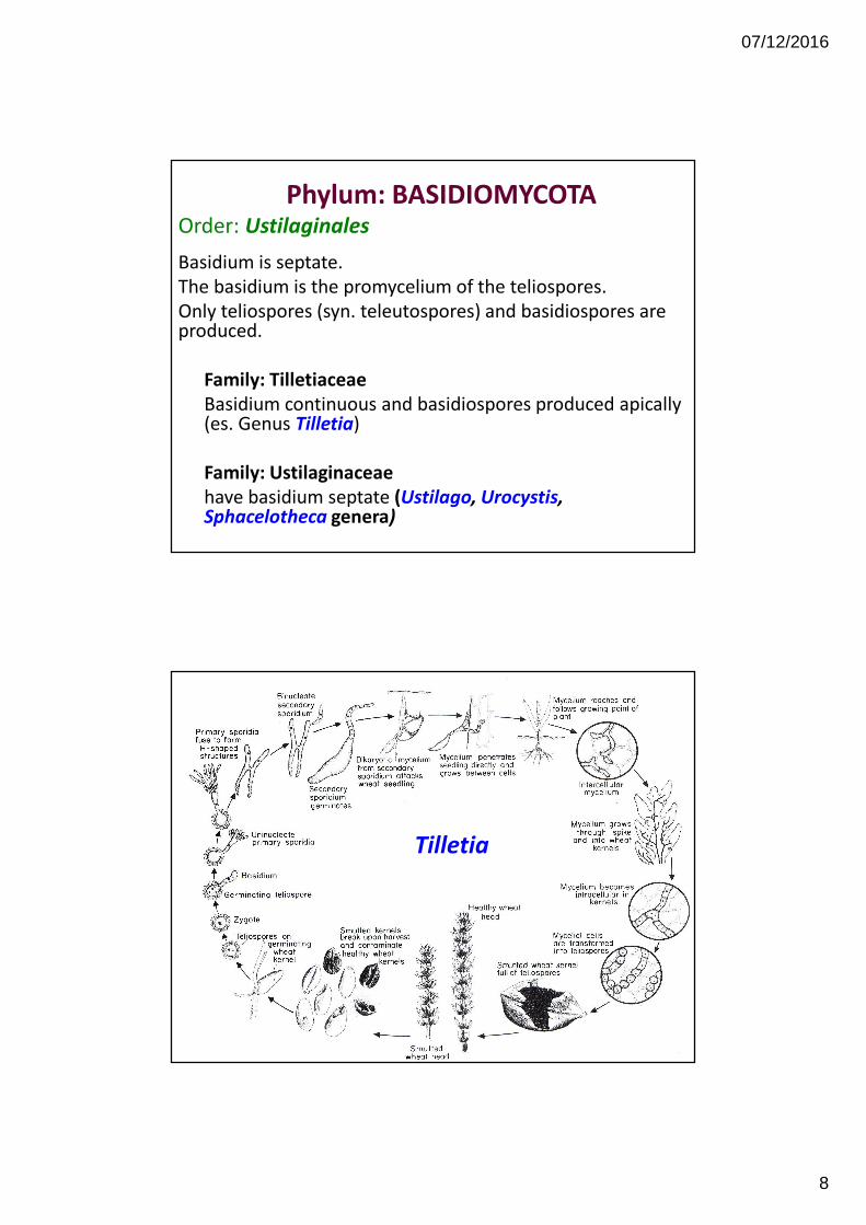
Phylum: BASIDIOMYCOTAOrder: Ustilaginales
Basidium is septate.
The basidium is the promycelium of the teliospores.
Only teliospores (syn. teleutospores) and basidiospores are produced.
Family: Tilletiaceae
Basidium continuous and basidiospores produced apically (es. Genus Tilletia)
Family: Ustilaginaceae
have basidium septate (Ustilago, Urocystis, Sphacelotheca genera)
Tilletia
Tilletia
07/12/2016
9
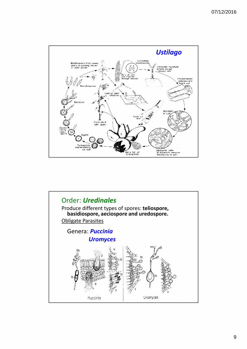
Ustilago
Order: UredinalesProduce different types of spores: teliospore,
basidiospore, aeciospore and uredospore.
Obligate Parasites
Genera: Puccinia
Uromyces
07/12/2016
10
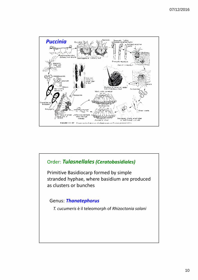
Puccinia
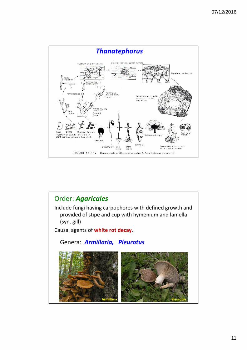
Order: Tulasnellales (Ceratobasidiales)
Primitive Basidiocarp formed by simple
stranded hyphae, where basidium are produced
as clusters or bunches
Genus: Thanatephorus
T. cucumeris è il teleomorph of Rhizoctonia solani
07/12/2016
11
Thanatephorus
Order: Agaricales
Include fungi having carpophores with defined growth and
provided of stipe and cup with hymenium and lamella
(syn. gill)
Causal agents of white rot decay.
Genera: Armillaria, Pleurotus
Armillaria Pleurotus
07/12/2016
12
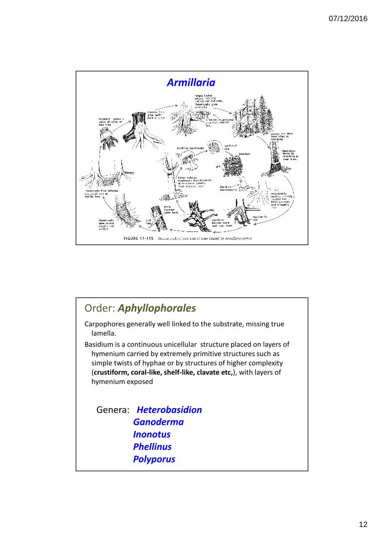
Armillaria
Order: Aphyllophorales
Carpophores generally well linked to the substrate, missing true
lamella.
Basidium is a continuous unicellular structure placed on layers of
hymenium carried by extremely primitive structures such as
simple twists of hyphae or by structures of higher complexity
(crustiform, coral-like, shelf-like, clavate etc,), with layers of
hymenium exposed
Genera: Heterobasidion
Ganoderma
Inonotus
Phellinus
Polyporus
07/12/2016
13
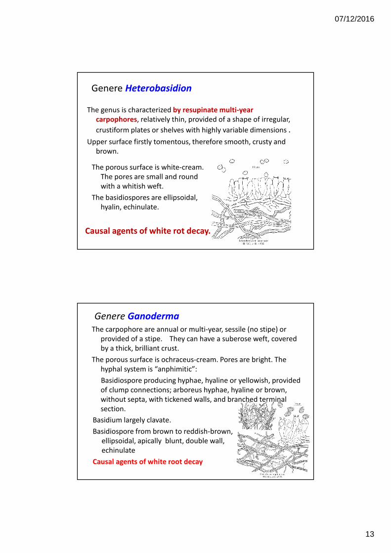
Genere Heterobasidion
The genus is characterized by resupinate multi-year
carpophores, relatively thin, provided of a shape of irregular,
crustiform plates or shelves with highly variable dimensions .
Upper surface firstly tomentous, therefore smooth, crusty and
brown.
The porous surface is white-cream.
The pores are small and round
with a whitish weft.
The basidiospores are ellipsoidal,
hyalin, echinulate.
Causal agents of white rot decay.
Genere Ganoderma
The carpophore are annual or multi-year, sessile (no stipe) or
provided of a stipe. They can have a suberose weft, covered
by a thick, brilliant crust.
The porous surface is ochraceus-cream. Pores are bright. The
hyphal system is “anphimitic”:
Basidiospore producing hyphae, hyaline or yellowish, provided
of clump connections; arboreus hyphae, hyaline or brown,
without septa, with tickened walls, and branched terminal
section.
Basidium largely clavate.
Basidiospore from brown to reddish-brown,
ellipsoidal, apically blunt, double wall,
echinulate
Causal agents of white root decay
07/12/2016
14
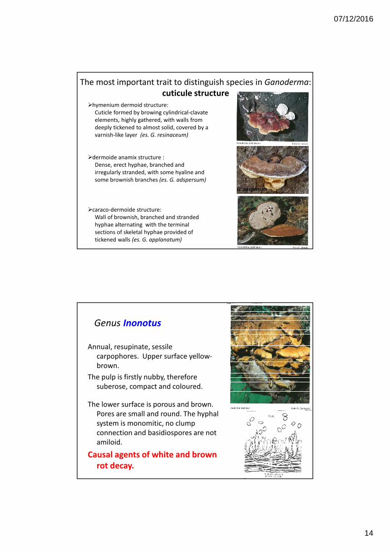
The most important trait to distinguish species in Ganoderma:
cuticule structure
�hymenium dermoid structure:
Cuticle formed by browing cylindrical-clavate
elements, highly gathered, with walls from
deeply tickened to almost solid, covered by a
varnish-like layer (es. G. resinaceum)
�dermoide anamix structure :
Dense, erect hyphae, branched and
irregularly stranded, with some hyaline and
some brownish branches (es. G. adspersum)
�caraco-dermoide structure:
Wall of brownish, branched and stranded
hyphae alternating with the terminal
sections of skeletal hyphae provided of
tickened walls (es. G. applanatum)
G. adspersum
Genus Inonotus
Annual, resupinate, sessile
carpophores. Upper surface yellow-
brown.
The pulp is firstly nubby, therefore
suberose, compact and coloured.
The lower surface is porous and brown.
Pores are small and round. The hyphal
system is monomitic, no clump
connection and basidiospores are not
amiloid.
Causal agents of white and brown
rot decay.
07/12/2016
15
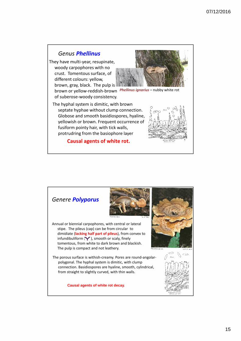
Genus Phellinus
They have multi-year, resupinate,
woody carpophores with no
crust. Tomentous surface, of
different colours: yellow,
brown, gray, black. The pulp is
brown or yellow-reddish-brown
of suberose-woody consistency.
The hyphal system is dimitic, with brown
septate hyphae without clump connection.
Globose and smooth basidiospores, hyaline,
yellowish or brown. Frequent occurrence of
fusiform pointy hair, with tick walls,
protrudring from the basiophore layer
Causal agents of white rot.
Phellinus ignarius – nubby white rot
Genere Polyporus
Annual or biennial carpophores, with central or lateral
stipe. The pileus (cap) can be from circular to
dimidiate (lacking half part of pileus), from convex to
infundibuliform ( ), smooth or scaly, finely
tomentous, from white to dark brown and blackish.
The pulp is compact and not leathery.
The porous surface is withish-creamy. Pores are round-angolar-
polygonal. The hyphal system is dimitic, with clump
connection. Basidiospores are hyaline, smooth, cylindrical,
from straight to slightly curved, with thin walls.
Causal agents of white rot decay.
07/12/2016
16
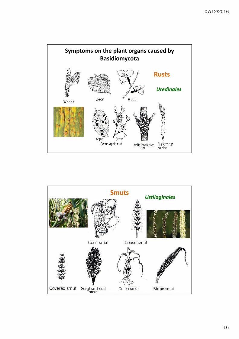
Symptoms on the plant organs caused by
Basidiomycota
Rusts
Uredinales
SmutsUstilaginales
07/12/2016
17
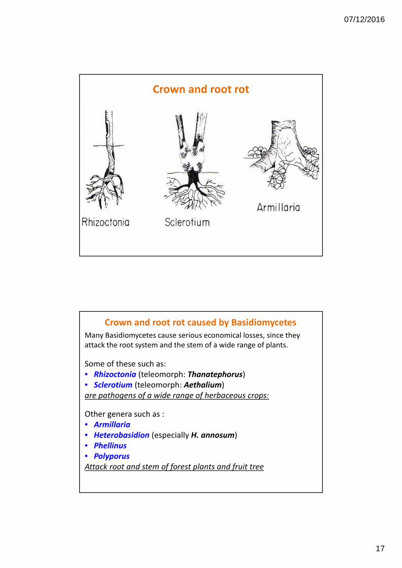
Crown and root rot
Crown and root rot caused by Basidiomycetes
Many Basidiomycetes cause serious economical losses, since they
attack the root system and the stem of a wide range of plants.
Some of these such as:
• Rhizoctonia (teleomorph: Thanatephorus)
• Sclerotium (teleomorph: Aethalium)
are pathogens of a wide range of herbaceous crops:
Other genera such as :
• Armillaria
• Heterobasidion (especially H. annosum)
• Phellinus
• Polyporus
Attack root and stem of forest plants and fruit tree
07/12/2016
18
Root rot and stem rot caused by
“fungi sterilia” Rhizoctonia and Sclerotium
Fungi of Rhizoctonia e Sclerotium genera have as main habitat the
soil. They can cause serious plant diseases on a wide range of both
different plant and organs: roots, stems, bulbs, tubers etc.
These two genera were well known as sterile fungi, since they were
thought for many years to produce only sclerotium being unable to
produce sexual or asexual spores.
More recently also these two genera were shown to produce
basidiospores, and therefore included in Basidiomycetes.
Rhizoctonia e Sclerotium spores are produced only in well selected
laboratory conditions and their natural occurrence is extremely rare.
For this reason they are extremely important as diagnostic tool.
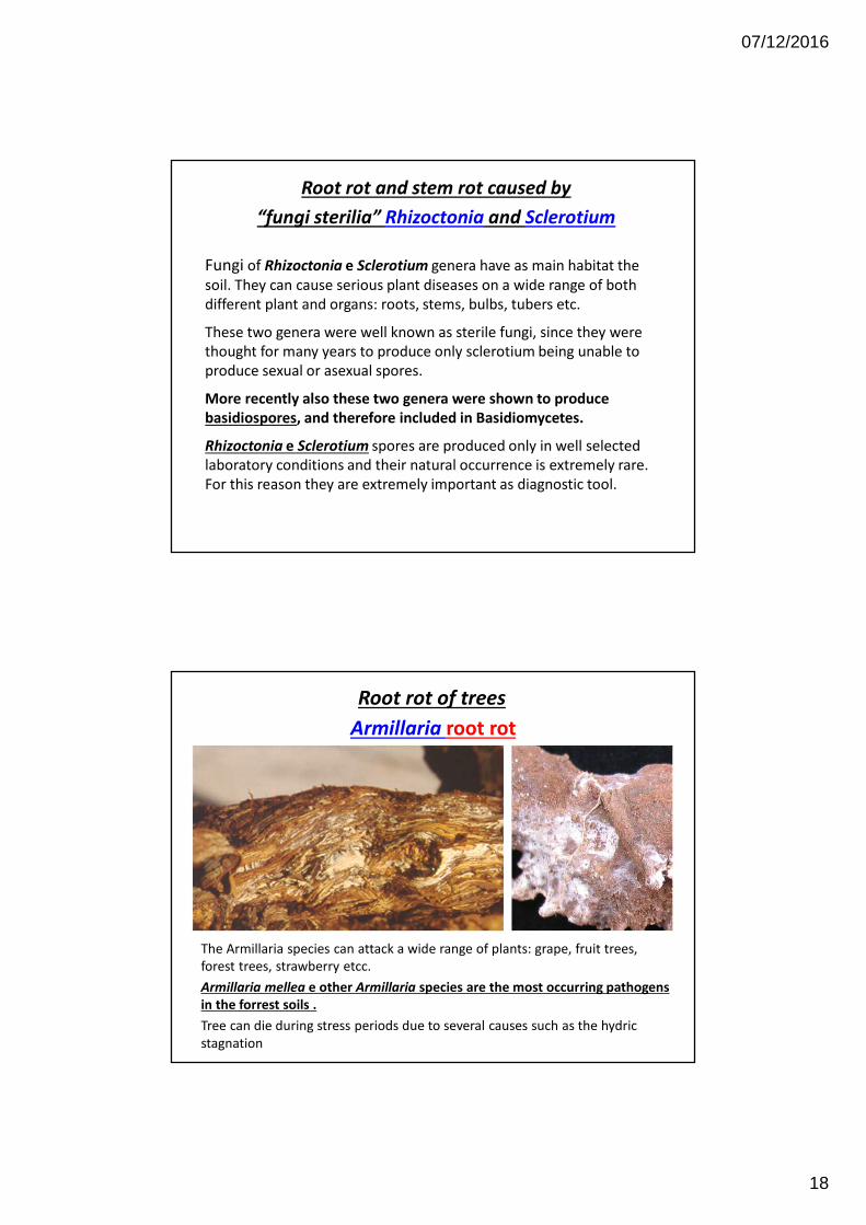
Armillaria root rot
Root rot of trees
The Armillaria species can attack a wide range of plants: grape, fruit trees,
forest trees, strawberry etcc.
Armillaria mellea e other Armillaria species are the most occurring pathogens
in the forrest soils .
Tree can die during stress periods due to several causes such as the hydric
stagnation
07/12/2016
19
Trees with root rot caused by Armillaria show similar symptoms of other
root rot:
• general distress
• Reduced growth,
• Yellowish leaves,
• Gradual or sudden death of the tree
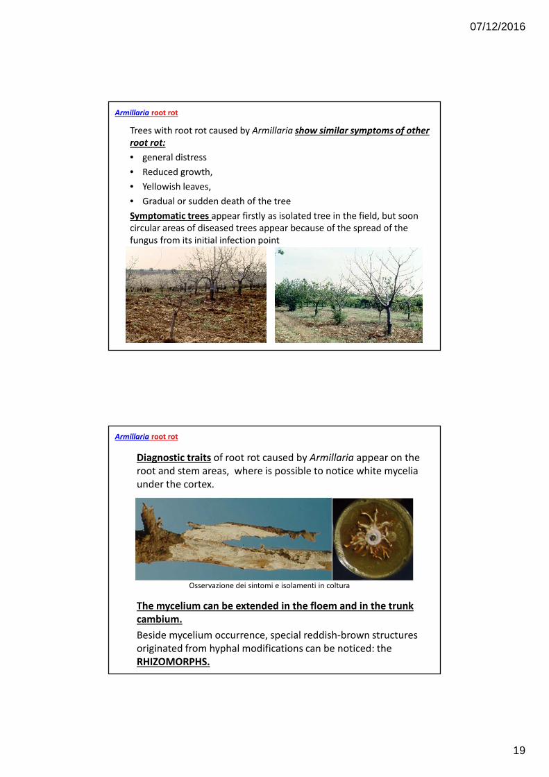
Symptomatic trees appear firstly as isolated tree in the field, but soon
circular areas of diseased trees appear because of the spread of the
fungus from its initial infection point
Armillaria root rot
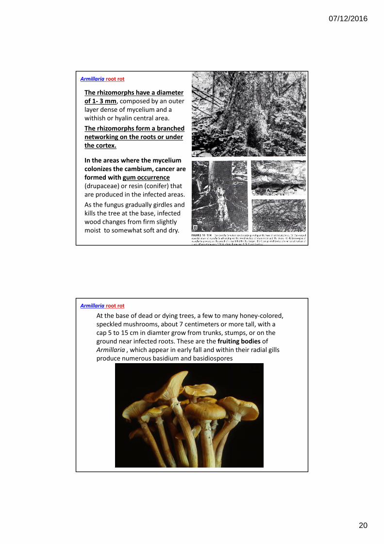
Diagnostic traits of root rot caused by Armillaria appear on the
root and stem areas, where is possible to notice white mycelia
under the cortex.
Osservazione dei sintomi e isolamenti in coltura
The mycelium can be extended in the floem and in the trunk
cambium.
Beside mycelium occurrence, special reddish-brown structures
originated from hyphal modifications can be noticed: the
RHIZOMORPHS.
Armillaria root rot
07/12/2016
20
The rhizomorphs have a diameter
of 1- 3 mm, composed by an outer
layer dense of mycelium and a
withish or hyalin central area.
The rhizomorphs form a branched
networking on the roots or under
the cortex.
In the areas where the mycelium
colonizes the cambium, cancer are
formed with gum occurrence
(drupaceae) or resin (conifer) that
are produced in the infected areas.
As the fungus gradually girdles and
kills the tree at the base, infected
wood changes from firm slightly
moist to somewhat soft and dry.
Armillaria root rot
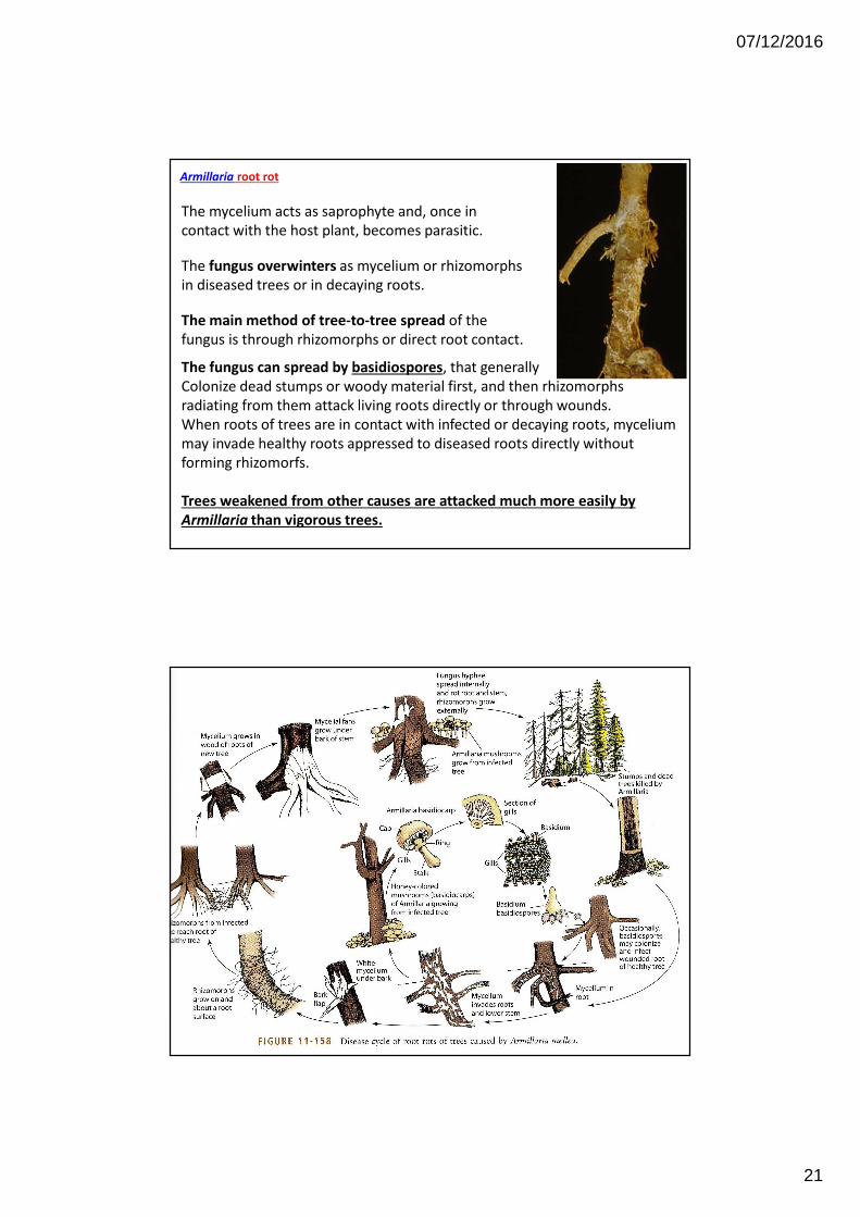
At the base of dead or dying trees, a few to many honey-colored,
speckled mushrooms, about 7 centimeters or more tall, with a
cap 5 to 15 cm in diamter grow from trunks, stumps, or on the
ground near infected roots. These are the fruiting bodies of
Armillaria , which appear in early fall and within their radial gills
produce numerous basidium and basidiospores
Armillaria root rot
07/12/2016
21
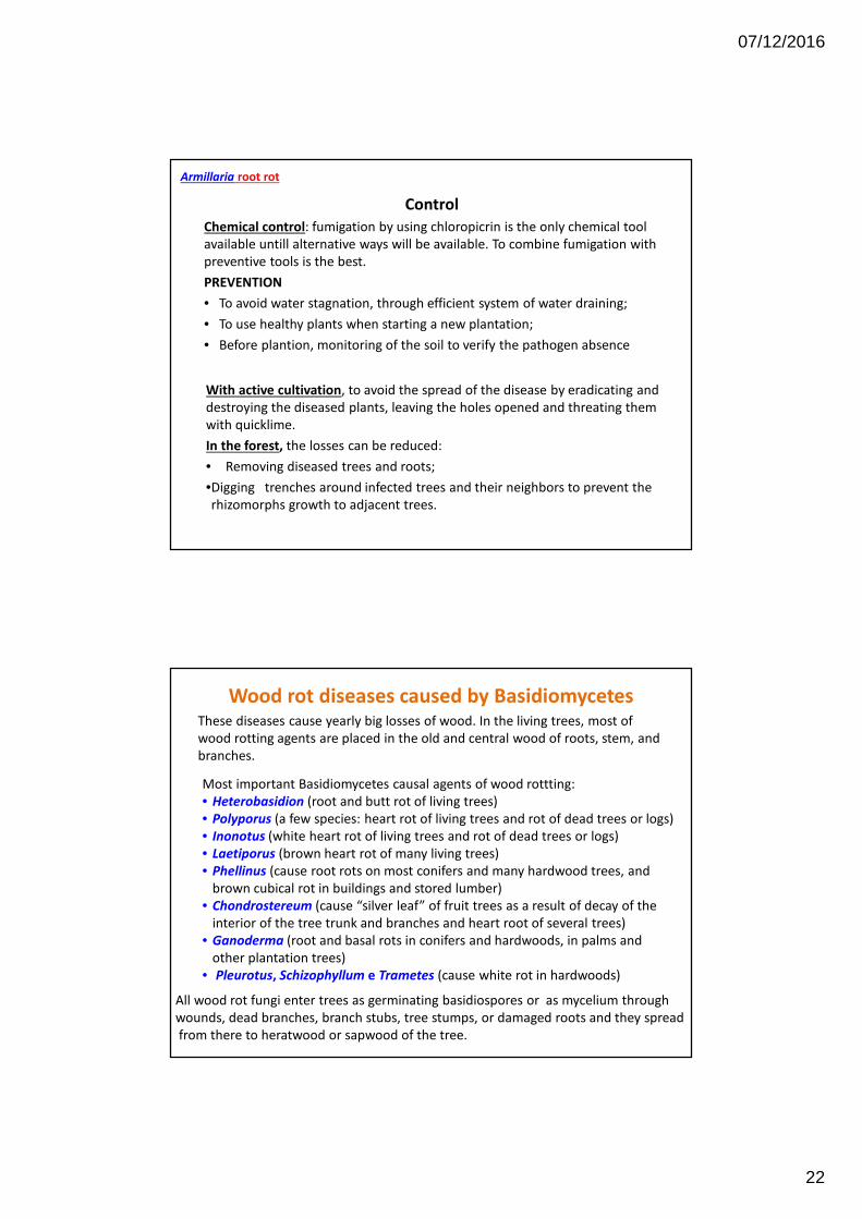
The mycelium acts as saprophyte and, once in
contact with the host plant, becomes parasitic.
The fungus overwinters as mycelium or rhizomorphs
in diseased trees or in decaying roots.
The main method of tree-to-tree spread of the
fungus is through rhizomorphs or direct root contact.
The fungus can spread by basidiospores, that generally
Colonize dead stumps or woody material first, and then rhizomorphs
radiating from them attack living roots directly or through wounds.
When roots of trees are in contact with infected or decaying roots, mycelium
may invade healthy roots appressed to diseased roots directly without
forming rhizomorfs.
Trees weakened from other causes are attacked much more easily by
Armillaria than vigorous trees.
Armillaria root rot
07/12/2016
22
Control
Chemical control: fumigation by using chloropicrin is the only chemical tool
available untill alternative ways will be available. To combine fumigation with
preventive tools is the best.
PREVENTION
• To avoid water stagnation, through efficient system of water draining;
• To use healthy plants when starting a new plantation;
• Before plantion, monitoring of the soil to verify the pathogen absence
With active cultivation, to avoid the spread of the disease by eradicating and
destroying the diseased plants, leaving the holes opened and threating them
with quicklime.
In the forest, the losses can be reduced:
• Removing diseased trees and roots;
•Digging trenches around infected trees and their neighbors to prevent the
rhizomorphs growth to adjacent trees.
Armillaria root rot
Wood rot diseases caused by BasidiomycetesThese diseases cause yearly big losses of wood. In the living trees, most of
wood rotting agents are placed in the old and central wood of roots, stem, and
branches.
Most important Basidiomycetes causal agents of wood rottting:
• Heterobasidion (root and butt rot of living trees)
• Polyporus (a few species: heart rot of living trees and rot of dead trees or logs)
• Inonotus (white heart rot of living trees and rot of dead trees or logs)
• Laetiporus (brown heart rot of many living trees)
• Phellinus (cause root rots on most conifers and many hardwood trees, and
brown cubical rot in buildings and stored lumber)
• Chondrostereum (cause “silver leaf” of fruit trees as a result of decay of the
interior of the tree trunk and branches and heart root of several trees)
• Ganoderma (root and basal rots in conifers and hardwoods, in palms and
other plantation trees)
• Pleurotus, Schizophyllum e Trametes (cause white rot in hardwoods)
All wood rot fungi enter trees as germinating basidiospores or as mycelium through
wounds, dead branches, branch stubs, tree stumps, or damaged roots and they spread
from there to heratwood or sapwood of the tree.
07/12/2016
23
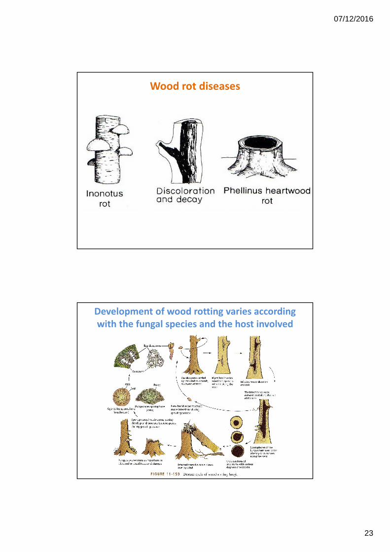
Wood rot diseases
Development of wood rotting varies according
with the fungal species and the host involved
07/12/2016
24
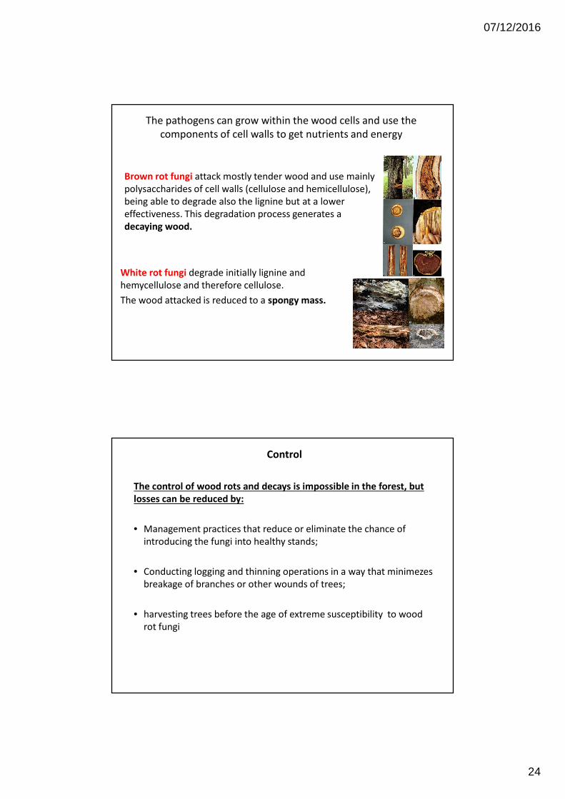
Brown rot fungi attack mostly tender wood and use mainly
polysaccharides of cell walls (cellulose and hemicellulose),
being able to degrade also the lignine but at a lower
effectiveness. This degradation process generates a
decaying wood.
The pathogens can grow within the wood cells and use the
components of cell walls to get nutrients and energy
White rot fungi degrade initially lignine and
hemycellulose and therefore cellulose.
The wood attacked is reduced to a spongy mass.
Control
The control of wood rots and decays is impossible in the forest, but
losses can be reduced by:
• Management practices that reduce or eliminate the chance of
introducing the fungi into healthy stands;
• Conducting logging and thinning operations in a way that minimezes
breakage of branches or other wounds of trees;
• harvesting trees before the age of extreme susceptibility to wood
rot fungi
07/12/2016
25
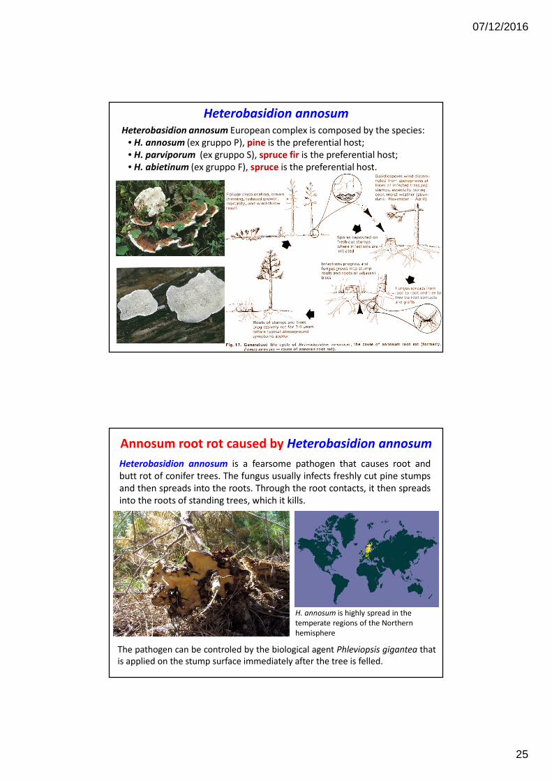
Heterobasidion annosum
Heterobasidion annosum European complex is composed by the species:
• H. annosum (ex gruppo P), pine is the preferential host;
• H. parviporum (ex gruppo S), spruce fir is the preferential host;
• H. abietinum (ex gruppo F), spruce is the preferential host.
Annosum root rot caused by Heterobasidion annosum
Heterobasidion annosum is a fearsome pathogen that causes root and
butt rot of conifer trees. The fungus usually infects freshly cut pine stumps
and then spreads into the roots. Through the root contacts, it then spreads
into the roots of standing trees, which it kills.
H. annosum is highly spread in the
temperate regions of the Northern
hemisphere
The pathogen can be controled by the biological agent Phleviopsis gigantea that
is applied on the stump surface immediately after the tree is felled.
07/12/2016
26
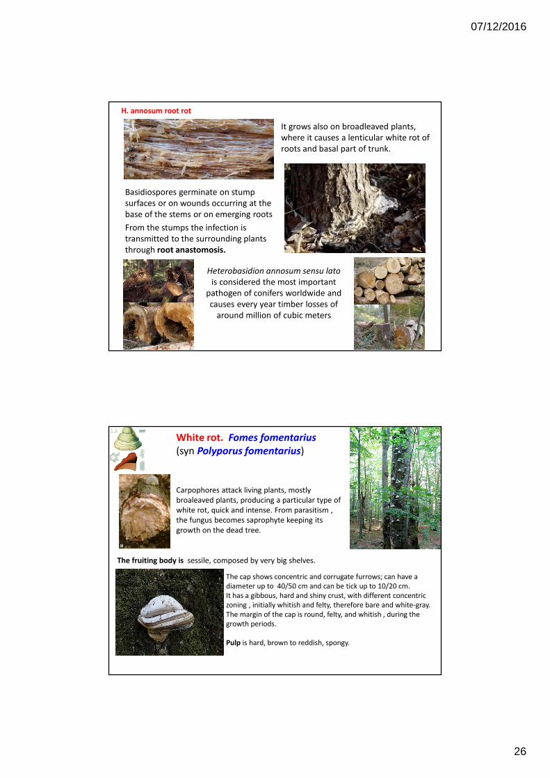
Heterobasidion annosum sensu lato
is considered the most important
pathogen of conifers worldwide and
causes every year timber losses of
around million of cubic meters
It grows also on broadleaved plants,
where it causes a lenticular white rot of
roots and basal part of trunk.
Basidiospores germinate on stump
surfaces or on wounds occurring at the
base of the stems or on emerging roots
From the stumps the infection is
transmitted to the surrounding plants
through root anastomosis.
H. annosum root rot
The cap shows concentric and corrugate furrows; can have a
diameter up to 40/50 cm and can be tick up to 10/20 cm.
It has a gibbous, hard and shiny crust, with different concentric
zoning , initially whitish and felty, therefore bare and white-gray.
The margin of the cap is round, felty, and whitish , during the
growth periods.
Pulp is hard, brown to reddish, spongy.
White rot. Fomes fomentarius
(syn Polyporus fomentarius)
Carpophores attack living plants, mostly
broaleaved plants, producing a particular type of
white rot, quick and intense. From parasitism ,
the fungus becomes saprophyte keeping its
growth on the dead tree.
The fruiting body is sessile, composed by very big shelves.
07/12/2016
27
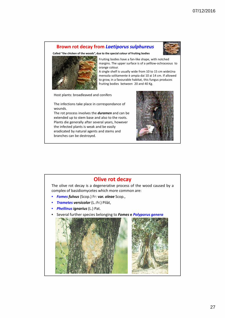
Brown rot decay from Laetiporus sulphureus
Host plants: broadleaved and conifers
The infections take place in correspondance of
wounds.
The rot process involves the duramen and can be
extended up to stem base and also to the roots.
Plants die generally after several years, however
the infected plants is weak and be easily
eradicated by natural agents and stems and
branches can be destroyed.
Fruiting bodies have a fan-like shape, with notched
margins. The upper surface is of a yelllow-ochraceous to
orange colour.
A single shelf is usually wide from 10 to 15 cm wideUna
mensola solitamente è ampia dai 10 ai 14 cm. If allowed
to grow, in a favourable habitat, this fungus produces
fruiting bodies between 20 and 40 Kg.
Called “the chicken of the woods”, due to the special colour of fruiting bodies
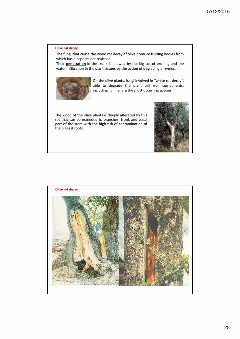
Olive rot decayThe olive rot decay is a degenerative process of the wood caused by a
complex of basidiomycetes which more common are:
• Fomes fulvus (Scop.) Fr: var. oleae Scop.,
• Trametes versicolor (L.:Fr.) Pilàt,
• Phellinus ignarius (L.) Pat.
• Several further species belonging to Fomes e Polyporus genera
07/12/2016
28
The wood of the olive plants is deeply alterated by thisrot that can be extended to branches, trunk and basalpart of the stem with the high risk of contamination ofthe biggest roots.
Olive rot decay
On the olive plants, fungi involved in “white rot decay”,
able to degrade the plant cell wall components,
including lignine, are the most occurring species.
The fungi that cause the wood rot decay of olive produce fruiting bodies from
which basidiospores are realesed.
Their penetration in the trunk is allowed by the big cut of pruning and the
water infiltration in the plant tissues by the action of degrading enzymes.
Olive rot decay
07/12/2016
29
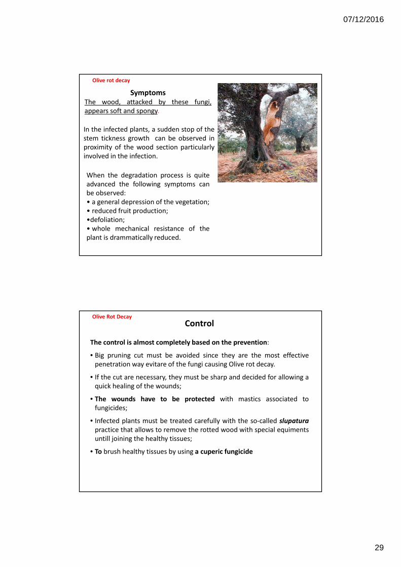
Symptoms
The wood, attacked by these fungi,
appears soft and spongy.
In the infected plants, a sudden stop of the
stem tickness growth can be observed in
proximity of the wood section particularly
involved in the infection.
When the degradation process is quite
advanced the following symptoms can
be observed:
• a general depression of the vegetation;
• reduced fruit production;
•defoliation;
• whole mechanical resistance of the
plant is drammatically reduced.
Olive rot decay
Control
The control is almost completely based on the prevention:
• Big pruning cut must be avoided since they are the most effective
penetration way evitare of the fungi causing Olive rot decay.
• If the cut are necessary, they must be sharp and decided for allowing a
quick healing of the wounds;
• The wounds have to be protected with mastics associated to
fungicides;
• Infected plants must be treated carefully with the so-called slupatura
practice that allows to remove the rotted wood with special equiments
untill joining the healthy tissues;
• To brush healthy tissues by using a cuperic fungicide
Olive Rot Decay

07/12/2016
30