Embed Size (px)
Citation preview
Int.J.Curr.Microbiol.App.Sci (2015) 4(3): 361-371
361
Original Research Article
Taxonomy and Toxicological Evaluation of Tetraodontiformes from Kasimedu, Chennai, India
M.Mohan raj1* and J.SeshSerebiah1
Department of Marine Studies and Coastal Resource Management (Autonomous), Tambaram, Chennai-59, India
*Corresponding author
A B S T R A C T
Introduction
Tetraodontiforms is an order of primarily marine and estuarine fish. The order includes many familiar species which are variously called puffers, balloonfish, blowfish, bubblefish, globefish, swellfish, toadfish, and toadies. They are morphologically similar to the closely related porcupine fish, which have large conspicuous spines (unlike the small, almost sandpaper-like spines of Tetraodontidae). The scientific name, Tetraodontidae, refers to the four large teeth, fused into an upper and lower plate, which are used for crushing
the shells of crustaceans and mollusks, and red worms, their natural prey. Puffer Fish are the second most poisonous vertebrate in the world, the first being a Golden Poison Frog. The skin and certain internal organs of many Tetraodontidae are highly toxic to humans, but nevertheless the meat of some species is considered a delicacy in both Japan (as fugu) and Korea (as bok-uh). If one is caught while fishing, it is recommended that thick gloves are worn to avoid poisoning and getting bitten when removing the hook.
ISSN: 2319-7706 Volume 4 Number 3 (2015) pp. 361-371 http://www.ijcmas.com
The present study discusses the Taxonomy and Toxicological Evaluation of Tetraodontiformes from coastal waters of Kasimedu, Chennai, India. The buffer fish has a pelagic, or open-ocean, life stage. Spawning occurs after males slowly push females to the water surface. The eggs are spherical and buoyant, floating in the water. Hatching occurs roughly after four days. Live specimens of Lagocephalus scleratus were procured from Kasimedu landing center, Chennai. Crude mucus toxin extraction was performed from buffer fish and toxicity test were done. The level of monocytes, neutrophils, Haemoglobin content, were not found in decreasing trend with increment of crude toxin in treated rat groups. But there is no change in esonophiles, basophiles.The level of blood glucose, urea, creatinine, PCV were found decreased in treated rat groups when compared with the control. The MCV had positive correlation with crude toxin administration.
K e y w o r d s
Tetraodontiformes, Medicinal use, Kasimedu, Survey, Treatment.
Int.J.Curr.Microbiol.App.Sci (2015) 4(3): 361-371
362
Members of the Tetraodontidae family are found near shore in temperate and tropical seas worldwide and completely absent from cold waters. Pufferfish are mostly found in coastal regions, though some are oceanic (e.g., Lagocephaluslagocephalus) or live in the deep sea (e.g., Sphoeroidespachygaster). The Tetraodontidae contains at least 121 species of puffers in 19 genera. They are typically small to medium in size, although a few species can reach lengths of 100 centimetres (39 in). A large number of puffers are found in brackish and fresh waters: at least 39 marine species enter brackish or freshwater to feed or breed (e.g., Arothronhispidus), and a further 28 species are completely freshwater fish in distribution and never enter the sea (e.g., Colomesusasellus). It is surprising that some select species are even known to live in entirely polluted water.
The puffers's unique and distinctive natural defenses are great due to their particular form of locomotion. Puffers use a combination of pectoral, dorsal, anal, and caudal fins for propulsion that make them highly maneuverable but very slow, and therefore comparatively easy targets for predators. As a defense mechanism, puffers have the ability to inflate rapidly, filling their extremely elastic stomachs with water (or air when outside the water) until they are almost spherical in shape. Thus, a hungry predator stalking the puffers may suddenly find itself facing what seems to be a much larger fish and pause, giving the puffers an opportunity to retreat to safety. When lifted out of water there is a risk that puffers inflate with air. This may result in problems deflating again afterwards. When this happens with aquarium specimens the recommended course of action for fishkeepers is to hold the puffer underwater by the tail, head upwards, and shake the fish gently until the air escapes out of the mouth.
Puffers are able to move their eyes independently, and many species can change the color or intensity of their patterns in response to environmental changes. In these respects they are somewhat similar to the terrestrial chameleon. Although most puffers are drab, many have bright colors and distinctive markings and make no attempt to hide from predators. Due to some unknown selection pressure, intronic and extragenic sequences have been drastically reduced within this family. As a result, they have the smallest-known genomes yet found amongst the vertebrate animals, while containing a genetic repertoire very similar to other fish and thus comparable to vertebrates generally. Since these genomes are relatively compact it is relatively fast and inexpensive to compile their complete sequences, as has been done for two species (Takifugurubripes and Tetraodonnigro viridis).
The buffer fish has a pelagic, or open-ocean, life stage. Spawning occurs after males slowly push females to the water surface. The eggs are spherical and buoyant, floating in the water. Hatching occurs roughly after four days. The larvae are predominately yellow with scattered red spots. They are well developed with a functional mouth, eyes, and a swim bladder. Larvae less than ten days old are covered with a thin shell. After the first ten days, the shell is lost and the spines begin to develop. The larvae undergo a metamorphosis approximately three weeks after hatching. During this time, all the fins and fin rays are present and the teeth are formed. The red and yellow colors of the larvae do not persist into the juvenile phase and are replaced by the olives and browns; characteristic of adults. Dark spots also appear on the juvenile's underside. Pelagic juveniles are often associated with floating sargassum, and these spots may serve as camouflage from predators such as
Int.J.Curr.Microbiol.App.Sci (2015) 4(3): 361-371
363
dolphin that swim below the seaweeds. Juveniles retain spotting until they move inshore and become adults. The juvenile buffer fish does not undergo another metamorphosis to become an adult. All changes now are external and include elongation of the spines and normal body growth
The puffer's toxin is called tetrodotoxin. Some puffers also produce a powerful neurotoxin in their internal organs, making them an unpleasant, possibly lethal, meal for any predatory fish that eats one. This neurotoxin is found primarily in the ovaries and liver, although smaller amounts exist in the intestines and skin, as well as trace amounts in muscle tissue and in its blood. This is likely an example of aposematism. Puffer's toxin evolved as a response to aquatic predators such as larger fish, rather than for use against humans. Note also, not all puffers are poisonous; Takifuguoblongus, for example, is one of the few puffers that is not poisonous. However, it should be noted that puffer's neurotoxin is not necessarily as toxic to other animals as it is to humans, and puffers are eaten routinely by some species of fish, such as lizardfish and tiger sharks. It is also found within other animals such as the blue-ringed octopus, cone snail, and in certain varieties of newt. Tetrodotoxin is produced within the puffers by bacteria, which are acquired through food. This means that puffers raised in captivity do not contain tetrodotoxin, and therefore are not poisonous until they come into contact with the bacteria. The puffer itself has immunity to the poison due to a mutation in the protein sequence of the Sodium ion channel on the cell membranes. Tetrodotoxin is an exceptionally lethal poison. Tetrodotoxin is said to be nearly 100 times deadlier than potassium cyanide . In animal studies with mice, 8 gtetrodotoxin per kilogram of body weight killed 50% of the mice (see also
LD50). It is estimated that a single puffer has enough poison to kill 30 adult humans.
Puffer poisoning usually results from consumption of incorrectly prepared puffer soup, chiri or occasionally from raw puffer meat, sashimi fugu. While chiri is much more likely to cause death, sashimi fugu often causes intoxication, light-headedness, and numbness of the lips, and is often eaten for this reason. Puffer's (tetrodotoxin) poisoning will cause deadening of the tongue and lips, dizziness, and vomiting. These are followed by numbness and prickling over the body, rapid heart rate, decreased blood pressure, and muscle paralysis. Death results from suffocation as diaphragm muscles are paralyzed. Patients who live longer than 24 hours are expected to survive, although the poison can cause comas lasting several days. Many people report being fully conscious during the entirety of the coma, and can often remember everything that was said while they were supposedly unconscious. In Voodoo, puffer's poison must be ingested by the victim for the black magic of creating "zombies," most likely because of the pseudocomatose effect. Pufferfish, called pakpao, are also consumed in Thailand, usually by mistake, at times these fish are eaten because they are cheaper to buy, and there is little awareness or monitoring of the situation. Patients are regularly hospitalized or die as there are no specific preparations to remove the toxin before eating. Treatment consists of supportive care and intestinal decontamination with gastric lavage and activated charcoal. Case reports suggest that anticholinesterases such as edrophonium may be effective. Saxitoxin, the cause of PSP (paralytic shellfish poisoning, red tide), can also be found in puffers. Cases of neurologic symptoms, including numbness and tingling of the lips and mouth, have been reported to arise rapidly after the
Int.J.Curr.Microbiol.App.Sci (2015) 4(3): 361-371
364
consumption of puffers caught in the area of Titusville, Florida. These symptoms are generally resolved within hours to days, although one affected individual required intubation for 72 hours. As a result of such cases, Florida banned the harvesting of puffers from certain bodies of water. A drug called Tectin that is derived from tetrodotoxin is being developed as a potent pain reliever. Administered in very small quantities it can bring relief to those suffering from intense chronic pain, such as that experienced by some cancer patients. Other uses, such as helping opiate addicts through withdrawal, are also being studied.
Materials and Methods
Study Area
The Tetraodontiforms were collected from Kasimedu landing center, Chennai. There are about 500 mechanized boats operating from the Madras fishing harbor. Except for 30 to 40 gill netters, all are trawlers. Besides mechanized boats, traditional craft and nets are operated from the harbour by traditional fishermen. The peak auctioning hours, when most of the boats land, are between 6.30 a.m. and 8.30 a.m. and again between 2.30 p.m. and 5.30 p.m. Except for a few vessels belonging to CMFRI, Fishery Survey of India, and TNFDC, all boats are privately owned. At the auctioning place, there are 50 or more sheds for processing exportable species, for packing, and for icing the fish. Nearly 100 more sheds are scattered around the harbour. People engaged in these activities live near the harbour, the main exception being some women fish traders. Before the fishing harbour was built, a traditional fishing village, Kasimedu, was the centre of activities in the area. Kasimedu s village council still exerts authority over the auctioning place. Members collect Rs.0.25 per basket of fish
bought here. All the catch, except the large fish and exportable species is auctioned. At times large fish may also be sold this way. The major marine species of commercial importance are big prawn, lobsters, quality fish like seer and pomfret, small fish like sardines, mackerels, cuttlefish, mussels, and trash fish (all the smaller varieties). Market outlets: Retail and wholesale city markets, suburban markets, other town and city markets in Tamil Nadu, other Indian town and city markets and foreign markets.
Taxonomical study
Specimens belong to the order tetraodontiforms were collected in Kasimedu Landing center, Chennai for the period of six months. All the species were identified up to species level with standard references (Fish base, 2009).
Toxicity studies
Specimen collection
Live specimens of Lagocephalus scleratus were procured from Kasimedu landing center, Chennai and transported to the laboratory where they were stored frozen at -20o C until use. The average size of the fish was 21 cm and 53 gm. Taxonomic identification of fish was done following Talwar and Jhingran (1991).
Extraction of crude mucus toxin
Mucus from 15 fishes were scraped off using a scalpel and was homogenised for 10 minutes in 25 ml phosphate buffer saline (pH 7.5). The mixture thus obtained was centrifuged twice at 15,000 rpm for 15 minutes. The residue was discarded and the supernatant was collected. This was then lyophilised in a freeze-dry system
Int.J.Curr.Microbiol.App.Sci (2015) 4(3): 361-371
365
(LABCONCO ) to obtain crude toxin and stored in refrigerator at 4oC.
Experimental animal
Adult male albino rats of wistar strain 4
6
weeks old, weighing 180
230g were
obtained from the laboratory animal center of Vapery Medical College. The animals were housed in large spacious cages, maintained in controlled environment of temperature, humidity and light/dark cycles. They were fed with standard pelleted diet obtained from Hindustan Lever Limited, Bangalore and water ad libitum. The experiment was carried out at Dept of Marine Studies and Coastal Resources Management, Tamil Nadu, India.
Acute toxicity studies
Mice were divided into 3 groups of 10 mice each. The crude toxic under study was injected intraperitoneally in different doses to different groups. The animals were then observed for signs of toxicity and mortality for every 2 hours and at the end of 9 days, number of animals dead was counted in each group.
Percentage death was then calculated and was transferred to probits using probit table. Probits were then plotted in Y-axis against the log dose of the drug in x-axis. The LD50
was determined by the dose that corresponds to probit 5. Mouse unit (M.U.) was calculated by modifying the method described by the A. O. A. C. (1995). In this case, 1 M.U. was defined as the amount of toxin required to kill a male strain of mice weighing 20gms in 100 seconds.
Mice bioassay
The sample for injection was prepared by diluting the lyophilised samples in phosphate buffer saline (pH 7.5) @ 1
mg/ml. This prepared mucus was injected intraperitoneally in doses varying from 0.1 to 0.35 ml.
Fifteen albino rats were taken and divided in to four groups. Apart control, Group I injected the crude toxin of 0.1 mg per mice per day. Group II injected the crude toxin of 0.25 mg per mice per day. The group III injected the crude toxin of 0.35 mg per mice per day. This dose was given to the animals for 9 days. The injected mice were kept under observation in cages and their activities were noted. The time of injection and the time of death were recorded. A control was maintained with each set of experiment by injecting an equal volume of 0.15 M phosphate buffer saline (pH 7.5). Triplicate sets were maintained for each dose administered. All the animals were sacrificed at 9th day, different viscera s were separated, grossly inspected and then subjected to histological studies. Physical observations were also made in elite organs such as liver, heart; lung and kidney. Blood was drawn from the heart directly and the blood cell parameters viz., RBC, WBC and differential counts such as polymorphs, lymphocytes, eosinbophiles, monocytes, basophiles, neutrophils and platelets were analyzed by using standard procedures. Besides that the content of hemoglobin, packed cell volume, blood glucose were analyzed based on the standard procedures. Moreover the urea, creatinine, Mean corpuscular volume (MCV) contents were also analyzed by using standard methods.
Histopathological study
Kidney and liver were excised from the mice which died upon envenomation and was fixed in 10% formalin. It was then rinsed in three changes of 70% (v/v) alcohol to remove the excess fixative. The tissues were dehydrated in ascending grades (50%,
Int.J.Curr.Microbiol.App.Sci (2015) 4(3): 361-371
366
70%, 90%, 100%) of alcohol for 1 hour each. The samples were then cleared in xylene for two hours and embedded in paraffin wax thrice, each time kept for 45 minutes. Once the samples were blocked, it was allowed to solidify and the surplus amount was trimmed off. Sections of 10 m thickness were made using a hand rotary microtome. The ribbon thus obtained was kept with its shiny side downwards onto the surface of warm water in the water bath where the temperature was maintained 2-3o
C lesser than the melting point of paraffin wax.
The best section among them was picked up on a microscopic slide. The excess water was removed using a blotting paper. Dewaxing was done by drying the slides in hot plate for 2-3 hours and by clearing them in xylene. Samples were then hydrated in descending grades of alcohol. The staining was done by using Delafield s haematoxylene for 7 minutes, dipped once in acid alcohol and then for 3 minutes in Scotts tap water. This were then passed through descending grades of alcohol (3 minutes each), followed by Eosin stain (3 min) and absolute alcohol (1 dip) and finally cleared in xylene. These were mounted on DPX. Prepared sections were examined and photographed on a phase contrast microscope.
Result and Discussion
A total of twelve species belong to the order tetraodontiforms were collected from the study area (Kasimedu landing center). The list of species, common name and distribution frequency are given in table 1.
The species Lagocephalus inermis, Arothronim maculatus, Torquigener hypselogeneion, and Lagocephaluslunaris were noticed higher distribution frequency
(100%) which means the availability of these species in all seasons. The species, Diodonholocanthus and Arothronstellatus showed restricted distribution (4%) and found only in chosen season. The collected species and its detailed taxonomical information given in plates (Plates 1-12).
A total amount of 190 mg lyophilised mucus toxins were extracted using phosphate buffer saline (pH 7.5) from 10 numbers of Lagocephalusscleratus(4.226 kg total wt of the fishes). A quantity of 1mg lyophilised toxin were obtained from 10ml of the thehomogenised samples of skin mucus extract. Primor and Zlotkin (1975) reported that in the skin of Paradachirusmarmoratus, the same constituent was responsible for both the lethal and hemolytic activities. The minimum lethal dose (MLD) of the lyophilised mucus toxin was found to be 1.75ml at a concentration of 1mg/ml at which the death occurred in six hours. Sub-lethal doses ( 0.85, 0.80, 0.75 ) elicited symptoms similar to that of the lethal dose, but the mouse were found to recover in a few minutes or hours. The general symptoms shown were the dragging of hind limbs, scratching of the snout area, movement along the corners of the cage, excessive urination and defaecation, apathy, palpitation, respiratory distress, convulsions and loss of balance. In the case of the lethal doses, apart from the general symptoms described above, violent jumping was also noticed. The toxin contains two lethal factors, as the symptoms shown by the mice on envenomation resembles those reported by Shiomiet al., (1986) in the case of Plotosuslineatus. Similar symptoms were also shown with the goby toxin, Gobiuscriniger in mice (Noguchi and Hashimoto, 1971). There is no hyperactivity shown by the mice in the present study with the findings of Bhimachar (1994); Dattaet
Int.J.Curr.Microbiol.App.Sci (2015) 4(3): 361-371
367
al., (1982) with the toxin from H. fossils on catfishes and frogs respectively.
Microscopic examination of the prepared sections of liver and kidney of the envenomated mice revealed moderate reduction in cells regeneration. Tubular necrosis and prominence of pyknoticnucleii (condensed nucleii) in certain areas were observed in the kidney. The liver revealed intracellular vacuolization, nuclear condenstion (pyknosis) and degeneration of hepatocytes. Necrosis was also pronounced at certain areas. The results are shown in Plates 13-14.
Histopathological observation points out that the main target organs were the kidney and the liver. Moderately damage to the kidney cells confirms that the kidney was affected by the toxin, next being the liver in decreasing order. The damage to the hepatocytes may be due to the storage of the toxin in the liver for detoxification. It is possible that the toxin, which is manifested by the symptoms like palpitation and respiratory distress, only indirectly affects the heart. The induced respiratory failure may not be due to the direct effect on heart, but may be due to the combined effect of hypertension, cardiogenic shock and CNS manifestation as shown in the case of Plotosuscanius (Auddy, 1994).
The symptoms like apathy, paralysis, loss of balance and convulsions indicates that the toxin might have affected the CNS. Deo (1995) and Absuma (1997) also reported these symptoms in H. fossils. Tetanic convulsions in frogs due to H. fossils venom was reported by Bhimachar (1944). The convulsions incurred may be due to the presence of prostaglandin like substances in the toxin; Datta (1982) has explained the relation between prostaglandin like substances and convulsions. Presence of yet
another chemical called acetylcholine along with a toxic component that affects the neuromuscular transmission, without affecting the heart function is also suspected in the epidermal secretion because of the similarity in symptoms to that of the lionfish spine toxin (Cohen and Olek, 1989).
The blood parameters such as RBC and WBC were analyzed in both control and treated group of mice and found that, the WBC and RBC were showing increasing trend in all mice groups (Fig.3 and 4). It understood that the count of RBC and WBC would be increased by the increased level of toxin. The drastic changes noticed between the group 1 and 2 with group 3 is due to one to two fold increased level of crude toxin administration to the group 3.
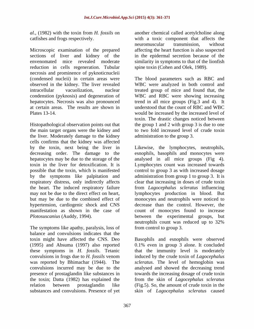
Likewise, the lymphocytes, neutrophils, esnophils, basophils and monocytes were analysed in all mice groups (Fig 4). Lymphocytes count was increased towards control to group 3 as with increased dosage administration from group 1 to group 3. It is clear that increasing in doses of crude toxin from Lagocephalus scleratus influencing lymphocytes production in blood. But monocytes and neutrophils were noticed to decrease than the control. However, the count of monocytes found to increase between the experimental groups, but neutrophils count was reduced up to 32% from control to group 3.
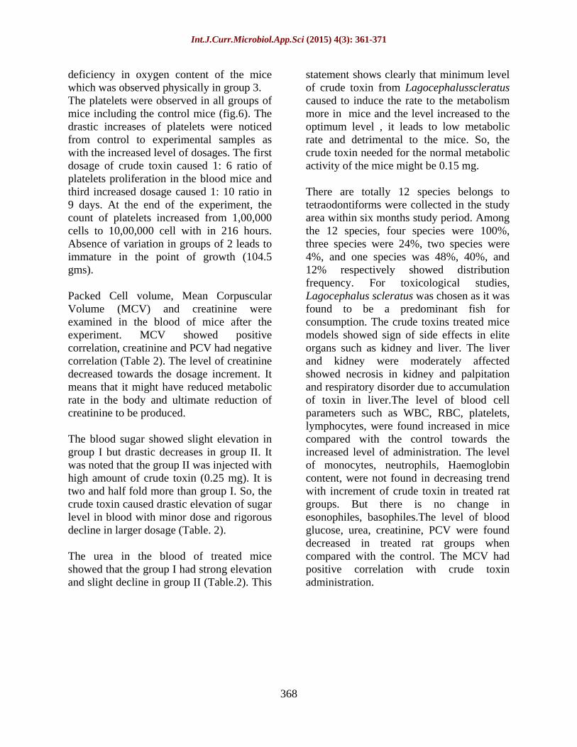
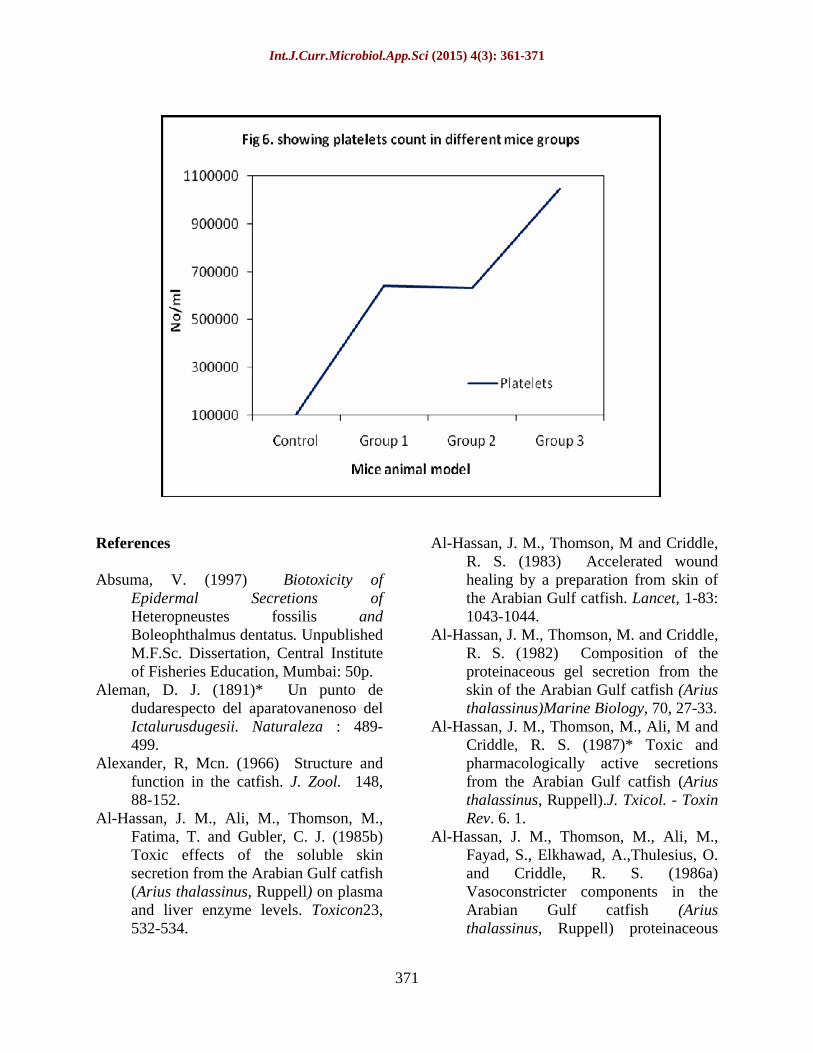
Basophils and esnophils were observed 0.1% even in group 3 alone. It concluded that the immunity level is moderately induced by the crude toxin of Lagocephalus scleratus. The level of hemoglobin was analysed and showed the decreasing trend towards the increasing dosage of crude toxin from the skin of Lagocephalus scleratus (Fig.5). So, the amount of crude toxin in the skin of Lagocephalus scleratus caused
Int.J.Curr.Microbiol.App.Sci (2015) 4(3): 361-371
368
deficiency in oxygen content of the mice which was observed physically in group 3. The platelets were observed in all groups of mice including the control mice (fig.6). The drastic increases of platelets were noticed from control to experimental samples as with the increased level of dosages. The first dosage of crude toxin caused 1: 6 ratio of platelets proliferation in the blood mice and third increased dosage caused 1: 10 ratio in 9 days. At the end of the experiment, the count of platelets increased from 1,00,000 cells to 10,00,000 cell with in 216 hours. Absence of variation in groups of 2 leads to immature in the point of growth (104.5 gms).
Packed Cell volume, Mean Corpuscular Volume (MCV) and creatinine were examined in the blood of mice after the experiment. MCV showed positive correlation, creatinine and PCV had negative correlation (Table 2). The level of creatinine decreased towards the dosage increment. It means that it might have reduced metabolic rate in the body and ultimate reduction of creatinine to be produced.
The blood sugar showed slight elevation in group I but drastic decreases in group II. It was noted that the group II was injected with high amount of crude toxin (0.25 mg). It is two and half fold more than group I. So, the crude toxin caused drastic elevation of sugar level in blood with minor dose and rigorous decline in larger dosage (Table. 2).
The urea in the blood of treated mice showed that the group I had strong elevation and slight decline in group II (Table.2). This
statement shows clearly that minimum level of crude toxin from Lagocephalusscleratus caused to induce the rate to the metabolism more in mice and the level increased to the optimum level , it leads to low metabolic rate and detrimental to the mice. So, the crude toxin needed for the normal metabolic activity of the mice might be 0.15 mg.
There are totally 12 species belongs to tetraodontiforms were collected in the study area within six months study period. Among the 12 species, four species were 100%, three species were 24%, two species were 4%, and one species was 48%, 40%, and 12% respectively showed distribution frequency. For toxicological studies, Lagocephalus scleratus was chosen as it was found to be a predominant fish for consumption. The crude toxins treated mice models showed sign of side effects in elite organs such as kidney and liver. The liver and kidney were moderately affected showed necrosis in kidney and palpitation and respiratory disorder due to accumulation of toxin in liver.The level of blood cell parameters such as WBC, RBC, platelets, lymphocytes, were found increased in mice compared with the control towards the increased level of administration. The level of monocytes, neutrophils, Haemoglobin content, were not found in decreasing trend with increment of crude toxin in treated rat groups. But there is no change in esonophiles, basophiles.The level of blood glucose, urea, creatinine, PCV were found decreased in treated rat groups when compared with the control. The MCV had positive correlation with crude toxin administration.
Int.J.Curr.Microbiol.App.Sci (2015) 4(3): 361-371
369
Table.1 showing the list of species, common name and its distribution frequency
Table.2 Level of PCV, blood glucose, urea, creatinine and MCV in different groups of mice Groups
S.No Name of the species (family) Common Name Distribution
Frequency (%)
01.
Lagocephalusscleratus (Tetroadontidae)
Silver stripe blausop puffer fish
48
02. Lagocephalusinermis (Tetroadontidae)
Smooth blaasop puffer fish 100
03. Lagocephaluslunaris (Tetroadontidae)
Green Rough-Backed puffer fish
100
04. Chelonodonpatoca (Tetroadontidae)
Milk spotted puffer fish 40
05. Torquigenerhypselogeneion (Tetroadontidae)
Orange-spotted toad fish 100
06. Arothronstellatus (Tetroadontidae)
Starry toad fish 4
07. Arothronimmaculatus (Tetroadontidae)
Immaculate puffer fish 100
08. Arothronreticularis (Tetroadontidae)
Reticulated puffer fish 12
09. Diodonholocanthus (Diodontidae)
Long-spine porcupine fish 4
10 Paramonacanthuschoirocephalus
(Monacanthidae) Pig faced leather jacket fish
24
11 Tetrosomusgibbosus (Ostraciidae)
Humpback turret fish 24
12 Triacanthusbiaculeatus (Triacanthidae)
Short-Nosed Tripos-fish 24
Mice Groups Blood Glucose (mg/dl)
PCV (%) Urea (mg/dl)
Creatinine (mg/dl)
MCV
Control 116.1 49 23.38 1 48.3
Group 1 140.2 45.7 28.39 0.9 49.7
Group 2 92 47.1 22.32 0.9 49.1
Group 3 - 42.6 - - 51.2
Int.J.Curr.Microbiol.App.Sci (2015) 4(3): 361-371
370
Int.J.Curr.Microbiol.App.Sci (2015) 4(3): 361-371
371
References
Absuma, V. (1997) Biotoxicity of Epidermal Secretions of Heteropneustes fossilis and Boleophthalmus dentatus. Unpublished M.F.Sc. Dissertation, Central Institute of Fisheries Education, Mumbai: 50p.
Aleman, D. J. (1891)* Un punto de dudarespecto del aparatovanenoso del Ictalurusdugesii. Naturaleza : 489-499.
Alexander, R, Mcn. (1966) Structure and function in the catfish. J. Zool. 148, 88-152.
Al-Hassan, J. M., Ali, M., Thomson, M., Fatima, T. and Gubler, C. J. (1985b) Toxic effects of the soluble skin secretion from the Arabian Gulf catfish (Arius thalassinus, Ruppell) on plasma and liver enzyme levels. Toxicon23, 532-534.
Al-Hassan, J. M., Thomson, M and Criddle, R. S. (1983) Accelerated wound healing by a preparation from skin of the Arabian Gulf catfish. Lancet, 1-83: 1043-1044.
Al-Hassan, J. M., Thomson, M. and Criddle, R. S. (1982) Composition of the proteinaceous gel secretion from the skin of the Arabian Gulf catfish (Arius thalassinus)Marine Biology, 70, 27-33.
Al-Hassan, J. M., Thomson, M., Ali, M and Criddle, R. S. (1987)* Toxic and pharmacologically active secretions from the Arabian Gulf catfish (Arius thalassinus, Ruppell).J. Txicol. - Toxin Rev. 6. 1.
Al-Hassan, J. M., Thomson, M., Ali, M., Fayad, S., Elkhawad, A.,Thulesius, O. and Criddle, R. S. (1986a) Vasoconstricter components in the Arabian Gulf catfish (Arius thalassinus, Ruppell) proteinaceous
Int.J.Curr.Microbiol.App.Sci (2015) 4(3): 361-371
372
skin secretion. Toxicon, 24 , 1009-1014.
Al-Hassan, J. M., Thomson, M., Al-Lahham, A. and Criddle, R. S. (1986b)* A hemolytic factor secreted from the skin of the Arabian Gulf catfish Arius thalassinus. Fed.Proc. 45, 1795.
Al-Hassan, J. M., Thomson, M., Criddle, K. R., Summers, B. and Criddle, R. S. (1985a) Catfish epidermal secretions in response to threat or injury, a novel defense response to threat or injury; a novel defense response. Marine Biology,88, 117-123.
Allman, G. J. (1840)* On the stinging property of the lesser weever fish (Trachinusvipera) . Ann. Mag. Nat. Hist. 6, 161-165.
Alnaqeeb, M. A., Al-Hassan, J. M., Ali, M., Thomson, H. and Criddle, R. S. (1989) Histological observations on organs from rabbits injected with the skin toxin of the Arabian Gulf catfish (Arius bilineatus, Valenciennes). Toxicon, 27, 789-795.
Amour, D and Smith , (1941)* J.Pharmac.Exp. Therapy, 72 :74.
AOAC (1995) Method 959.08. Seafood Toxins. Paralytic Shellfish poison. Chapter 49.Official Methods of Analysis. Association of Official Analytical Chemists, Virginia, USA: 46-48p.
Auddy, B., Alam, M. I. and Gomes, A. (1994) Pharmacological actions of the Indian catfish (Plotosuscanius, Hamilton). Ind. J. Med. Res. 99, 47-51.
Auerbach, P. S. (1987) Natural microbiological hazards of the aquatic environment. Ciin.Dermatol.5, 52-61.
Austin, L., Cairncross, K. D. and McCallum, I. A. N. (1961) Some pharmacological actions of the venom of the stone fish Synanceahorrida Arch. Int. Pharmacodyn. 131, 339-347.
Baily, R. M. and Taylor, W. R. (1950)* Schilbeodeshildebrandi, a new ameiurid catfish from Mississippi. Copeia, 1950, 31.
Bhatti, H. K (1938) The integument and dermal skeleton of Siluroidea. Trans. Zoo. Soc. Lond. 24, 1.
Bhimachar, B. W. (1944) Poison glands in the pectoral spines of two catfishes - H.fossiles(Bloch) and Plotosusarab (Forsk) with remarks on the nature of their venom. Proc. Indian Acad. Sci. 19, 65-70.
Birkhead, W. S. (1972) Toxicity of stings of Arid and Ictalurid Catfishes. Copeia, 4, 790-807.
Bottard, A. (1889) Lespoissons venimeux. Octane Doin, Paris: 198p.
Boylan, D. B. and Scheuer, P. J. (1967) Pahutoxin: a fish poison. Science.155, 52.
Burgees. W. E (1989) An Atlas of Freshwater and Marine catfishes - A priliminary survey of the Siluriformes. T. F. H. Publications, Ine, USA.
Burkholder, P. (1963) Drugs from the sea. Armed Forces Chem. J. 27, 1-8.
Calmette, A. (1908)* Venomous Animals and Antitetnus Serum Therapeutics. (Eng.Transl.) John Bale, Sons and Danielsson, Ltd, London: 403p.
Cameron, A, M. and Endean, R (1971) The axillary glands of the plotosid catfish Cnidoglanismacrocephalus. Toxicon, 9, 345-352.
Cameron, A. M. and Endean, R. (1973) Epidermal secretions and the evolution of venom glands in fishes. Toxicon,11, 401-410.
Carlisle, D. B. (1962) On the venom of the lesser weever fish, Trachinusvipera. J. Marine. Bio. Assoc. U. K. 42: 156-162.
Carlson, R. W., Schaeffer, R. C. and Russel, F. E. (1970) Studies on the venom of the California scorpion fish
Int.J.Curr.Microbiol.App.Sci (2015) 4(3): 361-371
373
Scorpaenaguttata Girard 1. Proc. West .Pharmac. Soc. 131, 110-112.
Cavazzani, E. (1892)* L' ichtyotoxique chez le "Petromyzonmarinus". Arch. Ital. Biol. 18, 182-186.
Choromanski, J. M,. Murray, T. F. and Weber, L. J. (1984) Responses of the isolated buffalo sculpin heart to stabilized venom of the lion fish (Pterosisvolitens). Proc. West. Pharmac. Soc. 27, 229-232.
Citterio, V. (1925)* L'apparatovulnerante di Amiruscatus (L).AttiSoc. Ital. Sci. Nat. 64, 1-8.
Clark, E. (1974) The Red Sea's shark proof fish. Natl. Georg. Mag. 146, 719-729.
Clark, E. and Chao, S. (1973) A toxic secretion of the Red Sea flat fish Paradachirusmarmoratus (Lacepede). Bull. Sea Fish Res.Stn.Israel, 60: 53-56.
Clarke, T. L. (1918) Some observations of fish poisoning in the Virgin Islands. West Indian Bull.17, 56-57.
Cohen, A, S. and Olek, A. J. (1989) An extract of lionfish (Pteroisvolitens) spine tissue contains acetyl choline and a toxin that affects neuromuscular transmission. Toxicon, 27, 1367-1377.
Collette, B. B. (1956) A review of the venomous toadfishes, SubfimilyThalassophyninae.Copeia, 4: 846-864.
Coutiere, H. (1899)* Poissonsvenimeux at poissonsvenemeux. These Agreg.Carre et Naud, Paris: 220p.
Datta, A., Gomes, A., Sarangi, B., Kar, P. K and Lahiri, S. C. (1982)* Pharmacodynamic actions of crude venom of the Indian catfish H.fossilis. Ind. J. Med. Res. 76, 892.
Day, F. (1889) The Fauna of British India including Ceylon and Burma: Fishes. Vol-1. London Taylor and Francis: 548p.
Deo, D. A. (1995) Biotoxins from the Freshwater Catfish Heteropneus tesfossilis (Bloch,1794). Unpublished M.Sc. Dissertation, Central Institute of Fisheries Education, Bombay: 53p.
Duges, A. (1891)* Apartovenenoso del barge (I. dugesi, Bean). Naturaleza, 2, 1: 405-408.
Evans, H. M. (1907a)* Observation on the poisonous species of the weever fish (Trichinusdraco) Brit. Med. J. 1, 73-76.
Evans, H. M. (1907b)* Observation on the poisonous species of the weever fish (Trachinusdraco). Trans. Norfolk. Norwich. Nat. Soc. 8, 355-368.
Evans, H. M. (1920). The poison of the spiny dog fish.Brit. Med .J. 1 (3087), 287-288.
Evans, H. M. (1923) The defensive spines of fishes, living and fossil, and the glandular structure in connection there with observations on the nature of fish venoms. Phil. Trans. Roy. Soc. London. B. 212, 1-33.
Felix, S., Rachel, I., and Venugopalan, A. T. (1994) Catfish-its scope in the growing industrial aquaculture of India. Seafood Export Journal, 25, 25-33.
Fletcher, T. C. and Grant, P. T. (1969)* Immunoglobins in the serum and mucous of plaice, Pleuronectes platessa.Biochem. J. 115, 65.
Gail, R. and Rageau, T. (1956)* Premieressur un poison marine venimeuxdeba Nouvelle caledonie la synacee (Synaneejaverrucossa,Bloch). Bull. Soc. Pathol. Ecotique, 49, 846-854.
Gemter.A. (1864)* Catalogue of the Fishes of the British Museum, 7: 1-52.
Halstead, B. W. (1980) Dangerous Marine Animals. Cornell Maritime Press, Centreville, New York.
Int.J.Curr.Microbiol.App.Sci (2015) 4(3): 361-371
374
Halstead, B. W. and Courville, D. A. (1967)
Poisonous and Venomous Marine Animals of the World, Vol. 2. U.S. Govt. Printing Office, Washington. D. C: 1006p.
Halstead, B. W. and Courville, D. A. (1970) Poisonous and Venomous Marine Animals of the World.Vol. 3. U.S. Govt. Printing Office, Washington. D. C: 1006p.
Halstead, B. W. and Smith, R. L. (1954)* Presence of an axillary venom gland in the oriental catfish Plotosuslineatus.Copeia, 1954, 153.
Halstead, B. W., Kuninbbu, L. S and Herbard, H. G. (1953) Catfish stings and the venom apparatus of the Mexican catfish Galeichthysfelis(Linnaeus). Trans. Am. Microscop. Soc. 72, 297-314.
Hashimoto, Y. (1979) Marine Toxins and other Bioactive Marine Metabolites. Japan Scientific Societies Press. Tokyo: 369p.
Hashimoto, Y. and Kamiya, H. (1969)* Occurrence of toxic substances in the skin of sea bass, Pogonopercapunctata. Toxicon, 7: 65.
Hashimoto, Y. and Oshima, Y. (1972)* Separation of grammistins A, B and C from soapfishPogonoporepunctata. Toxicon10, 279.
Herre, A. W. (1952) A review of the scorpaenoid fishes of Philippines and adjacent seas. Philippine. J. Sci. 80, 381-482.
Hori, K., Fusetani, N., Hashimoto, K., Aida, K and Randall, J. E. (1979) Occurence of a grammistin like mucous toxin in the cling fish Diademichthyslineatus. Toxicon, 17, 418-424.
In:Ichthyology. John Wiley and Sons.Inc.,New York: 506p.
Jhingran, V. G. (1991) Fish and Fisheries of India. Hindustan Publishing Corporation, Delhi: 727p.
Kitzan, S. M. and Sweeney, P. R. (1968)* A light electron microscope study of the structure of Protopterusannectens epidermis. I. Mucus production. Can. J. Zool. 46, 787.
Kobert, R. (1894)* Compendium der praktischentoxikologie, p: 90-93. F. Enke, stuttgart.
Kobert, R. (1902)* Uehergiftfische und fischgifte. Med. Woche(19): 199-201.
Lagler, K. F., Bardach, J. E., Miller, R. R. and Passiono, D. M. (1977)
Lahiri, S. B.,.Purohit, S. K. and Ghosal, A. K. (1983) Electrophoraetic patterns of the crude extract and serum of the Indian venomous catfish H.fossilis(Bloch). Indian J. Amim. Sci. 53, 693-695.
Liang, S. P. and Pan, X. (1995) Lectin-like peptide isolated from the venom of the Chinese bird spider Selenocosmiahuwena. Toxicon, 33, 876-879.
Liguori, V. R., Ruggieri, G. D., Baslow, M. H., Stempien, M. F. and Nigrelli, R. E. (1963)* Antibiotic and toxic activity of the mucous of the Pacific golden striped bass Grammistessexlineatus. Am. Zool. 3, 546.
Maretzki, A., Del Castillo J. (1967)* A toxin secreted from soapfish, Ripticussaponaceus. A preliminary report.Toxicon,4 : 245.
Marwick, C. (1998) Nature's Agents Help Heal Humans-Some Now Take Steps to Reciprocate. JAMA.Medical News and Perspective: 279. No.21, p. 1679 - 1681.
Nair, M. S. R., Leong, I. and Nayar. M. S. B. (1982) Ichthyotoxins from the oyster toad fish Opsantus tau (Linnaeus). Toxicon, 20, 933-935.
Nigrelli, R. F. (1935)* On the effect of fish mucus on Epibdellamelleni, a monogenetic trematode of marine fishes.J. Parasit.21, 438.
Int.J.Curr.Microbiol.App.Sci (2015) 4(3): 361-371
375
Nigrelli, R. F. (1937)* Further studies on
suceptability and acquired immunity of marine fishes to Epibdellamellehi,a monogenetic trematode. Zoologica, 22, 185.
Nigrelli, R. F., Jakowska, S. and Padnos, M. (1955) Pathogenecity of epibionts in fishes. J. Protozool. 2, 7.
Noguchi, T., Kao, H. and Hashimato, Y. (1971) Toxicity of the goby, Gobiuscriniger. Bulletin of the Japanese Society of ScientificFisheries,37, 642-647.
Norman, J. R. (1931) A History of Fishes, Ernest Benn Ltd. London. 463p.
Norman, J. R. and Greenwood, P. H. (1975) A History of Fishes. Ernest BennLtd. London: 467p.
Pani Prasad, K. and Venkateshvaran, K. (ed) (1997) Training Manual on AdvancedTechniques in Marine Biotoxinology. Central Institute Of Fisheries Education, Mumbai: 70p.
Patten, B. M. (1975)* More on catfish stings.JAMA, 232: 248p.
Pawlowsky, E. N (1914)* Uber den bau der giftdrusenbeiPlotosus und anderenfischen, Zool. Jahrb. 38, 427-442.
Pawlowsky, E. N. (1913)* Sur La stracture des glandes avenin de certainspoissons t en particular de celles de Plotosus. Compt. Rend. Soc. Biol. 74, 1033-1036.
Pawlowsky, E. N. (1927)* Gifttiere und ihregiftigkeit.Gustav Fischer, Jena: 515p.
Peterson, G. L. (1977) A simplification of the protein assay method of Lowry et al. which is more generally applicable. Analyt.Biochem.83, 346-356.
Phisalix, M. (1922)* Animauxvenimeux et venims.Ivol.Masson et cie. Paris.
Porta, A. (1905)* Ricercheanatomichesull' apparecchiovelenifero di alcunipesci. Anat. Anz. 26, 232-247.
Primor, N. and Zlotkin, E. (1975) Ichthyotoxic and hemolytic action of the skin secretion of flatfish, Paradacharismarmoratus (Solidae). Toxicon, 13, 227-231.
Prokhoroff, P. (1884)* On the poisonous character of certain lampreys. (In Russian) Vrach. St. Petersb. 5, 54-55.
Randall, J. E. (1967)* Food habits of reef fishes of the West Indies. Stud. trop. Oceanogr. Miami, 5, 665.
Randall, J. E., Aida, K., Hibiya, T., Mitsuura, N., Kamiya, H. and Hashimoto, Y. (1971)* Grammistin ,the skin toxin of soapfishes, and its significance in the classification of the Grammistidae. Publs.Seto. Mar. Bio. Lab. 19: 157.
Randall, J. E., Aida, K., Oshima, Y., Hori. K. ,Hashimoto. Y. (1981) Occurrence of a crinotoxin and hemagglutination in the skin mucous of the moray eel Lyncodontusnudivomer. Marine Biology.62, 179-184.
Ray, C. and Coates, C. W. (1958)* A case of poisoning by the lionfish, Pteroisvolitans. Copeia, 1958, 235.
Reed, H. D. (1900)* The structure of the poison glands of Schilbeodesgyrinus. Proc. Am. Sci. 232-233.
Reed, H. D. (1906)* Notes on the poison organs in fishes .Science,24, 293.
Reed, H. D. (1907)* The poison glands of Noturus and Schilbeodes. Am. Naturalist.41, 553-556.
Reed, H. D. (1924a)* The morphology and growth of the spines of siluroid fishes. J. Morphol. 38, 431-451.
Reed, H. D. (1924b)* The morphology of the dermal glands in nematognathus fishes. Z. Morphol. Anthoropol.24, 227-264.
Reed, H. D. and T. J. Lloyd. (1916)* The nature of the spines in catfishes. Trans. Am. Fish. Soc. 45, 202-205.
Int.J.Curr.Microbiol.App.Sci (2015) 4(3): 361-371
376
Rosen, M. W. and Cornford, N. E. (1971)
Fluid secretion of fishe slimes. Nature,Lond. 234, 49.
Russel, F. E. (1953) The sting ray. .Eng. Sci. 17, 15-18.
Russel, F. E. and Emery, J. A. (1960) Venom of the weevers Trachinusdraco and T. vipera. Ann. N.YAcad. Sci. 90: 805-819.
Russel, F. E., Fairchild, M. D., Michaelson, J. (1958) Some properties of the venom of the stingray. Med. Arts. Sci. 12, 78-86.
Saunders, P. R. (1959) Venom of the stonefish, Synanacejaverrucosa. Science,29, 272-274.
Saunders, P. R. and Taylor, P. B. (1959) Venom of the lionfish Pteroisvolitans. Am. J. Physiol. 197, 437-440.
Shephard, K. L. (1994) Functions for fish mucus. Reviews in Fish Biologyand Fisheries, 4, 401-429.
Shiomi, K., Takamiya, M., Yamanaka, H., Kikuchi, T. and Suzuki, Y. (1988) Toxins in the skin secretion of the oriental catfish (P.lineatus). Immunological properties and immuno-cytochemical identification of producing cells.Toxicon, 26, 353-361.
Shiomi. K., Takamiya, M., Yamanaka, H., Kikuchi, T. and Konno, K. (1986)* Hemolytic, lethal and edema forming activities of the skin secretion from oriental catfish (Plotosuslineatus). Toxicon, 24, 1015.
Starks, E. C. (1930)* The primary shoulder girdle of the bony fishes. StanfordUniv. Publ. Biol. Ser. 6, !-92.
Talwar, P. K and Jhingran, A. G. (1991) Inland Fishes of India and AdjacentCountries. Vol2. Oxford and IBH ,Co.Pvt, Ltd. New Delhi: 689p.
Tange, Y. (1955)* Beitargzurkenntnis der morphologie des giftapparatesbei den japanischenfischen, XV, Uber den
giftapparatbeiP.anguillaris(Lacepade). Yokohama Med. Bull.6, 424-437.
Thomson, D. A. (1964) Ostracitoxin: An ichthyotoxic stress secretion of the boxfish Ostracionlentiginosus, Science, 146, 244-255.
Thomson, M., Al-Hassan, J. M., Fayad, S., Al-Saleh, J. and Ali, M. (1998) Purification of a toxic factor from Gulf catfish epidermal secretions. Toxicon, 36, 859-865.
Toyoshima, T. (1918) *Serological study of toxin of the fish P.anguillarisLacepade. (In Japanese) J, Japan Protoz. Soc. 6, 45-270.
Whitear, M. (1970) The skin surface of bony fishes. J. Zool. Lond. 160, 437-454.
Wiener, S. (1959) The production and assay of stonefish antivenom. Med. J. Australia. 2, 715-719.