Embed Size (px)
Citation preview

Taxonomic studies in the Schizymeniaceae (Nemastomatales, Rhodophyta): on the
identity of Schizymenia sp. in the Azores and the generic placement of
Nemastoma confusum
DANIELA GABRIEL1,3*, TOM SCHILS
4, MANUELA I. PARENTE2,5, STEFANO G.A. DRAISMA
6,7, ANA I. NETO2,8
AND SUZANNE FREDERICQ3
1CIBIO-Acores, 2Departamento de Biologia, Universidade dos Acores, 9501-801 Ponta Delgada, Acores, Portugal3Department of Biology, University of Louisiana at Lafayette, Lafayette, LA 70504-2451, USA
4Marine Laboratory, University of Guam, Mangilao, GU 96923, USA5Departamento de Ciencias e Engenharia do Ambiente, Instituto do Mar – IMAR, Faculdade de Ciencias e Tecnologia,
Universidade Nova de Lisboa, Quinta da Torre, 2829-516 Caparica, Portugal6Nationaal Herbarium Nederland – Universiteit Leiden, 2333 CC Leiden, The Netherlands
7Institute of Ocean & Earth Sciences, University of Malaya, Kuala Lumpur 50603, Malaysia8CIIMAR (Centro Interdisciplinar de Investigacao Marinha e Ambiental), Universidade do Porto, Rua dos Bragas,
289-4050-123 Porto, Portugal
GABRIEL D., SCHILS T., PARENTE M.I., DRAISMA S.G.A., NETO A.I. AND FREDERICQ S. 2011. Taxonomic studies in theSchizymeniaceae (Nemastomatales, Rhodophyta): on the identity of Schizymenia sp. in the Azores and the genericplacement of Nemastoma confusum. Phycologia 50: 109–121. DOI: 10.2216/09-67.1
Comparative rbcL sequence analysis indicates that the species going under the name Schizymenia dubyi in the Azoresshould be referred to as S. apoda. Sequences of Schizymenia specimens from China and Namibia were also identified asS. apoda, of which the type locality is the Cape Province in South Africa. Schizymenia dubyi, described from AtlanticFrance, is clearly a distinct species that we here report for Japan and Sicily in the Mediterranean Sea. Both Schizymenia
species, along with an unreported species from Japan, are distinct from S. pacifica described from Washington, in thePacific Coast of North America. Secondary pit connections were observed in gametophytes of S. apoda from the Azores,a previously unknown character for the Nemastomatales. Examination of type material of Nemastoma confusum
indicates that this species, currently placed in the Nemastomataceae, should be transferred to the genus Platoma in theSchizymeniaceae. A morphological comparison between Platoma confusum (Kraft & John) comb. nov. with descriptionsof P. cyclocolpum and P. chrysymenioides suggests that the three species are closely related.
KEY WORDS: Azores, Nemastoma, Nemastomatales, Platoma, rbcL, Rhodophyta, Schizymenia, Schizymeniaceae,Systematics, Taxonomy
INTRODUCTION
Schizymenia J. Agardh (Schizymeniaceae) has the largest
geographical distribution of all nemastomatalean genera.
The 10 species of Schizymenia that are currently recognised
are found in temperate, subtropical and tropical waters,
ranging from the subantarctic islands to Alaska (Guiry &
Guiry 2009). The genus Schizymenia is distinguished from
other genera in the Schizymeniaceae by the presence of
diagnostic gland cells resembling large, inverted tears,
located terminally on cortical filaments (Dixon & Irvine
1977).
The type of the genus, Schizymenia dubyi (Chauvin ex
Duby) J. Agardh, was established by Agardh based on
Halymenia dubyi Chauvin ex Duby from Atlantic France,
and it is the most studied and best-characterised member of
the Schizymeniaceae. The genus also contains the only
known tetrasporophyte of Schizymeniaceae found in nature
(Masuda & Guiry 1994). Extensive culture studies per-
formed on S. dubyi (Ardre 1977, 1980) and S. pacifica
(Kylin) Kylin (DeCew et al. 1992) revealed a heteromorphic
life history where the tetraspophytic phase was identified as
a crustose species originally attributed to the generitype of
Haematocelis, H. rubens J. Agardh.
Kylin (1956) circumscribed the genus Schizymenia as
follows: ‘with blade-like thalli that bear gland cells in the
cortex, produce carpospores towards the exterior of the
thallus, with the base of the carposporophyte lying deep
inside the medulla, and in which nearly all cells of the
gonimoblasts become carpospores. Additional characters
include weakly developed sterile filaments, or lack thereof,
surrounding the carposporophyte, and a weakly defined
but distinct ostiole located above the slightly raised
carposporangial mass’. The shape and size of the blades,
along with vegetative features such as short cortical branch
systems with subspherical inner cells, may also serve to
distinguish species (Abbott 1967; Womersley 1994).
The species of Schizymenia occurring in the Azores were
originally reported by Trelease (1897) as S. undulata J.
Agardh (Terceira Island) and S. obovata J. Agardh (Corvo
Island), both species currently regarded as synonyms of S.
dubyi, described from Atlantic France, and S. apoda (J.
Agardh) J. Agardh described from Atlantic South Africa,* Corresponding author ([email protected]).
Phycologia (2011) Volume 50 (2), 109–121 Published 3 March 2011
109

respectively (Guiry & Guiry 2009). After extensive studies
on the Azorean marine flora, Neto (1994) reported that
only S. dubyi occurred in this archipelago.
The macroalgal communities of the Azores have been
studied since the mid-1800s (Seubert 1844), but early
publications were restricted to species inventories. Since
Feldmann (1942), more attention has been given to the
ecology of Azorean macroalgae, and several papers have
been published by Neto and coauthors over the past
15 years (see Tittley et al. 2001, 2009; Tittley & Neto 2005).
Life history studies and morphological information (Par-
ente et al. 2003a, b; Toste et al. 2003a, b; Gabriel et al. 2009,
2010) are scanty. The floristic studies reveal a mixed flora
with a strong component of cold-water species together
with a few tropical and subtropical taxa (Neto 1997).
The recently resurrected order Nemastomatales (Kylin)
Saunders & Kraft comprises two families, the Nemasto-
mataceae and Schizymeniaceae (Masuda & Guiry 1995;
Saunders & Kraft 2002; Gavio et al. 2005), both
represented in the marine flora of the Azores. Nemastoma
confusum Kraft & John (Fredericq et al. 1992; Neto 1994),
Itonoa marginifera (J. Agardh) Masuda & Guiry (Larkum
1960; Neto 1994) and Predaea feldmanii Børgesen (Freder-
icq et al. 1992; Neto 1994; Tittley & Neto 1994) are the
three reported species of the Nemastomataceae. Platoma
cyclocolpum (Montagne) F. Schmitz (South & Tittley 1986;
Neto 1991, 1994) and S. dubyi (Schmidt 1929; Palminha
1957; South & Tittley 1986; Neto 1994; Tittley & Neto
1994) are the listed species of the Schizymeniaceae.
The red algal genus Nemastoma J. Agardh (1842)
currently comprises nine species predominantly inhabiting
tropical to subtropical waters worldwide (Huisman 1999;
Rodriguez-Prieto et al. 2004). Despite its wide distribution
range, representatives of the genus are not abundantly
present in the respective floras, and most of the species are
known only from their original description (Kraft & John
1976). The reproductive features of Nemastoma species
other than those described in N. dichotomum J. Agardh
have been poorly studied and require re-examination
(Masuda & Guiry 1994). Nemastoma confusum was
described by Kraft & John (1976) based on gelatinous
specimens from Ghana. The specific epithet alludes to
initial misidentification of the species as Predaea feldmannii
when cystocarps could not be observed.
As part of a revision of the Schizymeniaceae, the present
paper reports on a molecular-based reappraisal of Schizy-
menia species and provides morphological and ecological
observations of the Azorean Schizymenia species. We also
present reasons for transferring N. confusum to the genus
Platoma.
MATERIAL AND METHODS
Algal material used in the present study was collected since
1990 at different sites of the Archipelago of the Azores.
Collections were made from April to September, in the
intertidal. Reference collections were made by storing
samples in a 5% formalin/seawater solution, pressed as
herbarium sheets or dried in silica gel. Collections are
deposited in the Herbarium of the Department of Biology,
University of the Azores. Additional samples of Nemasto-
matales from the Ghent University Herbarium (GENT,
Belgium) and the Herbarium of the University of Louisiana
at Lafayette (LAF, USA) were used for comparison.
The observations of N. confusum presented in this work
are based on five well-preserved slides of the isotype from
Ghana (John no. 7260) used for the original description of
this species (Kraft & John 1976, p. 332). The material was
obtained through the kindness of Dr Gerry Kraft
(University of Melbourne Herbarium – MELU) as a loan
to Dr Willem Prud’homme van Reine from the National
Herbarium of the Netherlands, Leiden (L).
A combination of stereo- and compound microscopes
was used to describe morphological and anatomical
characters and reproductive structures for the diagnosis.
Species identifications were based on the original descrip-
tions and on a critical analysis of the literature. Represen-
tative collections of microscope slides and photomicro-
graphs were made. Microscope slides of squash mounts
were prepared from liquid-preserved material, stained with
1% aniline blue with HCL acidification, and mounted in
50% Karo corn syrup–water solution (containing a few
drops of 2% phenol). Photomicrographs were taken using
various digital cameras connected to light microscopes.
Measurements of cells and other structures were made
using a micrometer eye piece (presented in the text as length
per width).
DNA samples were prepared using the DNAeasy Plant
Minikit (Qiagen, Valencia, CA). Silica gel dried specimens
and extracted DNA samples are deposited at LAF and
stored at 220uC. Plastid-encoded rbcL was selected to infer
a phylogeny of Schizymenia and related nemastomatalean
genera. Protocols for DNA extraction, gene amplification
and sequencing are described in Gavio & Fredericq (2002).
PCR primers (F7-R753, F57-R557, F645-R1150, F993-
RrbcSstart) and sequencing primers (F7, F57, F645, F993,
R376, R557, R753, R1150, RrbcSstart) are listed in Lin et
al. (2001) and Gavio & Fredericq (2002).
A total of 24 rbcL sequences were used in this study,
including newly generated sequences of which the vouchers
are located at LAF, and of sequences downloaded from
GenBank. A data set of available members of Schizymenia
was assembled, and two other taxa in the Schizymeniaceae,
P. cyclocolpum and Titanophora pikeana (Dickie) Feld-
mann, were added as the out-group based on a global
phylogenetic analyses of the Nemastomatales (Gavio et al.
2005). The information about the taxa, collection sites and
collectors is listed in Table 1.
The generated rbcL sequences were compiled, edited and
aligned using Sequencher software (Gene Codes Corp.,
Ann Arbor, MI, USA) and exported for phylogenetic
analysis in PAUP* v.4.0 beta 10 (Swofford 2003) and
MacClade v.4 (Maddison & Maddison 2000).
Phylogenetic analyses were conducted with the maximum
parsimony (MP) and maximum likelihood (ML) algorithms
as implemented in PAUP and PhyML v2.4.4 (Guindon &
Gascuel 2003), respectively, and the Bayesian inference as
implemented in MrBayes 3.0 (Hall 2001; Huelsenbeck &
Ronquist 2001). Parsimony trees obtained under the Fitch
criterion of equal weights for all substitutions (Fitch 1971)
110 Phycologia, Vol. 50 (2), 2011

Table
1.
Lis
to
fsp
ecie
su
sed
inth
erb
cLse
qu
ence
an
aly
sis
wit
hco
llec
tio
nin
form
ati
on
an
dG
enB
an
kacc
essi
on
nu
mb
er.
Sp
ecie
sC
oll
ecti
on
iden
tifi
cati
on
Co
llec
tio
nlo
cali
tyC
oll
ecti
on
data
Gen
Ban
kacc
essi
on
nu
mb
er
Pla
tom
acyclo
colp
um
(Mo
nta
gn
e)S
chm
itz
SM
G-0
4-2
02
Po
nta
Garc
a,
Vil
aF
ran
ca,
Sao
Mig
uel
,A
zore
s(8
-md
epth
)D
.G
ab
riel
&P
.M
ad
eira
,20
Au
g.
2004
FJ8
68809
Sch
izym
enia
apoda
(J.
Agard
h)
J.A
gard
hL
AF
-7-6
-93-1
-1S
wak
op
mu
nd
,N
am
ibia
M.H
.H
om
mer
san
d,
6Ju
l.1993
AY
294401
Sch
izym
enia
apoda
(J.
Agard
h)
J.A
gard
hL
AF
-6-9
4-1
-1T
aip
ing
Cap
e,S
han
do
ng
pro
v.,
Ch
ina
M.H
.H
om
mer
san
d,
Jun
.1994
AY
294392
Sch
izym
enia
apoda
(J.
Agard
h)
J.A
gard
hG
RW
-04-8
7B
arr
oV
erm
elh
o,
Gra
cio
sa,
Azo
res
D.
Gab
riel
,10
Jun
.2004
FJ8
78860
Sch
izym
enia
apoda
(J.
Agard
h)
J.A
gard
hG
RW
-04-8
8B
arr
oV
erm
elh
o,
Gra
cio
sa,
Azo
res
D.
Gab
riel
,10
Jun
.2004
FJ8
78861
Sch
izym
enia
apoda
(J.
Agard
h)
J.A
gard
hG
RW
-06-7
86
Baıa
da
Fo
nte
,G
raci
osa
,A
zore
s9
Jul.
2006
FJ8
78862
Sch
izym
enia
apoda
(J.
Agard
h)
J.A
gard
hS
MG
-04-1
64
Pra
iad
oP
op
ulo
,S
ao
Mig
uel
,A
zore
sM
.I.
Pare
nte
,14
Jun
.2004
FJ8
78863
Sch
izym
enia
apoda
(J.
Agard
h)
J.A
gard
hS
MG
-04-1
65
Pra
iad
oP
op
ulo
,S
ao
Mig
uel
,A
zore
sM
.I.
Pare
nte
,14
Jun
.2004
FJ8
78864
Sch
izym
enia
apoda
(J.
Agard
h)
J.A
gard
hS
MG
-05-1
45
Sao
Ro
qu
e,S
ao
Mig
uel
,A
zore
sD
.G
ab
riel
,9
Au
g.
2005
FJ8
78865
Sch
izym
enia
apoda
(J.
Agard
h)
J.A
gard
hS
MG
-05-2
26
Mo
stei
ros,
Sao
Mig
uel
,A
zore
sD
.G
ab
riel
&M
.I.
Pare
nte
,07
Sep
.2005
FJ8
78866
Sch
izym
enia
apoda
(J.
Agard
h)
J.A
gard
hS
MG
-05-2
59
Sao
Vic
ente
,S
ao
Mig
uel
,A
zore
sD
.G
ab
riel
,21
Sep
.2005
FJ8
78867
Sch
izym
enia
apoda
(J.
Agard
h)
J.A
gard
hS
MG
-06-8
4S
ao
Mig
uel
,A
zore
sD
.G
ab
riel
,2006
FJ8
78868
Sch
izym
enia
apoda
(J.
Agard
h)
J.A
gard
hP
G07220
Pra
iad
oP
op
ulo
,S
ao
Mig
uel
,A
zore
sD
.G
ab
riel
&M
.P
are
nte
2007
GQ
495983
Sch
izym
enia
dubyi
(Ch
au
vin
exD
ub
y)
J.A
gard
hL
AF
-6-2
2-9
3-1
-1P
igh
et,
Bri
ttan
y,
Fra
nce
J.C
ab
ioch
,22
Jun
.1993
AY
294389
Sch
izym
enia
dubyi
(Ch
au
vin
exD
ub
y)
J.A
gard
hL
AF
-7-1
0-9
4-1
-1Jo
do
gah
am
a,
Miy
ak
o,
Iwate
-ken
,Ja
pan
M.H
.H
om
mer
san
d&
M.
Yo
shiz
ak
i,10
Jul.
1994
AY
294388
Sch
izym
enia
dubyi
(Ch
au
vin
exD
ub
y)
J.A
gard
hL
AF
-7-1
0-9
4-1
-2Jo
do
gah
am
a,
Miy
ak
o,
Iwate
-ken
,Ja
pan
M.H
.H
om
mer
san
d&
M.
Yo
shiz
ak
i,10
Jul.
1994
FJ8
78869
Sch
izym
enia
dubyi
h(C
hau
vin
exD
ub
y)
J.A
gard
PG
08865
Mo
nd
ello
,S
icil
y,
Italy
M.
Pare
nte
&R
.S
ou
sa2008
GQ
495984
Sch
izym
enia
dubyi
(Ch
au
vin
exD
ub
y)
J.A
gard
hP
G08887
Mo
nd
ello
,S
icil
y,
Italy
M.
Pare
nte
&R
.S
ou
sa2008
GQ
495985
Sch
izym
enia
paci
fica
(Kyli
n)
Kyli
nL
AF
-7-2
6-9
5-1
-1K
an
ah
aB
ay,
W.
Juan
Isla
nd
,W
A,
US
AM
.J.
Wyn
ne,
26
Jul.
1995
AY
294393
Sch
izym
enia
paci
fica
(Kyli
n)
Kyli
nL
AF
-4-1
5-9
4-1
-1V
an
cou
ver
,B
riti
shC
olu
mb
ia,
Can
ad
a(d
rift
)S
.C.
Lin
dst
rom
,15
Ap
r.1994
AY
294394
Sch
izym
enia
paci
fica
(Kyli
n)
Kyli
nL
AF
-7-2
4-9
8-1
-1(T
C101)
No
rth
Bo
ard
man
St.
Park
,O
R,
US
AT
.O.
Ch
o&
G.I
.G
ayle
,24
Jul.
1998
FJ8
78870
Sch
izym
enia
paci
fica
(Kyli
n)
Kyli
nL
AF
-7-2
4-9
8-1
-2(T
C130)
No
rth
Bo
ard
man
St.
Park
,O
R,
US
AT
.O.
Ch
o&
G.I
.G
ayle
,24
Jul.
1998
FJ8
78871
Sch
izym
enia
sp.
cf.
novae-z
elandia
eJ.
Agard
hL
AF
-9-5
-93-1
-1T
ok
aw
a,
Ch
osh
i,C
hib
aP
ref.
,Ja
pan
S.
Fre
der
icq
&M
.Y
osh
izak
i,2
Sep
.1993
AY
294391
Tit
anophora
pik
eana
(Dic
kie
)J.
Fel
dm
an
nL
AF
-2-1
1-0
1-1
-1D
eep
Sp
on
ge
Ree
f,S
od
wan
aB
ay,
Kw
aZ
ulu
-Nata
l,S
ou
thA
fric
a(2
7-m
dep
th)
S.
Fre
der
icq
&O
.D
eCle
rck
,11
Feb
.2001
AY
294364
Gabriel et al.: Schizymenia sp. in the Azores and ‘Nemastoma’ confusum 111

were inferred from a heuristic search, excluding uninfor-
mative characters consisting of 1000 random sequence
additions holding 10 trees at each step, MULPARS and
tree-bisection-reconnection (TBR) algorithms with the
MULTREES (saving multiple trees) and STEEPEST
DESCENT option. Support for nodes in the MP and ML
analyses were assessed by calculating bootstrap proportion
values (Felsenstein 1985) based on 1000 and 100 resam-
plings, respectively, retaining groups with a frequency
greater than 50%, MULPARS and TBR algorithms.
The optimal models of sequence evolution to fit the data
alignment estimated by hierarchical likelihood ratio tests
performed by Modeltest v.3.6 (Posada & Crandall 1998)
was the GTR + I + G (general time reversible model with a
proportion of invariable sites and gamma distribution split
into four categories) for the data set. The Akaike
information criterion parameters applied in the likelihood
analysis were as follows: assumed nucleotide frequencies
A 5 0.3045; C 5 0.1632; G 5 0.2103; T 5 0.3220;
substitution rate matrix A–C substitutions 5 0.0, A–G 5
6.0541, A–T 5 2.3094, C–G 5 0.8317, C–T5 17.2925, G–T
5 1.0; proportion of sites assumed to be invariable 5 0.485;
and rates for variable sites assumed to follow a gamma
distribution with shape parameter 5 0.625.
For the Bayesian analysis, four chains of the Markov
chain Monte Carlo were run, sampling one tree every 100
generations for 2,000,000 generations starting with a
random tree for the rbcL data sets, with the following
parameters applied: nst 5 6 and Rates 5 invgamma. The
first 20,000 generations were discarded as the ‘burn-in’
period to reach equilibrium. A 50% consensus tree
(majority rule as implemented by PAUP) was computed
from the trees saved after the burn-in point. Reliability of
the Bayesian consensus tree is given by the frequency at
which each node appears among all saved trees after the
burn-in generation. This frequency corresponds to the
posterior probability of the clades (Hall 2001).
RESULTS
Schizymenia ‘dubyi’ (Chauvin ex Duby) J. Agardh (1851,
p. 171) from the Azores
HABIT AND VEGETATIVE STRUCTURE: Plants foliose, liver-
red to brownish-red in colour, simple or broadly lanceolate
with irregular lobes and proliferations from the margins,
sometimes undulate, soft fleshy, and slippery (Figs 1–4), up
to 20 cm tall. The occasionally perforated blades are
attached with a short stipe to the substratum by a small
discoid holdfast, from which other blades can emerge,
usually growing within dense assemblages of geniculate
Corallinaceae (Fig. 4). Cystocarps on the blade surface are
easily distinguished by visible ostioles.
The cortex is composed of anticlinal fascicles of
dichotomously branched moniliform filaments (Figs 5–8,
10), with occasionally darkly staining elongated gland cells
(Fig. 6, arrow) reaching the thallus surface. Innermost
cortical cells are spherical, surface cells are more elongated
(Figs 5–8). Inner cortical cells laterally produce primordia
that extend into medullary rhizoidal filaments (Figs 6, 7).
Occasionally, inner cortical cells are linked by secondary pit
connections (Fig. 8, arrow). The medulla is composed of
primary filiform filaments as well as secondarily formed
rhizoidal filaments that are laxly to densely interwoven with
one another and that may undergo localised swellings
(Fig. 9).
PRE- AND EARLY POSTFERTILISATION STRUCTURES: Thalli
are monoecious. Carpogonial branches (Figs 10, 11)
develop outwards from an inner cortical cell bearing
subdichotomies of cortical filaments. The generative
cortical cell becomes the supporting cell of a three-celled
carpogonial branch (Fig. 10). With maturation, a straight
trichogyne develops from the carpogonium growing to-
wards the thallus surface (Fig. 11). Simultaneously, the
cortical cells linked to the supporting cell expand in size and
become dark staining subsidiary auxiliary cells (a.k.a.
nutritive auxiliary cells; Fig. 11).
POSTFERTILISATION STRUCTURES: The formation of con-
necting filaments was not observed. The generative auxiliary
cell cuts off same-sized gonimoblast cells that are surround-
ed by a loose network of vegetative filaments linked to the
auxiliary cell that form an involucrum-like structure around
the carposporophyte (Fig. 12, arrows). A surface ostiole
develops at the distal end of the gonimoblast (Fig. 13).
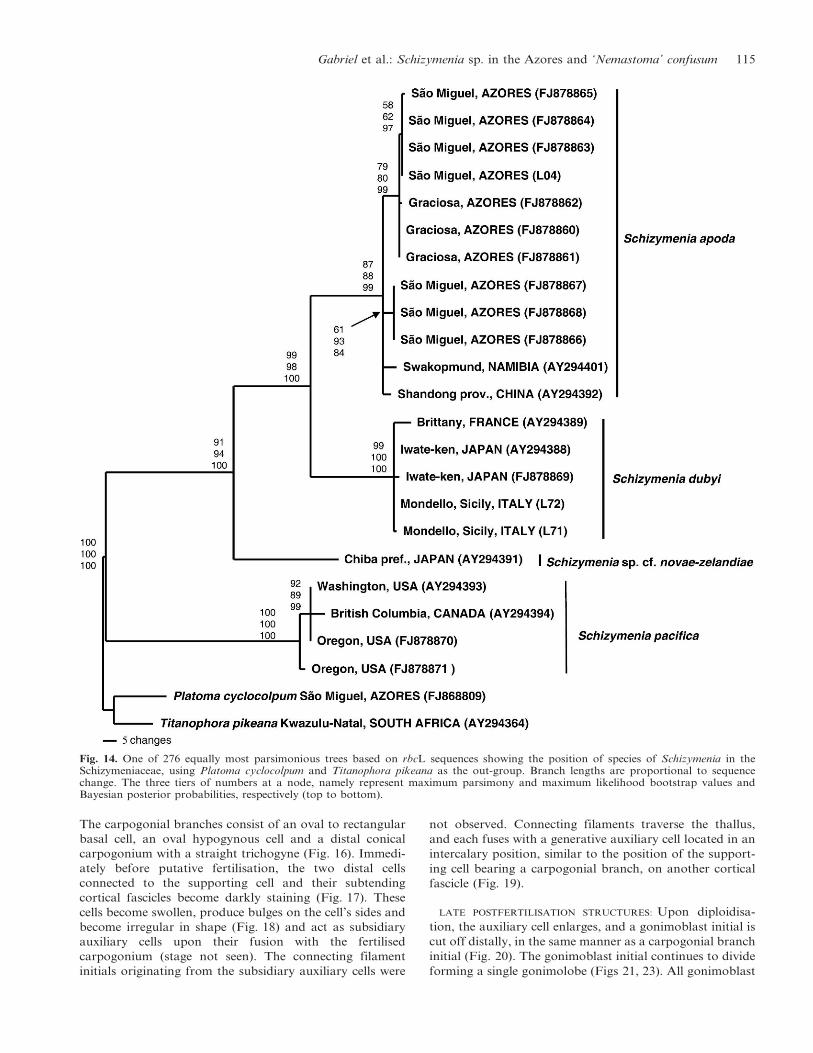
MOLECULAR EVIDENCE: All Schizymenia vouchers from the
Azores belong to a single species, nested in a clade that is
distinct from, but sister to, S. dubyi from Atlantic France
and Japan (Fig. 14). The Azorean samples are conspecific
with Schizymenia apoda from Namibia and China. An
unreported Schizymenia species from Japan is sister to S.
dubyi and S. apoda. A fourth species is S. pacifica (Kylin)
Kylin occurring along the North American West Coast
(Washington and Oregon, USA, and Canada). The
uncorrected pairwise difference between S. dubyi and S.
apoda is 0.015, between S. dubyi and S. sp. is 0.034, between
S. apoda and S. sp. is 0.033, between S. pacifica and S.
dubyi is 0.050, between S. pacifica and S. apoda is 0.046 and
between S. pacifica and S. sp. 0.048.
Nemastoma confusum Kraft & John (1976, p. 332) from
Ghana (based on isotype J-7260)
VEGETATIVE STRUCTURE: The thallus consists of narrow
medullary filaments that are sparsely branched and loosely
aggregated, sometimes comprising X- and Y-shaped cells,
and pseudodichotomously branched cortical filaments, with
cells decreasing in size and becoming more darkly stained
with aniline blue closer to the surface (Fig. 15). Occasion-
ally, adventitious rhizoidal filaments are formed in the inner
cortex, growing inward where they intertwine with the
medullary filaments. Gland cells are subspherical and
intercalary on cortical filaments (Fig. 15), occurring singly
at different densities throughout the blade.
PRE- AND EARLY POSTFERTILISATION STRUCTURES: Mature
three-celled carpogonial branches develop at the distal end
of an inner cortical cell (supporting cell), in an intercalary
position at the dichotomy of a cortical fascicle (Figs 16, 17).
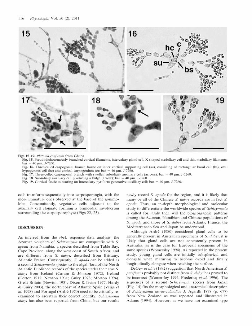
112 Phycologia, Vol. 50 (2), 2011

Figs 1–7. Habit of Schizymenia apoda from the Azores.Fig. 1. Cystocarpic specimen from Sao Miguel, SMG-01-30; bar 5 1 cm.Fig. 2. Cystocarpic specimen from Sao Miguel, SMG-04-170; bar 5 1 cm.Fig. 3. Cystocarpic specimen from Graciosa, GRW-04-87; bar 5 1 cm.Fig. 4. Cystocarpic specimen from Sao Miguel growing on Corallina elongata.Fig. 5. Vegetative morphology showing regularly branched cortical fascicles subtending a medullary filament; bar 5 100 mm. GRW-04-88.Fig. 6. Cortical branches with elongated gland cell (arrow) and filiform medullary filaments; bar 5 40 mm. SMG-06-84.Fig. 7. Primary medullary (m) and secondary rhizoidal filament (r) connected to base of cortical fascicle; bar 5 40 mm. SMG-06-84.
Gabriel et al.: Schizymenia sp. in the Azores and ‘Nemastoma’ confusum 113

Figs 8–13. Schizymenia apoda from the Azores.Fig. 8. Secondary pit connection (arrow) linking inner cortical cell with cell in neighboring cell file; bar 5 25 mm. GRW-04.88.Fig. 9. Filiform rhizoidal filaments and medullary cells connected by secondary pit connections (arrow); bar 5 100 mm. GRW-04.88.Fig. 10. Immature carpogonial branch (icb) borne on supporting cell (arrow) cut off laterally from inner cortical cell; bar 5 25 mm. GRW-04.88.Fig. 11. Squashed three-celled carpogonial branch consisting of rounded basal cell, flat hypogenous cell and carpogonium withtrichogyne (t). Note that neighboring cortical cells have expanded and started to stain darkly, which marks their transformation intosubsidiary auxiliary cells (sac); bar 5 25 mm. GRW-04.88.Fig. 12. Young gonimoblasts (g) borne on auxiliary cell (aux), and incoming connecting filament (cf). Note that the basal cells of corticalfilaments surrounding the gonimoblasts have elongated (arrows), forming a ‘rudimentary’ pericarp; bar 5 25 mm. GRW-04.88.Fig. 13. Surface ostiole of the carposporophyte showing carposporangia; bar 5 100 mm. SMG-04-164.
114 Phycologia, Vol. 50 (2), 2011
The carpogonial branches consist of an oval to rectangular
basal cell, an oval hypogynous cell and a distal conical
carpogonium with a straight trichogyne (Fig. 16). Immedi-
ately before putative fertilisation, the two distal cells
connected to the supporting cell and their subtending
cortical fascicles become darkly staining (Fig. 17). These
cells become swollen, produce bulges on the cell’s sides and
become irregular in shape (Fig. 18) and act as subsidiary
auxiliary cells upon their fusion with the fertilised
carpogonium (stage not seen). The connecting filament
initials originating from the subsidiary auxiliary cells were
not observed. Connecting filaments traverse the thallus,
and each fuses with a generative auxiliary cell located in an
intercalary position, similar to the position of the support-
ing cell bearing a carpogonial branch, on another cortical
fascicle (Fig. 19).
LATE POSTFERTILISATION STRUCTURES: Upon diploidisa-
tion, the auxiliary cell enlarges, and a gonimoblast initial is
cut off distally, in the same manner as a carpogonial branch
initial (Fig. 20). The gonimoblast initial continues to divide
forming a single gonimolobe (Figs 21, 23). All gonimoblast
Fig. 14. One of 276 equally most parsimonious trees based on rbcL sequences showing the position of species of Schizymenia in theSchizymeniaceae, using Platoma cyclocolpum and Titanophora pikeana as the out-group. Branch lengths are proportional to sequencechange. The three tiers of numbers at a node, namely represent maximum parsimony and maximum likelihood bootstrap values andBayesian posterior probabilities, respectively (top to bottom).
Gabriel et al.: Schizymenia sp. in the Azores and ‘Nemastoma’ confusum 115
cells transform sequentially into carposporangia, with the
more immature ones observed at the base of the gonimo-
lobe. Concomitantly, vegetative cells adjacent to the
auxiliary cell elongate forming a primordial involucrum
surrounding the carposporophyte (Figs 22, 23).
DISCUSSION
As inferred from the rbcL sequence data analysis, the
Azorean vouchers of Schizymenia are conspecific with S.
apoda from Namibia, a species described from Table Bay,
Cape Province, along the west coast of South Africa, and
are different from S. dubyi, described from Brittany,
Atlantic France. Consequently, S. apoda can be added as
a second Schizymenia species to the algal flora of the North
Atlantic. Published records of the species under the name S.
dubyi from Iceland (Caram & Jonsson 1972), Ireland
(Cotton 1912; Newton 1931; Guiry 1978; Morton 1994),
Great Britain (Newton 1931; Dixon & Irvine 1977; Hardy
& Guiry 2003), the north coast of Atlantic Spain (Veiga et
al. 1998) and Portugal (Ardre 1970) need to be critically re-
examined to ascertain their correct identity. Schizymenia
dubyi has also been reported from China, but our results
newly record S. apoda for the region, and it is likely that
many or all of the Chinese S. dubyi records are in fact S.
apoda. Thus, an in-depth morphological and molecular
study to differentiate the worldwide species of Schizymenia
is called for. Only then will the biogeographic patterns
among the Azorean, Namibian and Chinese populations of
S. apoda and those of S. dubyi from Atlantic France, the
Mediterranean Sea and Japan be understood.
Although Ardre (1980) considered gland cells to be
generally present in Australian specimens of S. dubyi, it is
likely that gland cells are not consistently present in
Australia, as is the case for European specimens of the
same species (Womersley 1994). As reported in the present
study, young gland cells are initially subspherical and
elongate when maturing to become ovoid and finally
longitudinally elongate when reaching the surface.
DeCew et al.’s (1992) suggestion that North American S.
pacifica is probably not distinct from S. dubyi has proved to
be incorrect (Womersley 1994; Fredericq et al. 1996). The
sequences of a second Schizymenia species from Japan
(Fig. 14) fits the morphological and anatomical description
of Schizymenia novae-zelandiae J. Agardh 1878 (p. 677)
from New Zealand as was reported and illustrated in
Adams (1994). However, as we have not examined type
Figs 15–19. Platoma confusum from Ghana.Fig. 15. Pseudodichotomously branched cortical filaments, intercalary gland cell, X-shaped medullary cell and thin medullary filaments;bar 5 40 mm. J-7260.Fig. 16. Three-celled carpogonial branch borne on inner cortical supporting cell (su), consisting of rectangular basal cell (bs), ovalhypogynous cell (hc) and conical carpogonium (c); bar 5 40 mm. J-7260.Fig. 17. Three-celled carpogonial branch with swollen subsidiary auxiliary cells (arrows); bar 5 40 mm. J-7260.Fig. 18. Subsidiary auxiliary cell producing a bulge (arrow); bar 5 40 mm. J-7260.Fig. 19. Cortical fascicles bearing an intercalary pyriform generative auxiliary cell; bar 5 40 mm. J-7260.
116 Phycologia, Vol. 50 (2), 2011

material of S. novae-zelandiae, the Japanese specimen is
here referred to as Schizymenia sp. Figure 24 presents an
updated overview of Schizymenia distributions in the world.
In detailed studies of S. dubyi, Ardre (1977, 1980)
discovered that the species is actually heteromorphic in
which the life cycle comprises an erect gametophyte
alternating with a crustose tetrasporophyte that bears
zonate tetrasporangia and resembles Haematocelis rubens
J. Agardh (Hansen 1989). Until the present study,
secondary pit connections have been reported only from
the crustose ‘Haematocelis’ tetrasporophyte in Nemasto-
matales; it is herein newly reported that they occasionally
occur in the gametophyte of S. apoda, linking cells of
adjacent inner cortical branches and between medullary X-
cells.
In the course of a revision of the Nemastomatales, we
examined the vegetative and reproductive morphology of
the red alga N. confusum, a species whose taxonomic status
should be reinvestigated (e.g. Masuda & Guiry 1994; Gavio
et al. 2005). When this species was erected, all nemastoma-
talean taxa were placed in the Nemastomataceae; the
morphologically similar genera Nemastoma, Platoma and
Predaea were separated based on a combination of features.
The presence of nutritive cell clusters on cells adjacent to
the auxiliary cell, a diagnostic character of the genus
Predaea, separates Predaea huismanii from N. confusum
(Sanson et al. 1991). Although N. confusum shares features
with both Nemastoma and Platoma, the species was placed
in the former genus mainly on the basis of the presence of
gland cells (Itono & Tsuda 1980).
Kajimura (1997) suggested that N. confusum was
probably a member of Platoma lacking a stipe and noticed
its resemblance to some specimens of Platoma izunosimense
Segawa. Masuda & Guiry (1994) noted that N. confusum
was closely related to P. cyclocolpum based on the following
morphological similarities: supporting cells and auxiliary
cells originate from the basal cells of cortical fascicles, the
gonimoblast develops directly from the auxiliary cell and
gland cells are formed in an intercalary position on cortical
fascicles.
Based on the illustrations and original description of N.
confusum, Gavio et al. (2005) also concluded that the
species should be transferred to a related genus once the
mode of connecting filament initiation has been clarified.
Gavio et al. (2005) contended that most species placed in
Nemastoma may in fact be species of Predaea because the
Figs 20–23. Platoma confusum from Ghana.Fig. 20. Auxiliary cell borne intercalary in inner cortex bearing a gonimoblast initial; bar 5 40 mm. J-7260.Fig. 21. Young gonimoblast develops distally from the opposite side of the incoming connecting filament (cf); bar 5 4.5 mm. J-7260.Fig. 22. Young gonimolobe on an inflated auxiliary cell (ac); bar 5 100 mm. J-7260.Fig. 23. Close-up of Figure 22 showing a connecting filament (cf) and elongated cortical cells surrounding the gonimolobe (arrows); bar5 40 mm. J-7260.
Gabriel et al.: Schizymenia sp. in the Azores and ‘Nemastoma’ confusum 117

original drawings by Berthold (1884) of the type N.
dichotomum from the Bay of Naples depict auxiliary cells
encompassing one or two swollen cells toward the ends of
filaments. Such rhizoidal auxiliary filaments were the basis
for the recognition of the monotypic Adelophycus (as
Adelophyton) Kraft, a genus that thus may have to be
merged with Nemastoma (Gavio et al. 2005).
Nemastoma confusum had been considered a ‘male’ plant
of P. feldmannii described by Børgesen from Saint Helena
(Kraft & John 1976) or perhaps a species of Predaea lacking
auxiliary nutritive cells (Gavio et al. 2005). Upon examina-
tion of type material of N. confusum, we conclude that this
species belongs to the Schizymeniaceae instead of the
Nemastomataceae based on the early prefertilisation events
in which the subsidiary auxiliary cells inflate, become darkly
staining, produce bulges and become irregular in outline, the
diagnostic feature of this family (Gavio et al. 2005).
Among the Schizymeniaceae (i.e. Platoma, Schizymenia,
Titanophora and Wetherbeella), N. confusum is readily
distinguished from species of Titanophora by a lack of
calcified thallus incrustation and from Schizymenia by a
lack of a compact cortex bearing elongate gland cells
terminally on cortical fascicles. The genus Wetherbeella was
erected on the basis of DNA sequence analysis to
accommodate two ‘Platomas’ from Australia that lacked
gland cells and formed a sister lineage to the other three
genera in the family (Saunders & Kraft 2002). Since we did
not have access to suitable material of N. confusum for
molecular phylogenetic studies, we were not able to
compare it with Wetherbeella using rbcL sequences.
A comparison between N. confusum and descriptions of
Platoma suggests that this species is closely related to P.
cyclocolpum and P. chrysymenioides Gavio, Hickerson &
Fredericq (Gabriel et al. 2010). Platoma cyclocolpum has a
great morphological plasticity covering apparently distinct
morphotypes even when environmental conditions did not
vary between specimens (Huisman 1999). Phylogenetic
studies showed that specimens going under the name P.
cyclocolpum worldwide are often misidentified members of
P. chrysymenioides (Gabriel et al. 2010). The distinction
between P. cyclocolpum, P. heteromorphum, P. izunosimense
and P. chrysymenioides lies in the behaviour of the fertilised
carpogonium before fusion with the subsidiary auxiliary
cells and the origin of the connecting filament (Schils &
Coppejans 2002; Gavio et al. 2005; Gabriel et al. 2010).
Since we were unable to observe this ephemeral stage in the
slides we had access to, we suggest the new combination
Platoma confusum, transferring the species to the genus
Platoma while keeping the specific epithet.
Taxonomic conclusions
Schizymenia apoda (J. Agardh) J. Agardh 1851, p. 175
Figs 2–9
TYPE LOCALITY: Table Bay, Cape Province, South Africa.
BASIONYM: Platymenia apoda J. Agardh 1848, p. 47.
SYNONYMS: Platymenia undulata var. obovata J. Agardh
1848, p. 47 (fide Silva 1980; Silva et al. 1996).
Schizymenia obovata J. Agardh 1851, p. 175 (fide Silva
1980, Silva et al. 1996).
DISTRIBUTION: South Africa, Tristan da Cunha (Stegenga
et al. 1997), Namibia (Fredericq et al. 1996), (as S. dubyi):
Fig. 24. Global distribution patterns of Schizymenia species.
118 Phycologia, Vol. 50 (2), 2011

The Azores (Neto 1994; Tittley & Neto 1994), Madeira
(Levring 1974).
Schizymenia dubyi (Chauvin ex Duby) J. Agardh 1851, p. 169
TYPE LOCALITY: Cherbourg, Manche, Atlantic France.
BASIONYM: Halymenia dubyi Chauvin ex Duby 1830,
p. 944.
SYNONYMS: Haematocelis rubens J. Agardh 1851, pp. 496–
497 (fide Ardre 1980).
Turnerella atlantica Kylin 1930: 40 (fide Womersley &
Kraft in Womersley 1994).
Schizymenia minor (J. Agardh) J. Agardh 1851: 172 (fide
Guiry & Guiry 1990).
Haematophloea crouaniorum P.L. Crouan & H.M.
Crouan 1858, pp. 5–6 (fide Guiry & Guiry 1990).
DISTRIBUTION: Atlantic France (Feldmann 1954; Ardre
1977, 1980), Japan (Fredericq et al. 1996), Mediterranean
Sea (this paper).
Platoma confusum (Kraft & John) Gabriel & Fredericq,
comb. nov.
BASIONYM: Nemastoma confusum Kraft & John, British
Phycological Journal 11, pp. 332–335, figs. 1–9, 1976.
TYPE LOCALITY: Vernon Bank, Ghana.
DISTRIBUTION: Ghana (Lawson & John 1987; John et al.
2003, 2004).
ACKNOWLEDGEMENTS
We thank the Fundacao para a Ciencia e Tecnologia (FCT)
for PhD grant SFRH/BD/12541/2003, the SYNTHESYS
Program from the European Union for the Research Grant
NL-TAF-4691, Centro de Investigacao de Recursos Na-
turais da Universidade dos Acores (CIRN/UA) and
Direccao Regional da Ciencia e da Tecnologia (DRCT/
Acores) for the travel grants M3.2.1/I/133/2007 and M3.2.1/
I/062/2008. The National Science Foundation grants DEB-
0315995, DEB-0328491, DEB 0743024, DEB-0919508 and
DEB-0937978 are thanked for financial support. We also
would like to thank Joao Brum, Nuno Alvaro, Patrıcia
Madeira and Sandra Monteiro for their help in collecting
Azorean specimens and Ian Tittley (Herbarium NHM,
London), Willem Prud’homme van Reine (NHN, Leiden),
Olivier de Clerck (Herbarium Ghent) and other collectors
listed in Table 1 for providing vouchers used in this study.
REFERENCES
ABBOTT I.A. 1967. Studies in some foliose red algae of the Pacificcoast II. Schizymenia. Bulletin of the Japanese Society forPhycology 66: 161–174.
ADAMS N.M. 1994. Seaweeds of New Zealand. An Illustrated Guide.Canterbury University Press, Canterbury. 360 pp.
AGARDH J.G. 1842. Algae maris Mediterranei et Adriatici,observationes in diagnosin specierum et dispositionem generum.Fortin, Masson et Cie, Paris. 164 pp.
AGARDH J.G. 1848. Om de Kapska artena af slagtet Iridaea.Ofversigt af Kongelige Vetenskaps-Akademiens Forhandlingar,Stockholm 5: 46–49.
AGARDH J.G. 1851. Species genera et ordines algarum, seudescriptiones succinctae specierum, generum et ordinum, quibusalgarum regnum constituitur. Volumen secundum: algas flor-ideas complectens. Part 1. Lundae [Lund]. XII + 351 pp.
AGARDH J.G. 1878. De algis Novae Zelandiae marinis ActaUniversitatis Lundensis. Lunds Universitets Arsskrift. Afdelningenfor Mathematik och Naturvetenskap 14: 1–32.
ARDRE F. 1970. Contribution a l’etude des algues marines duPortugal. I. La flore. Portugalia Acta Biologica Series B 10:137–555.
ARDRE F. 1977. Sur le cycle du Schizymenia dubyi (Chauvin exDuby) J. Agardh (Nemastomacee, Gigartinale). Revue Algologi-que, Nouvelle Serie 12: 73–86.
ARDRE F. 1980. Observations sur le cycle de developpement duSchizymenia dubyi (Rhodophycee, Gigartinale) en culture, etremarques sur certains genres de Nemastomacees. Cryptogamie,Algologie 1: 111–140.
BERTHOLD G. 1884. Die Cryptonemiaceen des Golfes von Neapel.Fauna und Flora des Golfes von Neapel 12: 1–127.
CARAM B. & JONSSON S. 1972. Nouvelle inventaire des alguesmarines de l’Islande. Acta Botanica Islandica 1: 5–31.
COTTON A.D. 1912. Clare Island Survey. Marine algae. Proceedingsof the Royal Irish Academy 31B(15): 1–178.
CROUAN P.L. & CROUAN H.M. 1858. Note sur quelques alguesmarines nouvelles de la rade de Brest. Annales des SciencesNaturelles, Botanique 4: 69–75.
DECEW T.C., SILVA P.C. & WEST J.A. 1992. Culture studies on therelationship between Schizymenia and Haematocelis (Gigar-tinales, Rhodophyceae) from the Pacific coast of North America.Journal of Phycology 28: 558–566.
DIXON P.S. & IRVINE L.M. 1977. Seaweeds of the BritishIsles. Volume 1. Rhodophyta. Part 1. Introduction, Nemaliales,Gigartinales. British Museum (Natural History), London. 252pp.
DUBY J.E. 1830. Aug. Pyrami de Candolle Botanicon gallicum seuSynopsis plantarum in Flora gallica descriptarum. Editio secunda.Ex herbariis et schedis Candollianis propriisque digestum. Parssecunda plantas cellulares continens. Desray, Paris. 1068 pp.
FELDMANN J. 1942. Remarque sur les Nemastomacees. Bulletin dela Societe Botanique de France 89: 104–113.
FELDMANN J. 1954. Inventaire de la flore marine de Roscoff.Algues, champignons, lichens et spermatophytes. TravauxStation Biologique de Roscoff, Nouvelle Serie 6, supplement:152.
FELSENSTEIN J. 1985. Confidence limits on phylogenies: anapproach using the bootstrap. Evolution 39: 783–791.
FITCH W.M. 1971. Toward defining the course of evolution:minimal change for a specific tree topology. Systematic Zoology20: 406–416.
FREDERICQ S., SERRAO E. & NORRIS J.N. 1992. New records of redalgae from the Azores. Arquipelago 10A: 1–4.
FREDERICQ S., HOMMERSAND M.H. & FRESHWATER D.W. 1996.The molecular systematics of some agar-and carrageenan-containing marine red algae based on rbcL sequence analysis.Hydrobiologia 326/327: 125–135.
GABRIEL D., SCHILS T., NETO A.I., PARAMIO L. & FREDERICQ S.2009. Predaea feldmannii subsp. azorica (Nemastomataceae,Nemastomatales), a new subspecies of red algae from the Azores.Cryptogamie, Algologie 30: 251–270.
GABRIEL D., PARENTE M.I., NETO A.I., RAPOSO M., SCHILS T.,PARAMIO L. & FREDERICQ S. 2010. A phylogenetic appraisal ofthe genus Platoma (Nemastomatales, Rhodophyta), includinglife history and morphological observations on P. cyclocolpumfrom the Azores. Phycologia 49: 2–21.
GAVIO B. & FREDERICQ S. 2002. Grateloupia turuturu (Halymenia-ceae, Rhodophyta) is the correct name of the non-native speciesin the Atlantic known as Grateloupia doryphora. EuropeanJournal of Phycology 37: 349–360.
GAVIO B., HICKERSON E. & FREDERICQ S. 2005. Platomachrysymenioides sp. nov. (Schizymeniaceae), and Sebdenia integrasp. nov. (Sebdeniaceae), two new red algal pecies from the
Gabriel et al.: Schizymenia sp. in the Azores and ‘Nemastoma’ confusum 119

northwestern Gulf of Mexico, with a phylogenetic assessment ofthe Cryptonemiales-complex (Rhodophyta). Gulf of MexicoScience 23: 38–57.
GUINDON S. & GASCUEL O. 2003. A simple, fast, and accuratealgorithm to estimate large phylogenies by maximum likelihood.Systematic Biology 52: 696–704.
GUIRY M.D. 1978. An appraisal of the Irish benthic marine algalflora. British Phycological Journal 13: 1–200.
GUIRY M.D. & GUIRY G.M. 2009. AlgaeBase. Worldwideelectronic publication, National University of Ireland, Galway.http://www.algaebase.org (11 August 2009).
HALL B.G. 2001. Phylogenetic trees made easy. Sinauer Associates,Sunderland, Massachusetts. 179 pp.
HANSEN G.I. 1989. Schizymenia dawsonii and its relation tothe genus Sebdenia (Sebdeniaceae, Rhodophyta). Taxon 38:54–59.
HARDY F.G. & GUIRY M.D. 2003. A check-list and atlas of theseaweeds of Britain and Ireland. British Phycological Society,London. 435 pp.
HUELSENBECK J.P. & RONQUIST F.R. 2001. MrBayes. Bayesianinference of phylogeny. Biometrics 17: 754–755.
HUISMAN J.M. 1999. Vegetative and reproductive morphologyof Nemastoma damaecorne (Gigartinales, Rhodophyta)from Western Australia. Australian Systematic Botany 11:721–728.
ITONO H. & TSUDA R.T. 1980. Titanophora marianensis sp. nov.(Nemastomataceae, Rhodophyta) from the Sea of Japan.Phycologia 26: 419–428.
JOHN D.M., LAWSON G.W. & AMEKA G.K. 2003. The marinemacroalgae of the Tropical West Africa Subregion. Beihefte zurNova Hedwigia 125: 1–217.
JOHN D.M., PRUD’HOMME VAN REINE W.F., LAWSON G.W.,KOSTERMANS T.B. & PRICE J.H. 2004. A taxonomic andgeographical catalogue of the seaweeds of the western coast ofAfrica and adjacent islands. Beihefte zur Nova Hedwigia 127:1–339.
KAJIMURA M. 1997. The morphology of Platoma izunosimense(Shizymeniaceae, Rhodophyta). Botanica Marina 40: 477–485.
KRAFT G.T. & JOHN D.M. 1976. The morphology and ecologyof Nemastoma and Predaea species (Nemastomataceae,Rhodophyta) from Ghana. British Phycological Journal 11:331–344.
KYLIN H. 1930. Uber die Entwicklungsgeschichte der Florideen.Lunds Universitets Arsskrift, Ny Foljd, Andra Afdelningen.26(6): 103 pp.
KYLIN H. 1956. Die Gattungen der Rhodophyceen. C.W.K.Gleerups Forlag, Lund. 673 pp.
LARKUM A.W. 1960. Botany (Algae). Azores Expedition 1959,Final Report. The Exploration Board, Imperial College ofScience and Technology, London.
LAWSON G.W. & JOHN D.W. 1987. The marine algae and coastalenvironment of tropical West Africa (second edition). Beiheftezur Nova Hedwigia 93: 1–415.
LEVRING T. 1974. The marine algae of the Archipelago of Madeira.Boletim do Museu Municipal do Funchal 28: 5–111.
LIN S.M., FREDERICQ S. & HOMMERSAND M.H. 2001. Systematicsof the Delesseriaceae (Ceramiales, Rhodophyta) based on thelarge subunit rDNA and rbcL sequences, including the Phyco-dryoideae, subfam. nov. Journal of Phycology 37: 881–899.
MADDISON D.R. & MADDISON W.P. 2000. MacClade 4: Analysis ofphylogeny and character evolution. Version 4.0. Sinauer Associ-ates, Sunderland, Massachusetts.
MASUDA M. & GUIRY M.D. 1994. The reproductive morphologyof Platoma cyclocolpum (Nemastomataceae, Gigartinales) fromGran Canaria, Canary Islands. Cryptogamie, Algologie 15:191–212.
MASUDA M. & GUIRY M.D. 1995. Reproductive morphologyof Itonoa marginifera (J. Agardh) gen. et comb. nov. (Nemas-tomataceae, Rhodophyta). European Journal of Phycology 30:57–67.
MORTON O. 1994. Marine algae of Northern Ireland. UlsterMuseum, Belfast. 123 pp.
NETO A.I. 1991. Estudo da vegetacao macrofitobentonica da costade Sao Roque, ilha de Sao Miguel, Acores. 1u Encontro Nacionalde Macroalgas Marinhas, LNETI, Lisboa 13–14 de Dezembro1990: 73–90.
NETO A.I. 1994. Checklist of the benthic marine macro algae of theAzores. Arquipelago 12A: 15–34.
NETO A.I. 1997. Studies on algal communities of Sao Miguel,Azores. PhD thesis. Universidade dos Acores, Ponta Delgada.309 pp.
NEWTON L. 1931. A handbook of the British seaweeds. BritishMuseum (Natural History), London. 478 pp.
PALMINHA F.P. 1957. Contribuicoes para o estudo das algasmarinhas portuguesas. II. (Litoral Algarvio). Boletim daSociedade Portuguesa de Ciencias Naturais Series 2 22: 68–74.
PARENTE M.I., NETO A.I. & FLETCHER R.L. 2003a. Morphologyand life history studies of Endarachne binghamiae (Scytosipho-naceae, Phaeophycota) from the Azores. Aquatic Botany 76:109–116.
PARENTE M.I., NETO A.I. & FLETCHER R.L. 2003b. Morphologyand life history studies of Scytosiphon lomentaria (Scytosipho-naceae, Phaeophyceae) from the Azores. Journal of Phycology39: 353–359.
POSADA D. & CRANDALL K.A. 1998. Modeltest: testing the modelof DNA substitution. Bioinformatics 14: 817–818.
RODRIGUEZ-PRIETO C., VERGES A., SANCHEZ N., POLO L. &VERLAQUE M. 2004. The morphology and reproductive struc-tures of Mediterranean species of the genus Nemastoma J.Agardh, nom. cons. (Nemastomataceae, Nemasomatales): Ne-mastoma dichotomum and N. dumontioides. Botanica Marina 47:38–52.
SANSON M., REYES J. & AFONSO-CARRILLO J. 1991. Contributionto the seaweed flora of the Canary Islands: new records ofFlorideophyceae. Botanica Marina 34: 527–536.
SAUNDERS G.W. & KRAFT G.T. 2002. Two new Australianspecies of Predaea (Nemastomataceae, Rhodophyta) withtaxonomic recommendations for an emended Nemastomatalesand expanded Halymeniales. Journal of Phycology 38:1245–1260.
SCHILS T. & COPPEJANS E. 2002. Gelatinous red algae of theArabian Sea, including Platoma heteromorphum sp. nov.(Gigartinales, Rhodophyta). Phycologia 41: 254–267.
SCHMIDT O.C. 1929. Beitrage zur Kenntnis der Meeresalgen derAzoren. I. Hedwigia 69: 95–113.
SEUBERT M. 1844. Flora Azorica. Adolph Marcus, Bonn.50 pp.
SILVA P.C. 1980. Remarks on algal nomenclature. VI. Taxon 29:121–145.
SILVA P.C., BASSON P.W. & MOE R.L. 1996. Catalogue of thebenthic marine algae of the Indian Ocean. University ofCalifornia Publications in Botany 79: 1–1259.
SOUTH G.R. & TITTLEY I. 1986. A checklist and distributionalindex of the benthic marine algae of the North Atlantic Ocean.British Museum (Natural History) and Huntsman MarineLaboratory, London and St. Andrews, New Brunswick, Canada.76 pp.
STEGENGA H., BOLTON J.J. & ANDERSON R.J. 1997. Seaweeds of theSouth African West Coast. Contributions from the BolusHerbarium No 18, Cape Town. 655 pp.
SWOFFORD D.L. 2003. PAUP*: phylogenetic analysis usingparsimony (and other methods). Version 4.0. Sinauer Associates,Sunderland, Massachusetts.
TITTLEY I. & NETO A.I. 1994. ‘Expedition Azores 1989’: benthicmarine algae (seaweeds) recorded from Faial and Pico.Arquipelago 12A: 1–13.
TITTLEY I. & NETO A.I. 2005. The marine algal (seaweed) flora ofthe Azores: additions and amendments. Botanica Marina 48:248–255.
TITTLEY I., NETO A.I., FARNHAM W.F. & PARENTE M.I. 2001.Additions to the marine algal (seaweed) flora of the Azores.Botanica Marina 44: 215–220.
TITTLEY I., NETO A.I. & PARENTE M.I. 2009. The marine algal(seaweed) flora of the Azores: additions and amendments 3.Botanica Marina 52: 7–14.
120 Phycologia, Vol. 50 (2), 2011

TOSTE M.F., PARENTE M.I., NETO A.I. & FLETCHER R.L. 2003a.Life history and phenology of Hydroclathrus clathratus (Scyto-siphonaceae, Phaeophyceae) in the Azores. Cryptogamie Algolo-gie 24: 209–218.
TOSTE M.F., PARENTE M.I., NETO A.I. & FLETCHER R.L. 2003b.Life history of Colpomenia sinuosa (Scytosiphonaceae,Phaeophyceae) in the Azores. Journal of Phycology 39:1268–1274.
TRELEASE W. 1897. Botanical observations on the Azores. AnnualReport of the Michigan Botanical Garden 8: 77–220.
VEIGA A.J., CREMADES J. & BARBARA I. 1998. A catalogue of themarine benthic algae of the Sisargas Islands (N.W. Iberian
Peninsula, Spain). Boletim Museu Municipal do Funchal 5,supplement: 481–493.
WOMERSLEY H.B.S. 1994. The marine benthic flora of southernAustralia – Part IIIA – Bangiophyceae and Florideophyceae(Acrochaetiales, Nemaliales, Gelidiales, Hildenbrandiales andGigartinales sensu lato). Australian Biological Resources Study,Canberra. 508 pp.
Received 20 August 2009; Accepted 7 June 2010
Associate editor: Juliet Brodie
Gabriel et al.: Schizymenia sp. in the Azores and ‘Nemastoma’ confusum 121