Embed Size (px)
Citation preview
RESEARCH PAPER
Taxonomic revision of the tribe Zoraidini (Hemiptera:Fulgoromorpha: Derbidae) from KoreaMohammad Atikur RAHMAN,1,2 Yong Jung KWON1 and Sang Jae SUH1
1 School of Applied Biosciences, Kyungpook National University, Daegu, Korea2 Department of Entomology, Patuakhali Science and Technology University, Dumki, Patuakhali, Bangladesh
Correspondence
Yong Jung Kwon, School of AppliedBiosciences, College of Agriculture and LifeSciences, Kyungpook National University,Daegu 702 701, Korea.Email: [email protected]
Received 29 January 2012;accepted 19 July 2012.
doi: 10.1111/j.1748-5967.2012.00463.x
Abstract
The Korean planthopper tribe Zoraidini is revised taxonomically. Five genera arerecognized in the Korean fauna: Diostrombus Uhler, 1896, Losbanosia Muir, 1917,Pamendanga Distant, 1906, Shirakiana Metcalf, 1945, Zoraida Kirkaldy, 1900.Among them, the genus Shirakiana is recorded for the first time in Korea. Ninespecies, four of which are recognized new to Korea: S. infumata (Matsumura),Z. koannania Matsumura, Z. hubeiensis Chou et Huang, and Z. kuwayamae (Mat-sumura). Previous record of one species, Z. pterophoroides (Westwood, 1851), isremoved from the list of Korean fauna because it was erroneously reported, basedon a misidentification. All species are described and illustrated, and identificationkeys to genera and species are provided.
Key words: Auchenorrhyncha, Derbid planthopper, Fulgoroidea, taxonomy.
Introduction
Derbidae Spinola, 1839 is one of the largest and most mor-phologically differentiated families in Fulgoromorpha andcomprises the subfamilies Cedusinae Emeljanov, 1992, Der-binae Spinola, 1839 and Otiocerinae Muir, 1917. The tribeZoraidini Muir, 1918 is one of the nine tribes in the sub-family Otiocerinae with several hundred species worldwide(Emeljanov 1995; Szwedo 2006). This group of insect faunais poorly known in Korea. Previously, only the followingfive species belonging to this tribe were described primarilybased on external morphology by Lee (1979): Diostrombuspolitus Uhler, Nomuraida hibarensis Matsumura, Pamen-danga matsumurae (Muir), Zoraida horishana Matsumuraand Zoraida pterophoroides (Westwood). Later, Kwon andHuh (2001) provided a revised checklist including one morespecies, Zoraida albicans Anufriev, 1968, that belongs tothis tribe from Korea and then no further work was done.The aim of this study, therefore, is to provide a taxonomicrevision of Korean Zoraidini with the description of malegenital structures because these provide a better means todifferentiate taxa at the species level.
Recent investigation of specimens of this tribe revealedthe presence of following other four species which are new
to Korea: Shirakiana infumata (Matsumura), Zoraida koan-nania Matsumura, Zoraida hubeiensis Chou et Huang andZoraida kuwayamae (Matsumura). The voucher specimensof Zoraida pterophoroides (Westwood 1851), previouslyexamined by Lee and Kwon (1979) and Kwon and Huh(2001), have been rechecked recently, and found thatZoraida hubeiensis Chou et Huang, 1985 was erroneouslytreated as Zoraida pterophoroides (Westwood 1851).Depending on the above findings and the unavailability ofthe species in the present investigation, the previous recordof Zoraida pterophoroides (Westwood 1851) is removedfrom the list of Korean fauna. All recorded and unrecordedspecies are redescribed in this paper with illustrations andkey to the genera and species are provided.
Materials and methods
The terminology used in this study follows Yang and Wu(1993) and Zelazny and Webb (2011). The genital segmentsof examined specimens were observed in glycerin jellyusing a stereoscopic microscope (Olympus SZX 12,Olympus, San Diego,CA, USA). Photographs of the speci-men were made using JUJAK 5.5 (DIXI 3000, DIXI Optics,Daejeon, Korea) digital camera. Illustrations were scanned
Entomological Research 42 (2012) 227–242
bs_bs_banner
© 2012 The AuthorsEntomological Research © 2012 The Entomological Society of Korea and Wiley Publishing Asia Pty Ltd
with HP Scanjet 4850 (Hewlett-Packard Company, Houston,TX, USA). Image and plate compositions are producedusing the software Helicon Focus 5.1 (Helicon Soft Ltd.,Kharkov, Ukraine) and Adobe Photoshop CS3 (Adobe, SanJose, CA, USA), respectively. Spinal formula means thenumbers of apical spines of the hind tibiae and 1st and 2nd
hind tarsomeres.Specimens examined in the present study are deposited in
the collection of the School of Applied Bio-sciences,Kyungpook National University (KNU), Daegu, Republic ofKorea.
Taxonomy
Family Derbidae Spinola, 1839Subfamily Otiocerinae Muir, 1917Tribe Zoraidini Muir, 1918
Key to genera of the tribe Zoraidini from Korea
1. Antennae very short and conical shape. Mesonotum withindistinct carinae ......................... Diostrombus Uhler
– Antennae long and elongate. Mesonotum with distinctcarinae ............................................................2
2. Third median sector of forewing furcate.............................................Pamendanga Distant
– Third median sector of forewing not furcate .............33. Second and third sectors of forewing without cross veins
and posterior margin at near middles with three crossveins in an oblique line ................Shirakiana Metcalf
– Second or third sector of forewing with cross veins andposterior margin at near middles without such an o bliqueline of cross veins ..............................................4
4. Ventral margin of forewing evenly curved before apex ofclaval and cubital veins, and margin not undulate................................................. Zoraida Kirkaldy
– Ventral margin of forewing angulately produced but apexof claval and cubital veins, and margin undulat..................................................Losbanosia Muir
Genus Diostrombus Uhler, 1896
Diostrombus Uhler, 1896: p. 283. Type species: Diostrom-bus politus Uhler, 1896.Diagnosis. Head roundish above, including the large eyes,prominent in front, where it is grooved on the middle line, alittle wider above, and carinate on each lateral margin.Rostrum extended to the posterior coxae. Antennae short,second antennomere claviform, completed by a very slenderbristle. Eyes broadly circular. Mesonotum large, tumidlyconvex, scutellar portion very broadly curved, carinae veryfeeble or absent. Costal area of forewing cover contractedfor a long space beyond the middle followed near the tip by
two diagonal veins, ScRA and RP form an areol cell, Ms1and Cu1 connected by a small cross vein, all median sectorsingle, usually 6. Hind wings much less than half as long asforewings, acute at apex. Abdomen contracted near the base,last ventral segment armed each side with a long, curvedappendage.
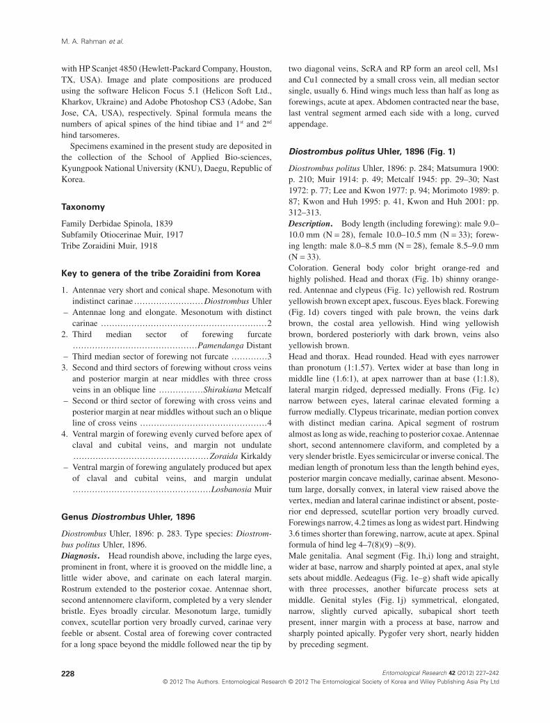
Diostrombus politus Uhler, 1896 (Fig. 1)
Diostrombus politus Uhler, 1896: p. 284; Matsumura 1900:p. 210; Muir 1914: p. 49; Metcalf 1945: pp. 29–30; Nast1972: p. 77; Lee and Kwon 1977: p. 94; Morimoto 1989: p.87; Kwon and Huh 1995: p. 41, Kwon and Huh 2001: pp.312–313.Description. Body length (including forewing): male 9.0–10.0 mm (N = 28), female 10.0–10.5 mm (N = 33); forew-ing length: male 8.0–8.5 mm (N = 28), female 8.5–9.0 mm(N = 33).Coloration. General body color bright orange-red andhighly polished. Head and thorax (Fig. 1b) shinny orange-red. Antennae and clypeus (Fig. 1c) yellowish red. Rostrumyellowish brown except apex, fuscous. Eyes black. Forewing(Fig. 1d) covers tinged with pale brown, the veins darkbrown, the costal area yellowish. Hind wing yellowishbrown, bordered posteriorly with dark brown, veins alsoyellowish brown.Head and thorax. Head rounded. Head with eyes narrowerthan pronotum (1:1.57). Vertex wider at base than long inmiddle line (1.6:1), at apex narrower than at base (1:1.8),lateral margin ridged, depressed medially. Frons (Fig. 1c)narrow between eyes, lateral carinae elevated forming afurrow medially. Clypeus tricarinate, median portion convexwith distinct median carina. Apical segment of rostrumalmost as long as wide, reaching to posterior coxae.Antennaeshort, second antennomere claviform, and completed by avery slender bristle. Eyes semicircular or inverse conical. Themedian length of pronotum less than the length behind eyes,posterior margin concave medially, carinae absent. Mesono-tum large, dorsally convex, in lateral view raised above thevertex, median and lateral carinae indistinct or absent, poste-rior end depressed, scutellar portion very broadly curved.Forewings narrow, 4.2 times as long as widest part. Hindwing3.6 times shorter than forewing, narrow, acute at apex. Spinalformula of hind leg 4–7(8)(9) -8(9).Male genitalia. Anal segment (Fig. 1h,i) long and straight,wider at base, narrow and sharply pointed at apex, anal stylesets about middle. Aedeagus (Fig. 1e–g) shaft wide apicallywith three processes, another bifurcate process sets atmiddle. Genital styles (Fig. 1j) symmetrical, elongated,narrow, slightly curved apically, subapical short teethpresent, inner margin with a process at base, narrow andsharply pointed apically. Pygofer very short, nearly hiddenby preceding segment.
M. A. Rahman et al.
228 Entomological Research 42 (2012) 227–242© 2012 The Authors. Entomological Research © 2012 The Entomological Society of Korea and Wiley Publishing Asia Pty Ltd
Material examined: 1 male, Songnisan, Chungcheongbuk-do, Korea, 9.ix.2001, Y. J. Kwon; 1 male, 1 female,Daejeon city, Chungcheongnam-do, Korea, 29.vii.1993, S.L. An; 1 male, Chunghwa-myeon, Chungcheongnam-do,Korea, 19.viii.1993, S. L. An; 1 male, Dansan Myeon,Gyeongsangbuk-do, Korea, 17.viii.1982, Y. J. Kwon; 1female, same locality, 13.viii.1983, Y. J. Kwon; 1 male, 1female, Mt. Hwanghaksan, Gyeongsangbuk-do, Korea,27.viii.1985, Y. J. Kwon; 2 males, 3 females, Yecheon,Gyeongsangbuk-do, Korea, 3.ix.2008,Y. J. Kwon; 15 males,Gunwi, Gyeongsangbuk-do, Korea, 17.vii.2011,Y. J. Kwon;17 females, Gunwi, Gyeongsangbuk-do, Korea, 17.vii.2011,Y. J. Kwon; 1 male, 3 females, Surisan, Gyeonggi-do, Korea,21.viii.1996, Y. J. Kwon; 1 male, Mt. Naejangsan,Jeollabuk-do, Korea, 14.viii.1981, Y. J. Kwon; 2 males, 1
female, Cheonhwangsan, Jeollabuk-do, Korea, 12.ix.1999,Y. J. Kwon; 1 male, 2 females, Gwanchon, Jeollabuk-do,Korea, 29.vii.2006, E. Y. Huh; 1 male, Pyeonjang,Jeollabuk-do, Korea, 29.vii.2006, E. Y. Huh; 1 male, 2females, Taein, Jeollabuk-do, Korea, 30.vii.2006, E.Y.Huh; 1 female, Chilbo, Jeollabuk-do, Korea, 30.vii.2006, E.Y. Huh.Distribution. Korea, Japan, China, Taiwan.Host plant: Avena sativa (cf. Kor. Soc. Plant Prot. 1986),Graminae spp. (cf. Lee & Kwon 1977, 1979), Hordeum spp.(cf. Kor. Soc. Plant Prot. 1986), Hordeum vulgare var. hex-astichon (cf. Kor. Soc. Plant Prot. 1986), H. vulgare var.nudum (cf. Kor. Soc. Plant Prot. 1986), Oryza sativa (cf. Lee& Kwon 1977, 1979; Kor. Soc. Plant Prot. 1986), Oryzasativa var. terrestris (cf. Kor. Soc. Plant Prot. 1986), Secale
Figure 1 Diostrombus politus Uhler.(a) Dorsal habitus; (b) vertex, pronotumand mesonotum; (c) frons and clypeus;(d) forewing and hind wing; (e) aedeagus(left lateral view); (f) ditto (right lateralview); (g) ditto (ventral view); (h) analsegment (dorsal view); (i) ditto (lateralview); (j) genital styles (latero-ventralview). Scale bars (a, d) 2.0 mm; (b, c)1.0 mm; (e–j) 0.5 mm.
Review of Zoraidini from Korea
229Entomological Research 42 (2012) 227–242© 2012 The Authors. Entomological Research © 2012 The Entomological Society of Korea and Wiley Publishing Asia Pty Ltd
cereale (cf. Kor. Soc. Plant Prot. 1986), Solanum tuberosum(cf. Lee & Kwon 1977, 1979), Triticum aestivum (cf. Kor.Soc. Plant Prot. 1986).
Genus Losbanosia Muir, 1917
Losbanosia Muir, 1917: p. 85. Type species: Losbanosiabakeri Muir, 1917.Nomuraida Matsumura, 1935: p. 79. Type species: Nomu-raida hibarensis Matsumura, 1935, by monotypy, syno-nymised by Chou et al. 1985: p. 50; Szwedo andAdamczewska 2004: pp. 1–11.Diagnosis. Vertex small, flat, trapeziform, strongly nar-rowing forward. Frons linear. Antennae long, cylindricalsecond antennomere. Postclypeus with sharp median carinaand weak lateral carinae. Mesonotum swollen, with 3carinae. Ventral margin of forewing angularly producedbetween apex of claval and cubital veins, thus formingdorsal margin nearly parallel with ventral margin, ventralmargin undulate.
Losbanosia hibarensis (Matsumura, 1935) (Fig. 2)
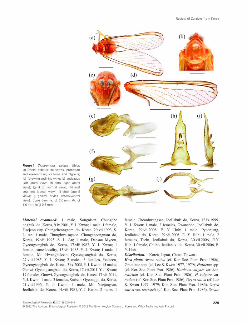
Nomuraida hibarensis Matsumura, 1935: pp. 79–80; Doi1936: p. 102; Nast 1972: p. 78; Lee and Kwon 1979: p. 961;Anufriev & Emeljanov 1988: p. 475; Kwon and Huh 1995:p. 41, Kwon and Huh 2001: pp. 312–313.Losbanosia hibarensis: Chou et al. 1985: p. 50; Yang andWu 1993: pp. 37–38; Liang and Suwa 1998: p. 149.Description. Body length (including forewing): male12.6–13.0 mm (N = 05), forewing length: male 11.5–11.8 mm (N = 05).Coloration. General color reddish brownish (Fig. 2a).Vertex reddish brown with yellow brown lateral margin.Frons, clypeus, antennae and eyes reddish brown (Fig. 2b).Rostrum yellow except apex, fuscous. Pronotum reddishbrown with yellow margin and granules. Mesonotum usuallyyellowish brown. Forewings (Fig. 2c) hyaline, with castane-ous stripe along anterior margin, posterior margin of thestripe projecting backwards in the shape of 3 teeth, veinspartly red, partly brown. Hind wings with castaneous api-cally, veins reddish brown. Thorax brown to yellowishbrown. Legs yellow to yellowish brown. Margins of abdomi-nal segments carmine-red, dorsally brown with granules.Genital segment carmine-red.Head and thorax. Head with eyes narrower than pronotum(1:1.57). Vertex wider at base than long in middle line(2.5:1), at apex narrower than at base (1:2.5), flat, trapezi-form, excavated medially, not surpassing before eyes. Fronslinear between eyes, longer in middle line than wide atwidest part (4.2:1), disc depressed in entire length, wide atapex. Clypeus tricarinate, median carina sharp and promi-nent. Antennae long, second antennomere cylindrical, flag-
ellum originated from subapical point. The median length ofpronotum as long as the median length of vertex (1:1), lengthbehind eyes greater than median length (1.9:1), pronotumwith several pits. Mesonotum tricarinate, scutellar portionbroadly developed. Forewings (Fig. 2c) narrow, 3.4 times aslong as widest part, median cells short, their length andwidth about equal Hindwing 3.1 times shorter than forew-ing. Spinal formula of hind leg 5–5–5(4).Male genitalia. Anal segment in lateral profile (Fig. 2j)long, wider at basal half, slender apical half, in dorsal view(Fig. 2i) concave both lateral margin medially, longer inmiddle line than wide at base (2.75:1), bluntly pointed atapex, anal style sets near middle. Aedeagus (Fig. 2d,e) shaftlonger and curved apically, flagellum terminating with along process, directed cephalad. Genital styles (Fig. 2g,h)large, elongated, apical half wider than basal half in dorso-lateral view (Fig. 2h), inner margin with a small process atmiddle, subapically and apically lobed, inner ventral marginincised at middle. Medioventral process of pygofer (Fig. 2f)narrow at apical half, hairy, apex acute, wide at base.Materials examined. 1 male, Cheamsan, Jeollanam-do,Korea, 28.vii.1999, Y. J. Kwon; 1 male, Chogyesan,Jeollanam-do, Korea, 10.ix.1998; 1 male, Gayasan,Gyeongsangnam-do, Korea, 12.viii.1996; 1 male, Yecheon,Gyeongsangbuk-do, Korea, 2.viii.2008, same collector.Distribution. Korea, Japan, Taiwan, China, Russia.Remarks. This species was reported as Nomuraida hiba-rensis Matsumura from Korea previously but it has alreadybeen synonymized to genus Losbanosia by Chou et al. 1985.
Genus Pamendanga Distant, 1906
Pamendanga Distant, 1906: p. 298. Type species: Pamen-danga rubilinea Distant, 1906 (India).Paraproutista Muir, 1913: p. 77. Type species: Parap-routista ceramensis Muir, 1913Diagnosis. Vertex obtuse-angulate, strongly depressed.Face narrow, laminate, convex and centrally carinate.Clypeus shorter than face and tricarinate. Antennae long,second segment cylindrical, flagellum arising apically. Thirdmedian sector of forewing furcate.
Pamendanga matsumurae (Muir, 1918) (Fig. 3)
Pamendanga rubilinea (nec Distant): Matsumura, 1914: p.297.Paraproutista matsumurae Muir, 1918: p. 422.Paraproutista variegata Muir, 1914: p. 51, synonymizedwith Paraproutista matsumurae Muir, 1918 byYang and Wu1993.Pamendanga matsumurai: Ishihara 1965: p. 130; Anufrievand Emeljanov 1988: p. 476.Pamendanga rubilinea (nec Distant): Lee and Kwon 1977:p. 94.
M. A. Rahman et al.
230 Entomological Research 42 (2012) 227–242© 2012 The Authors. Entomological Research © 2012 The Entomological Society of Korea and Wiley Publishing Asia Pty Ltd
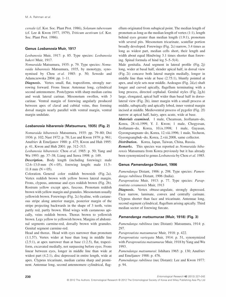
Pamendanga matsumurae: Nast 1972: p. 77; Kwon and Lee1979: p. 66; Morimoto 1989: p. 87; Kwon and Huh 2001: p.314.Description. Body length (including forewing): male 8.0–8.2 mm (N = 02), female 8.9–9.2 mm (N = 04); forewinglength: male 7.0–7.2 mm (N = 02), female 8.0–8.2(N = 04).Coloration. General color pale yellow to yellow brown(Fig. 3a). Vertex and frons yellowish brown. Clypeus yellow.
Rostrum yellow except apex, fuscous. Eyes brown to darkbrown. Antennae yellowish brown. Pronotum and mesono-tum pale yellow. Forewings (Fig. 3a) semihyaline, withnumerous, small, scattered brown spots forming bands trans-versely, apical portion of sub-costal and radial veins reddish,other veins pale yellow, transverse veins surrounded withbrown color. Hind wing hyaline, with scattered brown spots,veins fuscous spreading out into the membrane. Thorax withventral area yellowish brown. Legs pale yellow. Abdomen
Figure 2 Losbanosia hibarensis (Mat-sumura). (a) Dorsal habitus; (b) frons andclypeus; (c) forewing wing; (d) aedeagus(left lateral view); (e) ditto (right lateralview); (f) pygofer (ventral view); (g) geni-tal styles (latero-ventral view); (h) ditto(dorsal view); (i) anal segment (dorsalview); (j) anal segment (lateral view).Scale bars (a, c) 2.0 mm; (b) 1.0 mm; (d–j)0.5 mm.
Review of Zoraidini from Korea
231Entomological Research 42 (2012) 227–242© 2012 The Authors. Entomological Research © 2012 The Entomological Society of Korea and Wiley Publishing Asia Pty Ltd
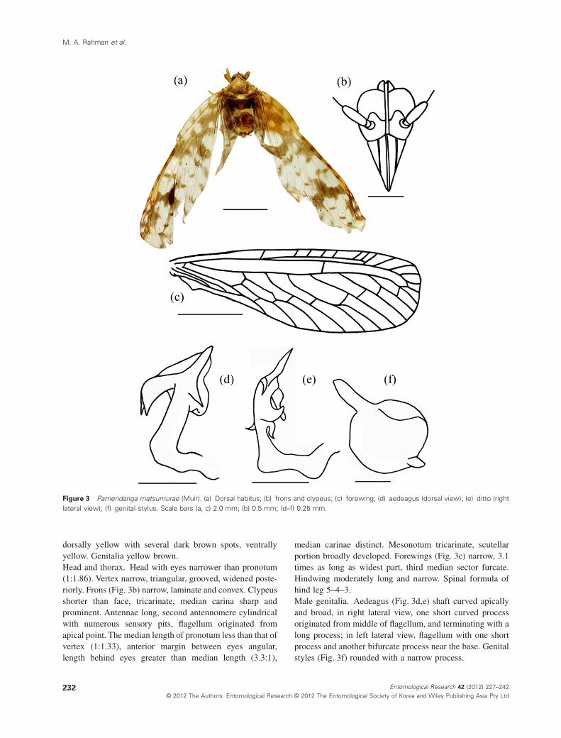
dorsally yellow with several dark brown spots, ventrallyyellow. Genitalia yellow brown.Head and thorax. Head with eyes narrower than pronotum(1:1.86). Vertex narrow, triangular, grooved, widened poste-riorly. Frons (Fig. 3b) narrow, laminate and convex. Clypeusshorter than face, tricarinate, median carina sharp andprominent. Antennae long, second antennomere cylindricalwith numerous sensory pits, flagellum originated fromapical point. The median length of pronotum less than that ofvertex (1:1.33), anterior margin between eyes angular,length behind eyes greater than median length (3.3:1),
median carinae distinct. Mesonotum tricarinate, scutellarportion broadly developed. Forewings (Fig. 3c) narrow, 3.1times as long as widest part, third median sector furcate.Hindwing moderately long and narrow. Spinal formula ofhind leg 5–4–3.Male genitalia. Aedeagus (Fig. 3d,e) shaft curved apicallyand broad, in right lateral view, one short curved processoriginated from middle of flagellum, and terminating with along process; in left lateral view, flagellum with one shortprocess and another bifurcate process near the base. Genitalstyles (Fig. 3f) rounded with a narrow process.
Figure 3 Pamendanga matsumurae (Muir). (a) Dorsal habitus; (b) frons and clypeus; (c) forewing; (d) aedeagus (dorsal view); (e) ditto (rightlateral view); (f) genital stylus. Scale bars (a, c) 2.0 mm; (b) 0.5 mm; (d–f) 0.25 mm.
M. A. Rahman et al.
232 Entomological Research 42 (2012) 227–242© 2012 The Authors. Entomological Research © 2012 The Entomological Society of Korea and Wiley Publishing Asia Pty Ltd
Material examined. 1 male, Gyeryongsan,Chungcheongnam-do, Korea, 20.viii.1978, Y. J. Kwon; 1female, Hakgasan, Gyeongsanbuk-do, Korea, 21.viii.1998;2 females, Seoraksan, Gangwon-do, Korea, 15.ix.1984; 1male, Deogyusan, Jeollabuk-do, Korea, 14.viii.1991; 1female, Naejangsan, Jeollabuk-do, Korea, 14.viii.1981, allsame collector.Distribution. Korea, Japan, Taiwan, Russia.Host plants. Acer spp. (cf. Lee & Kwon 1979; For. Res.Ins. 1995; Lee & Chung 1997), A. buergerianum (cf. For.Res. Ins. 1995), A. cissifalium (cf. For. Res. Ins. 1995),A. ginnala (cf. For. Res. Ins. 1995), A. japonicum (cf. For.Res. Ins. 1995), A. mono (cf. For. Res. Ins. 1995), A. ne-gundo (cf. For. Res. Ins. 1995), A. okamotoanum (cf. For.Res. Ins. 1995), A. palmatum (cf. For. Res. Ins. 1995),A. pseudo-sieboldianum (cf. For. Res. Ins. 1995), A. pseudo-sieboldianum koreanum (cf. For. Res. Ins. 1995), A. saccha-rinum (cf. For. Res. Ins. 1995), A. tegmentosum (cf. For.Res. Ins. 1995), A. triflorum (cf. For. Res. Ins. 1995),A. tschonoskii rubripes (cf. For. Res. Ins. 1995), A. ukurun-duense (cf. For. Res. Ins. 1995), Carpinus cordate (cf. Lee &Kwon 1979), Tilia spp. (cf. Kor. Soc. Plant Prot. 1986),T. amurensis (cf. Kor. Soc. Plant Prot. 1986), T. mandshu-rica (cf. Kor. Soc. Plant Prot. 1986), T. miqueliena (cf. Kor.Soc. Plant Prot. 1986), T. semicostata (cf. Kor. Soc. PlantProt. 1986), Ulmus spp. (cf. For. Res. Ins. 1995; Lee &Chung 1997), U. davidiana (cf. For. Res. Ins. 1995), U. da-vidiana japonica (cf. For. Res. Ins. 1995), U. laciniata (cf.For. Res. Ins. 1995), U. parvifolia (cf. For. Res. Ins. 1995),U. pumila (cf. For. Res. Ins. 1995).
Genus Shirakiana Metcalf, 1945
Shirakiana Metcalf, 1945: 57. Type species: Shirakianainfumata (Matsumura 1914).Shirakia Matsumura, 1914: 303. Type species: Shirakiainfumata Matsumura, 1914, by monotypy.Diagnosis. Vertex at middle near hind margin with moreor less rectangular broken transverse carina. Antennaelonger than face. Clypeus with three distinct carinae. Mes-onotum tricarinate, median carina with three terminalswhere lateral two granulated. Forewing near middle with3 cross veins in an oblique line, between second andthird sectors without cross veins, claval vein ending hindmargin.
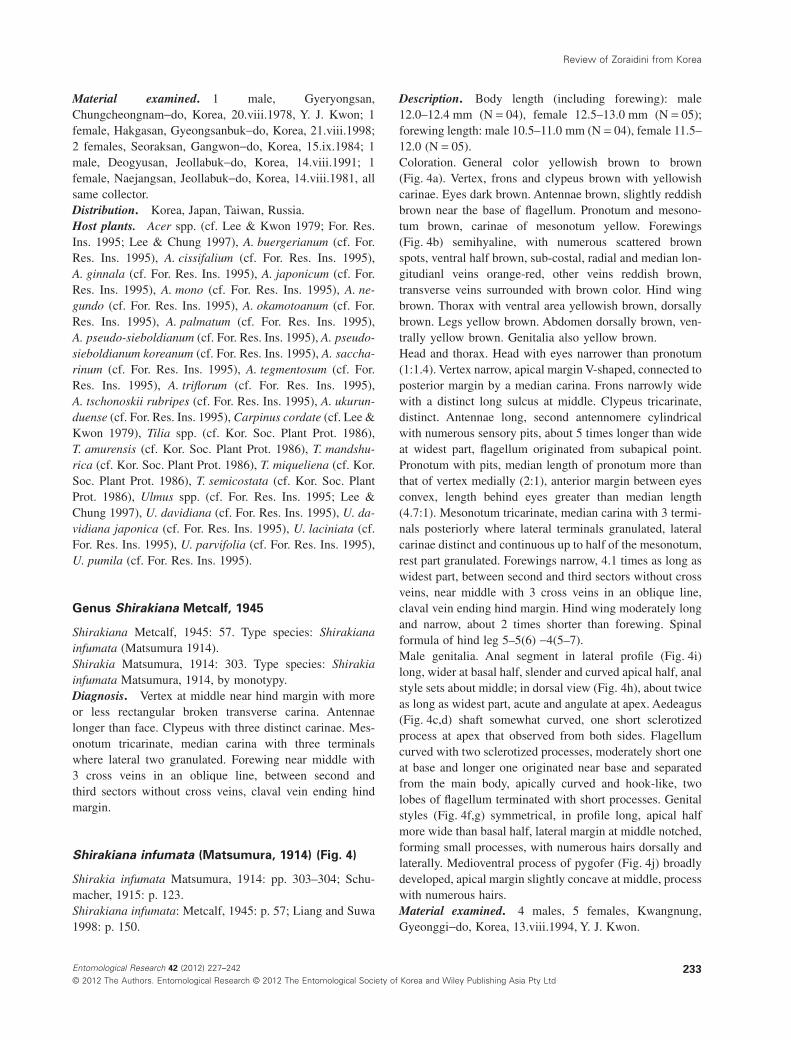
Shirakiana infumata (Matsumura, 1914) (Fig. 4)
Shirakia infumata Matsumura, 1914: pp. 303–304; Schu-macher, 1915: p. 123.Shirakiana infumata: Metcalf, 1945: p. 57; Liang and Suwa1998: p. 150.
Description. Body length (including forewing): male12.0–12.4 mm (N = 04), female 12.5–13.0 mm (N = 05);forewing length: male 10.5–11.0 mm (N = 04), female 11.5–12.0 (N = 05).Coloration. General color yellowish brown to brown(Fig. 4a). Vertex, frons and clypeus brown with yellowishcarinae. Eyes dark brown. Antennae brown, slightly reddishbrown near the base of flagellum. Pronotum and mesono-tum brown, carinae of mesonotum yellow. Forewings(Fig. 4b) semihyaline, with numerous scattered brownspots, ventral half brown, sub-costal, radial and median lon-gitudianl veins orange-red, other veins reddish brown,transverse veins surrounded with brown color. Hind wingbrown. Thorax with ventral area yellowish brown, dorsallybrown. Legs yellow brown. Abdomen dorsally brown, ven-trally yellow brown. Genitalia also yellow brown.Head and thorax. Head with eyes narrower than pronotum(1:1.4). Vertex narrow, apical margin V-shaped, connected toposterior margin by a median carina. Frons narrowly widewith a distinct long sulcus at middle. Clypeus tricarinate,distinct. Antennae long, second antennomere cylindricalwith numerous sensory pits, about 5 times longer than wideat widest part, flagellum originated from subapical point.Pronotum with pits, median length of pronotum more thanthat of vertex medially (2:1), anterior margin between eyesconvex, length behind eyes greater than median length(4.7:1). Mesonotum tricarinate, median carina with 3 termi-nals posteriorly where lateral terminals granulated, lateralcarinae distinct and continuous up to half of the mesonotum,rest part granulated. Forewings narrow, 4.1 times as long aswidest part, between second and third sectors without crossveins, near middle with 3 cross veins in an oblique line,claval vein ending hind margin. Hind wing moderately longand narrow, about 2 times shorter than forewing. Spinalformula of hind leg 5–5(6) -4(5–7).Male genitalia. Anal segment in lateral profile (Fig. 4i)long, wider at basal half, slender and curved apical half, analstyle sets about middle; in dorsal view (Fig. 4h), about twiceas long as widest part, acute and angulate at apex. Aedeagus(Fig. 4c,d) shaft somewhat curved, one short sclerotizedprocess at apex that observed from both sides. Flagellumcurved with two sclerotized processes, moderately short oneat base and longer one originated near base and separatedfrom the main body, apically curved and hook-like, twolobes of flagellum terminated with short processes. Genitalstyles (Fig. 4f,g) symmetrical, in profile long, apical halfmore wide than basal half, lateral margin at middle notched,forming small processes, with numerous hairs dorsally andlaterally. Medioventral process of pygofer (Fig. 4j) broadlydeveloped, apical margin slightly concave at middle, processwith numerous hairs.Material examined. 4 males, 5 females, Kwangnung,Gyeonggi-do, Korea, 13.viii.1994, Y. J. Kwon.
Review of Zoraidini from Korea
233Entomological Research 42 (2012) 227–242© 2012 The Authors. Entomological Research © 2012 The Entomological Society of Korea and Wiley Publishing Asia Pty Ltd
Distribution. Korea (new record), Taiwan.Remarks. This species can be distinguished easily fromany other species of Zoraidini by the morphological featuresof forewings (Second and third sectors of forewing withoutcross veins and posterior margin at near middles with threecross veins in an oblique line, with numerous scatteredbrown spots) and the shape of male genitalia. It is reportedhere for the first time in Korea.
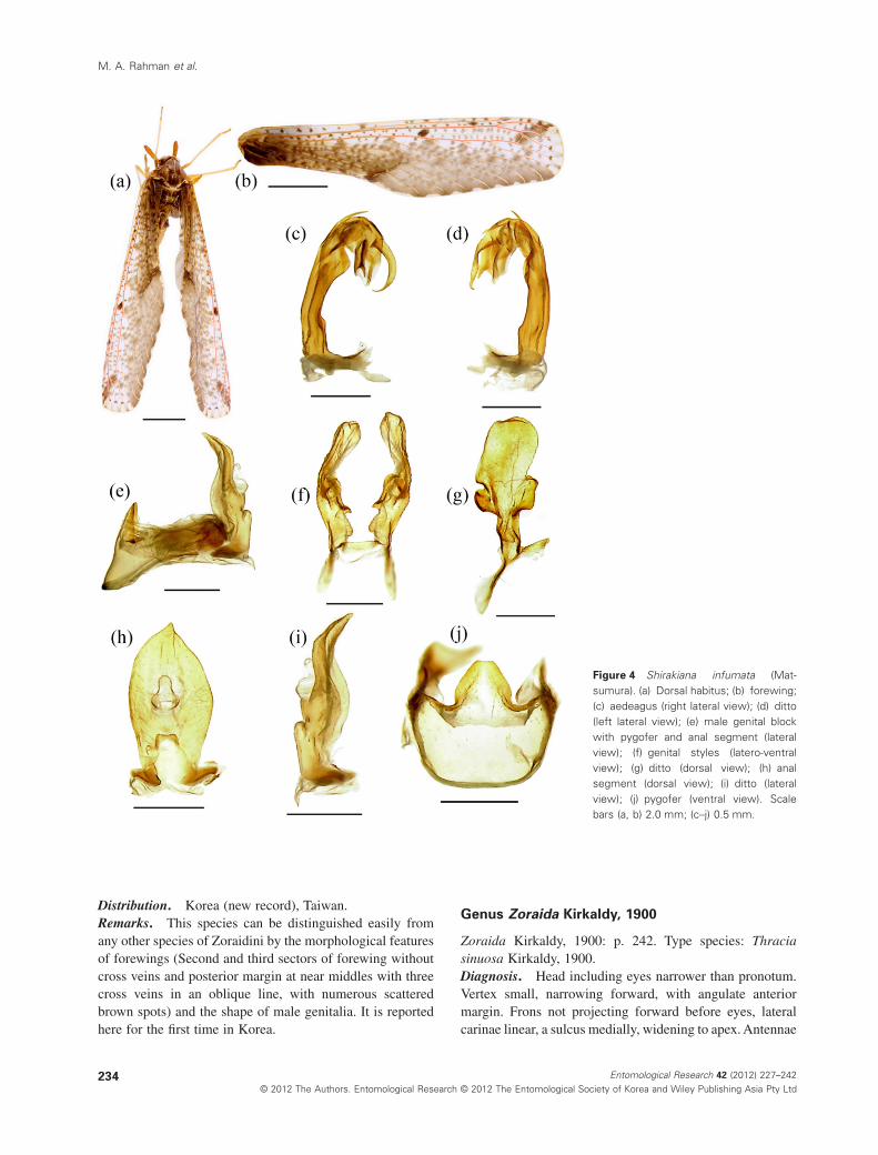
Genus Zoraida Kirkaldy, 1900
Zoraida Kirkaldy, 1900: p. 242. Type species: Thraciasinuosa Kirkaldy, 1900.Diagnosis. Head including eyes narrower than pronotum.Vertex small, narrowing forward, with angulate anteriormargin. Frons not projecting forward before eyes, lateralcarinae linear, a sulcus medially, widening to apex. Antennae
Figure 4 Shirakiana infumata (Mat-sumura). (a) Dorsal habitus; (b) forewing;(c) aedeagus (right lateral view); (d) ditto(left lateral view); (e) male genital blockwith pygofer and anal segment (lateralview); (f) genital styles (latero-ventralview); (g) ditto (dorsal view); (h) analsegment (dorsal view); (i) ditto (lateralview); (j) pygofer (ventral view). Scalebars (a, b) 2.0 mm; (c–j) 0.5 mm.
M. A. Rahman et al.
234 Entomological Research 42 (2012) 227–242© 2012 The Authors. Entomological Research © 2012 The Entomological Society of Korea and Wiley Publishing Asia Pty Ltd
long, cylindrical or flattened, flagellum attached subapically.Postclypeus with 3 sharp carinae. Eyes shallowly incisedventrally. Ocelli absent. Pronotum with hind margin deeplyangulated medially, median carina distinct. Mesonotumswollen, with 3 carinae. First median sector of forewingbranched, with 3–6 veins reaching to hind margin. Malegenitalia with medioventral process.
Key to species of the genus Zoraida from Korea
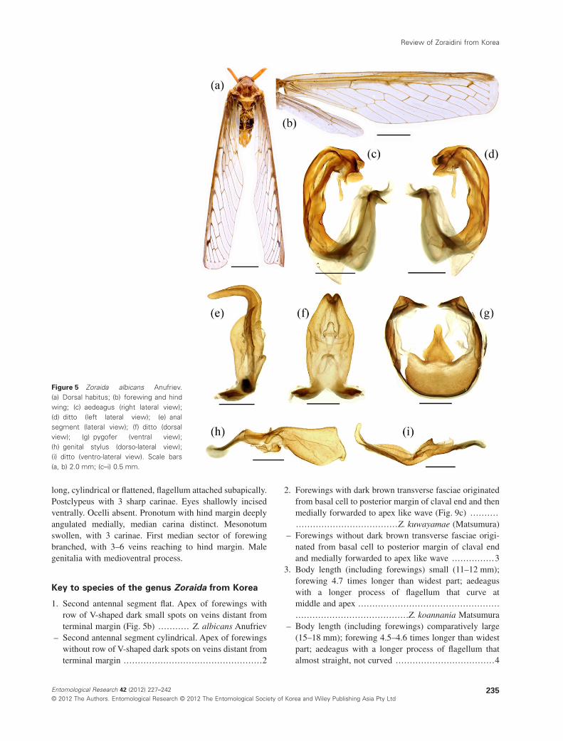
1. Second antennal segment flat. Apex of forewings withrow of V-shaped dark small spots on veins distant fromterminal margin (Fig. 5b) ........... Z. albicans Anufriev
– Second antennal segment cylindrical. Apex of forewingswithout row of V-shaped dark spots on veins distant fromterminal margin .................................................2
2. Forewings with dark brown transverse fasciae originatedfrom basal cell to posterior margin of claval end and thenmedially forwarded to apex like wave (Fig. 9c) ..............................................Z. kuwayamae (Matsumura)
– Forewings without dark brown transverse fasciae origi-nated from basal cell to posterior margin of claval endand medially forwarded to apex like wave ............... 3
3. Body length (including forewings) small (11–12 mm);forewing 4.7 times longer than widest part; aedeaguswith a longer process of flagellum that curve atmiddle and apex ..........................................................................................Z. koannania Matsumura
– Body length (including forewings) comparatively large(15–18 mm); forewing 4.5–4.6 times longer than widestpart; aedeagus with a longer process of flagellum thatalmost straight, not curved ...................................4
Figure 5 Zoraida albicans Anufriev.(a) Dorsal habitus; (b) forewing and hindwing; (c) aedeagus (right lateral view);(d) ditto (left lateral view); (e) analsegment (lateral view); (f) ditto (dorsalview); (g) pygofer (ventral view);(h) genital stylus (dorso-lateral view);(i) ditto (ventro-lateral view). Scale bars(a, b) 2.0 mm; (c–i) 0.5 mm.
Review of Zoraidini from Korea
235Entomological Research 42 (2012) 227–242© 2012 The Authors. Entomological Research © 2012 The Entomological Society of Korea and Wiley Publishing Asia Pty Ltd
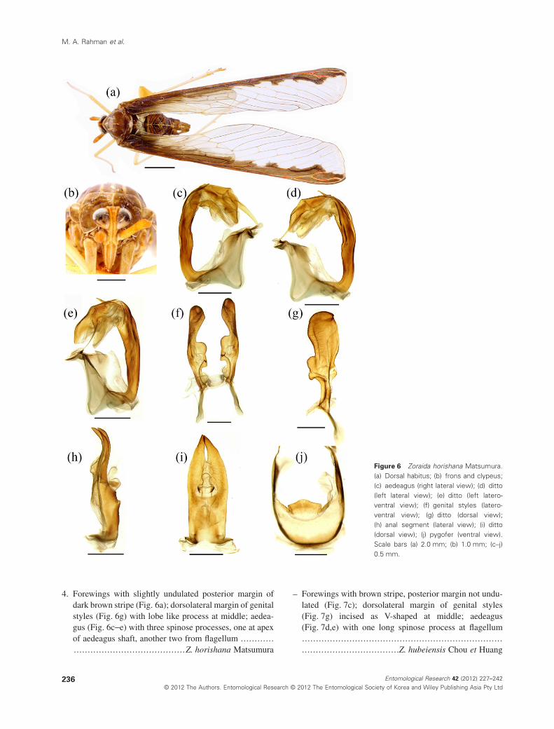
4. Forewings with slightly undulated posterior margin ofdark brown stripe (Fig. 6a); dorsolateral margin of genitalstyles (Fig. 6g) with lobe like process at middle; aedea-gus (Fig. 6c-e) with three spinose processes, one at apexof aedeagus shaft, another two from flagellum ....................................................Z. horishana Matsumura
– Forewings with brown stripe, posterior margin not undu-lated (Fig. 7c); dorsolateral margin of genital styles(Fig. 7g) incised as V-shaped at middle; aedeagus(Fig. 7d,e) with one long spinose process at flagellum...........................................................................................................Z. hubeiensis Chou et Huang
Figure 6 Zoraida horishana Matsumura.(a) Dorsal habitus; (b) frons and clypeus;(c) aedeagus (right lateral view); (d) ditto(left lateral view); (e) ditto (left latero-ventral view); (f) genital styles (latero-ventral view); (g) ditto (dorsal view);(h) anal segment (lateral view); (i) ditto(dorsal view); (j) pygofer (ventral view).Scale bars (a) 2.0 mm; (b) 1.0 mm; (c–j)0.5 mm.
M. A. Rahman et al.
236 Entomological Research 42 (2012) 227–242© 2012 The Authors. Entomological Research © 2012 The Entomological Society of Korea and Wiley Publishing Asia Pty Ltd
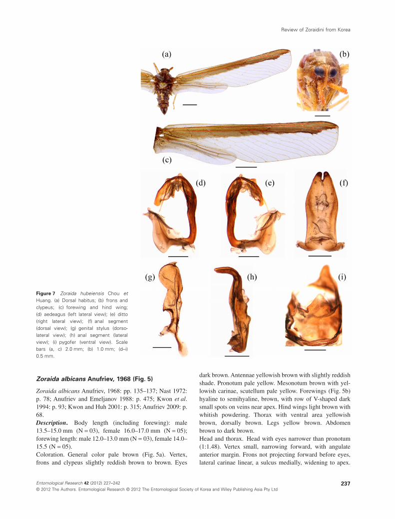
Zoraida albicans Anufriev, 1968 (Fig. 5)
Zoraida albicans Anufriev, 1968: pp. 135–137; Nast 1972:p. 78; Anufriev and Emeljanov 1988: p. 475; Kwon et al.1994: p. 93; Kwon and Huh 2001: p. 315; Anufriev 2009: p.68.Description. Body length (including forewing): male13.5–15.0 mm (N = 03), female 16.0–17.0 mm (N = 05);forewing length: male 12.0–13.0 mm (N = 03), female 14.0–15.5 (N = 05).Coloration. General color pale brown (Fig. 5a). Vertex,frons and clypeus slightly reddish brown to brown. Eyes
dark brown. Antennae yellowish brown with slightly reddishshade. Pronotum pale yellow. Mesonotum brown with yel-lowish carinae, scutellum pale yellow. Forewings (Fig. 5b)hyaline to semihyaline, brown, with row of V-shaped darksmall spots on veins near apex. Hind wings light brown withwhitish powdering. Thorax with ventral area yellowishbrown, dorsally brown. Legs yellow brown. Abdomenbrown to dark brown.Head and thorax. Head with eyes narrower than pronotum(1:1.48). Vertex small, narrowing forward, with angulateanterior margin. Frons not projecting forward before eyes,lateral carinae linear, a sulcus medially, widening to apex.
Figure 7 Zoraida hubeiensis Chou etHuang. (a) Dorsal habitus; (b) frons andclypeus; (c) forewing and hind wing;(d) aedeagus (left lateral view); (e) ditto(right lateral view); (f) anal segment(dorsal view); (g) genital stylus (dorso-lateral view); (h) anal segment (lateralview); (i) pygofer (ventral view). Scalebars (a, c) 2.0 mm; (b) 1.0 mm; (d–i)0.5 mm.
Review of Zoraidini from Korea
237Entomological Research 42 (2012) 227–242© 2012 The Authors. Entomological Research © 2012 The Entomological Society of Korea and Wiley Publishing Asia Pty Ltd
Clypeus tricarinate, median carina ridged and distinct. Fronsslightly shorter than clypeaus (1:1.2). Antennae long, secondantennomere flat with numerous sensory pits, about 7 timeslonger than wide at widest part, flagellum originated fromsubapical point. Pronotum length behind eyes greater thanmedian length (2:1). Mesonotum tricarinate, median carinadistinctly terminated to posterior end, lateral carinae half oflength distinctly terminated at the end. Forewings narrow,4.1 times as long as widest part, apex of forewings with rowof V-shaped small spots on veins distant from terminalmargin. Hind wing moderately long and narrow, about 2.2times shorter than forewing. Spinal formula of hind leg5–5–5.Male genitalia. Anal segment in lateral profile (Fig. 5e)long, wider at basal half, apical half slender and horizontallycurved; in dorsal view (Fig. 5f), longer in middle line thanwidest part (2:1), anal style sets at middle. Aedeagus shaftcurved; flagellum, right lateral view (Fig. 5c), with one shortsclerotized process at apex, and left lateral view (Fig. 5d),with comparatively long sclerotized process at apex. Genitalstyles (Fig. 5h,i) angulate and acute at apex, hook-shapedwide lobe at near base. Medioventral process of pygofer(Fig. 5g) broadly developed, apical half narrow, concave,blunt at apex, dorsocaudal process asymmetrical.Material examined. 2 females, Juwangsan,Gyeongsanbuk-do, Korea, 28. vii. 1984, Y. J. Kwon; 1female, Yecheon, Gyeongsanbuk-do, Korea, 2.viii.2008; 1female, same locality, 7.viii.2008; 1 male, Dongducheon,Gyeonggi-do, Korea, 22.viii.2006; 1 female, Seolaksan,Gangwon-do, 9.viii.1976; 2 males, Paegunsan,Jeollanam-do, 13.viii.1999, all same collector.Distribution. Korea, Russia.Host plant. Unknown.
Zoraida horishana Matsumura, 1914 (Fig. 6)
Zoraida horishana Matsumura, 1914: p. 302; Doi 1933: p.89; Nast 1972: p. 78; Lee and Kwon 1979: p. 960; Anufrievand Emeljanov 1988: p. 475; Morimoto 1989: p. 87; Yangand Wu 1993: pp. 49–51; Liang and Suwa 1998: p. 149;Kwon and Huh 1995: p. 41, Kwon and Huh 2001: pp.315–316.Description. Body length (including forewing): male15.0–17.0 mm (N = 06), female 16.5–17.5 mm (N = 03);forewing length: male 14.0–15.5 mm (N = 06), female 15.0–16.0 (N = 03).Coloration. General color brown to dark brown (Fig. 6a).Vertex and frons slightly reddish brown to brown, clypeusyellowish brown. Eyes dark brown to black. Antennaeslightly reddish yellow. Pronotum, mesonotum brown withyellowish carinae, metanotum dark brown base with whitishend. Forewings (Fig. 6a) hyaline, with dark veins and cas-taneous longitudinal stripe along anterior margin occupying
whole subcostal cell. Hind wings also hyaline. Thorax yel-lowish brown to brown. Legs yellow brown. Abdomenbrown to dark brown.Head and thorax. Head with eyes narrower than pronotum(1:1.6). Vertex small, narrowing forward, with angulateanterior margin, vertex medially shorter than pronotum(1:3). Frons not projecting forward before eyes, lateralcarinae linear, a sulcus medially, widening to apex. Clypeustricarinate, distinct and equally elevated. Frons slightlyshorter than clypeaus (1:1.2). Antennae long, second anten-nomere cylindrical with numerous sensory pits, about 5times longer than wide at widest part, flagellum originatedfrom subapical point. Pronotum granulated medially, lengthbehind eyes greater than median length (1.66:1). Mesono-tum tricarinated. Forewings narrow, 4.48 times as long aswidest part, with castaneous longitudinal stripe along ante-rior margin occupying whole subcostal cell, posteriormargin of the stripe undulated, running behind anteriormedial vein. Hind wing moderately long and narrow, about2.14 times shorter than forewing. Spinal formula of hindleg 5–5–5.Male genitalia. Anal segment in lateral profile (Fig. 6h)long, wider at more than half of length, near apical partslender and slightly curved; in dorsal view (Fig. 6i), longerin middle line than widest part (2:42), medially furcate atapex, anal style sets at middle. Aedeagus (Fig. 6c-e) shaftslightly curved with one sclerotized spinose process at apex;flagellum membraneous with two blade-like long processes,one originated from base and another from near apex.Genital styles (Fig. 6f,g) with apical portion rounded, smallfinger shaped process at apex, ventrally incised at middle,dorsolateral margin with basal part somewhat membraneouswith small lobe-like process at its surface. Medioventralprocess of pygofer (Fig. 6j) broadly developed at base,apical half narrow, convex, blunt at apex.Material examined. 1 male, Hwanghaksan,Gyeongsanbuk-do, Korea, 28. ix. 1991, Y. J. Kwon; 1 male,Hakkasan, Gyeongsanbuk-do, Korea, 21.viii.1998; 1 male,2 females, Yecheon, Gyeongsanbuk-do, Korea, 7.viii.2008;1 male, Gwangneung, Gyeonggi-do, Korea, 14.viii.1994; 1female, Bukhansan B, Gyeonggi-do, Korea, 21.viii.2001; 1male, Sinbulsan B, Gyeongsangnam-do, Korea,30.vii.2003; 1 male, Cheamsan, Jeollanam-do, Korea,28.vii.1999, all same collector.Distribution. Korea, Japan, Taiwan, Russia.Host plant. Unknown.
Zoraida hubeiensis Chou et Huang, 1985 (Fig. 7)
Zoraida hubeiensis: Chou et al. 1985: p. 57.Description. Body length (including forewing): male16.5 mm (N = 01), female 18.0 mm (N = 01); forewinglength: male 15.0 mm (N = 01), female 16.5 (N = 01).
M. A. Rahman et al.
238 Entomological Research 42 (2012) 227–242© 2012 The Authors. Entomological Research © 2012 The Entomological Society of Korea and Wiley Publishing Asia Pty Ltd
Coloration. General color brown to dark brown (Fig. 7a).Vertex, frons and clypeus yellowish brown (Fig. 7b). Eyesdark brown to black. Antennae brownish yellow. Pronotum,mesonotum brown with yellowish carinae, metanotum yel-lowish white. Apex of rostrum black. Forewings (Fig. 7c)hyaline, with castaneous longitudinal stripe along anteriormargin occupying subcostal cell to median longitudinalvein, subcostal area near base hyaline, longitudinal veins onstripe reddish. Hind wings also hyaline with light brownishshade along subcostal area. Thorax and abdomen brown todark brown, dorsally yellowish lining on middle segments.Legs yellow brown. Genital segment brown to dark brown.Head and thorax. Head with eyes narrower than pronotum(1:1.7). Vertex small, narrowing forward, with angulate ante-rior margin, vertex medially shorter than pronotum (1:2.5).Frons not projecting forward before eyes, lateral carinaelinear, a sulcus medially, widening to apex. Clypeus tricari-nate, distinct and equally elevated. Frons slightly shorterthan clypeaus (1:1.27). Antennae long, second antennomerecylindrical with numerous sensory pits, about 6.3 timeslonger than wide at widest part, flagellum originated fromsubapical point. Pronotum granulated medially, lengthbehind eyes greater than median length (1.6:1). Mesonotumtricarinate, lateral carinae half of length distinctly terminatedat the end. Forewings narrow, 4.6 times as long as widestpart, with castaneous longitudinal stripe along anteriormargin occupying whole subcostal cell except basal part,posterior margin of the stripe almost straight, running behindanterior medial vein. Hind wing moderately long andnarrow, about 2.2 times shorter than forewing. Spinalformula of hind leg 5–6(7) -5.Male genitalia. Anal segment in lateral profile (Fig. 7h)long, wider at basal half, apical half slender and horizontallycurved; in dorsal view (Fig. 7f), longer in middle line thanwidest part (1:2.36), anal style sets at near middle. Aedeagus(Fig. 7d,e) shaft slightly curved; flagellum, right lateralview, with one short finger-shaped lobe at base and anotherfolded lobe overlapped, and in left lateral view, with a long,slender, sclerotized process extended to apex. Genital styles(Fig. 7g) angulate and beak-shaped at apex, dorsolateralmargin of genital styles incised deeply at middle and base,ventral margin with a hook-like small process basally.Medioventral process of pygofer (Fig. 7i) broadly developedat base, apical half narrow, and blunt at apex, dorsocaudalprocess asymmetrical.Material examined. 1 male, Dongducheon, Gyeonggi-do,Korea, 22.vii.2006, Y. J. Kwon; 1 female, Seolaksan,Gangwon-do, Korea, 21.viii.1974, all same collector.Distribution. Korea (new record), China.Host plant. Unknown.Remarks. While studying the derbid specimens in theInsect Collection of the Kyungpook National University,Korea, we found that this species was erroneously treated as
Zoraida pterophoroides (Westwood, 1851) based on a misi-dentification. Therefore, previous record of Zoraida ptero-phoroides is removed from the list of Korean fauna, and theopportunity of reporting a new record, Zoraida hubeiensisChou et Huang, 1985, has been taken in this paper.
Zoraida koannania Matsumura, 1914 (Fig. 8)
Zoraida koannania Matsumura, 1914: p. 302; Schumacher1915: p. 123; Yang and Wu 1993: p. 67; Liang and Suwa1998: p. 150.Description. Body length (including forewing): male11.0 mm (N = 02), female 12.3 mm (N = 01); forewinglength: male 10.0 mm (N = 02), female 11.0 (N = 01).Coloration. General color brown to dark brown (Fig. 8a).Vertex, frons and clypeus yellowish brown (Fig. 8b). Eyesdark brown. Antennae brownish yellow with shade of red.Pronotum, mesonotum dark brown with yellow browncarinae, metanotum yellow. Rostrum golden yellow exceptapex, fuscous. Forewings hyaline, with dark brown costalstripe, longitudinal veins brown, apical four veins near apexwith dark spots, cross vein dark brown. Hind wings alsohyaline with brownish veins. Ventral aspect of thorax andabdomen yellow, dorsal aspect dark brown. Legs yellowbrown. Genital segment yellowish brown.Head and thorax. Head with eyes narrower than pronotum(1:1.63). Vertex small, narrowing forward, with angulateanterior margin, vertex medially shorter than pronotum(1:1.33). Frons not projecting forward before eyes, lateralcarinae linear, a sulcus medially, widening to apex. Clypeustricarinate, distinct and equally elevated. Frons slightlyshorter than clypeaus (1:1.3). Antennae long, second anten-nomere cylindrical with numerous sensory pits, about 4times longer than wide at widest part, flagellum originatedfrom subapical point. Pronotum granulated medially, lengthbehind eyes greater than median length (2.5:1). Mesonotumtricarinated. Forewings narrow, 4.7 times as long as widestpart, costal stripe with lower margin very slightly undulated.Hind wing moderately long and narrow, about 2.21 timesshorter than forewing. Spinal formula of hind leg5–5–5(6).Male genitalia. Anal segment in lateral profile (Fig. 8e)long, wider at basal half, apical half slender and horizontallycurved; in dorsal view (Fig. 8f), longer in middle line thanwidest part at base (1:1.73), anal style sets at near middle.Aedeagus (Fig. 8c,d) shaft gently curved; flagellum bilobed,right lateral view, with a short process at base, and in leftlateral view, with a long, sclerotized process curved atmiddle and apex. Genital styles (Fig. 8i,j) pentagonal-shaped, wide at apical half, a hook-like process laterally nearat base. Medioventral process of pygofer (Fig. 8g) broadlydeveloped at middle, pentagonal-shaped and bluntly devel-oped at apex.
Review of Zoraidini from Korea
239Entomological Research 42 (2012) 227–242© 2012 The Authors. Entomological Research © 2012 The Entomological Society of Korea and Wiley Publishing Asia Pty Ltd
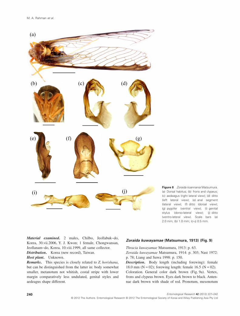
Material examined. 2 males, Chilbo, Jeollabuk-do,Korea, 30.vii.2006, Y. J. Kwon; 1 female, Chongwansan,Jeollanam-do, Korea, 10.viii.1999, all same collector.Distribution. Korea (new record), Taiwan.Host plant. Unknown.Remarks. This species is closely related to Z. horishana,but can be distinguished from the latter in: body somewhatsmaller, metanotum not whitish, costal stripe with lowermargin comparatively less undulated, genital styles andaedeagus shape different.
Zoraida kuwayamae (Matsumura, 1913) (Fig. 9)
Thracia kuwayamae Matsumura, 1913: p. 63.Zoraida kuwayamae Matsumura, 1914: p. 303; Nast 1972:p. 78; Liang and Suwa 1998: p. 150.Description. Body length (including forewing): female18.0 mm (N = 02); forewing length: female 16.5 (N = 02).Coloration. General color dark brown (Fig. 9a). Vertex,frons and clypeus brown. Eyes dark brown to black. Anten-nae dark brown with shade of red. Pronotum, mesonotum
Figure 8 Zoraida koannania Matsumura.(a) Dorsal habitus; (b) frons and clypeus;(c) aedeagus (right lateral view); (d) ditto(left lateral view); (e) anal segment(lateral view); (f) ditto (dorsal view);(g) pygofer (ventral view); (i) genitalstylus (dorso-lateral view); (j) ditto(ventro-lateral view). Scale bars (a)2.0 mm; (b) 1.0 mm; (c–j) 0.5 mm.
M. A. Rahman et al.
240 Entomological Research 42 (2012) 227–242© 2012 The Authors. Entomological Research © 2012 The Entomological Society of Korea and Wiley Publishing Asia Pty Ltd
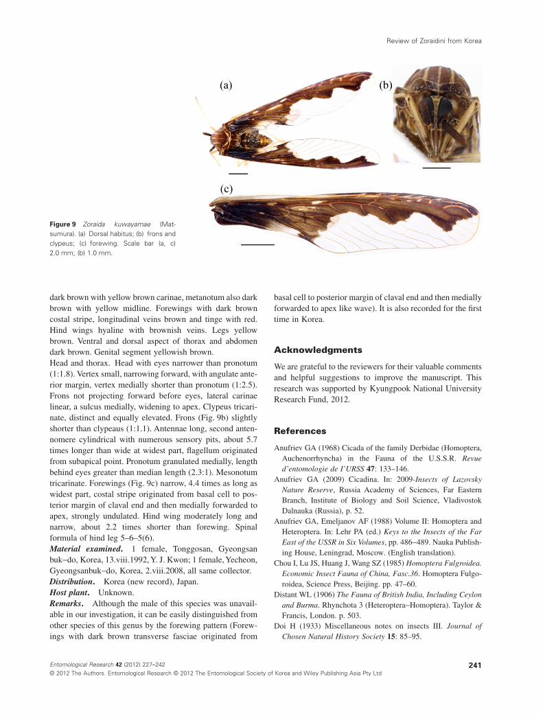
dark brown with yellow brown carinae, metanotum also darkbrown with yellow midline. Forewings with dark browncostal stripe, longitudinal veins brown and tinge with red.Hind wings hyaline with brownish veins. Legs yellowbrown. Ventral and dorsal aspect of thorax and abdomendark brown. Genital segment yellowish brown.Head and thorax. Head with eyes narrower than pronotum(1:1.8). Vertex small, narrowing forward, with angulate ante-rior margin, vertex medially shorter than pronotum (1:2.5).Frons not projecting forward before eyes, lateral carinaelinear, a sulcus medially, widening to apex. Clypeus tricari-nate, distinct and equally elevated. Frons (Fig. 9b) slightlyshorter than clypeaus (1:1.1). Antennae long, second anten-nomere cylindrical with numerous sensory pits, about 5.7times longer than wide at widest part, flagellum originatedfrom subapical point. Pronotum granulated medially, lengthbehind eyes greater than median length (2.3:1). Mesonotumtricarinate. Forewings (Fig. 9c) narrow, 4.4 times as long aswidest part, costal stripe originated from basal cell to pos-terior margin of claval end and then medially forwarded toapex, strongly undulated. Hind wing moderately long andnarrow, about 2.2 times shorter than forewing. Spinalformula of hind leg 5–6–5(6).Material examined. 1 female, Tonggosan, Gyeongsanbuk-do, Korea, 13.viii.1992, Y. J. Kwon; 1 female, Yecheon,Gyeongsanbuk-do, Korea, 2.viii.2008, all same collector.Distribution. Korea (new record), Japan.Host plant. Unknown.Remarks. Although the male of this species was unavail-able in our investigation, it can be easily distinguished fromother species of this genus by the forewing pattern (Forew-ings with dark brown transverse fasciae originated from
basal cell to posterior margin of claval end and then mediallyforwarded to apex like wave). It is also recorded for the firsttime in Korea.
Acknowledgments
We are grateful to the reviewers for their valuable commentsand helpful suggestions to improve the manuscript. Thisresearch was supported by Kyungpook National UniversityResearch Fund, 2012.
References
Anufriev GA (1968) Cicada of the family Derbidae (Homoptera,Auchenorrhyncha) in the Fauna of the U.S.S.R. Revued’entomologie de I’URSS 47: 133–146.
Anufriev GA (2009) Cicadina. In: 2009-Insects of LazovskyNature Reserve, Russia Academy of Sciences, Far EasternBranch, Institute of Biology and Soil Science, VladivostokDalnauka (Russia), p. 52.
Anufriev GA, Emeljanov AF (1988) Volume II: Homoptera andHeteroptera. In: Lehr PA (ed.) Keys to the Insects of the FarEast of the USSR in Six Volumes, pp. 486-489. Nauka Publish-ing House, Leningrad, Moscow. (English translation).
Chou I, Lu JS, Huang J, Wang SZ (1985) Homoptera Fulgroidea.Economic Insect Fauna of China, Fasc.36. Homoptera Fulgo-roidea, Science Press, Beijing. pp. 47–60.
Distant WL (1906) The Fauna of British India, Including Ceylonand Burma. Rhynchota 3 (Heteroptera-Homoptera). Taylor &Francis, London. p. 503.
Doi H (1933) Miscellaneous notes on insects III. Journal ofChosen Natural History Society 15: 85–95.
Figure 9 Zoraida kuwayamae (Mat-sumura). (a) Dorsal habitus; (b) frons andclypeus; (c) forewing. Scale bar (a, c)2.0 mm; (b) 1.0 mm.
Review of Zoraidini from Korea
241Entomological Research 42 (2012) 227–242© 2012 The Authors. Entomological Research © 2012 The Entomological Society of Korea and Wiley Publishing Asia Pty Ltd
Doi H (1936) Miscellaneous notes on insects VII. Journal ofChosen Natural History Society 21: 102–108.
Emeljanov AF (1995) On the system and phylogeny of the familyDerbidae (Homoptera, Cicadina). Entomologicheskoeobozrenie 73(4): 783–811. [In Russian. English translationpublished in Entomological review 75(2): 70-100].
Forest Research Institute (1995) A List of Insect Pests of Treesand Shrubs in Korea. Samjeong Publishing, Seoul. p. 360.
Ishihara T (1965) Homoptera. Iconographia Insectorum Japoni-corum. Colore Naturali Edita, 3, Hokyuryukan, Tokyo, pp.109–136.
Kirkaldy GW (1900) Rhynchota miscellanea. Entomologist 33:296–297.
Korean Society of Plant Protection (1986) A List of Plant Dis-eases, Insect Pests, and Weeds in Korea, 2nd edn. Suweon,Seoul. p. 633. (in Korean).
Kwon YJ, Huh EY (1995) A check list of the Auchenorrhynchafrom Chejudo (Homoptera): 19-54. In: Park KT (ed.), Biodi-versity (Insects) of Hallasan I. Insecta Koreana Suplement 5:1-214.
Kwon YJ, Huh EY (2001) Suborder Acuchenorrhyncha. Eco-nomic Insects of Korea 19. Insecta Koreana Supplement 26:320-329.
Kwon YJ, Lee CE, Lee WK, Huh EY (1994) Order 21. Homop-tera: 82-113. In: H.S. Ryu et al., Check list of Insects fromKorea. Entomological Society of Korea & Korean Society ofApplied Entomology, 744 pp. Kon-Kuk Univ. Press, Seoul. (inKorean).
Lee BY, Chung YJ (1997) Insect Pests of Trees and Shrubs inKorea. Seong An Dang Publishing, Seoul.p.459. (in Korean).
Lee CE (1979) Illustrated Flora and Fauna of Korea, Vol. 23(Insecta VII). Ministry of Education, Seoul.p.1070.
Lee CE, Kwon YJ (1977) Studies on the spittlebugs, Leafhoppersand planthoppers (Auchenorrhyncha, Homoptera, Hemiptera).Nature & Life 7(2): 55–111.
Lee CE, Kwon YJ (1979) A check list of Auchenorrhyncha fromKorea (Homoptera). In: Lee CE (ed.) Illustrated Flora &Fauna of Korea 23(Insecta VII): 799-1018.
Liang AP, Suwa M (1998) Type specimens of Matsumura’sspecies of Fulgoroidea (excluding Delphacidae) in theHokkaido University insect collection, Japna (Hemiptera:Fulgomorpha). Insecta Matsumurana 54: 133–166.
Matsumura S (1900) Uebersicht der Fulgoriden Japans. Ento-mologische Nachrichten 26(13, 14): 205–213.
Matsumura S (1913) Thousand Insects of Japan. Additamenta 1:1–184.
Matsumura S (1914) Beitrag zur kenntnis der Fulgoriden Japans.Annales Musei Nationalis Hungarici 12: 261–305.
Matsumura S (1935) A new genus and new species of Derbidaefrom Fukushima. Insecta Matsumurana 10(1, 2): 79–80.
Metcalf ZP (1945) Part IV. Derbidae. In: Metcalf ZP (ed.) 1954-General Catalogue of Homoptera, Fascicule IV, pp. 1–212.North Carolina State College, Raleigh(USA).
Morimoto K (1989) 21. Hemiptera, (a) Homoptera. In: HirashimaY and Daigaku K (eds) A Check List of Japanese Insects, Vol.1.pp. 82–151. Kyushu University, Fukuoka. (in Japanese).
Muir F (1913) On some new species of leafhoppers. Part II.Derbidae. Bulletin of the Hawaiian Sugar Planters’ Associa-tion. Division of Entomology 12: 28–92.
Muir F (1914) On some Derbidae from Formosa and Japan.Proceedings of the Hawaiian Entomological Society 3: 42–52.
Muir F (1917) The Derbidae of the Philippine Islands. PhilippineJournal of Science 12: 49–105.
Muir F (1918) Notes on the Derbidae in the British MuseumCollection II Derbidae. Entomologist’s Monthly Magazine 54:228–249.
Nast J (1972) Palaearctic Auchenorrhyncha (Homoptera), AnAnnotated Check List. Polish Academy of Sciences, Warszawa.pp. 77–80.
Schumacher F (1915) Der gegenwärtige Stand unserer Kenntnisvon der Homopteren-Fauna der Insel Formosa unter beson-derer Berücksichtigung von Sauter’schem Material. Mitteilun-gen aus dem Zoologischen Museum in Berlin 8: 73–134.
Szwedo J (2006) First fossil record of Cedusini in the EoceneBaltic amber with notes on the tribe (Hemiptera: Fulgoromor-pha: Derbidae). Russian Entomological Journal 15(3): 327–333.
Szwedo J, Adamczewska M (2004) Notes on some oriental Der-bidae (Hemiptera: Fulgoroidea) with description of newspecies. Zootaxa 597: 1–11.
Uhler PR (1896) Summary of the Hemiptera of Japan presented tothe United States National Museum by Professor Mitzukuri.Proceedings of the United States National Museum, Washing-ton 19: 255–297.
Yang C-T, Wu R-H (1993) Derbidae of Taiwan (Homoptera:Fulgoroidea). Department of Entomology, National ChungHsing University, Taichung, Taiwan.p.230.
Zelazny B, Webb MD (2011) Revision of the planthopper tribeRhotanini (Hemiptera: Auchenorrhyncha: Derbidae). Zootaxa3071: 1–307.
M. A. Rahman et al.
242 Entomological Research 42 (2012) 227–242© 2012 The Authors. Entomological Research © 2012 The Entomological Society of Korea and Wiley Publishing Asia Pty Ltd