Embed Size (px)
Citation preview

Neurotherapeutics: The Journal of the American Society for Experimental NeuroTherapeutics
Tau-Based Treatment Strategies in Neurodegenerative Diseases
Anja Schneider*† and Eckhard Mandelkow‡
*Department of Psychiatry and Psychotherapy, Von-Siebold-Strasse 5, University of Goettingen, 37075 Goettingen, Germany;†Max-Planck-Institute for Experimental Medicine, Hermann-Rein-Strasse 3, 37075 Goettingen, Germany; and ‡Max-Planck-Unit
for Structural Molecular Biology, c/o DESY, Notkestrasse 85, 22607 Hamburg, Germany
Summary: Neurofibrillary tangles are a characteristic hallmarkof Alzheimer’s and other neurodegenerative diseases, such asPick’s disease (PiD), progressive supranuclear palsy (PSP),corticobasal degeneration (CBD), and frontotemporal dementiaand parkinsonism linked to chromosome 17 (FTDP-17). Thesediseases are summarized as tauopathies, because neurofibrillarytangles are composed of intracellular aggregates of the micro-tubule-associated protein tau. The molecular mechanisms oftau-mediated neurotoxicity are not well understood; however,pathologic hyperphosphorylation and aggregation of tau play acentral role in neurodegeneration and neuronal dysfunction.The present review, therefore, focuses on therapeutic ap-
proaches that aim to inhibit tau phosphorylation and aggrega-c/o DESY, Notkestrasse 85, 22607 Hamburg, Germany. E-mail:[email protected].
Vol. 5, 443–457, July 2008 © The American Society for Experimen
tion or to dissolve preexisting tau aggregates. Further experi-mental therapy strategies include the enhancement of tauclearance by activation of proteolytic, proteasomal, or autopha-gosomal degradation pathways or anti-tau directed immuno-therapy. Hyperphosphorylated tau does not bind microtubules,leading to microtubule instability and transport impairment.Pharmacological stabilization of microtubule networks mightcounteract this effect. In several tauopathies there is a shifttoward four-repeat tau isoforms, and interference with thesplicing machinery to decrease four-repeat splicing might beanother therapeutic option. Key Words: Tau, Alzheimer’sdisease, phosphorylation, aggregation, neurodegeneration,
therapy.INTRODUCTION
Cell biology and function of tauThe microtubule-associated protein (MAP) tau is ex-
pressed mainly in neurons, where it regulates microtu-bule assembly and stability.1,2 The synthesis of tau isupregulated along with tubulin during neuronal differen-tiation; in mature neurons, tau occurs mostly in the axonand is largely excluded from the somatodendritic com-partment, whereas other MAPs (e.g., MAP2) predomi-nate in dendrites.3,4 Microtubules must be able to dy-namically grow and shrink, to support cellular shapechanges. This is a prerequisite to establishing neuronalpolarity and axonal outgrowth.5,6 In addition, microtu-bules serve as tracks for the transport of cellular cargoes(vesicles, organelles) along axons and dendrites.7 Boththe dynamics and transport properties of microtubulesare regulated by MAPs, such as tau. Their interactionsare in turn regulated by post-translational modifications,such as phosphorylation.8,9
Address correspondence and reprint requests to: Eckhard Man-delkow, Ph.D., Max-Planck-Unit for Structural Molecular Biology,
Due to its content of hydrophilic residues, tau is a highlysoluble protein.10 It represents a prototype of the class ofnatively unfolded (or intrinsically unstructured) proteins.11
Six isoforms of tau are expressed in the adult human CNSby alternative splicing of exons 2, 3, and 10 of the singleMAPT gene (alias TAU) on chromosome 17q21.3; MAPTcomprises 16 exons (FIG. 1B).12,13 Tau isoforms differ bythe absence or presence of one or two N-terminal inserts(N1, N2), as well as exclusion or inclusion of the second offour pseudo-repeats (�31 residues each) in the microtu-bule-binding domain (3R, 4R) (FIG. 1A).10,13 Tau-micro-tubule binding is mediated by the repeat domain and re-quires the presence of both N- and C-terminally flankingproline-rich domains which target tau to the microtubulesurface.14 The number of repeats can modulate tau–mi-crotubule affinity.15–17 Consistent with the weaker mi-crotubule binding of three-repeat (3R) tau isoforms, only3R tau is expressed during neurogenesis, when highermicrotubule dynamics are required.18 Phosphorylation ofthe repeat region and, to a lesser extent, also of theflanking regions impairs the tau–microtubule interactionand leads to tau detachment from microtubules.19,20
Whereas the microtubule interaction domain of tau
resides in the C-terminal half (the assembly fragment),tal NeuroTherapeutics, Inc. 443
SCHNEIDER AND MANDELKOW444
FIG. 1. Diagram of tau domains, mutations, phosphorylation sites, and antibody epitopes. (A) Human tau contains between 352 and 441residues, depending on the splicing isoform. The isoforms are generated by the presence or absence of inserts I1, I2, and R2. The N-terminalhalf constitutes the projection domain, which protrudes away from the microtubule surface. The C-terminal half constitutes the assemblydomain, which binds and stabilizes microtubules. The core of this domain is formed by the three or four repeats of �31 residues each (R1–R4;R2 may be absent in the three-repeat isoforms of tau). The repeat domain is also responsible for the aggregation of tau into PHFs. The diagramshows many of the mutations observed in cases of FTDP-17. Most of them map to the repeat domain and may influence microtubule bindingor PHF aggregation. �K280 and P301L are two FTDP-17 mutations which are prone to aggregation. (B) Diagram of the tau gene in the regioncovering the repeat domain (exons 9–13). The MAPT gene consists of 16 exons. Exons 1 and 14 are not translated. The repeat domain isencoded by exons 9–12. Exclusion or inclusion of exon 10 gives rise to three-repeat or four-repeat tau isoforms. Most intronic tau mutationslocalize to the 5= region of intron 10, they increase the incorporation of exon 10 (increased expression of 4R-isoforms), but they are silent onthe protein level. Exon 10 mutations can also shift splicing toward four-repeat tau (for details, see http://www.alzforum.org).227 (C) Diagramof the longest tau isoform, tau441, with phosphorylation sites, kinases, and antibody epitopes. Tau isoforms differ by the presence or absenceof 2 N-terminal inserts N1, N2) and a second repetitive amino acid sequence, R2. Phosphorylation of S214 by PKA and S262/S356 by MARK,PKA, or other kinases leads to tau detachment from the microtubule. Phosphorylation of SP/TP sites in the flanking regions have a modulating
effect on tau-microtubule affinity. Epitopes of different diagnostic phospho-epitopes are indicated.Neurotherapeutics, Vol. 5, No. 3, 2008
TREATMENT STRATEGIES FOR TAUOPATHIES 445
the N-terminal half projects away from the microtu-bule surface.21 Its functions are not well defined. Itcould act as a spacer, or interact with other proteins—for example, signaling molecules such as kinases,phosphatases, heat shock proteins (HSPs) and othercytoskeletal elements.22–25 Because MAPs and micro-tubule motors both bind to the surface of microtubules,they can potentially interfere with each other. Thus,the overexpression of tau can retard motor-dependenttransport, particularly in the anterograde direc-tion.26 –29 This can lead to oxidative damage and syn-aptic degeneration.30,31
TauopathiesFilamentous or amorphous aggregates of hyperphos-
phorylated tau have been described in the context ofvarious neurodegenerative diseases. These tauopathiescover a wide spectrum of neurodegenerative diseaseswith prominent tau pathology, including dementia syn-dromes (Alzheimer’s disease [AD] and Pick’s disease),which can be accompanied by parkinsonism (progressivesupranuclear palsy [PSP], corticobasal degeneration[CBD], frontotemporal dementia and parkinsonismlinked to chromosome 17 [FTDP-17]), and motor neurondiseases (amyotrophic lateral sclerosis–Parkinson–de-mentia complex, or ALS-PDC).
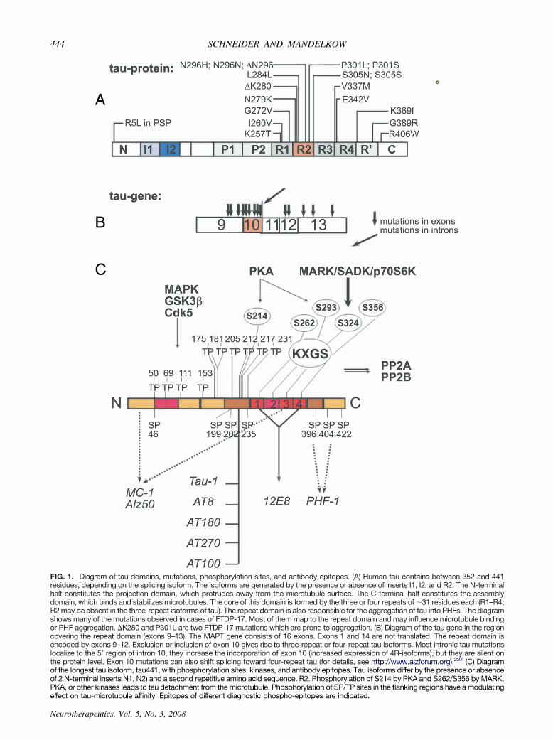
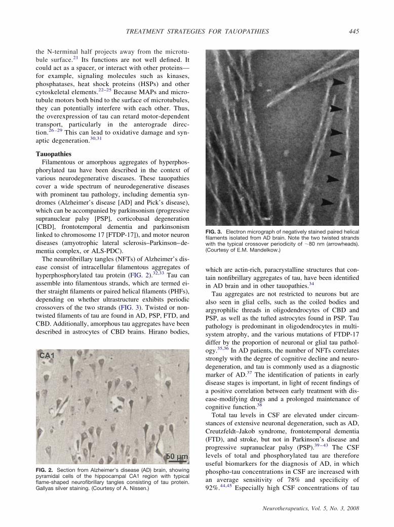
The neurofibrillary tangles (NFTs) of Alzheimer’s dis-ease consist of intracellular filamentous aggregates ofhyperphosphorylated tau protein (FIG. 2).32,33 Tau canassemble into filamentous strands, which are termed ei-ther straight filaments or paired helical filaments (PHFs),depending on whether ultrastructure exhibits periodiccrossovers of the two strands (FIG. 3). Twisted or non-twisted filaments of tau are found in AD, PSP, FTD, andCBD. Additionally, amorphous tau aggregates have beendescribed in astrocytes of CBD brains. Hirano bodies,
FIG. 2. Section from Alzheimer’s disease (AD) brain, showingpyramidal cells of the hippocampal CA1 region with typical
flame-shaped neurofibrillary tangles consisting of tau protein.Gallyas silver staining. (Courtesy of A. Nissen.)which are actin-rich, paracrystalline structures that con-tain nonfibrillary aggregates of tau, have been identifiedin AD brain and in other tauopathies.34
Tau aggregates are not restricted to neurons but arealso seen in glial cells, such as the coiled bodies andargyrophilic threads in oligodendrocytes of CBD andPSP, as well as the tufted astrocytes found in PSP. Taupathology is predominant in oligodendrocytes in multi-system atrophy, and the various mutations of FTDP-17differ by the proportion of neuronal or glial tau pathol-ogy.35,36 In AD patients, the number of NFTs correlatesstrongly with the degree of cognitive decline and neuro-degeneration, and tau is commonly used as a diagnosticmarker of AD.37 The identification of patients in earlydisease stages is important, in light of recent findings ofa positive correlation between early treatment with dis-ease-modifying drugs and a prolonged maintenance ofcognitive function.38
Total tau levels in CSF are elevated under circum-stances of extensive neuronal degeneration, such as AD,Creutzfeldt–Jakob syndrome, frontotemporal dementia(FTD), and stroke, but not in Parkinson’s disease andprogressive supranuclear palsy (PSP).39–43 The CSFlevels of total and phosphorylated tau are thereforeuseful biomarkers for the diagnosis of AD, in whichphospho-tau concentrations in CSF are increased withan average sensitivity of 78% and specificity of
FIG. 3. Electron micrograph of negatively stained paired helicalfilaments isolated from AD brain. Note the two twisted strandswith the typical crossover periodicity of �80 nm (arrowheads).(Courtesy of E.M. Mandelkow.)
92%.44,45 Especially high CSF concentrations of tau
Neurotherapeutics, Vol. 5, No. 3, 2008

SCHNEIDER AND MANDELKOW446
phosphorylated at T181, T231, S199, and S396/404(FIG. 1B) have been suggested to be reliable predic-tors for the conversion of mild cognitive impairmentinto AD.46 – 48 Noninvasive diagnostic methods fortauopathies include positron emission tomography im-aging with plaque-binding and especially tangle-bind-ing tracers such as FDDNP, similar to the amyloid-binding radiotracer Pittsburgh compound B.49,50
PATHOPHYSIOLOGY
In AD, tau pathology is accompanied by extracellularamyloid plaques and intracellular NFTs that are distrib-uted mostly in the entorhinal region, hippocampus, andcortex. This is in contrast to other tauopathies, in whichamyloid plaques are absent and NFTs are preferentiallylocalized in different brain regions, such as the brainstem and basal ganglia (in PSP) or in the frontotemporalcortex (in FTDP-17). The identification of tau mutationsin FTDP-17 families and in sporadic FTD has providedcompelling evidence for a causative role for tau in neu-rodegeneration, rather than tau pathology being a mereside effect observed in neurodegenerative diseases.51–53
Tau mutations in FTDP-17 are either intronic, local-ized close to the splice-donor site following exon 10 andresulting in overproduction of four-repeat tau isoforms,or comprise missense, deletion, or silent mutations in thecoding regions (FIG. 1B).52,53 The mutations lie mostlyin or near the repeat domain, which constitutes the coreof the microtubule binding domain and also the PHFassembly domain; the mutations result in reduced micro-tubule binding ability or in enhanced tau aggregation intoPHFs (R5L, K257T, G272V, �K280, P301L, P301S,V337M, and R406W).54–60 Mutations in exon 10 canaffect both protein properties and exon 10 splicing (e.g.,�K280, �N296 and N296H), the latter shifting the iso-form ratio of three- to four-repeat tau from approxi-mately 1:1 in healthy individuals toward the four-repeatisoforms.61,62 Similar findings have been obtained inPSP and CBD, two tauopathies that share the MAPT H1haplotype as a risk factor.63,64 Filamentous tau aggre-gates in PSP and CBD consist of four-repeat isoforms.65
Why tau, a highly soluble protein, aggregates intoPHFs is still a matter of debate. In AD brains, tau isredistributed from the axonal to the somatodendriticcompartment and is hyperphosphorylated at 25 differentsites at least, which are known to interfere with theaffinity of tau–microtubule binding (FIG. 1C).66 Patho-logic hyperphosphorylation might thus lead to tau-mi-crotubule detachment, subsequent instability of microtu-bule networks, impaired axonal transport and synapticdegeneration (FIG. 4). In addition to this loss-of-functioneffect, caused by microtubule instability, a toxic gain-of-function was assigned to aggregated PHF-tau (FIG.
4).67–69 Moreover, hyperphosphorylated, nonaggregatedNeurotherapeutics, Vol. 5, No. 3, 2008
tau might be toxic by itself, as suggested by recent find-ings in Drosophila fruit fly and mouse models.70–73
Prevention of mis-splicing, hyperphosphorylation, andaggregation of tau are therefore promising therapeutictargets to interfere with tau-based toxicity and neurode-generation. New approaches currently under intense in-vestigation include various kinase and aggregation inhib-itors, microtubule-stabilizing agents, enhancers of tauclearance (e.g., HSP90 inhibitors, immunotherapy), andrecently described techniques to shift the ratio of mis-spliced tau toward three-repeat isoforms.
TAU-BASED THERAPEUTIC APPROACHES
Antiphosphorylation strategiesThe largest tau isoform contains 45 serine (S), 35
threonine (T), and 5 tyrosine (Y) residues (FIG. 1C).Among these, 17 sites are part of serine/proline (SP) orthreonine/proline (TP) motifs and represent targets ofproline-directed SP/TP protein kinases such as glycogensynthase kinase 3� (GSK-3�), cyclin-dependent kinases(cdk5, cdc2), mitogen-activated protein kinase (MAPK),
FIG. 4. Possible mechanisms of tau toxicity. (1) Microtubulesstabilized by tau protein support axonal transport by motor pro-teins. (2) Excess bound tau can reduce the attachment of motorproteins and thus impair axonal traffic. (3) In AD, tau becomeshyperphosphorylated by several protein kinases (e.g., MARK)and detaches from microtubules. The microtubules break down(resulting in perturbed axonal transport) and the detached tauself-assembles into paired helical filaments. (4) The PHFs bundletogether and form neurofibrillary tangles that obstruct the cellinterior.27
stress-activated kinases (JNK, p38), and others.74,75 Ad-

TREATMENT STRATEGIES FOR TAUOPATHIES 447
ditional sites are targeted by different kinases, includingcAMP-dependent protein kinase (PKA), protein kinase C(PKC), calcium/calmodulin dependent protein kinase II(CaMKII), serum- and glucocorticoid-inducible kinase(SGK), protein kinase B (PKB), microtubule-affinityregulating kinase (MARK), and SAD kinase (SADK).76
The KIGS or KCGS motifs in the repeat domain (S262,S293, S324, S356) can be phosphorylated by MARK,PKA, PKB, SADK, CaMKII, and p70S6K.77–80 PKAadditionally phosphorylates other sites including S214, aphospho-epitope that is especially upregulated duringmitosis.81 In AD brain, tau is hyperphosphorylated atnearly all phosphorylation sites, with approximately ninephosphates per molecule, in contrast to the two to threephosphorylated residues observed in healthy controlbrains.82
Various antibodies directed against these phospho-epitopes preferentially recognize AD brain-derived tauand are used as diagnostic tools. Phosphorylation ofS214 or the KXGS sites within the repeat domainstrongly inhibits tau–microtubule binding, thereby rais-ing soluble tau concentrations and increasing microtu-bule instability. Tau-microtubule detachment seems to bean early event in the course of the disease, becausepretangle tau is phosphorylated at S262/S356 (12E8epitope) in AD brains and in mouse models of tauopa-thy.83,84 Similarly, S214 phosphorylation was found toprecede PHF formation in a triple transgenic mousemodel of mutant APP, presenilin, and the FTDP-17 taumutation P301L.85 However, phosphorylation of theKXGS motifs, especially S262, has been shown to in-hibit the aggregation of tau into PHFs in vitro.86 Ratherthan promoting tau aggregation, the detachment frommicrotubules might render free cytosolic tau more acces-sible to phosphorylation by different SP/TP directed ki-nases than microtubule-bound tau would be.
There is a widespread assumption that hyperphos-phorylation at SP or TP motifs leads to aggregation(although this is a matter of debate). Independently ofaggregation, phosphorylation may be toxic to neurons(e.g., by microtubule destabilization, repeat domainphosphorylation) or by other mechanisms (e.g., acti-vation of the mTOR pathway, SP/TP phosphoryla-tion).87 Most approaches to interfering with tau phos-phorylation rely on inhibiting different kinases oractivation of phosphatases.88
MARK. The KXGS motifs in the repeat region oftau are phosphorylated by MARK/Par-1. The decreasedmicrotubule affinity of KXGS phosphorylated tau andsubsequent microtubule instability77 might be the mech-anism by which MARK exerts its function in cell differ-entiation, neurite outgrowth, and maintenance of axonaltransport.89–91 The PHF tau is hyperphosphorylated at
S262/S356, and fluorescence resonance energy transferexperiments revealed active MARK in close proximity tophosphorylated S262/S356 residues in AD brain PHFs.92
MARK/Par-1 mediated S262/S356 phosphorylationwas shown to enhance the toxic phenotype caused byhuman tau expression in the Drosophila fruit fly ret-ina, which was absent in a S262A/S356A expressingmutant.72 In addition, S262A/S356A mutants wereless phosphorylated at SP/TP sites that become hyper-phosphorylated during disease progression.83 Thisfinding hints toward a priming effect of S262/S356phosphorylation for subsequent pathological tauhyperphosphorylation.
MARK/Par-1 belongs to the calcium/calmodulin-de-pendent protein kinases. Its activity depends on phos-phorylation by activating kinases (MARK kinase/thou-sand and one amino acid kinase [MARKK/TAO-1] orLKB1).91 Inhibitory regulation of MARK can addition-ally be achieved by binding to PAK5 or to scaffoldingproteins or by subcellular localization.91,93 The activat-ing MARKK is a member of the Ste20 family of kinasesand phosphorylates MARK at a threonine residue in theactivation loop. Other functions of MARKK/TAO-1 in-volve mitosis progression and MAPK/p38 signalingupon stress induced activation.94,95 By these mecha-nisms, apart from the toxic effects of S262/S356 phos-phorylated tau, MARKK might contribute to neurode-generation and cell death.
Binding of testis-specific protein kinase 1 (TESK1)inhibits MARKK activity by protein–protein interac-tion.96,97 TESK1 is similar to LIM kinase, in that itphosphorylates cofilin and plays a role in the organiza-tion of the actin cytoskeleton.97–99 Note that active LIMkinase (LIMK) is also a downstream effector of A�-mediated toxicity.100 Pharmacological inhibitors ofMARK/MARKK include staurosporine (unselectively)and hymenialdisine, which also inhibits GSK3�.90,101
Given the important and early role of MARK phosphor-ylation in the pathogenesis of tauopathies, the applicationof MARK inhibitors would be a worthwhile approach.
PKA. cAMP-dependent protein kinase A (PKA)has been shown to phosphorylate tau at S262/S356 andS214 in vitro and in vivo.102,103 Phosphorylation by PKAresults in reduced microtubule-binding affinity and pro-motes the subsequent phosphorylation of different diag-nostic phospho-epitopes. PKA phosphorylation at S214is required to prime tau for its sequential phosphorylationby GSK-3�, thereby giving rise to the AD-specificAT100 phospho-epitope.102 Activation of PKA by fors-kolin resulted in spatial memory deficits in rats.103 Iso-quinoline sulfonamide derivatives and staurosporines areknown to inhibit PKA with low selectivity and alsoaffect PKG, PKC, and the myosin light chain kinase(MLCK). PKA inhibitors with higher selectivity are KT-5720 and PKI. PKI binds to the catalytic subunit of PKA
and promotes its nuclear export, thereby preventingNeurotherapeutics, Vol. 5, No. 3, 2008

SCHNEIDER AND MANDELKOW448
cAMP response element regulated gene expression.104
However, none of these inhibitors have been tested inAD model systems to date.
Cdk5. Cyclin dependent kinase 5 (Cdk5) accumu-lates in tangle-bearing neurons of AD brains, and kinaseactivity was found to be upregulated in AD.105 Cdk5 isregulated by the neuron-specific activator p35. The my-ristoylated and membrane-bound domain of p35 can becleaved by calpain to p25. This leads to a cytosolicredistribution of p25-bound cdk5 and aberrant tau phos-phorylation.106 Overexpression of p25 in a rodent modelresulted in increased tau phosphorylation and neurode-generation and SP/TP phosphorylation by cdk5 increasedtau toxicity in a Drosophila fruit fly model.71,107,108 Re-cent evidence suggests that cdk5/p25 might be a down-stream effector of A� mediated toxicity, leaving cdk5 asa promising therapeutic target.109 One must be aware,however, that the inhibition of cdk5 was accompanied byactivation of GSK-3� in neuronal cell culture and thatp35 knock-out mice revealed increased GSK-3� activityand tau phosphorylation.110,111
Cdk5 inhibition can be achieved by active site-di-rected inhibitors, by 2,6,9-trisubstituted purines andaloisines, which are small-molecule inhibitors that inter-fere with the cdk5/p25 complex formation (Cdk5 inhib-itory peptide [CIP]), and by calpain inhibitors, whichprevent p25 generation.101,112,113 The use of active siteinhibitors is hampered by their lack of selectivity towardcdk5. Indirubins, which belong to the chemical classindolinones, also inhibit GSK-3. Likewise, purines arenot highly selective for cdk5 and can inhibit ERK2. Thepurine roscovitine has been tested in p25 transgenicmice, and reduced tau phosphorylation and neurodegen-eration was demonstrated upon roscovitine treatment.Roscovitine shows a higher selectivity toward cdk5 thanolomoucine, another purine inhibitor of cyclin-dependentkinases, which has been tested successfully in NiemannPick type C (NPC) mice, and aminopurvalanol, whichefficiently inhibited cdk in vitro.114 Less cdk5 specificityis achieved by flavopiridol, indirubin, 1-aza-9-oxaflu-orenes, and aminothiazoles, all of which inhibit cyclin-dependent kinases in general in addition to GSK3.115 Thecdk5 inhibitory peptide (CIP) interferes with cdk5/p25binding and is more selective toward cdk5 than to othercyclin kinases. Calpain inhibitors have shown beneficialresults in AD models, however their clinical applicationis restricted by their poor ability to cross the blood–brainbarrier (BBB).116,117 In contrast, roscovitine has beendescribed to penetrate the BBB at a sufficient rate.118
GSK3. The GSK-3 isoforms � and � are encodedby two different genes.119 Both isoforms are involved inglucose metabolism, cell proliferation, wnt signaling,and apoptosis.120,121 GSK-3� activity is regulated byphosphorylation; S9 phosphorylation inhibits kinase ac-
tivity, and Y216 phosphorylation activates GSK-3�.121Neurotherapeutics, Vol. 5, No. 3, 2008
Tau has been identified as an important substrate ofGSK-3 in vitro and in vivo and can be phosphorylated atprimed (e.g., T231) and unprimed epitopes (PHF-1,Tau-1, and AT8).122–124 Priming involves the prior phos-phorylation of a serine or threonine residue, four aminoacids C-terminal to the target phosphorylation site.125
Active GSK-3 was detected in pretangle neurons andNFTs in AD brains, and coexpression of the GSK-3homolog shaggy with tau led to tau hyperphosphoryla-tion, filamentous tau aggregation, and neurotoxicity inDrosophila fruit fly.126,127 In line with these findings,GSK-3� overexpressing transgenic mice showed in-creased tau phosphorylation and deficits in spatial mem-ory.128,129 Tau phosphorylation by GSK-3 can be en-hanced by A� -treatment, which thus provides a possiblelink between tau and A� pathology.130 GSK-3� activityis negatively regulated by phosphorylation at S9 throughdifferent kinases (e.g., AKT–protein kinase B).
Various small-molecule inhibitors such as flavopiri-dol, paullones, and aloisines are directed against theATP-binding site of GSK-3 and therefore display a lowselectivity toward GSK-3� or -�, and most of them alsotarget cdk5. The recently described staurosporine-de-rived bisazaindolylmaleimide derivatives as well aspyrazolopyrazines and pyrazolopyridines are character-ized by a higher selectivity toward GSK-3�. Oral admin-istration of the aminothiazole AR-A014418 resulted indecreased tau aggregation in a transgenic mouse modelof P301L tau.131,132
Lithium, a well-characterized mood-stabilizer, com-petes with magnesium for GSK-3 binding; it leads toreduced tau-phosphorylation, aggregation and axonal de-generation in transgenic mice.133,134 Valproate, anothermood-stabilizing drug (and an antiepileptic), also inhibitsGSK-3� and is currently being tested in clinical ADtrials.
Phosphatases. Tau is dephosphorylated by proteinphosphatase-2A (PP-2A) and to a minor extent by PP-1and PP-2B (calcineurin).23,135 In AD brain, the mRNAlevels of PP-2A and PP-1 are reduced compared withcontrols.136,137 A decrease in phosphatase activity mightresult in impaired tau dephosphorylation, as well as en-hanced tau phosphorylation, because various tau-directedkinases are activated by phosphorylation themselves. In-deed, inhibition of PP-2A by okadaic acid resulted inAD-like tau hyperphosphorylation in rat brain slice cul-tures.138 To date, no pharmacologic approaches to acti-vate PP-2A have been reported. However, memantine, anN-methyl-D-aspartate (NMDA) receptor antagonist thatis well established for the treatment of AD, has beendescribed as antagonizing the okadaic acid–mediated in-hibition of PP-2A in hippocampal slice cultures.139
These findings further support the choice of phospha-
tases as potential pharmacological targets in AD.
TREATMENT STRATEGIES FOR TAUOPATHIES 449
Antiaggregation strategiesTau, a natively unfolded protein, is highly soluble and
does not readily aggregate into filaments. In vitro, poly-anionic cofactors such as glycosaminoglycans, acidicpeptides, RNA, or fatty acid micelles can promote PHFassembly.140–143 Aggregation is further promoted byhigh tau concentrations. In vitro studies have shown thattau phosphorylation is not necessary to drive tau intoPHFs.140,141 On the contrary, phosphorylation of theKXGS motifs in the repeat region inhibits tau aggrega-tion in vitro.86 Tau comprises two hydrophobic hexapep-tide motifs144,145 in the repeat domains (275VQIINK280at the beginning of R2 and 306VQIVYK311 at the be-ginning of R3) that form a cross �-structure and make upthe PHF core. The N- and C-terminal flanking regionsare thought to form the PHF fuzzy coat.146 Proteolyticcleavage of the inhibitory N- and C-termini exposes theaggregation-prone hexapeptide motifs, thereby greatlyfacilitating the nucleation of filaments.144,147
There has been debate on what form of tau is the toxicspecies for neurons. Elevation of tau, phosphorylation oftau at different sites, and truncation by different pro-teases have been investigated.148–150 Recent findingspoint to toxic functions of tau aggregates. For example,in stable tau cell lines, toxicity paralleled tau aggregationand removal of aggregates rescued the toxic pheno-type.151,152 Similarly, in inducible transgenic mice, onlythe proaggregation tau was toxic; antiaggregation tauwas not.68,69 Thus, preventing the buildup of aggregatesby small-molecule inhibitors could be a promising strat-egy in the treatment of tauopathies. The non-neurolepticphenothiazine methylene blue, which penetrates theBBB, and its desmethyl derivatives have been describedto inhibit tau aggregation, with Ki values in the nanomo-lar range.153 Recent high-throughput screens yielded var-ious potential drug candidates. Members of different sub-stance classes, such as anthraquinones, N-phenylamines,phenylthiazolhydrazides, and rhodanines, can inhibit tauaggregation and, even more importantly, disassemble ex-isting filaments.67,154–156 The respective lead substanceswere identified in a high-throughput in vitro screen ofapproximately 200,000 compounds followed by chemi-cal modification and retesting in a tau-expressing stableneuronal cell line with marked tau aggregation (FIG. 5).157
PTH. The phenylthiazolyl-hydrazides (PTH) (FIG.5) were found by in silico screening of 77 primary screenhits. From this, 49 PTH derivatives were synthesized anda PTH lead structure as well as a structure–activity rela-tionship could be verified. The structure–activity rela-tionship distinguishes PTHs from nonselective inhibitorswithout a structure–activity relationship.155 The tau–PTH interaction was studied by saturation transfer dif-ference NMR and appeared to be hydrophobic.158 Theexact structural interaction of tau and PTH still needs to
be determined. One could envision interference with nu-cleation or elongation steps of tau assembly, either me-diated by steric inhibition of tau–tau interaction, stabili-zation of a non–aggregation prone tau conformation orinteraction with polyanionic cofactors of aggregation.
Rhodanines and anthraquinones. The recentlypublished substance class of rhodanines is preferable toanthraquinones, due to reduced cytotoxicity and absenceof mutagenicity. Notably, a rhodanine derivative, thealdose reductase inhibitor epalrestat, showed no toxicside effects in a long-term clinical trial.159,160 Differentrhodanine derivatives inhibit tau aggregation and pro-mote filament disassembly in a cell culture model, withefficacies in the nanomolar range and in the absence ofobvious cytotoxicity or interference with microtubulestability.156
N-phenylamines. N-phenylamines were proved tohave a comparable activity as rhodanines, but show alower pharmacological potency in vitro.67 It will now beinteresting to test these substances in mouse models forbioavailability and pharmacokinetics.
Thiacarbocyanines. Another class of small-mole-cule inhibitors of tau aggregation are thiacarbocyaninedyes, such as N744, which were identified by in vitroscreens.161,162 A multivalent cyane derivative, the cyclicbis-thiacarbocyanine, inhibits tau aggregation in vitrowith approximately a fourfold higher potency than themonovalent cyane itself.163 The mechanism by which
FIG. 5. Tau mutant expression, aggregation, and inhibition in acellular model. The left panels show the expression of the taurepeat domain with the �K280 mutation after addition of doxy-cycline in an inducible N2a cell line, stained with tau antibody(red). Tau aggregates are formed and can be detected by thefluorescence of thioflavine S (right, green). Top row, control with-out aggregation inhibitor compound, bottom row, with inhibitorBSc3551 (a phenylthiazolyl hydrazide derivative). Note thestrong reduction of aggregation by the compound. Scale bar: 20�m. (Modified from Pickhardt et al.155)
multivalent ligands interfere with protein aggregation
Neurotherapeutics, Vol. 5, No. 3, 2008
SCHNEIDER AND MANDELKOW450
has been speculated to involve their binding to oli-gomers, thereby preventing incorporation of new mole-cules into filaments.164 Alternatively, these small-mole-cule inhibitors might decrease the concentration ofaggregation-prone proteins by sequestration into pro-tein–inhibitor complexes.165 Developing multivalentcyanes, which are characterized by an improved potencydue to an increased local concentration of ligands, mighttherefore be a promising strategy.
Tau clearanceAnother possible approach to prevent tau toxicity
could be the augmentation of its cellular clearance, aswell as the degradation of tau aggregates.
Proteasome and autophagosome activation. Ubiq-uitin–proteasome and autophagy–lysosomal pathwaysare the two major means of protein degradation withinthe cell.166 Oligomers or higher-order aggregates cannotenter the proteasome for steric reasons, but will be de-graded by autophagy. During autophagy, a double mem-brane is formed around the intracellular aggregate. Thisautophagosome then fuses with the lysosome (autolyso-some), where acidic lysosomal hydrolases degrade thecontents of the vesicle.167 Autophagy has been describedfor intracellular protein aggregates such as polyQ-hun-tingtin and �-synuclein aggregates, and also fortau.168,169 Rapamycin inhibits mTOR (mammalian targetof rapamycin), thereby promoting autophagy. In a Dro-sophila fruit fly model expressing wild-type or R406Wmutant tau, treatment with rapamycin induced autoph-agic tau degradation and diminished tau-induced toxi-city.169 It has been speculated that the nonclassic, pro-teasome-independent K63 ubiquitin pathway is involvedin the autophagic route of tau. This notion is supportedby the finding that P301L tau-positive inclusions in aneuronal cell line were cleared after stimulation of auto-phagy, a mechanism that was impaired by coexpressionof the ubiquitin mutant K63R.170
Note, however, that the beneficial effect on tau clear-ance by stimulating autophagy might be counteracted byincreased A� production in the autophagosomal system,as has been proposed by Rubinsztein et al.171,172
The chaperones HSP27 and HSP90 are involved inphosphorylation-dependent proteasomal degradation oftau.24,173 HSP90-interacting proteins are degraded by theproteasome upon pharmacological inhibition of HSP90.Phosphorylation of the flanking regions enhanced theproteasomal degradation of tau in transfected cell linesthat were treated with HSP90 inhibitors.173 Phosphory-lation of S262/S356 protected tau against proteasomaldegradation. The HSP90 inhibitor geldanamycin is notfeasible for clinical studies because of hepatotoxic sideeffects, but its less toxic derivatives 17-AAG and 17-DMAG have been tested as anticancer treatment in a
clinical setting.174 Their pharmacological properties,Neurotherapeutics, Vol. 5, No. 3, 2008
however, suggest a poor ability to cross the BBB. Re-cently, several HSP90 inhibitors of low molecular weighthave been described, which might reach sufficient ther-apeutic CNS concentrations.173
PHF-tau has been shown to be ubiquitinated at least atfour different sites, and through different lysine linkageson ubiquitin.175,176 Tau is predominantly monoubiquiti-nated, and only a minor part of PHF-tau is linked topolyubiquitin chains by Lys42. Conjugation sites for mo-noubiquitination have been identified within the repeatregion. They include Lys254, Lys257, Lys311, andLys317, with Lys311 being a major linkage site.176 TheHSP70 co-chaperone CHIP (the acronym stands for thecarboxyl terminus of the Hsc70-interacting protein)functions as the primary E3-ligase in ubiquitin-depen-dent tau clearance.177–180 In line with the important func-tion of CHIP in tau clearance, increased tau accumula-tion was reported in CHIP knock-out mice.178 Activationof the co-chaperone CHIP could prove to be an attractivedrug target, especially because it also stabilizes fulllength APP against secretase cleavage. In addition, CHIPdirects APP to proteasomal degradation, by promotingAPP ubiquitination. It also accelerates A�42 removal andcan protect against A� toxicity.181
Proteases. It has been assumed that cytosolic pro-teases might also play a role in tau degradation. Re-cently, the puromycin-sensitive aminopeptidase PSA, a100-kDa M1 metalloprotease, was identified by agenomic approach screening for modifiers of tauopa-thy.182 Besides its function in protein turnover, PSA isinvolved in cell cycle regulation, and a wide range ofPSA substrates are known, including enkephalins, cho-lecystokinin, and somatostatin, among many oth-ers.183,184 Tau has been identified as a substrate of PSAin vitro. PSA mediated amino-terminal tau degradationof AD brain-derived soluble or insoluble tau was im-paired, relative to control tau.185 At first sight, activationof PSA appears to be an attractive therapeutic target, butregarding the large number of physiologic processes inwhich PSA is involved, side effects are likely to occur.
When activating tau proteolysis, it has to be consid-ered that proteolytic tau fragments of the repeat domainmight act as a seed for tau aggregation. A stepwiseproteolysis of the tau repeat domain has been describedin an inducible FTDP-17�280 mutant tau cell line.151
There, a PHF nucleating fragment was generated bysubsequent cleavage, first between K257/S258, followedby cleavage at I360 or K353. The responsible proteasehas not yet been identified, and it is not known whetherit is upregulated in AD. This could, however, be a prom-ising target for pharmacological inhibition.
Two cysteine proteases, caspase-3 and calpain-1, havebeen described as downstream targets of A�-inducedtoxicity in cell culture models. In cultured hippocampal
neurons, A�-induced toxicity was mediated by calpain-1
TREATMENT STRATEGIES FOR TAUOPATHIES 451
activation and generation of a 17-kD tau fragment. Bothtau fragmentation and neurotoxic effects of A� could bediminished by calpain-1 inhibition.186 To date, however,no direct proof for the suggested neurotoxicity of the17-kD tau fragment has been provided.
Tau can be cleaved in vitro by the serine-aspartyl pro-tease caspase-3 at Asp421, which increased and acceleratedthe aggregation of both truncated and full-length tau. Inaddition, the caspase-3 cleaved tau fragment exerted toxiceffects in tissue-culture experiments.187–189 Caspase-3 isactivated in AD, and PHFs from AD-brains were recog-nized by antibodies directed against the caspase-3cleavage site of tau.150,187,188 The caspase-3 specificinhibitor AcDEVD-CHO inhibited tau cleavage andreduced A�-mediated toxicity in organotypic rat brainslices.190
Tau vaccinationIntriguing new treatment strategies, currently being
investigated in multicenter clinical trials, involve passiveand active immunization against A�. Approximately0.1% of total circulating IgG is found within the CNS.191
IgG is assumed to enter the CNS via BBB-deficientregions and BBB transcytosis, and CNS entry might beenhanced by an increased BBB permeability, as has beendescribed for AD.192–195 Similar vaccination approacheshave been tested for a tau-directed immunotherapy inmice, even though tau aggregates are intracellular (incontrast to amyloid plaques).196,197 Neuronal pinocyticuptake of antibodies has been reported, partially medi-ated by the neuronal Fc� or Thy1.1 receptors.198–204 Thisincreases the probability of targeting intracellular tau byan antibody-mediated approach. However, no explana-tion has been provided as to how tau antibodies, once inendosomal compartments, interact with the cytosolic tauprotein, nor how tau immunocomplexes, once formed,are cleared by Fc�-receptors or astrocytes and microglia-mediated phagocytosis.
Despite such concerns, one could imagine immunome-diated clearance of extracellular PHFs, the so-calledghost tangles, and tau-based immunotherapy was furtherencouraged by the reduction of intracellular �-synucleinaggregates in synuclein vaccinated transgenic mice.205 Inthe case of tau, P301L transgenic mice were immunizedwith a phospho-tau peptide (containing the phosphory-lated PHF-1 epitope).196 These mice responded with an-tibody synthesis, less of histochemically detectable tauaggregates, and slower progression of behavior deficits,compared with untreated mice. The feasibility of tau-immunotherapy must be considered critically, however,given that vaccination of C57BL/6 mice with recombi-nant human tau protein resulted in the onset of neurofi-brillary tangle pathology, axon damage, demyelination,and gliosis.197 This is an important finding, especially in
the light of the first clinical A� immunization trial, whichwas terminated after the detection of profound neuroin-flammation in some immunized patients.206
Isoform approachesIn approximately half of the FTDP-17 mutations, a
twofold to sixfold excess of four-repeat tau over three-repeat isoforms was observed, whereas both isoforms areexpressed in equal proportions in normal adultbrain.53,207 The hypothesis that four-repeat tau isoformsmight play a pathogenic role is further strengthened bythe predominance of filamentous tau aggregates, consist-ing of four-repeat isoforms, in FTDP-17 mutations and inPSP and CBD brain.208–211 In addition, sporadic tauopa-thies are strongly correlated with the MAPT H1 haplo-type, from which approximately 1.4-fold more four-re-peat tau isoforms are generated than from the H2haplotype.63,64,212 No difference in the ratio of four- andthree-repeat isoforms was found in total postmortem ADbrain tau, but a roughly 1.5-fold excess of four-repeat tauhas been reported in the temporal cortices of ADbrains.207,213,214
Four-repeat tau binds microtubules with a higher af-finity than three-repeat tau, and an isoform imbalance infavor of four-repeat isoforms might impair kinesin-de-pendent anterograde traffic, because tau and kinesinsbind to the microtubule surface in a competitive fash-ion.28,30,146 Shifting the tau isoform ratio from four-repeat to three-repeat isoforms by alternative splicingwas therefore proposed as a therapeutic option in tauopa-thies. Promising results have already been achieved, withantisense oligonucleotides directed against the splicejunction of exon 10 in PC12 cells leading to reducedexon 10 splicing.215
Spliceosome-mediated RNA trans-splicing (SMarT) isanother method for mRNA reprogramming, and has beentested in tau transfected cells.216,217 Trans-splicing oc-curs between the 5= splice site of a pre-mRNA and the 3=splice site of a second mRNA at the spliceosome. WithSMarT, the endogenous target pre-mRNA becomestrans-spliced to an exogenously delivered pre-trans-splicing molecule. This contains a binding domain thatinteracts with the 3= end of the target pre-mRNA intron.As a proof of principle, an exogenous pre-trans-splicingRNA comprising human tau exons10 to 13 and a bindingsequence complementary to the 3= end of intron 9 weretransfected into cells expressing tau exons 9, 10, and 11.The resulting trans-spliced tau chimera contained exons9 to 13 with a correct and exact exon 9 to exon 10junction. Shifting the ratio from four- to three-repeat taucould be achieved in a similar way, with a pre-trans-splicing construct containing exons 11 to 13 togetherwith the intron 9 binding site.
Besides interfering with defective alternative splicing,the SMarT method has been successfully applied to ex-
press the correct transcripts in the case of dominantNeurotherapeutics, Vol. 5, No. 3, 2008

SCHNEIDER AND MANDELKOW452
mutations such as cystic fibrosis and hemophilia A.218,219
The application of this exciting method in animal modelsof tauopathy has not been studied to date, and might behindered by the difficulty of RNA entry into brain cells.
Microtubule-stabilizing drugsThe rationale behind microtubule-binding drug ap-
proaches in tauopathies is to compensate for the putativeloss-of-function of abnormally hyperphosphorylated oraggregated tau that no longer binds and stabilizes micro-tubules. PHF tau cannot stabilize microtubules, and thisleads to microtubule decay, transport impairment, andsynaptic degeneration (FIG. 4).146 Indeed, a microtubulestability marker, acetylated �-tubulin, is reduced in ADneurons. In tissue culture, phosphorylation of tau repeatsresulted in transport impairment, and deficits in fast an-terograde transport were described in tau transgenicmice.27,220
One major advantage of microtubule-stabilizing strat-egies is the availability of approved and well-establishedchemotherapeutic drugs such as taxol. Taxol and its de-rivative taxane analog TX67 stabilize microtubules andpromote tubulin polymerization.221 Weekly taxol treat-ment of tau transgenic mice over 12 weeks restored fastanterograde transport, increased the number of axonalmicrotubules, and diminished motor deficits.222 Othermicrotubule-stabilizing approaches are reviewed byMichelis et al.223,224
The neuronal tubulin-preferring agent NAP was re-ported to display less side effects than the non–cell-type-specific taxol, which inhibits cell mitosis in general.225
The octapeptide NAPVSIPQ (NAP) has a preference forneuronal tubulin and crosses the BBB readily when ad-ministered intranasally. Continuous NAP treatment of atriple transgenic mouse model characterized by A� andtau-based pathology reduced the levels of hyperphospho-rylated soluble and insoluble tau and enhanced the cog-nitive function of these mice.85,225
PERSPECTIVES
Compelling evidence supports the crucial role of tau inthe pathogenic cascade in AD and other tauopathies (Fig.4). Therapeutic approaches to interfere with tau-medi-ated toxicity are still in the stage of scientific validationand clearly more work will be needed before any of theseexperimental strategies will find their way into clinicalapplication. Interest in tau is growing, especially afterdiscovery of several tau mutations in FTDP-17 and inlight of recent reports that suggested tau as a downstreammediator of a�-conferred toxicity.186,226 Counteractingtau neurotoxicity will therefore be a promising strategy,and even more so when combined with anti-A� directed
approaches.Neurotherapeutics, Vol. 5, No. 3, 2008
Acknowledgments: We are grateful to Dr. Eva-Maria Man-delkow for suggestions and critical reading of the manuscript.
REFERENCES
1. Weingarten MD, Lockwood AH, Hwo SY, Kirschner MW. Aprotein factor essential for microtubule assembly. Proc Natl AcadSci U S A 1975;72:1858–1862.
2. Cleveland DW, Hwo SY, Kirschner MW. Purification of tau, amicrotubule-associated protein that induces assembly of micro-tubules from purified tubulin. J Mol Biol 1977;116:207–225.
3. Drubin DG, Caput D, Kirschner MW. Studies on the expressionof the microtubule-associated protein, tau, during mouse braindevelopment, with newly isolated complementary DNA probes.J Cell Biol 1984;98:1090–1097.
4. Binder LI, Frankfurter A, Rebhun LI. The distribution of tau inthe mammalian central nervous system. J Cell Biol 1985;101:1371–1378.
5. Kirschner M, Mitchison T. Beyond self-assembly: from microtu-bules to morphogenesis. Cell 1986;45:329–342.
6. Caceres A, Kosik KS. Inhibition of neurite polarity by tau anti-sense oligonucleotides in primary cerebellar neurons. Nature1990;343:461–463.
7. Sheetz MP, Vale R, Schnapp B, et al. Vesicle movements andmicrotubule-based motors. J Cell Sci Suppl 1986;5:181–188.
8. Sloboda RD, Rudolph SA, Rosenbaum JL, Greengard P. CyclicAMP-dependent endogenous phosphorylation of a microtubule-associated protein. Proc Natl Acad Sci U S A 1975;72:177–181.
9. Mandell JW, Banker GA. Microtubule-associated proteins, phos-phorylation gradients, and the establishment of neuronal polarity.Perspect Dev Neurobiol 1996;4:125–135.
10. Lee G, Cowan N, Kirschner M. The primary structure and het-erogeneity of tau protein from mouse brain. Science 1988;239:285–288.
11. Schweers O, Schönbrunn-Hanebeck E, Marx A, Mandelkow E.Structural studies of tau protein and Alzheimer paired helicalfilaments show no evidence for �-structure. J Biol Chem 1994;269:24290–24297.
12. Andreadis A, Brown WM, Kosik KS. Structure and novel exonsof the human tau gene. Biochemistry 1992;31:10626–10633.
13. Goedert M, Spillantini MG, Jakes R, Rutherford D, Crowther RA.Multiple isoforms of human microtubule-associated protein tau:sequences and localization in neurofibrillary tangles of Alzhei-mer’s disease. Neuron 1989;3:519–526.
14. Preuss U, Biernat J, Mandelkow EM, Mandelkow E. The ‘jaws’model of tau-microtubule interaction examined in CHO cells.J Cell Sci 1997;110:789–800.
15. Butner KA, Kirschner MW. Tau protein binds to microtubulesthrough a flexible array of distributed weak sites. J Cell Biol1991;115:717–730.
16. Goode BL, Feinstein SC. Identification of a novel microtubulebinding and assembly domain in the developmentally regulatedinter-repeat region of tau. J Cell Biol 1994;124:769–782.
17. Panda D, Samuel JC, Massie M, Feinstein SC, Wilson L. Differ-ential regulation of microtubule dynamics by three- and four-repeat tau: implications for the onset of neurodegenerative dis-ease. Proc Natl Acad Sci U S A 2003;100:9548–9553.
18. Nothias F, Boyne L, Murray M, Tessler A, Fischer I. The expres-sion and distribution of tau proteins and messenger RNA in ratdorsal root ganglion neurons during development and regenera-tion. Neuroscience 1995;66:707–719.
19. Biernat J, Gustke N, Drewes G, Mandelkow EM, Mandelkow E.Phosphorylation of Ser262 strongly reduces binding of tau tomicrotubules: distinction between PHF-like immunoreactivityand microtubule binding. Neuron 1993;11:153–163.
20. Bramblett GT, Goedert M, Jakes R, Merrick SE, Trojanowski JQ,Lee VM. Abnormal tau phosphorylation at Ser396 in Alzheimer’sdisease recapitulates development and contributes to reduced mi-crotubule binding. Neuron 1993;10:1089–1099.
21. Hirokawa N, Shiomura Y, Okabe S. Tau proteins: the molecular
structure and mode of binding on microtubules. J Cell Biol 1988;107:1449–1459.
TREATMENT STRATEGIES FOR TAUOPATHIES 453
22. Chen J, Kanai Y, Cowan NJ, Hirokawa N. Projection domains ofMAP2 and tau determine spacings between microtubules in den-drites and axons. Nature 1992;360:674–677.
23. Sontag E, Nunbhakdi-Craig V, Lee G, Bloom GS, Mumby MC.Regulation of the phosphorylation state and microtubule-bindingactivity of Tau by protein phosphatase 2A. Neuron 1996;17:1201–1207.
24. Shimura H, Miura-Shimura Y, Kosik KS. Binding of tau to heatshock protein 27 leads to decreased concentration of hyperphos-phorylated tau and enhanced cell survival. J Biol Chem 2004;279:17957–17962.
25. Selden SC, Pollard TD. Phosphorylation of microtubule-associ-ated proteins regulates their interaction with actin filaments.J Biol Chem 1983;258:7064–7071.
26. Ebneth A, Godemann R, Stamer K, Illenberger S, Trinczek B,Mandelkow E. Overexpression of tau protein inhibits kinesin-dependent trafficking of vesicles, mitochondria, and endoplasmicreticulum: implications for Alzheimer’s disease. J Cell Biol 1998;143:777–794.
27. Mandelkow EM, Stamer K, Vogel R, Thies E, Mandelkow E.Clogging of axons by tau, inhibition of axonal traffic and starva-tion of synapses. Neurobiol Aging 2003;24:1079–1085.
28. Seitz A, Kojima H, Oiwa K, Mandelkow EM, Song YH, Man-delkow E. Single-molecule investigation of the interference be-tween kinesin, tau and MAP2c. EMBO J 2002;21:4896–4905.
29. Dixit R, Ross JL, Goldman YE, Holzbaur EL. Differential regu-lation of dynein and kinesin motor proteins by tau. Science 2008;319:1086–1089.
30. Stamer K, Vogel R, Thies E, Mandelkow E, Mandelkow EM. Taublocks traffic of organelles, neurofilaments, and APP vesicles inneurons and enhances oxidative stress. J Cell Biol 2002;156:1051–1063.
31. Thies E, Mandelkow EM. Missorting of tau in neurons causesdegeneration of synapses that can be rescued by the kinaseMARK2/Par-1. J Neurosci 2007;27:2896–2907.
32. Brion JP, Flament-Durand J, Dustin P. Alzheimer’s disease andtau proteins. Lancet 1986;2(8515):1098.
33. Grundke-Iqbal I, Iqbal K, Quinlan M, Tung YC, Zaidi MS,Wisniewski HM. Microtubule-associated protein tau: a compo-nent of Alzheimer paired helical filaments. J Biol Chem 1986;261:6084–6089.
34. Hirano A. Hirano bodies and related neuronal inclusions. Neuro-pathol Appl Neurobiol 1994;20:3–11.
35. Arima K. Ultrastructural characteristics of tau filaments intauopathies: immuno-electron microscopic demonstration of taufilaments in tauopathies. Neuropathology 2006;26:475–483.
36. Berry RW, Quinn B, Johnson N, Cochran EJ, Ghoshal N, BinderLI. Pathological glial tau accumulations in neurodegenerativedisease: review and case report. Neurochem Int 2001;39:469–479.
37. Thal DR, Holzer M, Rüb U, et al. Alzheimer-related tau-pathol-ogy in the perforant path target zone and in the hippocampalstratum oriens and radiatum correlates with onset and degree ofdementia. Exp Neurol 2000;163:98–110.
38. Lane RM, Potkin SG, Enz A. Targeting acetylcholinesterase andbutyrylcholinesterase in dementia. Int J Neuropsychopharmacol2006;9:101–124.
39. Kapaki E, Kilidireas K, Paraskevas GP, Michalopoulou M, Pat-souris E. Highly increased CSF tau protein and decreased �-amy-loid1-42 in sporadic CJD: a discrimination from Alzheimer’s dis-ease? J Neurol Neurosurg Psychiatry 2001;71:401–403.
40. Blennow K, Wallin A, Agren H, Spenger C, Siegfried J, Van-mechelen E. Tau protein in cerebrospinal fluid: a biochemicalmarker for axonal degeneration in Alzheimer disease? Mol ChemNeuropathol 1995;26:231–245.
41. Sjögren M, Minthon L, Davidsson P, et al. CSF levels of tau,�-amyloid1-42 and GAP-43 in frontotemporal dementia, othertypes of dementia and normal aging. J Neural Transm 2000;107:563–579.
42. Formichi P, Battisti C, Radi E, et al. Cerebrospinal fluid tau, A�,
and phosphorylated tau protein for the diagnosis of Alzheimer’sdisease. J Cell Physiol 2006;208:39–46.43. Ganzer S, Arlt S, Schoder V, et al. CSF-tau, CSF-A�1-42, ApoE-genotype and clinical parameters in the diagnosis of Alzheimer’sdisease: combination of CSF-tau and MMSE yields highest sen-sitivity and specificity. J Neural Transm 2003;110:1149–1160.
44. Blennow K, Hampel H. CSF markers for incipient Alzheimer’sdisease. Lancet Neurol 2003;2:605–613.
45. Hampel H, Mitchell A, Blennow K, et al. Core biological markercandidates of Alzheimer’s disease: perspectives for diagnosis,prediction of outcome and reflection of biological activity. J Neu-ral Transm 2004;111:247–272.
46. Diniz BS, Pinto JA Jr, Forlenza OV. Do CSF total tau, phosphor-ylated tau, and �-amyloid 42 help to predict progression of mildcognitive impairment to Alzheimer’s disease? A systematic re-view and meta-analysis of the literature. World J Biol Psychiatry2008 Jan 29 (Epub ahead of print).
47. Ewers M, Buerger K, Teipel SJ, et al. Multicenter assessment ofCSF-phosphorylated tau for the prediction of conversion of MCI.Neurology 2007;69:2205–2212.
48. Buerger K, Ewers M, Andreasen N, et al. Phosphorylated taupredicts rate of cognitive decline in MCI subjects: a comparativeCSF study. Neurology 2005;65:1502–1503.
49. Small GW, Kepe V, Ercoli LM, et al. PET of brain amyloid andtau in mild cognitive impairment. N Engl J Med 2006;355:2652–2663.
50. Klunk WE, Engler H, Nordberg A, et al. Imaging brain amyloidin Alzheimer’s disease with Pittsburgh Compound-B. Ann Neurol2004;55:306–319.
51. Poorkaj P, Bird TD, Wijsman E, et al. Tau is a candidate gene forchromosome 17 frontotemporal dementia [Erratum in: Ann Neu-rol 1998;44:428]. Ann Neurol 1998;43:815–825.
52. Hutton M, Lendon CL, Rizzu P, et al. Association of missenseand 5=-splice-site mutations in tau with the inherited dementiaFTDP-17. Nature 1998;393:702–705.
53. Spillantini MG, Murrell JR, Goedert M, Farlow MR, Klug A,Ghetti B. Mutation in the tau gene in familial multiple systemtauopathy with presenile dementia. Proc Natl Acad Sci U S A1998;95:7737–7741.
54. Hasegawa M, Smith MJ, Goedert M. Tau proteins with FTDP-17mutations have a reduced ability to promote microtubule assem-bly. FEBS Lett 1998;437:207–210.
55. Hong M, Zhukareva V, Vogelsberg-Ragaglia V, et al. Mutation-specific functional impairments in distinct tau isoforms of hered-itary FTDP-17. Science 1998;282:1914–1917.
56. Dayanandan R, Van Slegtenhorst M, Mack TG, et al. Mutationsin tau reduce its microtubule binding properties in intact cells andaffect its phosphorylation. FEBS Lett 1999;446:228–232.
57. Nacharaju P, Lewis J, Easson C, et al. Accelerated filamentformation from tau protein with specific FTDP-17 missense mu-tations. FEBS Lett 1999;447:195–199.
58. Goedert M, Jakes R, Crowther RA. Effects of frontotemporaldementia FTDP-17 mutations on heparin-induced assembly of taufilaments. FEBS Lett 1999;450:306–311.
59. Gamblin TC, King ME, Dawson H, et al. In vitro polymerizationof tau protein monitored by laser light scattering: method andapplication to the study of FTDP-17 mutants. Biochemistry 2000;39:6136–6144.
60. Barghorn S, Zheng-Fischhöfer Q, Ackmann M, et al. Structure,microtubule interactions, and paired helical filament aggregationby tau mutants of frontotemporal dementias. Biochemistry 2000;39:11714–11721.
61. D’Souza I, Poorkaj P, Hong M, et al. Missense and silent tau genemutations cause frontotemporal dementia with parkinsonism-chromosome 17 type, by affecting multiple alternative RNAsplicing regulatory elements. Proc Natl Acad Sci U S A 1999;96:5598–5603.
62. Grover A, DeTure M, Yen SH, Hutton M. Effects on splicing andprotein function of three mutations in codon N296 of tau in vitro.Neurosci Lett 2002;323:33–36.
63. Baker M, Litvan I, Houlden H, et al. Association of an extendedhaplotype in the tau gene with progressive supranuclear palsy.Hum Mol Genet 1999;8:711–715.
64. Houlden H, Baker M, Morris HR, et al. Corticobasal degeneration
Neurotherapeutics, Vol. 5, No. 3, 2008

SCHNEIDER AND MANDELKOW454
and progressive supranuclear palsy share a common tauhaplotype. Neurology 2001;56:1702–1706.
65. Togo T, Sahara N, Yen SH, et al. Argyrophilic grain disease is asporadic 4-repeat tauopathy. J Neuropathol Exp Neurol 2002;61:547–556.
66. Anderton BH, Betts J, Blackstock WP, et al. Sites of phosphor-ylation in tau and factors affecting their regulation. Biochem SocSymp 2001;(67):73–80.
67. Khlistunova I, Biernat J, Wang Y, et al. Inducible expression ofTau repeat domain in cell models of tauopathy: aggregation istoxic to cells but can be reversed by inhibitor drugs. J Biol Chem2006;281:1205–1214.
68. Eckermann K, Mocanu MM, Khlistunova I, et al. The �-propen-sity of Tau determines aggregation and synaptic loss in induciblemouse models of tauopathy. J Biol Chem 2007;282:31755–31765.
69. Mocanu MM, Nissen A, Eckermann K, et al. The potential for�-structure in the repeat domain of tau protein determines aggre-gation, synaptic decay, neuronal loss, and coassembly with en-dogenous Tau in inducible mouse models of tauopathy. J Neuro-sci 2008;28:737–748.
70. Steinhilb ML, Dias-Santagata D, Fulga TA, Felch DL, FeanyMB. Tau phosphorylation sites work in concert to promote neu-rotoxicity in vivo. Mol Biol Cell 2007;18:5060–5068.
71. Steinhilb ML, Dias-Santagata D, Mulkearns EE, et al. S/P andT/P phosphorylation is critical for tau neurotoxicity in Drosoph-ila. J Neurosci Res 2007;85:1271–1278.
72. Nishimura I, Yang Y, Lu B. PAR-1 kinase plays an initiator rolein a temporally ordered phosphorylation process that confers tautoxicity in Drosophila. Cell 2004;116:671–682.
73. Santacruz K, Lewis J, Spires T, et al. Tau suppression in aneurodegenerative mouse model improves memory function. Sci-ence 2005;309:476–481.
74. Buée L, Bussière T, Buée-Scherrer V, Delacourte A, Hof PR. Tauprotein isoforms, phosphorylation and role in neurodegenerativedisorders. Brain Res Brain Res Rev 2000;33:95–130.
75. Gong CX, Liu F, Grundke-Iqbal I, Iqbal K. Post-translationalmodifications of tau protein in Alzheimer’s disease. J NeuralTransm 2005;112:813–838.
76. Yang YC, Lin CH, Lee EH. Serum- and glucocorticoid-induciblekinase 1 (SGK1) increases neurite formation through microtubuledepolymerization by SGK1 and by SGK1 phosphorylation of tau.Mol Cell Biol 2006;26:8357–8370.
77. Drewes G, Ebneth A, Preuss U, Mandelkow EM, Mandelkow E.MARK, a novel family of protein kinases that phosphorylatemicrotubule-associated proteins and trigger microtubule disrup-tion. Cell 1997;89:297–308.
78. Kishi M, Pan YA, Crump JG, Sanes JR. Mammalian SAD kinasesare required for neuronal polarization. Science 2005;307:929–932.
79. Pei JJ, Khatoon S, An WL, et al. Role of protein kinase B inAlzheimer’s neurofibrillary pathology. Acta Neuropathol 2003;105:381–392.
80. Pei JJ, Braak H, An WL, et al. Up-regulation of mitogen-activatedprotein kinases ERK1/2 and MEK1/2 is associated with the pro-gression of neurofibrillary degeneration in Alzheimer’s disease.Brain Res Mol Brain Res 2002;109:45–55.
81. Illenberger S, Zheng-Fischhöfer Q, Preuss U, et al. The endoge-nous and cell cycle-dependent phosphorylation of tau protein inliving cells: implications for Alzheimer’s disease. Mol Biol Cell1998;9:1495–1512.
82. Morishima-Kawashima M, Hasegawa M, Takio K, et al. Hyper-phosphorylation of tau in PHF. Neurobiol Aging 1995;16:365–371; discussion 371–380.
83. Augustinack JC, Schneider A, Mandelkow EM, Hyman BT. Spe-cific tau phosphorylation sites correlate with severity of neuronalcytopathology in Alzheimer’s disease. Acta Neuropathol 2002;103:26–35.
84. Drewes G, Trinczek B, Illenberger S, et al. Microtubule-associ-ated protein/microtubule affinity-regulating kinase (p110mark): anovel protein kinase that regulates tau-microtubule interactions
and dynamic instability by phosphorylation at the Alzheimer-specific site serine 262. J Biol Chem 1995;270:7679–7688.Neurotherapeutics, Vol. 5, No. 3, 2008
85. Oddo S, Caccamo A, Shepherd JD, et al. Triple-transgenic modelof Alzheimer’s disease with plaques and tangles: intracellular A�and synaptic dysfunction. Neuron 2003;39:409–421.
86. Schneider A, Biernat J, von Bergen M, Mandelkow E, Man-delkow EM. Phosphorylation that detaches tau protein from mi-crotubules (Ser262, Ser214) also protects it against aggregationinto Alzheimer paired helical filaments. Biochemistry 1999;38:3549–3558.
87. Khurana V, Lu Y, Steinhilb ML, Oldham S, Shulman JM, FeanyMB. TOR-mediated cell-cycle activation causes neurodegenera-tion in a Drosophila tauopathy model. Curr Biol 2006;16:230–241.
88. Kosik KS, Ahn J, Stein R, Yeh LA. Discovery of compounds thatwill prevent tau pathology. J Mol Neurosci 2002;19:261–266.
89. Mandelkow EM, Thies E, Trinczek B, Biernat J, lkow E. MARK/PAR1 kinase is a regulator of microtubule-dependent transport inaxons. J Cell Biol 2004;167:99–110.
90. Biernat J, Wu YZ, Timm T, et al. Protein kinase MARK/PAR-1is required for neurite outgrowth and establishment of neuronalpolarity. Mol Biol Cell 2002;13:4013–4028.
91. Timm T, Li XY, Biernat J, et al. MARKK, a Ste20-like kinase,activates the polarity-inducing kinase MARK/PAR-1. EMBO J2003;22:5090–5101.
92. Chin JY, Knowles RB, Schneider A, Drewes G, Mandelkow EM,Hyman BT. Microtubule-affinity regulating kinase (MARK) istightly associated with neurofibrillary tangles in Alzheimer brain:a fluorescence resonance energy transfer study. J NeuropatholExp Neurol 2000;59:966–971.
93. Matenia D, Griesshaber B, Li XY, et al. PAK5 kinase is aninhibitor of MARK/Par-1, which leads to stable microtubules anddynamic actin. Mol Biol Cell 2005;16:4410–4422.
94. Draviam VM, Stegmeier F, Nalepa G, et al. A functional genomicscreen identifies a role for TAO1 kinase in spindle-checkpointsignalling. Nat Cell Biol 2007;9:556–564.
95. Raman M, Earnest S, Zhang K, Zhao Y, Cobb MH. TAO kinasesmediate activation of p38 in response to DNA damage. EMBO J2007;26:2005–2014.
96. Toshima J, Toshima JY, Takeuchi K, Mori R, Mizuno K. Cofilinphosphorylation and actin reorganization activities of testicularprotein kinase 2 and its predominant expression in testicularSertoli cells. J Biol Chem 2001;276:31449–31458.
97. Johne C, Matenia D, Li XY, Timm T, Balusamy K, MandelkowEM. Spred1 and TESK1: two new interaction partners of thekinase MARKK/TAO1 that link the microtubule and actin cy-toskeleton. Mol Biol Cell 2008;19:1391–1401.
98. LaLonde DP, Brown MC, Bouverat BP, Turner CE. Actopaxininteracts with TESK1 to regulate cell spreading on fibronectin.J Biol Chem 2005;280:21680–21688.
99. Tsumura Y, Toshima J, Leeksma OC, Ohashi K, Mizuno K.Sprouty-4 negatively regulates cell spreading by inhibiting thekinase activity of testicular protein kinase. Biochem J 2005;387:627–637.
100. Heredia L, Helguera P, de Olmos S, et al. Phosphorylation ofactin-depolymerizing factor/cofilin by LIM-kinase mediates amy-loid �-induced degeneration: a potential mechanism of neuronaldystrophy in Alzheimer’s disease. J Neurosci 2006;26:6533–6542.
101. Meijer L, Thunnissen AM, White AW, et al. Inhibition of cyclin-dependent kinases, GSK-3� and CK1 by hymenialdisine, a ma-rine sponge constituent. Chem Biol 2000;7:51–63.
102. Zheng-Fischhöfer Q, Biernat J, Mandelkow EM, Illenberger S,Godemann R, Mandelkow E. Sequential phosphorylation of Tauby glycogen synthase kinase-3� and protein kinase A at Thr212and Ser214 generates the Alzheimer-specific epitope of antibodyAT100 and requires a paired-helical-filament-like conformation.Eur J Biochem 1998;252:542–552.
103. Liu SJ, Zhang JY, Li HL, et al. Tau becomes a more favorablesubstrate for GSK-3 when it is prephosphorylated by PKA in ratbrain. J Biol Chem 2004;279:50078–50088.
104. Shuntoh H, Sakamoto N, Matsuyama S, et al. Molecular structureof the C� catalytic subunit of rat cAMP-dependent protein kinase
and differential expression of C� and C� isoforms in rat tissuesand cultured cells. Biochim Biophys Acta 1992;1131:175–180.
TREATMENT STRATEGIES FOR TAUOPATHIES 455
105. Pei JJ, Grundke-Iqbal I, Iqbal K, Bogdanovic N, Winblad B,Cowburn RF. Accumulation of cyclin-dependent kinase 5 (cdk5)in neurons with early stages of Alzheimer’s disease neurofibril-lary degeneration. Brain Res 1998;797:267–277.
106. Lee MS, Kwon YT, Li M, Peng J, Friedlander RM, Tsai LH.Neurotoxicity induces cleavage of p35 to p25 by in. Nature2000;405:360–364.
107. Noble W, Olm V, Takata K, et al. Cdk5 is a key factor in tauaggregation and tangle formation in vivo. Neuron 2003;38:555–565.
108. Cruz JC, Tseng HC, Goldman JA, Shih H, Tsai LH. AberrantCdk5 activation by p25 triggers pathological events leading toneurodegeneration and neurofibrillary tangles. Neuron 2003;40:471–483.
109. Lopes JP, Oliveira CR, Agostinho P. Role of cyclin-dependentkinase 5 in the neurodegenerative process triggered by amyloid-�and prion peptides: implications for Alzheimer’s disease andprion-related encephalopathies. Cell Mol Neurobiol 2007;27:943–957.
110. Morfini G, Szebenyi G, Brown H, et al. A novel CDK5-dependentpathway for regulating GSK3 activity and kinesin-driven motilityin neurons. EMBO J 2004;23:2235–2245.
111. Hallows JL, Chen K, DePinho RA, Vincent I. Decreased cyclin-dependent kinase 5 (cdk5) activity is accompanied by redistribu-tion of cdk5 and cytoskeletal proteins and increased cytoskeletalprotein phosphorylation in p35 null mice. J Neurosci 2003;23:10633–10644.
112. Johnson K, Liu L, Majdzadeh N, et al. Inhibition of neuronalapoptosis by the cyclin-dependent kinase inhibitor GW8510:identification of 3= substituted indolones as a scaffold for thedevelopment of neuroprotective drugs. J Neurochem 2005;93:538–548.
113. Camins A, Verdaguer E, Folch J, Canudas AM, Pallàs M. Therole of CDK5/P25 formation/inhibition in neurodegeneration.Drug News Perspect 2006;19:453–460.
114. Rosania GR, Merlie J Jr, Gray N, Chang YT, Schultz PG, HealdR. A cyclin-dependent kinase inhibitor inducing cancer cell dif-ferentiation: biochemical identification using Xenopus egg ex-tracts. Proc Natl Acad Sci U S A 1999;96:4797–4802.
115. Tsai LH. The inducible p25 transgenic mouse as an Alzheimer’sdisease model. Presented at: Alzheimer’s disease: from molecularmechanisms to drug discovery, Cancun, Mexico, Dec 11-17,2004.
116. Higuchi M, Iwata N, Saido TC. Understanding molecular mech-anisms of proteolysis in Alzheimer’s disease: progress towardtherapeutic interventions. Biochim Biophys Acta 2005;1751:60–67.
117. Saez ME, Ramirez-Lorca R, Moron FJ, Ruiz A. The therapeuticpotential of the calpain family: new aspects. Drug Discov Today2006;11:917–923.
118. Vita M, Abdel-Rehim M, Olofsson S, et al. Tissue distribution,pharmacokinetics and identification of roscovitine metabolites inrat. Eur J Pharm Sci 2005;25:91–103.
119. Woodgett JR. cDNA cloning and properties of glycogen synthasekinase-3. Methods Enzymol 1991;200:564–577.
120. Grimes CA, Jope RS. The multifaceted roles of glycogen syn-thase kinase 3� in cellular signaling. Prog Neurobiol 2001;65:391–426.
121. Doble BW, Woodgett JR. GSK-3: tricks of the trade for a multi-tasking kinase. J Cell Sci 2003;116:1175–1186.
122. Lovestone S, Reynolds CH, Latimer D, et al. Alzheimer’s dis-ease-like phosphorylation of the microtubule-associated proteintau by glycogen synthase kinase-3 in transfected mammaliancells. Curr Biol 1994;4:1077–1086.
123. Hong M, Lee VM. Insulin and insulin-like growth factor-1 reg-ulate tau phosphorylation in cultured human neurons. J BiolChem 1997;272:19547–19553.
124. Muñoz-Montaño JR, Moreno FJ, Avila J, Diaz-Nido J. Lithiuminhibits Alzheimer’s disease-like tau protein phosphorylation inneurons. FEBS Lett 1997;411:183–188.
125. Li T, Paudel HK. Glycogen synthase kinase 3� phosphorylatesAlzheimer’s disease-specific Ser396 of microtubule-associated
protein tau by a sequential mechanism. Biochemistry 2006;45:3125–3133.
126. Pei JJ, Tanaka T, Tung YC, Braak E, Iqbal K, Grundke-Iqbal I.Distribution, levels, and activity of glycogen synthase kinase-3 inthe Alzheimer disease brain. J Neuropathol Exp Neurol 1997;56:70–78.
127. Jackson GR, Wiedau-Pazos M, Sang TK, et al. Human wild-typetau interacts with wingless pathway components and producesneurofibrillary pathology in Drosophila. Neuron 2002;34:509–519.
128. Hernández F, Borrell J, Guaza C, Avila J, Lucas JJ. Spatiallearning deficit in transgenic mice that conditionally over-expressGSK-3� in the brain but do not form tau filaments. J Neurochem2002;83:1529–1533.
129. Lucas JJ, Hernández F, Gómez-Ramos P, Morán MA, Hen R,Avila J. Decreased nuclear �-catenin, tau hyperphosphorylationand neurodegeneration in GSK-3� conditional transgenic mice.EMBO J 2001;20:27–39.
130. Alvarez G, Muñoz-Montaño JR, Satrústegui J, Avila J, BogónezE, Díaz-Nido J. Regulation of tau phosphorylation and protectionagainst �-amyloid-induced neurodegeneration by lithium: possi-ble implications for Alzheimer’s disease. Bipolar Disord 2002;4:153–165.
131. Churcher I. Tau therapeutic strategies for the treatment of Alz-heimer’s disease. Curr Top Med Chem 2006;6:579–595.
132. Bhat R, Xue Y, Berg S, et al. Structural insights and biologicaleffects of glycogen synthase kinase 3-specific inhibitor AR-A014418. J Biol Chem 2003;278:45937–45945.
133. Phiel CJ, Klein PS. Molecular targets of lithium action. Annu RevPharmacol Toxicol 2001;41:789–813.
134. Noble W, Planel E, Zehr C, et al. Inhibition of glycogen synthasekinase-3 by lithium correlates with reduced tauopathy and degen-eration in vivo. Proc Natl Acad Sci U S A 2005;102:6990–6995.
135. Sun L, Liu SY, Zhou XW, et al. Inhibition of protein phosphatase2A- and protein phosphatase 1-induced tau hyperphosphorylationand impairment of spatial memory retention in rats. Neuroscience2003;118:1175–1182.
136. Vogelsberg-Ragaglia V, Schuck T, Trojanowski JQ, Lee VM.PP2A mRNA expression is quantitatively decreased in Alzhei-mer’s disease hippocampus. Exp Neurol 2001;168:402–412.
137. Gong CX, Shaikh S, Wang JZ, Zaidi T, Grundke-Iqbal I, Iqbal K.Phosphatase activity toward abnormally phosphorylated tau: de-crease in Alzheimer disease brain. J Neurochem 1995;65:732–738.
138. Pei JJ, Gong CX, An WL, et al. Okadaic-acid-induced inhibitionof protein phosphatase 2A produces activation of mitogen-acti-vated protein kinases ERK1/2, MEK1/2, and p70 S6, similar tothat in Alzheimer’s disease. Am J Pathol 2003;163:845–858.
139. Li L, Sengupta A, Haque N, Grundke-Iqbal I, Iqbal K. Meman-tine inhibits and reverses the Alzheimer type abnormal hyper-phosphorylation of tau and associated neurodegeneration. FEBSLett 2004;566:261–269.
140. Kampers T, Friedhoff P, Biernat J, Mandelkow EM, MandelkowE. RNA stimulates aggregation of microtubule-associated proteintau into Alzheimer-like paired helical filaments. FEBS Lett 1996;399:344–349.
141. Goedert M, Jakes R, Spillantini MG, Hasegawa M, Smith MJ,Crowther RA. Assembly of microtubule-associated protein tauinto Alzheimer-like filaments induced by sulphated glycosamino-glycans. Nature 1996;383:550–553.
142. Arrasate M, Pérez M, Valpuesta JM, Avila J. Role of glycosami-noglycans in determining the helicity of paired helical filaments.Am J Pathol 1997;151:1115–1122.
143. Wilson DM, Binder LI. Free fatty acids stimulate the polymer-ization of tau and amyloid � peptides: in vitro evidence for acommon effector of pathogenesis in Alzheimer’s disease. Am JPathol 1997;150:2181–2195.
144. von Bergen M, Friedhoff P, Biernat J, Heberle J, Mandelkow EM,Mandelkow E. Assembly of � protein into Alzheimer pairedhelical filaments depends on a local sequence motif(306VQIVYK311) forming � structure. Proc Natl Acad Sci U S A2000;97:5129–5134.
145. von Bergen M, Barghorn S, Li L, et al. Mutations of tau protein
Neurotherapeutics, Vol. 5, No. 3, 2008

SCHNEIDER AND MANDELKOW456
in frontotemporal dementia promote aggregation of paired helicalfilaments by enhancing local �-structure. J Biol Chem2001;276:48165–48174.
146. Mandelkow E, von Bergen M, Biernat J, Mandelkow EM. Struc-tural principles of tau and the paired helical filaments of Alzhei-mer’s disease. Brain Pathol 2007;17:83–90.
147. Wille H, Drewes G, Biernat J, Mandelkow EM, Mandelkow E.Alzheimer-like paired helical filaments and antiparallel dimersformed from microtubule-associated protein tau in vitro. J CellBiol 1992;118:573–584.
148. Avila J, Santa-María I, Pérez M, Hernández F, Moreno F. Tauphosphorylation, aggregation, and cell toxicity. J Biomed Bio-technol 2006;2006:74539.
149. Mi K, Johnson GV. The role of tau phosphorylation in the patho-genesis of Alzheimer’s disease. Curr Alzheimer Res 2006;3:449–463.
150. Cotman CW, Poon WW, Rissman RA, Blurton-Jones M. The roleof caspase cleavage of tau in Alzheimer disease neuropathology.J Neuropathol Exp Neurol 2005;64:104–112.
151. Wang YP, Biernat J, Pickhardt M, Mandelkow E, MandelkowEM. Stepwise proteolysis liberates tau fragments that nucleate theAlzheimer-like aggregation of full-length tau in a neuronal cellmodel. Proc Natl Acad Sci U S A 2007;104:10252–10257.
152. Bandyopadhyay B, Li G, Yin H, Kuret J. Tau aggregation andtoxicity in a cell culture model of tauopathy. J Biol Chem 2007;282:16454–16464.
153. Wischik CM, Edwards PC, Lai RY, Roth M, Harrington CR.Selective inhibition of Alzheimer disease-like tau aggregation byphenothiazines. Proc Natl Acad Sci U S A 1996;93:11213–11218.
154. Pickhardt M, Gazova Z, von Bergen M, et al. Anthraquinonesinhibit tau aggregation and dissolve Alzheimer’s paired helicalfilaments in vitro and in cells. J Biol Chem 2005;280:3628–3635.
155. Pickhardt M, Larbig G, Khlistunova I, et al. Phenylthiazolyl-hydrazide and its derivatives are potent inhibitors of tau aggre-gation and toxicity in vitro and in cells. Biochemistry 2007;46:10016–10023.
156. Bulic B, Pickhardt M, Khlistunova I, et al. Rhodanine-based tauaggregation inhibitors in cell models of tauopathy. Angew ChemInt Ed Engl 2007;46:9215–9219.
157. Pickhardt M, Biernat J, Khlistunova I, et al. N-phenylamine de-rivatives as aggregation inhibitors in cell models of tauopathy.Curr Alzheimer Res 2007;4:397–402.
158. Meyer B, Klein J, Mayer M, et al. Saturation transfer differenceNMR spectroscopy for identifying ligand epitopes and bindingspecificities. Ernst Schering Res Found Workshop 2004;(44):149–167.
159. Zeiger E, Shelby MD, Ivett J, McFee AF. Mutagenicity testing of5-(4–nitrophenyl)-2,4-pentadien-1-al (spy dust) and its metabo-lites in vitro and in vivo. Environ Mutagen 1987;9:269–280.
160. Hotta N, Sakamoto N, Shigeta Y, Kikkawa R, Goto Y; DiabeticNeuropathy Study Group in Japan. Clinical investigation of ep-alrestat, an aldose reductase inhibitor, on diabetic neuropathy inJapan: multicenter study. J Diabetes Complications 1996;10:168–172.
161. Necula M, Chirita CN, Kuret J. Cyanine dye N744 inhibits taufibrillization by blocking filament extension: implications for thetreatment of tauopathic neurodegenerative diseases. Biochemistry2005;44:10227–10237.
162. Chirita C, Necula M, Kuret J. Ligand-dependent inhibition andreversal of tau filament formation. Biochemistry 2004;43:2879–2887.
163. Honson NS, Jensen JR, Darby MV, Kuret J. Potent inhibition oftau fibrillization with a multivalent ligand. Biochem Biophys ResCommun 2007;363:229–234.
164. May BC, Fafarman AT, Hong SB, et al. Potent inhibition ofscrapie prion replication in cultured cells by bis-acridines. ProcNatl Acad Sci U S A 2003;100:3416–3421.
165. Necula M, Kayed R, Milton S, Glabe CG. Small molecule inhib-itors of aggregation indicate that amyloid � oligomerization andfibrillization pathways are independent and distinct. J Biol Chem2007;282:10311–10324.
166. Ravikumar B, Duden R, Rubinsztein DC. Aggregate-prone pro-
Neurotherapeutics, Vol. 5, No. 3, 2008
teins with polyglutamine and polyalanine expansions aredegraded by autophagy. Hum Mol Genet 2002;11:1107–1117.
167. Klionsky DJ, Emr SD. Autophagy as a regulated pathway ofcellular degradation. Science 2000;290:1717–1721.
168. Webb JL, Ravikumar B, Atkins J, Skepper JN, Rubinsztein DC.�-Synuclein is degraded by both autophagy and the proteasome.J Biol Chem 2003;278:25009–25013.
169. Berger Z, Ravikumar B, Menzies FM, et al. Rapamycin alleviatestoxicity of different aggregate-prone proteins. Hum Mol Genet2006;15:433–442.
170. Tan JM, Wong ES, Kirkpatrick DS, et al. Lysine 63-linked ubiq-uitination promotes the formation and autophagic clearance ofprotein inclusions associated with neurodegenerative diseases.Hum Mol Genet 2008;17:431–439.
171. Rubinsztein DC, Ravikumar B, Acevedo-Arozena A, Imarisio S,O’Kane CJ, Brown SD. Dyneins, autophagy, aggregation andneurodegeneration. Autophagy 2005;1:177–178.
172. Rubinsztein DC, DiFiglia M, Heintz N, et al. Autophagy and itspossible roles in nervous system diseases, damage and repair.Autophagy 2005;1:11–22.
173. Dickey CA, Dunmore J, Lu B, et al. HSP induction mediatesselective clearance of tau phosphorylated at proline-directed Ser/Thr sites but not KXGS (MARK) sites. FASEB J 2006;20:753–755.
174. Kamal A, Boehm MF, Burrows FJ. Therapeutic and diagnosticimplications of Hsp90 activation. Trends Mol Med 2004;10:283–290.
175. Cripps D, Thomas SN, Jeng Y, Yang F, Davies P, Yang AJ.Alzheimer disease-specific conformation of hyperphosphorylatedpaired helical filament-Tau is polyubiquitinated through Lys-48,Lys-11, and Lys-6 ubiquitin conjugation. J Biol Chem 2006;281:10825–10838.
176. Morishima-Kawashima M, Hasegawa M, Takio K, Suzuki M,Titani K, Ihara Y. Ubiquitin is conjugated with amino-terminallyprocessed tau in paired helical filaments. Neuron 1993;10:1151–1160.
177. Shimura H, Schwartz D, Gygi SP, Kosik KS. CHIP-Hsc70 com-plex ubiquitinates phosphorylated tau and enhances cell survival.J Biol Chem 2004;279:4869–4876.
178. Dickey CA, Yue M, Lin WL, et al. Deletion of the ubiquitinligase CHIP leads to the accumulation, but not the aggregation, ofboth endogenous phospho- and caspase-3-cleaved tau species.J Neurosci 2006;26:6985–6996.
179. Petrucelli L, Dickson D, Kehoe K, et al. CHIP and Hsp70 regulatetau ubiquitination, degradation and aggregation. Hum Mol Genet2004;13:703–714.
180. Sahara N, Murayama M, Mizoroki T, et al. In vivo evidence ofCHIP up-regulation attenuating tau aggregation. J Neurochem2005;94:1254–1263.
181. Kumar P, Ambasta RK, Veereshwarayya V, et al. CHIP and HSPsinteract with �-APP in a proteasome-dependent manner and in-fluence A� metabolism. Hum Mol Genet 2007;16:848–864.
182. Karsten SL, Sang TK, Gehman LT, et al. A genomic screen formodifiers of tauopathy identifies puromycin-sensitive aminopep-tidase as an inhibitor of tau-induced neurodegeneration. Neuron2006;51:549–560.
183. Constam DB, Tobler AR, Rensing-Ehl A, Kemler I, Hersh LB,Fontana A. Puromycin-sensitive aminopeptidase: sequence anal-ysis, expression, and functional characterization. J Biol Chem1995;270:26931–26939.
184. Hersh LB. Characterization of membrane-bound aminopeptidasesfrom rat brain: identification of the enkephalin-degrading amino-peptidase. J Neurochem 1985;44:1427–1435.
185. Sengupta S, Horowitz PM, Karsten SL, et al. Degradation of tauprotein by puromycin-sensitive aminopeptidase in vitro. Bio-chemistry 2006;45:15111–15119.
186. Park SY, Ferreira A. The generation of a 17 kDa neurotoxicfragment: an alternative mechanism by which tau mediates�-amyloid-induced neurodegeneration. J Neurosci 2005;25:5365–5375.
187. Gamblin TC, Chen F, Zambrano A, et al. Caspase cleavage of tau:
linking amyloid and neurofibrillary tangles in Alzheimer’s dis-ease. Proc Natl Acad Sci U S A 2003;100:10032–10037.
TREATMENT STRATEGIES FOR TAUOPATHIES 457
188. Rissman RA, Poon WW, Blurton-Jones M, et al. Caspase-cleav-age of tau is an early event in Alzheimer disease tangle pathology.J Clin Invest 2004;114:121–130.
189. Fasulo L, Ugolini G, Cattaneo A. Apoptotic effect of caspase-3cleaved tau in hippocampal neurons and its potentiation by tauFTDP-mutation N279K. J Alzheimers Dis 2005;7:3–13.
190. Chong YH, Shin YJ, Lee EO, Kayed R, Glabe CG, Tenner AJ.ERK1/2 activation mediates A� oligomer-induced neurotoxicityvia caspase-3 activation and tau cleavage in rat organotypic hip-pocampal slice cultures. J Biol Chem 2006;281:20315–20325.
191. Nerenberg ST, Prasad R. Radioimmunoassays for Ig classes G, A,M, D, and E in spinal fluids: normal values of different agegroups. J Lab Clin Med 1975;86:887–898.
192. Broadwell RD, Sofroniew MV. Serum proteins bypass the blood–brain fluid barriers for extracellular entry to the central nervoussystem. Exp Neurol 1993;120:245–263.
193. Zlokovic BV, Segal MB, Davson H, Lipovac MN, Hyman S,McComb JG. Circulating neuroactive peptides and the blood–brain and blood-cerebrospinal fluid barriers. Endocrinol Exp1990;24:9–17.
194. Poduslo JF, Curran GL. Amyloid � peptide as a vaccine forAlzheimer’s disease involves receptor-mediated transport at theblood–brain barrier. Neuroreport 2001;12:3197–3200.
195. LaRue B, Hogg E, Sagare A, et al. Method for measurement ofthe blood–brain barrier permeability in the perfused mouse brain:application to amyloid-� peptide in wild type and Alzheimer’sTg2576 mice. J Neurosci Methods 2004;138:233–242.
196. Asuni AA, Boutajangout A, Quartermain D, Sigurdsson EM.Immunotherapy targeting pathological tau conformers in a tanglemouse model reduces brain pathology with associated functionalimprovements. J Neurosci 2007;27:9115–9129.
197. Rosenmann H, Grigoriadis N, Karussis D, et al. Tauopathy-likeabnormalities and neurologic deficits in mice immunized withneuronal tau protein. Arch Neurol 2006;63:1459–1467.
198. Andoh T, Kuraishi Y. Expression of Fc� receptor I on primarysensory neurons in mice. Neuroreport 2004;15:2029–2031.
199. Fabian RH, Ritchie TC. Intraneuronal IgG in the central nervoussystem. J Neurol Sci 1986;73:257–267.
200. Fabian RH, Petroff G. Intraneuronal IgG in the central nervoussystem: uptake by retrograde axonal transport. Neurology 1987;37:1780–1784.
201. Dietzschold B, Kao M, Zheng YM, et al. Delineation of putativemechanisms involved in antibody-mediated clearance of rabiesvirus from the central nervous system [Erratum in: Proc NatlAcad Sci U S A 1992;89:9365]. Proc Natl Acad Sci U S A1992;89:7252–7256.
202. Aihara N, Tanno H, Hall JJ, Pitts LH, Noble LJ. Immunocyto-chemical localization of immunoglobulins in the rat brain: rela-tionship to the blood–brain barrier. J Comp Neurol 1994;342:481–496.
203. Mohamed HA, Mosier DR, Zou LL, et al. Immunoglobulin Fc�receptor promotes immunoglobulin uptake, immunoglobulin-me-diated calcium increase, and neurotransmitter release in motorneurons. J Neurosci Res 2002;69:110–116.
204. Lobo ED, Hansen RJ, Balthasar JP. Antibody pharmacokineticsand pharmacodynamics. J Pharm Sci 2004;93:2645–2668.
205. Masliah E, Rockenstein E, Adame A, et al. Effects of �-synucleinimmunization in a mouse model of Parkinson’s disease. Neuron2005;46:857–868.
206. Orgogozo JM, Gilman S, Dartigues JF, et al. Subacute meningo-encephalitis in a subset of patients with AD after A�42 immuni-zation. Neurology 2003;61:46–54.
207. Connell JW, Rodriguez-Martin T, Gibb GM, et al. Quantitativeanalysis of tau isoform transcripts in sporadic tauopathies. BrainRes Mol Brain Res 2005;137:104–109.
208. Ishizawa K, Ksiezak-Reding H, Davies P, et al. A double-labeling
immunohistochemical study of tau exon 10 in Alzheimer’s dis-ease, progressive supranuclear palsy and Pick’s disease. ActaNeuropathol 2000;100:235–244.
209. Buée L, Delacourte A. Comparative biochemistry of tau in pro-gressive supranuclear palsy, corticobasal degeneration, FTDP-17and Pick’s disease. Brain Pathol 1999;9:681–693.
210. Takanashi M, Mori H, Arima K, Mizuno Y, Hattori N. Expres-sion patterns of tau mRNA isoforms correlate with susceptiblelesions in progressive supranuclear palsy and corticobasal degen-eration. Brain Res Mol Brain Res 2002;104:210–219.
211. Chambers CB, Lee JM, Troncoso JC, Reich S, Muma NA. Over-expression of four-repeat tau mRNA isoforms in progressivesupranuclear palsy but not in Alzheimer’s disease. Ann Neurol1999;46:325–332.
212. Caffrey TM, Joachim C, Paracchini S, Esiri MM, Wade-MartinsR. Haplotype-specific expression of exon 10 at the human MAPTlocus. Hum Mol Genet 2006;15:3529–3537.
213. Boutajangout A, Boom A, Leroy K, Brion JP. Expression of taumRNA and soluble tau isoforms in affected and non-affectedbrain areas in Alzheimer’s disease. FEBS Lett 2004;576:183–189.
214. Glatz DC, Rujescu D, Tang Y, et al. The alternative splicing oftau exon 10 and its regulatory proteins CLK2 and TRA2-BETA1changes in sporadic Alzheimer’s disease. J Neurochem 2006;96:635–644.
215. Kalbfuss B, Mabon SA, Misteli T. Correction of alternative splic-ing of tau in frontotemporal dementia and parkinsonism linked tochromosome 17. J Biol Chem 2001;276:42986–42993.
216. Puttaraju M, Jamison SF, Mansfield SG, Garcia-Blanco MA,Mitchell LG. Spliceosome-mediated RNA trans-splicing as a toolfor gene therapy [Erratum in: Nat Biotechnol 1999;17:602 andNat Biotechnol 2001;19:277]. Nat Biotechnol 1999;17:246–252.
217. Rodriguez-Martin T, Garcia-Blanco MA, Mansfield SG, et al.Reprogramming of tau alternative splicing by spliceosome-medi-ated RNA trans-splicing: implications for tauopathies. Proc NatlAcad Sci U S A 2005;102:15659–15664.
218. Liu X, Jiang Q, Mansfield SG, et al. Partial correction of endog-enous �F508 CFTR in human cystic fibrosis airway epithelia byspliceosome-mediated RNA trans-splicing. Nat Biotechnol 2002;20:47–52.
219. Chao H, Mansfield SG, Bartel RC, et al. Phenotype correction ofhemophilia A mice by spliceosome-mediated RNA trans-splicing.Nat Med 2003;9:1015–1019.
220. Ishihara T, Hong M, Zhang B, et al. Age-dependent emergenceand progression of a tauopathy in transgenic mice overexpressingthe shortest human tau isoform. Neuron 1999;24:751–762.
221. Parness J, Horwitz SB. Taxol binds to polymerized tubulin invitro. J Cell Biol 1981;91:479–487.
222. Zhang B, Maiti A, Shively S, et al. Microtubule-binding drugsoffset tau sequestration by stabilizing microtubules and reversingfast axonal transport deficits in a tauopathy model. Proc NatlAcad Sci U S A 2005;102:227–231.
223. Michaelis ML, Seyb KI, Ansar S. Cytoskeletal integrity as a drugtarget. Curr Alzheimer Res 2005;2:227–229.
224. Michaelis ML. Ongoing in vivo studies with cytoskeletal drugs intau transgenic mice. Curr Alzheimer Res 2006;3:215–219.
225. Matsuoka Y, Jouroukhin Y, Gray AJ, et al. A neuronal microtu-bule interacting agent, NAP, reduces tau pathology and enhancescognitive function in a mouse model of Alzheimer’s disease.J Pharmacol Exp Ther 2008;325:146–153.
226. Roberson ED, Scearce-Levie K, Palop JJ, et al. Reducing endog-enous tau ameliorates amyloid �-induced deficits in an Alzhei-mer’s disease mouse model. Science 2007;316:750–754.
227. Hutton M. Missense and splice site mutations in tau associatedwith FTDP-17: multiple pathogenic mechanisms. Neurology
2001;56:S21–S25.Neurotherapeutics, Vol. 5, No. 3, 2008