Embed Size (px)
Citation preview
REVIEWpublished: 21 March 2019
doi: 10.3389/fimmu.2019.00543
Frontiers in Immunology | www.frontiersin.org 1 March 2019 | Volume 10 | Article 543
Edited by:
Liwu Li,
Virginia Tech, United States
Reviewed by:
Lubka T. Roumenina,
INSERM U1138 Centre de Recherche
des Cordeliers, France
Michael Kirschfink,
Universität Heidelberg, Germany
*Correspondence:
Markus Huber-Lang
Specialty section:
This article was submitted to
Molecular Innate Immunity,
a section of the journal
Frontiers in Immunology
Received: 11 January 2019
Accepted: 28 February 2019
Published: 21 March 2019
Citation:
Karasu E, Nilsson B, Köhl J,
Lambris JD and Huber-Lang M (2019)
Targeting Complement Pathways in
Polytrauma- and Sepsis-Induced
Multiple-Organ Dysfunction.
Front. Immunol. 10:543.
doi: 10.3389/fimmu.2019.00543
Targeting Complement Pathways inPolytrauma- and Sepsis-InducedMultiple-Organ Dysfunction
Ebru Karasu 1, Bo Nilsson 2, Jörg Köhl 3,4, John D. Lambris 5 and Markus Huber-Lang 1*
1 Institute for Clinical and Experimental Trauma-Immunology, University Hospital of Ulm, Ulm, Germany, 2Department of
Immunology, Genetics and Pathology (IGP), Laboratory C5:3, Uppsala University, Uppsala, Sweden, 3 Institute for Systemic
Inflammation Research (ISEF), University of Lübeck, Lübeck, Germany, 4Division of Immunobiology, Cincinnati Children’s
Hospital, Cincinnati, OH, United States, 5Department of Pathology & Laboratory Medicine, University of Pennsylvania School
of Medicine, Philadelphia, PA, United States
Exposure to traumatic or infectious insults results in a rapid activation of the complement
cascade as major fluid defense system of innate immunity. The complement system
acts as a master alarm system during the molecular danger response after trauma and
significantly contributes to the clearance of DAMPs and PAMPs. However, depending on
the origin and extent of the damaged macro- and micro -milieu, the complement system
can also be either excessively activated or inhibited. In both cases, this can lead to a
maladaptive immune response and subsequent multiple cellular and organ dysfunction.
The arsenal of complement-specific drugs offers promising strategies for various critical
conditions after trauma, hemorrhagic shock, sepsis, and multiple organ failure. The
imbalanced immune response needs to be detected in a rational and real-time manner
before the translational therapeutic potential of these drugs can be fully utilized. Overall,
the temporal-spatial complement response after tissue trauma and during sepsis remains
somewhat enigmatic and demands a clinical triad: reliable tissue damage assessment,
complement activation monitoring, and potent complement targeting to highly specific
rebalance the fluid phase innate immune response.
Keywords: trauma, sepsis, hemorrhagic shock, MODS, complement activation, complement dysregulation,
complement therapeutics, clinical trial
INTRODUCTION
Complement activation as a major innate defense strategy occurs early after trauma, hemorrhagicshock and during sepsis in both the experimental and clinical settings (1–6). A recent comparisonof severe trauma and septic patients in an intensive care unit (ICU) showed that duringsepsis, excessive activation of the complement cascade is detectable as evidenced by significantlyenhanced systemic C3a concentrations, whereas during trauma, complement activation is alsoexistent but less pronounced (7). In trauma, rapid consumption of key complement componentssuch as C3 or C5 seems to be the primary mechanisms (7) whereas in septic conditions,consumption of complement factors may occur later (if at all), secondary to the activation.Hyper-activated and consumed defense systems including the complement and coagulationcascade can result in imbalanced immune responses, impaired clearance of tissue debris andpathogens, dysregulated coagulation, perfusion disturbances, changes in tissue and cellularmicroenvironment, and barrier dysfunction. All these alterations culminate in multiple signaling-,
Karasu et al. Targeting Complement Pathways in Trauma
cellular-, and organ dysfunction (8, 9). Although highlyspecific complement inhibitors are available, translation of theseobservations into therapeutic strategies remains challenging andrequires differential considerations (10). The immune responseto damaged and infected tissue is not mono-dimensional; insteadit comprises various compartmentalized responses and organspecific outcomes (11, 12). For example, in experimental sepsisthere is a loss of C5aR expression on neutrophils whereas C5aRexpression on various organs is significantly enhanced (13). Thus,the complement reaction to danger associatedmolecular patterns(DAMPs) and pathogen-associated molecular patterns (PAMPs)needs to be reliably determined before any specific therapeuticintervention can be applied in the clinical setting of severesterile or infectious insults. Furthermore, underlying triggersand mechanisms of the multiple organ dysfunction syndrome(MODS) need to be detected and further explored. Severalhypotheses exist about the initiators and drivers of MODS. Maincontributors seem to be barrier failure (9), electrophysiologicalalterations (14), microcirculatory disturbances, inflammation-induced cell dysfunction, protein alterations and microbiomeshifts (15). Hibernation seems to be a contributing factor inthe development of MODS, which allows the cell to shut downenergy consuming functional efforts and therefore to preservethe cellular morphology (16). Although MODS can be inducedby distinct injuries and infectious insults, once established, itseems to follow common pathways. For the individual and alsothe society, MODS remains a major burden with a high lethalityrate and a high socio-economic impact and therefore requiresan improved understanding, comprehensivemechanistic insightsand basic as well as clinical research efforts (17).
POLYTRAUMA-INDUCED MODS—ROLEOF COMPLEMENT
Polytrauma comprises life-threatening multiple injuries thatactivate innate and adaptive immunity with multidimensionalconsequences for the host (9, 18). Although some reductionin the frequency of MODS after polytrauma has been notedin the last decades, it still remains a major cause of deathafter severe trauma (19). Within minutes after polytrauma thereis a significant increase in circulating complement activationproducts such as C3a, C5a, and sC5b-9 and a drop in complementhemolytic activity (2–4, 6, 20) (Figure 1). Of note, an enhancedC3a/C3 ratio in plasma early after trauma was prognostic forlethal outcome (6). Another study showed enhanced C3a levelsas an indicator for MODS (20). Activation via its amplification bythe alternative pathway is observed early after trauma measuredby Bb plasma levels, which was additionally correlated withinjury severity and the development of organ failure such as
Abbreviations: CReg, complement regulator; DAMP, danger-associated
molecular pattern; HMOX1, heme-oxygenase-1; HS, hemorrhagic shock;
MAC, membrane attack complex; MODS, multiple organ dysfunction;
MV, microvesicle; NHP, non-human primate; PAMP, pathogen-associated
molecular patterns; PICS, persistent inflammation, immunosuppression and
catabolism syndrome; PICS, persistent inflammation, immunosuppression
and catabolism syndrome; PMN, polymorphonuclear leukocyte; sCR2, soluble
complement receptor 2; TNF, tumor necrosis factor.
acute lung injury and acute renal failure (3). The underlyingmechanisms, however, are still elusive. For example, C3a mayhave a direct pathophysiological impact on the lungs as an“engine” of multiple organ failure. C3a alters not only themicrovascular and airway tonus (21) but induces direct pro-inflammatory effects (22) which may contribute to perfusiondisturbances and cellular dysfunction. In a murine model ofblunt thorax trauma, we have found enhanced C3 and heme-oxygenase-1 (HMOX1) transcriptional expression levels in thelungs early after injury (23). A recent analyses of 81 polytraumapatients also revealed an enhanced expression of HMOX1,which was associated with septic complications (24). In thiscontext, it is noteworthy that HMOX1 is downregulated inleukemic leukocytes by C3a and C5a resulting in an enhancedcellular mobility and infectious complications (25), indicatingsome interaction between HMOX1 and complement activationprocesses. Clinical trials, evaluating the effect of targeting at theC3or C3a/C3aR are lacking. Although in a different context,a recent study showed the potential of HMOX1 to protectendothelial cells against heme-mediated complement activation.Heme activates the alternative complement pathway and also up-regulates the cyto-protective and stress-response gene HMOX1in an organ-specific manner. While it was highly upregulatedin endothelial cells of large vessels, it was poorly upregulated inthe renal endothelium, which makes the renal endotheliummorevulnerably for complement over-activation and showed strongerC3 deposition (26).
On the C5 level, the generated anaphylatoxin C5a is a potentchemoattractant that enhances surface expression of intercellularadhesion molecules on the endothelium, and thereby effectivelyrecruits inflammatory cells to the injured site (27). The migratedcells of the first line of defense can sense, phagocyte andclear damaged tissue and induced repair processes (9). In arecent polytrauma study, leukocytes with low C5-expression onday 1 after trauma correlated with an increased risk for thedevelopment of nosocomial infections during the later course(28). The corresponding C5a receptors, C5aR1 and C5aR2, aredown-regulated on leukocytes early after trauma (29), somethingthat has been proposed as a sign of enhanced risk for infectiouscomplications (30). Of note, no clinical trial has been proposed ordesigned in regard to polytrauma using downstream modulationprincipals of the complement cascade, e.g., modulation of themembrane attack complex (MAC). This might represent aninteresting pharmaceutical target, especially since significantamounts of sC5-9 are generated early after polytrauma (2).
HEMORRHAGIC-SHOCK-INDUCEDMODS—ROLE OF COMPLEMENT
Hemorrhagic shock is a condition of disturbed tissue perfusion,resulting in the inadequate delivery of oxygen and nutrients andinadequate clearance of waste products, all of which are vitalfor regular cellular function. The involvement of complementis complex.
Complement activation products have been reported todirectly or indirectly alter the vascular tonus. A C3a-analog
Frontiers in Immunology | www.frontiersin.org 2 March 2019 | Volume 10 | Article 543
Karasu et al. Targeting Complement Pathways in Trauma
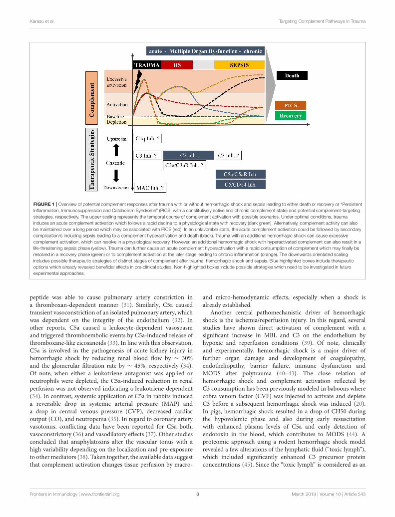
FIGURE 1 | Overview of potential complement responses after trauma with or without hemorrhagic shock and sepsis leading to either death or recovery or “Persistent
Inflammation, Immunosuppression and Catabolism Syndrome” (PICS; with a constitutively active and chronic complement state) and potential complement-targeting
strategies, respectively. The upper scaling represents the temporal course of complement activation with possible scenarios. Under optimal conditions, trauma
induces an acute complement activation which follows a rapid decline to a physiological state with recovery (dark green). Alternatively, complement activity can also
be maintained over a long period which may be associated with PICS (red). In an unfavorable state, the acute complement activation could be followed by secondary
complication/s including sepsis leading to a complement hyperactivation and death (black). Trauma with an additional hemorrhagic shock can cause excessive
complement activation, which can resolve in a physiological recovery. However, an additional hemorrhagic shock with hyperactivated complement can also result in a
life-threatening sepsis phase (yellow). Trauma can further cause an acute complement hyperactivation with a rapid consumption of complement which may finally be
resolved in a recovery phase (green) or to complement activation at the later stage leading to chronic inflammation (orange). The downwards orientated scaling
includes possible therapeutic strategies of distinct stages of complement after trauma, hemorrhagic shock and sepsis. Blue highlighted boxes include therapeutic
options which already revealed beneficial effects in pre-clinical studies. Non-highlighted boxes include possible strategies which need to be investigated in future
experimental approaches.
peptide was able to cause pulmonary artery constriction ina thromboxan-dependent manner (31). Similarly, C5a causedtransient vasoconstriction of an isolated pulmonary artery, whichwas dependent on the integrity of the endothelium (32). Inother reports, C5a caused a leukocyte-dependent vasospasmand triggered thromboembolic events by C5a-induced release ofthromboxane-like eicosanoids (33). In line with this observation,C5a is involved in the pathogenesis of acute kidney injury inhemorrhagic shock by reducing renal blood flow by ∼ 30%and the glomerular filtration rate by ∼ 45%, respectively (34).Of note, when either a leukotriene antagonist was applied orneutrophils were depleted, the C5a-induced reduction in renalperfusion was not observed indicating a leukotriene-dependent(34). In contrast, systemic application of C5a in rabbits induceda reversible drop in systemic arterial pressure (MAP) anda drop in central venous pressure (CVP), decreased cardiacoutput (CO), and neutropenia (35). In regard to coronary arteryvasotonus, conflicting data have been reported for C5a both,vasoconstrictory (36) and vasodilatory effects (37). Other studiesconcluded that anaphylatoxins alter the vascular tonus with ahigh variability depending on the localization and pre-exposureto other mediators (38). Taken together, the available data suggestthat complement activation changes tissue perfusion by macro-
and micro-hemodynamic effects, especially when a shock isalready established.
Another central pathomechanistic driver of hemorrhagicshock is the ischemia/reperfusion injury. In this regard, severalstudies have shown direct activation of complement with asignificant increase in MBL and C3 on the endothelium byhypoxic and reperfusion conditions (39). Of note, clinicallyand experimentally, hemorrhagic shock is a major driver offurther organ damage and development of coagulopathy,endotheliopathy, barrier failure, immune dysfunction andMODS after polytrauma (40–43). The close relation ofhemorrhagic shock and complement activation reflected byC3 consumption has been previously modeled in baboons wherecobra venom factor (CVF) was injected to activate and depleteC3 before a subsequent hemorrhagic shock was induced (20).In pigs, hemorrhagic shock resulted in a drop of CH50 duringthe hypovolemic phase and also during early resuscitationwith enhanced plasma levels of C5a and early detection ofendotoxin in the blood, which contributes to MODS (44). Aproteomic approach using a rodent hemorrhagic shock modelrevealed a few alterations of the lymphatic fluid (“toxic lymph”),which included significantly enhanced C3 precursor proteinconcentrations (45). Since the “toxic lymph” is considered as an
Frontiers in Immunology | www.frontiersin.org 3 March 2019 | Volume 10 | Article 543
Karasu et al. Targeting Complement Pathways in Trauma
important pathophysiological mechanism in the development ofadult respiratory distress syndrome and MODS, these findingsindicate C3 as a promising target for immune modulatoryapproaches (Figure 1).
SEPSIS-INDUCED MODS—ROLEOF COMPLEMENT
Sepsis was long considered as systemic inflammatory responsesyndrome with evidence of pathogenic microorganisms (46).In contrast, the new definitions describe sepsis rather as organdysfunction induced by a dysregulated host response to infection(47). This paradigm shift (48) may also change the focus oftherapeutic strategies toward support of organ functions, farbeyond the established eradication of the pathogens. However,it remains somehow enigmatic what “dysregulation” of the hostresponse means or if the “inadequacy” of the host response iscentral for development and progression of sepsis.
Multiple experimental sepsis studies have emphasized thedetrimental effects of excessive complement activation for thehost (Figure 1) (49, 50). This can be considered as a “complementparadox” since complement per se is central to the innateimmune defense against invading microorganisms. However, inthe context ofMODS development caused by sepsis, complementactivation seems to enhance rather than protecting against severalorgan dysfunctions, especially in the heart, lungs and kidneys,representing three central organs in MODS (51–53). The multi-organ gene expression profiles in experimental sepsis seems tobe either organ-specific, or common to more than one organ, ordistinctly opposite in some organs (54). Furthermore, a balancedpro- and anti-inflammatory genetic response was observedand a differential gene expression for mediators responsiblefor preventing tissue damage, e.g., protease inhibitors, oxidantneutralizing enzymes, decoy receptors, and proteins which canprotect tissue barriers (54). Concerning complement, pre-pro-complement C3 was highly expressed in all organs except in thebrain during the whole course of sepsis (54). However, geneticdeficiency of C3 resulted in significantly enhanced lethality incomparison to C3-sufficient mice most likely due to a lossof C3b-dependent opsonization of invaded pathogens (55). Incontrast, a blockade of C5a by various strategies in sepsis models,e.g., by anti-C5a antibodies, C5aR antibodies, small peptideC5aR1 antagonists, C5a-neutralizing mirror-image (l-)aptamerC5a aptamers, was coherently protective against biochemical andhistological evidence of MODS and in general improved survivalof sepsis (53, 55–61). All these experimental results demonstratethat C5a-C5aR interaction is clearly involved in the pathogenesisof MODS during sepsis and represents an important therapeuticsepsis target when the novel definitions of sepsis are applied(Figure 1) (47).
In translation to the clinical setting, several non-humanprimate experiments and studies in humans are in line withthe findings in the rodent sepsis model. Evidence of systemiccomplement activation with reduction of complement hemolyticactivity, C3 depletion and enhanced levels of C3a and C5a andcorresponding loss of C5aR on neutrophils have been described
in several human studies (62–64). Of note, the reduction ofC5aR1 and C5aR2 on neutrophils has been correlated with theoccurrence of infectious complications in ICU patients (30) andsepsis-induced MODS (62, 65, 66). Since loss of C5aR1 andC5aR2 has been concurrently correlated to the sequential organfailure assessment (SOFA) score (65), a flow-based rapid testing,might have a bedside monitoring potential to predict infectiousproblems and MODS development.
MODS—PATHOMECHANSISMS CAUSEDBY MULTIPLECOMPLEMENT DYSFUNCTIONS
Several pathomechanisms contribute to the development ofMODS, such as enhanced levels of DAMPs and PAMPs, reducedcytochrome P450 metabolism, macrophage activation syndrome,and cytokine-driven cellular dysfunction (67). Overall, it isclear that complement dysregulation contributes to MODS aftertrauma (Figure 2). Some of these aspects have been alreadymentioned and will be further discussed in this section.
Immune ParalysisThough severely injured patients receive modern ICUmanagement, many of them show signs of immunosuppressionknown as persistent inflammation-immunosuppressivecatabolism syndrome (PICS) (Figure 1) (68). Clinically,PICS patients suffer from persistent inflammation, immunesuppression and protein catabolism, which can lead to recurrentnosocomial infections with sepsis, MODS and death (68). Severeimmune suppression of the fluid-phase and cellular immuneresponse has been proposed as “immune paralysis” of the hostresponse to sterile and infectious insults. In a clinical case report,an inadequate response to infection with signs of systemicdepletion of complement (dropping C3 and C4 levels) has beenassociated with the development of acute kidney injury andmultiple organ failure in a 17-day old newborn (69). C5b-9 andC5a have been described as contributors to cell death, immuneparalysis, cardiac dysfunction, and multiple organ failure(Figure 2) (49, 70). In support, a baboon model of Escherichiacoli sepsis showed that blockade of C5 protected organs from“immune paralysis” and improved the sepsis survival rate(Table 1) (75).
Dysregulated Complement RegulatorsAdditional connection between MODS and signs ofcomplementopathy are supported by the fact that solubleand membrane-bound regulators of complement activity showalterations after trauma, sepsis and hemorrhagic shock. It iswell-established that severe tissue injury causes an excessivesystemic intravascular activation of the complement systemresulting in a loss over the control mechanisms (8). In thiscontext, the soluble form of the complement receptor 2 (sCR2)was shown to be present after nerve injury in rodents (80).After polytrauma in humans, leukocyte expression profiles ofthe complement regulators (CRegs) CD55 (decay acceleratingfactor), CD59 (membrane attack complex inhibitor), CD46
Frontiers in Immunology | www.frontiersin.org 4 March 2019 | Volume 10 | Article 543
Karasu et al. Targeting Complement Pathways in Trauma
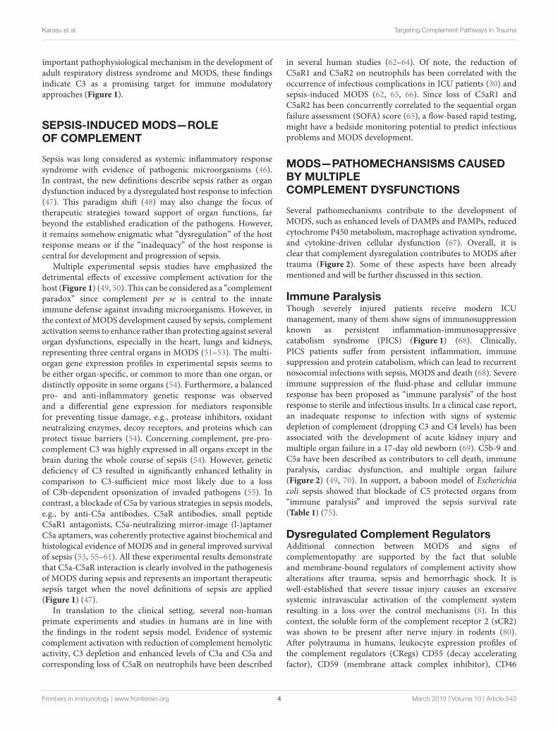
FIGURE 2 | Complement- mediated pathomechanisms in MODS development. Polytrauma, sepsis and hemorrhagic shock cause a critical complement
dysregulation, which causes dysfunction in multiple organs. This figure summarizes potential and known pathomechansims for MODS development caused by
complement dysregulation. Established complement involvement is highlighted in light green and proposed complement involvement is highlighted in gray.
(membrane cofactor protein), and CD35 (complement receptor1) were cell-type- and time-dependently altered, reflectingmanifestation of posttraumatic complementopathy (4, 29).Furthermore, an observational study including critically illpatients with multiple injuries and sepsis revealed that traumapatients with MODS have significantly lower C1-inhibitoractivities (Figure 2) (7). A rare mutation in the complementregulatory gene factor H has been also implicated with a severeform of complement-mediated hemolytic uremic syndrome withmultiple organ involvement, showing the general importance ofcomplement regulation for organ (dys)functions (81).
Alteration of Signaling Pathway ActivityExperimental studies showed an interplay of complement withspecific signaling pathways including the activation of PKC,MAPKs, and ERK (82). In a non-traumatic renal damagemodel, NF-κB contributed to enhanced complement activation(83), while in a similar mouse model the C5a/C5aR axisactivated MAPK signaling (84). During sepsis, the activation ofMAPKs and Akt signaling was complement-induced (51). Thesemechanistic insights require further elaboration to identify otherparticipatingmediators downstream to complement activation intrauma and hemorrhagic shock.
Changes in Protease ActivityExcessive systemic activation of proteases feed in thepathomechanisms of MODS and is known to create a viciouscycle of complement activation. Such proteases includeimmunoreactive trypsins and neutrophil elastase, which maydirectly interact with complement and have been extensivelydescribed in the case of polytrauma (9). A specific subtype ofMODS has been introduced as thrombocytopenia-associatedMODS, with low ADAMTS13 activity and defects in inhibitorycomplement regulators which may result in hyperactivationof coagulation and complement with resultant thrombosis
(85). This form of MODS has been successfully treated by C5blockade (eculizumab) and ADMTS13 reduction by plasmaexchange (67). Furthermore, an unspecific and unregulatedprotease hyperactivity may directly cleave various complementcomponents in a non-canonical manner, which representsa promising future research field for drug development. Inthis case, the intestine plays an important role in trauma andduring shock, which can proceed to a proposed autodigestionphenomenon, since digestive enzymes from the pancreas areactivated by enterokinases (86). Under physiological conditions,the auto-digestion is prevented by epithelial and mucosalbarriers. However, during shock digestive peptides can passthe mucus and reach the epithelial cell membranes, where theydisrupt junctional complexes, further activate complement andcontribute barrier and organ dysfunction (87).
Microcirculatory AlterationPatients who succumbed to traumatic-hemorrhagic shockshowed an impaired microcirculation for at least 72 h, whichalso was a reliable predictor for a high Sequential Organ FailureAssessment (SOFA) score (88). In an ischemia rat model,application of soluble complement receptor type 1 significantlyimproved microvascular perfusion in the liver assessed byin vivo microscopy, suggesting an essential role of complementin microcirculatory disorders (89). Activated complement withmicrovascular alterations can also cause a disruption of cellularbarriers leading to edema formation in lung, brain, and liver(84, 90, 91). However, hypothetical complement-caused damageof specific tight junction molecules in specific organs needs stillscientific verification. Such mechanisms of MODS developmentmay also be supported by simultaneous activation of complementand coagulation e.g., by complement-dependent generation ofthrombin, which is known to efficiently break down variousendothelial barriers. Furthermore, complement hyperactivationhas been associated with thrombotic microangiopathies causing
Frontiers in Immunology | www.frontiersin.org 5 March 2019 | Volume 10 | Article 543
Karasu et al. Targeting Complement Pathways in Trauma
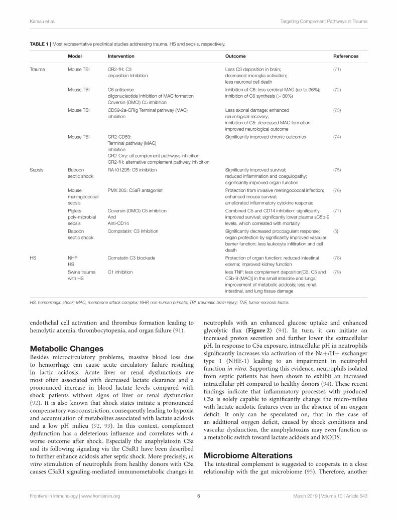
TABLE 1 | Most representative preclinical studies addressing trauma, HS and sepsis, respectively.
Model Intervention Outcome References
Trauma Mouse TBI CR2-fH: C3
deposition Inhibition
Less C3 deposition in brain;
decreased microglia activation;
less neuronal cell death
(71)
Mouse TBI C6 antisense
oligonucleotide Inhibition of MAC formation
Coversin (OMCI) C5 inhibition
Inhibition of C6: less cerebral MAC (up to 96%);
inhibition of C6 synthesis (> 80%)
(72)
Mouse TBI CD59-2a-CRIg Terminal pathway (MAC)
inhibition
Less axonal damage; enhanced
neurological recovery;
inhibition of C5: decreased MAC formation;
improved neurological outcome
(73)
Mouse TBI CR2-CD59:
Terminal pathway (MAC)
inhibition
CR2-Crry: all complement pathways inhibition
CR2-fH: alternative complement pathway inhibition
Significantly improved chronic outcomes (74)
Sepsis Baboon
septic shock
RA101295: C5 inhibition Significantly improved survival;
reduced inflammation and coagulopathy;
significantly improved organ function
(75)
Mouse
meningococcal
sepsis
PMX 205: C5aR antagonist Protection from invasive meningococcal infection;
enhanced mouse survival;
ameliorated inflammatory cytokine response
(76)
Piglets
poly-microbial
sepsis
Coversin (OMCI) C5 inhibition
And
Anti-CD14
Combined C5 and CD14 inhibition: significantly
improved survival; significantly lower plasma sC5b-9
levels, which correlated with mortality
(77)
Baboon
septic shock
Compstatin: C3 inhibition Significantly decreased procoagulant response;
organ protection by significantly improved vascular
barrier function; less leukocyte infiltration and cell
death
(5)
HS NHP
HS
Comstatin C3 blockade Protection of organ function; reduced intestinal
edema; improved kidney function
(78)
Swine trauma
with HS
C1 inhibition less TNF; less complement deposition[C3, C5 and
C5b-9 (MAC)] in the small intestine and lungs;
improvement of metabolic acidosis; less renal,
intestinal, and lung tissue damage
(79)
HS, hemorrhagic shock; MAC, membrane attack complex; NHP, non-human primate; TBI, traumatic brain injury; TNF, tumor necrosis factor.
endothelial cell activation and thrombus formation leading tohemolytic anemia, thrombocytopenia, and organ failure (91).
Metabolic ChangesBesides microcirculatory problems, massive blood loss dueto hemorrhage can cause acute circulatory failure resultingin lactic acidosis. Acute liver or renal dysfunctions aremost often associated with decreased lactate clearance and apronounced increase in blood lactate levels compared withshock patients without signs of liver or renal dysfunction(92). It is also known that shock states initiate a pronouncedcompensatory vasoconstriction, consequently leading to hypoxiaand accumulation of metabolites associated with lactate acidosisand a low pH milieu (92, 93). In this context, complementdysfunction has a deleterious influence and correlates with aworse outcome after shock. Especially the anaphylatoxin C5aand its following signaling via the C5aR1 have been describedto further enhance acidosis after septic shock. More precisely, invitro stimulation of neutrophils from healthy donors with C5acauses C5aR1 signaling-mediated immunometabolic changes in
neutrophils with an enhanced glucose uptake and enhancedglycolytic flux (Figure 2) (94). In turn, it can initiate anincreased proton secretion and further lower the extracellularpH. In response to C5a exposure, intracellular pH in neutrophilssignificantly increases via activation of the Na+/H+ exchangertype 1 (NHE-1) leading to an impairment in neutrophilfunction in vitro. Supporting this evidence, neutrophils isolatedfrom septic patients has been shown to exhibit an increasedintracellular pH compared to healthy donors (94). These recentfindings indicate that inflammatory processes with producedC5a is solely capable to significantly change the micro-milieuwith lactate acidotic features even in the absence of an oxygendeficit. It only can be speculated on, that in the case ofan additional oxygen deficit, caused by shock conditions andvascular dysfunction, the anaphylatoxins may even function asa metabolic switch toward lactate acidosis and MODS.
Microbiome AlterationsThe intestinal complement is suggested to cooperate in a closerelationship with the gut microbiome (95). Therefore, another
Frontiers in Immunology | www.frontiersin.org 6 March 2019 | Volume 10 | Article 543
Karasu et al. Targeting Complement Pathways in Trauma
contributor to MODS seems to be the alteration of the gutmicrobiome after trauma/hemorrhagic shock and/or sepsis. Themicrobiome, which is described by the phylogenetic compositionand taxon relative abundance of the bacteria, is significantlyaltered in the first 72 h after injury. This rapid change inintestinal microbiota represents a critical phenomenon that mayinfluence outcomes after severe trauma (15). The compositionof the microbiome may influence the activation/dysregulation ofcomplement pathways or dysregulated complement may changethe microbiome composition. In the skin, complement activationmodulates the inflammatory milieu by changing the cutaneousmicrobiota (96). Considering MODS, a mechanistic explanationfor complement-microbiome interaction still remains elusive andneeds further research.
Alterations in Microvesicle (MV) SheddingCommunication is essential for cellular homeostasis and vesicleshedding has been described to play a crucial role for maintainingproper immune cell function. Extracellular vesicle sheddingis altered after inflammation and is considered as a crucialcontributor to MODS after multiple injury and sepsis (97).Especially shedding of microvesicles (MV) have been implicatedin several inflammatory conditions including sepsis and trauma.Increased amounts of CD41+ and CD31+/CD41–/AnnexinV–MV after sepsis, released by activated platelets and leukocyteshave been shown to correlate with unfavorable outcomes (98).
Furthermore, MVs from patients with multiple organ failuresupport the coagulation system in triggering inflammation.In respect to complement, phosphatidylserine containing MVsalso serve as platform for complement activation (99). Besidesactivation of complement on their surfaces, MVs representtransport vehicles sending complement as cargo to neighboringas well as cells in distance (99). Hence, MV from different cellularorigins may contain complement receptors including C5aR butalso CRegs such as CD46 and CD59, suggesting a putative role forcomplement activity (99). In accordance with this, loss of C5aR1,C5aR2, and C3aR on neutrophils after multiple injury wasclinically present and was correlated to infectious complicationsand multiple organ dysfunction (Figure 2) (18).
Electrophysiological ChangesAnother concurrent theory of MODS addresseselectrophysiological changes of the cellular membrane whichhave recently been found in neutrophils from septic pigs(14). The anaphylatoxin C5a was able to alter the membranepotential of neutrophils but not in the case of neutrophils duringseptic MODS where the electrophysiological response to C5awas somehow frozen (Figure 2) (14). Whether complementinhibitory strategies will stabilize the cellular membraneelectrophysiology is currently under investigation.
Cellular HibernationTrauma causes an alarming stress situation for the whole bodywith an extensive inflammatory response (9). Some studiesindicate that trauma especially followed by additional sepsiscauses hibernation in the cellular as well as fluid phase ofinnate immunity including the complement system. Reflecting
the evolved habit of conserving physiological resources in theevent of environmental stress, with inflammation ensues a similarmechanism where energy-consuming processes are shut downin the organism. Supporting this evidence, hibernation has beenobserved in the septic heart with ongoing metabolic changesincluding the upregulation of specific glucose transportersin cardiomyocytes (100). Besides its effects on metabolism,hibernation is known to affect various immune functionincluding leukocyte migration, as well as adaptive immuneresponses and interestingly complement function, by loweringcomplement levels and reduced expression of C3 mRNA in theliver, which depicts a suggestive link post-shock. (101). However,another study demonstrated that hibernators are protected fromshock-induced injury, inflammation, and organ function (102).Strikingly, arctic ground squirrels challenged with cardiac arrestor hemorrhagic shock showed no markers of organ damage,systemic inflammation, or loss of acid/base balance as indicatedby a negative base excess. Neither reduced body temperature norhibernation season are components of this protection, indicatingstill unknown mechanisms involved (102).
Unfortunately, no supporting data is available indicating thatfuture research on the complement function during hibernation,especially following trauma and shock is needed. Further, theability to induce a fully reversible state of immune suppressionin humans by artificial hibernation might aid the treatment ofseveral inflammatory and immune-mediated diseases.
TARGETING COMPLEMENT PATHWAYSIN MODS
Despite improvements in trauma care, the morbidity andmortality of MODS remains very high.
Therefore, new therapeutic strategies are urgently needed.Since complement is critically involved in initiation andprogression of MODS, targeting complement as well asmolecules contributing to complement activation representpromising future clinical approaches. Other complement-associated inflammatory conditions already addressed such atargeting strategy. Above 20 complement-interfering drugs havebeen evaluated in clinical settings so far. A few of themreceived FDA approval for inflammatory indications includingeculizumab targeting the terminal complement pathway startingfrom C5, which is currently used for the treatment of paroxysmalnocturnal hemoglobinuria (PNH) (103). As existing complementtherapeutics cannot target every complement-driven diseasestatus, it is a requisite to evaluate the different complement stages,their relevance, and target them in a disease-specificmanner (10).
In the context of trauma, hemorrhagic shock and sepsis a fewpreclinical experimental studies focus on complement-targetingstudies (Table 1). Besides complement activation products, othermolecules including CRP, HMGB1, or mitochondrial DNAplay crucial roles in contributing to complement dysregulationafter sepsis and trauma, thus profoundly influencing secondaryoutcomes. Therefore, a rational approach seems to addressmolecules, which may further boost complement activity.Likewise, HMGB1 is a relevant danger molecule and complement
Frontiers in Immunology | www.frontiersin.org 7 March 2019 | Volume 10 | Article 543
Karasu et al. Targeting Complement Pathways in Trauma
has been described to regulate HMGB1 release from humanneutrophils (104) such that, complement inhibition has provedto be protective in blast-induced acute lung injury in rats byameliorating HMGB1-mediated inflammation (105).
Targeting Complement After (poly)TraumaIn polytrauma patients, the only study of complementintervention early after trauma with RCT quality used C1esterase inhibitor. However, this study has been terminated basedon the heterogeneity of the patients (106). Experimentally, themajority of the complement targeting studies were performedin mouse traumatic brain injury (TBI) models. In this case,various interventions to inhibit complement activity, such asinhibitors for MAC formation and C3 deposition inhibitors havebeen applied, analyzed and reviewed elsewhere (9, 107, 108).To further clarify the impact of distinct complement activationpathways on neuroinflammation, a recent study comparedthe effect of complement inhibitors targeting upstream as wellas downstream complement activity, respectively. This studybrought into light that instead of downstream complementactivation complexes, upstream complement activation productsof the alternative pathway predominantly modulate andpropagate chronic inflammation (74) (Table 1).
Targeting Complement inHemorrhagic ShockBased on the deduced potential of complement inhibitorsto improve micro-perfusion disturbances, ischemia/reperfusioninjury and the inflammatory response during hemorrhagicshock, various shock models have been tested for possiblebenefits of complement interventions. In a rat model of pressurecontrolled hemorrhagic shock, complement depletion by CVFimproved the recovery of the mean arterial pressure (MAP)post shock (109). In a pig model of pressure controlledhemorrhagic shock, application of a C1-inhibitor improvedthe functional performance and reduced C3 deposition ona multi-organ level including liver, small intestine and lungs(Table 1) (79). Limitation of organ injury and sustainedsurvival by C1 inhibitor therapy has also been reported fora porcine injury model mimicking battlefield injury (110).Further downstream, C3 deficient mice with bilateral femurfracture and hemorrhagic shock resulted in protective effects withreduced circulating DAMPs (e.g., dsDNA), decreased systemicinflammatory response and improved organ performance (e.g.,liver enzymes) (111). In a rat model of hemorrhagic shock,C3 depletion by CVF, and the soluble form of CR1 to inhibitC3 action restored vascular reactivity to norepinephrine in thesuperior mesenteric artery (112). In the clinical setting, C3inhibition has not been tested in hemorrhagic shock so farexcept in a recent study from our group, where non-humanprimates were modeled with severe hemorrhagic shock of 30mmHg MAP for 1 h, and a delayed application of CP40 wastested (78). C3 blockade by the compstatin compound CP40seems most promising since there was evidence of improvedrenal function, attenuated intestinal edema and reduced signsof systemic inflammation and coagulopathy (78). However, thetransfer into clinical reality is pending although C3 inhibition
seems rather safe in the case of hemorrhagic shock. Most likely,an early inhibition to avoid excessive C3 activation might beenough to improve the outcome (113).
On a C5 level, intestinal injury caused by hypovolemic shockin mice was ameliorated by application of a small peptideC5aR1 antagonist and C5 deficient littermates (114). Similarly,in a rodent model of ruptured aortic abdominal aneurysmawith shock, C5 blockade by an anti-C5 antibody revealed someprotective effects on remote lung injury with improvement ofthe bronchoalveolar permeability and myeloperoxidase (MPO)concentrations reflecting reduced neutrophil recruitment andactivation (115). Combined inhibition of the complementcomponent C5 and the Toll-like receptor co-factor CD14 ina porcine sepsis model, showed beneficial effects in regard tosurvival, hemodynamic parameters and systemic inflammationincluding complement activation (Figure 1, Table 1). Firstpreclinical studies in non-human primates indicate appearanceof sC5b-9 in the serum of traumatic-hemorrhagic shock andex vivo effectiveness of C5 inhibitors in prevention of hemolysis,e.g., the small, inhibitory peptide RA101348, commercially-available C5 inhibitory antibodies and Quidel’s A217 antibody(106). However, a RCT-clinical study with C5 inhibitorystrategies in hemorrhagic shock has not been performed so farmost likely based on the rareness and uncertain evidence of agreat impact on MODS development in the so far performedpreclinical non-human primate studies. Therefore, researchproviding information about the histological and biochemicaldata on the multi-organ level in non-human primate studies isnecessary for final assessment.
Targeting Complement in SepsisIn the case of septic shock, targeting the C5a/C5aR axis seemsto be of practical importance since it correlated with the diseaseseverity and mortality and showed promising improvementsin different sepsis models (Table 1). Concerning complement-targeted therapies in sepsis, application of RA101295, a 2-kDamacrocyclic peptide inhibitor of C5 cleavage, improved organperformance and survival in an E. coli-sepsis model of baboons(75) with lessened evidence of coagulopathy and preservedendothelial and barrier functions. Furthermore, the inhibition ofC5 cleavage lead to improved histomorphology of lungs, liver,kidneys, spleen, and adrenal glands suggesting improvement ofsepsis-induced MODS (75).
In the same sepsis model, systemic blockade of C3 bycompstatin also revealed organ protection on multiple levels.Compstatin showed evidence of sepsis-induced coagulopathyand preserved anti-coagulatory features of the endothelium.Furthermore, C3 blockade improved hemodynamics and heartfunction and biochemical damage markers of the kidney andliver, indicating protective effects in sepsis-induced MODS (5).For blocking the central complement component C3 duringdevelopment of sepsis-caused MODS, clinical trials might be safe(113) but these trials are pending and require a special focus onthe benefits or maladies for mental alterations during sepsis.
In terms of applicability in the clinical setting, it has beenrecently shown that blocking solely C5 activation by canonicalC5-convertase (e.g., by eculizumab) might not be specific enough
Frontiers in Immunology | www.frontiersin.org 8 March 2019 | Volume 10 | Article 543
Karasu et al. Targeting Complement Pathways in Trauma
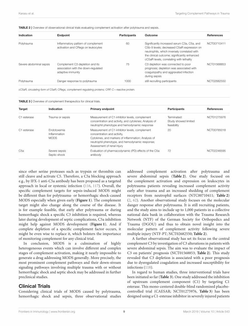
TABLE 2 | Overview of observational clinical trials evaluating complement activation after polytrauma and sepsis.
Indication Endpoint Participants Outcome References
Polytrauma Inflammatory pattern of complement
activation and CRegs on leukocytes
60 Significantly increased serum C3a, C5a, and
C5b-9 levels; decreased C5aR expression on
neutrophils, which inversely correlated with
the clinical outcome; significantly enhanced
cC5aR levels, correlating with lethality
NCT00710411
Severe abdominal sepsis Complement C3 depletion and its
association with the down-regulated
adaptive immunity
75 C3 depletion was connected to poor
prognosis; depletion was associated with
coagulopathy and aggravated infection
during sepsis
NCT01568853
Polytrauma Danger response to polytrauma 1000 still recruiting participants NCT02682550
cC5aR, circulating form of C5aR; CRegs, complement-regulating proteins; CRP, C—reactive protein.
TABLE 3 | Overview of complement therapeutics for clinical trials.
Target Indication Primary endpoint Participants References
C1 esterase Trauma or sepsis Measurement of C1-inhibitor levels, complement
concentration and activity, and cytokines; Analysis of
neutrophil phenotype and hemodynamic response
Terminated:
Study showed limited
feasibility
NCT01275976
C1 esterase Endotoxemia
Inflammation
MODS
Measurement of C1-inhibitor levels, complement
concentration and activity,
Cytokines, and markers of inflammation; Analysis of
neutrophil phenotype, and hemodynamic response;
Assessment of renal injury
20 NCT00785018
C5a Severe sepsis
Septic shock
Evaluation of pharmacodynamic (PD) effects of the C5a
antibody
72 NCT02246595
since other serine proteases such as trypsin or thrombin canstill cleave and activate C5. Therefore, a C5a blocking approache.g., by IFX-1 anti-C5a antibody has been proposed as a targetedapproach in local or systemic infection (116, 117). Overall, thespecific complement targets for sepsis-induced MODS mightbe different than for polytrauma- or hemorrhagic shock-causedMODS especially when given early (Figure 1). The complementtarget might also change along the course of the disease. Itis for example feasible, that early after polytrauma or duringhemorrhagic shock a specific C3 inhibition is required, whereaslater during development of septic complications, C5a inhibitionmight help against MODS development (Figure 1). And ifcomplete depletion of a specific complement factor occurs, itmight be even wise to replace it, which bolsters the importanceof monitoring complement for any clinical trial.
In conclusion, MODS is a culmination of highlyheterogeneous events which can involve different and complexstages of complement activation, making it nearly impossible togenerate a drug addressing MODS generally. More precisely, themost prominent complement pathways and their down-streamsignaling pathways involving multiple trauma with or withouthemorrhagic shock and septic shock may be addressed in furtherpreclinical studies.
Clinical TrialsConsidering clinical trials of MODS caused by polytrauma,hemorrhagic shock and sepsis, three observational studies
addressed complement activation after polytrauma andsevere abdominal sepsis (Table 2). One study focused onthe complement activation and expression on leukocytes inpolytrauma patients revealing increased complement activityearly after trauma and an increased shedding of complementreceptors from neutrophil surfaces (NTC00710411; Table 2)(2, 62). Another observational study focuses on the moleculardanger response after polytrauma. It is still recruiting patients,and the study aims to include up to 1,000 patients in a collectivenational data bank in collaboration with the Trauma ResearchNetwork (NTF) of the German Society for Orthopedics andTrauma (DGOU) and thus to obtain novel insight into themolecular pattern of complement activity following severemultiple injury (NTF-PT; NCT02682550; Table 2).
A further observational study has set its focus on the centralcomplement C3 by investigation of C3 alterations in patients withsevere abdominal sepsis. The aim was to evaluate the impact ofC3 on patients’ prognosis (NCT01568853; Table 2). This studyrevealed that C3 depletion is associated with a poor prognosisdue to dysregulated coagulation and increased susceptibility forinfections (118).
In regard to human studies, three interventional trials havebeen initiated so far (Table 3). One study addressed the inhibitionof upstream complement component (C1) by targeting C1esterase. This mono-centered double-blind randomized placebo-controlled trial (CAESAR; NCT01275976, Table 3) has beendesigned using a C1-esterase inhibitor in severely injured patients
Frontiers in Immunology | www.frontiersin.org 9 March 2019 | Volume 10 | Article 543
Karasu et al. Targeting Complement Pathways in Trauma
with femur fracture. Although the rationale of a complementblockade on this level is very well founded because it may alsoreveal beneficial effects by synchronically inhibition of excessiveactivation of the coagulation pathway (119), the study has beenterminated based on the heterogeneity of patients and challengesin recruitment. A further study has addressed the effect ofthe C1-esterase inhibitor on human endotoxemia by evaluatingits effect on inflammation and marker of organ dysfunction(VECTOR; NCT00785018, Table 3). Detailed results of the studyare still pending.
Another clinical trial has been designed and performedto study complement inhibition in early, newly developingseptic organ dysfunction (SCIENS; NCT02246595, Table 3)applying a monoclonal antibody against C5a, though thedetailed results of this trial have not been published so far.Regarding one cardinal clinical sign of sepsis, the occult orevident alterations of the mental status, inhibitory strategiesagainst C5a might reveal “Janus faced” effects (120). Onone hand, C5a inhibition could improve sepsis-impairedblood-brain-barrier, on the other hand, neuroprotectiveeffects by C5a might be compromised (120, 121). Therefore,alterations of the mental status need to be carefullyaddressed and monitored in any clinical trial using C5ainhibitory strategies.
It is important to note, that it is in general rather difficultto perform interventional studies on polytrauma patients sincean informed written consent cannot be provided and the legalrepresentatives are usually difficult to determine within the firsthours after severe injury. Therefore, innovative studies early afterpolytrauma addressing the complement cascade are rather rareand have not been performed on the C3 or C5 level yet.
CLINICAL PERSPECTIVES
Although various clinically relevant models of trauma,hemorrhagic shock and sepsis have been tested already in non-human primates for the benefit of complement interventions(75, 78, 106), clinical trials in these multidimensionalpathophysiologic conditions remain rare (119, 122). Whencomplement intervention strategies are designed for the clinics,
the targeted complement factor or activation product needsto be measured before the therapy can be applied. Especiallywhen complex intensive care is necessary which can altercomplement levels within a short time period, e.g., by infusionof blood products which contain highly variable concentrationsof complement (activation) factors (123) or by extra-corporalcirculation devices with large artificial surfaces which maydeplete key complement components (124, 125), the exactstatus of complement activation needs to be determined.This would allow precise and timely intervention either byinhibiting or supporting the complement response after traumaor during sepsis in order to rebalance the immune response.Whereas, various highly effective and specific complementintervention strategies have been developed within the lasttwo decades and are available now (10), in the context of thecomplex immune response after trauma, hemorrhagic shock andsepsis (9, 126), specific organ damage and function assessmentincluding the immune function at bedside seems far beyond.Therefore, functional monitoring of the organ and immunesystems can be considered as a prerequisite before complementinterventions move into clinical routine in diseases with acomplex pathophysiology. In conclusion, further scientificknowledge and translational efforts are demanded for targetingcomplement pathways in the setting of trauma, hemorrhagicshock and sepsis with the aim to offer causal therapy andimproved outcome.
AUTHOR CONTRIBUTIONS
EK and MH-L wrote the manuscript. All authors edited andcommented on the manuscript. All authors read and approvedthe final manuscript.
FUNDING
This review was supported by grants from the German ResearchFoundation (DFG) (EI 866/5-1) and by grants from the DFGwithin the Collaborative Research Center CRC1149/2 DangerResponse, Disturbance Factors and Regenerative Potential afterAcute Trauma to MH-L.
REFERENCES
1. Fosse E, Mollnes TE, Aasen AO, Trumpy JH, Stokke T. Complement
activation following multiple injuries. Acta Chir Scand. (1987) 153:325–30.
2. Burk AM, Martin M, Flierl MA, Rittirsch D, Helm M, Lampl L, et al.
Early complementopathy after multiple injuries in humans. Shock. (2012)
37:348–54. doi: 10.1097/SHK.0b013e3182471795
3. Ganter MT, Brohi K, Cohen MJ, Shaffer LA, Walsh MC, Stahl GL, et al.
Role of the alternative pathway in the early complement activation following
major trauma. Shock. (2007) 28:29–34. doi: 10.1097/shk.0b013e31803
42439
4. Amara U, Flierl MA, Rittirsch D, Klos A, Chen H, Acker B, et al. Molecular
intercommunication between the complement and coagulation systems.
J Immunol. (2010) 185:5628–36. doi: 10.4049/jimmunol.0903678
5. Silasi-Mansat R, Zhu H, Popescu NI, Peer G, Sfyroera G, Magotti P, et al.
Complement inhibition decreases the procoagulant response and confers
organ protection in a baboon model of Escherichia coli sepsis. Blood. (2010)
116:1002–10. doi: 10.1182/blood-2010-02-269746
6. Hecke F, Schmidt U, Kola A, Bautsch W, Klos A, Kohl J. Circulating
complement proteins in multiple trauma patients–correlation with injury
severity, development of sepsis, and outcome. Crit Care Med. (1997)
25:2015–24. doi: 10.1097/00003246-199712000-00019
7. Helling H, Stephan B, Pindur G. Coagulation and complement system
in critically ill patients. Clin Hemorheol Microcirc. (2015) 61:185–93.
doi: 10.3233/CH-151993
8. Rittirsch D, Redl H, Huber-Lang M. Role of complement in multiorgan
failure. Clin Dev Immunol. (2012) 2012:962927. doi: 10.1155/2012/962927
9. Huber-Lang M, Lambris JD, Ward PA. Innate immune responses to trauma.
Nat Immunol. (2018) 19:327–41. doi: 10.1038/s41590-018-0064-8
10. Ricklin D, Mastellos DC, Reis ES, Lambris JD. The renaissance
of complement therapeutics. Nat Rev Nephrol. (2018) 14:26–47.
doi: 10.1038/nrneph.2017.156
Frontiers in Immunology | www.frontiersin.org 10 March 2019 | Volume 10 | Article 543
Karasu et al. Targeting Complement Pathways in Trauma
11. Cavaillon JM, Annane D. Compartmentalization of the inflammatory
response in sepsis and SIRS. J Endotoxin Res. (2006) 12:151–70.
doi: 10.1179/096805106X102246
12. Cavaillon JM, Giamarellos-Bourboulis EJ. << Immunosuppression >> is
inappropriately qualifying the immune status of septic and sirs patients.
Shock. (2018). doi: 10.1097/SHK.0000000000001266. [Epubh ahead of print].
13. Riedemann NC, Guo RF, Neff TA, Laudes IJ, Keller KA, Sarma VJ, et al.
Increased C5a receptor expression in sepsis. J Clin Invest. (2002) 110:101–8.
doi: 10.1172/JCI0215409
14. Messerer DAC, Denk S, Fohr KJ, Halbgebauer R, Braun CK, Hones F,
et al. Complement C5a alters the membrane potential of neutrophils
during hemorrhagic shock. Mediators Inflamm. (2018) 2018:2052356.
doi: 10.1155/2018/2052356
15. Howard BM, Kornblith LZ, Christie SA, Conroy AS, Nelson MF, Campion
EM, et al. Characterizing the gut microbiome in trauma: significant changes
inmicrobial diversity occur early after severe injury.Trauma Surg Acute Care
Open. (2017) 2:e000108. doi: 10.1136/tsaco-2017-000108
16. Graetz TJ, Hotchkiss RS. Sepsis: preventing organ failure in sepsis - the search
continues. Nat Rev Nephrol. (2017) 13:5–6. doi: 10.1038/nrneph.2016.171
17. Coopersmith CM,De BD,DeutschmanCS, Ferrer R, Lat I,Machado FR, et al.
Surviving sepsis campaign: research priorities for sepsis and septic shock.
Crit Care Med. (2018) 46:1334–56. doi: 10.1097/CCM.0000000000003225
18. Chakraborty S, Karasu E, Huber-Lang M. Complement after trauma:
suturing innate and adaptive immunity. Front Immunol. (2018) 9:2050.
doi: 10.3389/fimmu.2018.02050
19. van Wessem KJP, Leenen LPH. Reduction in mortality rates of
postinjury multiple organ dysfunction syndrome: a shifting paradigm?
a prospective population-based cohort study. Shock. (2018) 49:33–8.
doi: 10.1097/SHK.0000000000000938
20. Roumen RM, Redl H, Schlag G, Zilow G, Sandtner W, Koller W, et al.
Inflammatory mediators in relation to the development of multiple organ
failure in patients after severe blunt trauma. Crit Care Med. (1995)
23:474–80. doi: 10.1097/00003246-199503000-00010
21. Bautsch W, Hoymann HG, Zhang Q, Meier-Wiedenbach I, Raschke U,
Ames RS, et al. Cutting edge: guinea pigs with a natural C3a-receptor defect
exhibit decreased bronchoconstriction in allergic airway disease: evidence
for an involvement of the C3a anaphylatoxin in the pathogenesis of asthma.
J Immunol. (2000) 165:5401–5. doi: 10.4049/jimmunol.165.10.5401
22. Asgari E, Le FG, Yamamoto H, Perucha E, Sacks SS, Kohl J, et al. C3a
modulates IL-1beta secretion in human monocytes by regulating ATP
efflux and subsequent NLRP3 inflammasome activation. Blood. (2013)
122:3473–81. doi: 10.1182/blood-2013-05-502229
23. Ehrnthaller C, Flierl M, Perl M, Denk S, Unnewehr H, Ward PA, et al.
The molecular fingerprint of lung inflammation after blunt chest trauma.
Eur J Med Res. (2015) 20:70. doi: 10.1186/s40001-015-0164-y
24. Rittirsch D, Schoenborn V, Lindig S, Wanner E, Sprengel K, Gunkel S, et al.
An integrated clinico-transcriptomic approach identifies a central role of the
heme degradation pathway for septic complications after trauma. Ann Surg.
(2016) 264:1125–34. doi: 10.1097/SLA.0000000000001553
25. Abdelbaset-Ismail A, Borkowska-Rzeszotek S, Kubis E, Bujko K,
Brzezniakiewicz-Janus K, Bolkun L, et al. Activation of the complement
cascade enhances motility of leukemic cells by downregulating expression of
HO-1. Leukemia. (2017) 31:446–58. doi: 10.1038/leu.2016.198
26. May O, Merle NS, Grunenwald A, Gnemmi V, Leon J, Payet C, et al.
Heme drives susceptibility of glomerular endothelium to complement
overactivation due to inefficient upregulation of heme oxygenase-1. Front
Immunol. (2018) 9:3008. doi: 10.3389/fimmu.2018.03008
27. Noris M, Remuzzi G. Overview of complement activation and regulation.
Semin Nephrol. (2013) 33:479–92. doi: 10.1016/j.semnephrol.2013.
08.001
28. Rittirsch D, Schoenborn V, Lindig S, Wanner E, Sprengel K, Gunkel S,
et al. Improvement of prognostic performance in severely injured patients
by integrated clinico-transcriptomics: a translational approach. Crit Care.
(2015) 19:414. doi: 10.1186/s13054-015-1127-y
29. Amara U, Kalbitz M, Perl M, Flierl MA, Rittirsch D, Weiss M, et al.
Early expression changes of complement regulatory proteins and C5A
receptor. (CD88) on leukocytes after multiple injury in humans. Shock.
(2010) 33:568–75. doi: 10.1097/SHK.0b013e3181c799d4
30. Morris AC, Brittan M, Wilkinson TS, McAuley DF, Antonelli J, McCulloch
C, et al. C5a-mediated neutrophil dysfunction is RhoA-dependent and
predicts infection in critically ill patients. Blood. (2011) 117:5178–88.
doi: 10.1182/blood-2010-08-304667
31. Morganroth ML, Schoeneich SO, Till GO, Ward PA, Horvath SJ, Glovsky
MM. C3a57-77, a C-terminal peptide, causes thromboxane-dependent
pulmonary vascular constriction in isolated perfused rat lungs. Am Rev
Respir Dis. (1990) 141:296–300. doi: 10.1164/ajrccm/141.2.296
32. Crowell RE, Van Epps DE, Reed WP. Responses of isolated pulmonary
arteries to the C5a anaphylatoxin. Am J Physiol. (1990) 259(5 Pt 2):H1325–9.
doi: 10.1152/ajpheart.1990.259.5.H1325
33. Fortin JP, Bouthillier J, St-Pierre SA, Marceau F. Contractile effect of
anaphylatoxin C5a and of a mimetic peptide on the human umbilical
artery: further evidence for leukocyte-dependent vasomotion. J Cardiovasc
Pharmacol. (2002) 40:815–21. doi: 10.1097/00005344-200212000-00002
34. Gulbins E, Schlottmann K, Rauterberg EW, Steinhausen
M. Effects of rC5a on the circulation of normal and split
hydronephrotic rat kidneys. Am J Physiol. (1993) 265(1 Pt 2):F96–103.
doi: 10.1152/ajprenal.1993.265.1.F96
35. Lundberg C, Marceau F, Hugli TE. C5a-induced hemodynamic and
hematologic changes in the rabbit. Role of cyclooxygenase products and
polymorphonuclear leukocytes. Am J Pathol. (1987) 128:471–83.
36. Rendig SV, Gray S, Amsterdam EA. Contractile actions of C5a on isolated
porcine coronary resistance and conductance arteries. Am J Physiol. (1997)
272(1 Pt 2):H12–6. doi: 10.1152/ajpheart.1997.272.1.H12
37. Schumacher WA, Fantone JC, Kunkel SE, Webb RC, Lucchesi BR. The
anaphylatoxins C3a and C5a are vasodilators in the canine coronary
vasculature in vitro and in vivo. Agents Actions. (1991) 34:345–9.
doi: 10.1007/BF01988727
38. Marceau F, Hugli TE. effect of C3a and C5a anaphylatoxins on guinea-
pig isolated blood vessels. J Pharmacol Exp Ther. (1984) 230:749–54.
doi: 10.1016/0162-3109(84)90019-5
39. Collard CD, Lekowski R, Jordan JE, Agah A, Stahl GL. Complement
activation following oxidative stress. Mol Immunol. (1999) 36:941–8.
doi: 10.1016/S0161-5890(99)00116-9
40. Wrba L, Ohmann JJ, Eisele P, Chakraborty S, Braumuller
S, Braun CK, et al. Remote intestinal injury early after
experimental polytrauma and hemorrhagic shock. Shock. (2018).
doi: 10.1097/SHK.0000000000001271. [Epubh ahead of print].
41. Halbgebauer R, Braun CK, Denk S, Mayer B, Cinelli P, Radermacher
P, et al. Hemorrhagic shock drives glycocalyx, barrier and organ
dysfunction early after polytrauma. J Crit Care. (2018) 44:229–37.
doi: 10.1016/j.jcrc.2017.11.025
42. Frith D, Goslings JC, Gaarder C, Maegele M, Cohen MJ, Allard S,
et al. Definition and drivers of acute traumatic coagulopathy: clinical
and experimental investigations. J Thromb Haemost. (2010) 8:1919–25.
doi: 10.1111/j.1538-7836.2010.03945.x
43. Rahbar E, Cardenas JC, Baimukanova G, Usadi B, Bruhn R, Pati
S, et al. Endothelial glycocalyx shedding and vascular permeability
in severely injured trauma patients. J Transl Med. (2015) 13:117.
doi: 10.1186/s12967-015-0481-5
44. Szebeni J, Baranyi L, Savay S, Gotze O, Alving CR, Bunger R, et al.
Complement activation during hemorrhagic shock and resuscitation in
swine. Shock. (2003) 20:347–55. doi: 10.1097/01.shk.0000082444.66379.17
45. Fang JF, Shih LY, Yuan KC, Fang KY, Hwang TL, Hsieh SY. Proteomic
analysis of post-hemorrhagic shock mesenteric lymph. Shock. (2010)
34:291–8. doi: 10.1097/SHK.0b013e3181ceef5e
46. Bone RC, Balk RA, Cerra FB, Dellinger RP, Fein AM, Knaus WA,
et al. Definitions for sepsis and organ failure and guidelines for the
use of innovative therapies in sepsis. The ACCP/SCCM consensus
conference committee. American college of chest physicians/society of
critical care medicine. Chest. (1992) 101:1644–55. doi: 10.1378/chest.101.
6.1644
47. Shankar-Hari M, Phillips GS, Levy ML, Seymour CW, Liu VX,
Deutschman CS, et al. Developing a new definition and assessing new
clinical criteria for septic shock: for the third international consensus
definitions for sepsis and septic shock (sepsis-3). JAMA. (2016) 315:775–87.
doi: 10.1001/jama.2016.0289
Frontiers in Immunology | www.frontiersin.org 11 March 2019 | Volume 10 | Article 543
Karasu et al. Targeting Complement Pathways in Trauma
48. Dickmann P, Scherag A, Coldewey SM, Sponholz C, Brunkhorst FM, Bauer
M. [Epistemology in the intensive care unit-what is the purpose of a
definition?: paradigm shift in sepsis research]. Anaesthesist. (2017) 66:622–5.
doi: 10.1007/s00101-017-0315-3
49. Ward PA. The dark side of C5a in sepsis. Nat Rev Immunol. (2004) 4:133–42.
doi: 10.1038/nri1269
50. Gerard C. Complement C5a in the sepsis syndrome–too much of a good
thing? N Engl J Med. (2003) 348:167–9. doi: 10.1056/NEJMcibr022995
51. Fattahi F, Kalbitz M, Malan EA, Abe E, Jajou L, Huber-Lang MS,
et al. Complement-induced activation of MAPKs and Akt during
sepsis: role in cardiac dysfunction. FASEB J. (2017) 31:4129–39.
doi: 10.1096/fj.201700140R
52. Kalbitz M, Fattahi F, Herron TJ, Grailer JJ, Jajou L, Lu H, et al. Complement
destabilizes cardiomyocyte function in vivo after polymicrobial sepsis
and in vitro. J Immunol. (2016) 197:2353–61. doi: 10.4049/jimmunol.
1600091
53. Huber-Lang MS, Riedeman NC, Sarma JV, Younkin EM, McGuire SR,
Laudes IJ, et al. Protection of innate immunity by C5aR antagonist in septic
mice. FASEB J. (2002) 16:1567–74. doi: 10.1096/fj.02-0209com
54. Chinnaiyan AM, Huber-Lang M, Kumar-Sinha C, Barrette TR, Shankar-
Sinha S, Sarma VJ, et al. Molecular signatures of sepsis: multiorgan
gene expression profiles of systemic inflammation. Am J Pathol. (2001)
159:1199–209. doi: 10.1016/S0002-9440(10)62505-9
55. Flierl MA, Rittirsch D, Chen AJ, Nadeau BA, Day DE, Sarma JV,
et al. The complement anaphylatoxin C5a induces apoptosis in
adrenomedullary cells during experimental sepsis. PLoS ONE. (2008)
3:e2560. doi: 10.1371/journal.pone.0002560
56. Flierl MA, Schreiber H, Huber-Lang MS. The role of complement, C5a and
its receptors in sepsis and multiorgan dysfunction syndrome. J Invest Surg.
(2006) 19:255–65. doi: 10.1080/08941930600778263
57. Flierl MA, Rittirsch D, Nadeau BA, Day DE, Zetoune FS, Sarma JV, et al.
Functions of the complement components C3 and C5 during sepsis. FASEB
J. (2008) 22:3483–90. doi: 10.1096/fj.08-110595
58. Rittirsch D, Flierl MA, Ward PA. Harmful molecular mechanisms
in sepsis. Nat Rev Immunol. (2008) 8:776–87. doi: 10.1038/
nri2402
59. Huber-Lang M, Sarma VJ, Lu KT, McGuire SR, Padgaonkar VA, Guo RF,
et al. Role of C5a in multiorgan failure during sepsis. J Immunol. (2001)
166:1193–9. doi: 10.4049/jimmunol.166.2.1193
60. Hoehlig K, Maasch C, Shushakova N, Buchner K, Huber-Lang M, Purschke
WG, et al. A novel C5a-neutralizing mirror-image (l-)aptamer prevents
organ failure and improves survival in experimental sepsis.Mol Ther. (2013)
21:2236–46. doi: 10.1038/mt.2013.178
61. Czermak BJ, Sarma V, Pierson CL, Warner RL, Huber-Lang M, Bless NM,
et al. Protective effects of C5a blockade in sepsis. Nat Med. (1999) 5:788–92.
doi: 10.1038/10512
62. Unnewehr H, Rittirsch D, Sarma JV, Zetoune F, Flierl MA, Perl M,
et al. Changes and regulation of the C5a receptor on neutrophils
during septic shock in humans. J Immunol. (2013) 190:4215–25.
doi: 10.4049/jimmunol.1200534
63. Solomkin JS, Jenkins MK, Nelson RD, Chenoweth D, Simmons
RL. Neutrophil dysfunction in sepsis. II. Evidence for the role of
complement activation products in cellular deactivation. Surgery. (1981)
90:319–27.
64. Stove S, Welte T, Wagner TO, Kola A, Klos A, Bautsch W, et al. Circulating
complement proteins in patients with sepsis or systemic inflammatory
response syndrome. Clin Diagn Lab Immunol. (1996) 3:175–83.
65. Xu R, Lin F, Bao C, Huang H, Ji C, Wang S, et al. Complement 5a receptor-
mediated neutrophil dysfunction is associated with a poor outcome in sepsis.
Cell Mol Immunol. (2016) 13:103–9. doi: 10.1038/cmi.2014.136
66. Huber-Lang M, Sarma JV, Rittirsch D, Schreiber H, Weiss M, Flierl
M, et al. Changes in the novel orphan, C5a receptor (C5L2), during
experimental sepsis and sepsis in humans. J Immunol. (2005) 174:1104–10.
doi: 10.4049/jimmunol.174.2.1104
67. Carcillo JA, Podd B, Aneja R, Weiss SL, Hall MW, Cornell TT,
et al. Pathophysiology of pediatric multiple organ dysfunction
syndrome. Pediatr Crit Care Med. (2017) 18(3 Suppl. 1):S32–45.
doi: 10.1097/PCC.0000000000001052
68. Mira JC, Brakenridge SC, Moldawer LL, Moore FA. Persistent inflammation,
immunosuppression and catabolism syndrome. Crit Care Clin. (2017)
33:245–58. doi: 10.1016/j.ccc.2016.12.001
69. Gaudreault-Tremblay MM, Litalien C, Patey N, Merouani A. Severe
acute kidney injury and multiple organ failure in a 17-day-old newborn:
when pathology makes the difference. Can J Kidney Health Dis. (2018)
5:2054358118804834. doi: 10.1177/2054358118804834
70. Markiewski MM, DeAngelis RA, Benencia F, Ricklin-Lichtsteiner
SK, Koutoulaki A, Gerard C, et al. Modulation of the antitumor
immune response by complement. Nat Immunol. (2008) 9:1225–35.
doi: 10.1038/ni.1655
71. Rich MC, Keene CN, Neher MD, Johnson K, Yu ZX, Ganivet A,
et al. Site-targeted complement inhibition by a complement receptor 2-
conjugated inhibitor (mTT30) ameliorates post-injury neuropathology in
mouse brains. Neurosci Lett. (2016) 617:188–94. doi: 10.1016/j.neulet.2016.
02.025
72. Fluiter K, Opperhuizen AL, Morgan BP, Baas F, Ramaglia V. Inhibition
of the membrane attack complex of the complement system reduces
secondary neuroaxonal loss and promotes neurologic recovery after
traumatic brain injury in mice. J Immunol. (2014) 192:2339–48.
doi: 10.4049/jimmunol.1302793
73. Ruseva MM, Ramaglia V, Morgan BP, Harris CL. An anticomplement agent
that homes to the damaged brain and promotes recovery after traumatic
brain injury in mice. Proc Natl Acad Sci USA. (2015) 112:14319–24.
doi: 10.1073/pnas.1513698112
74. Alawieh A, Langley EF, Weber S, Adkins D, Tomlinson S. Identifying the
role of complement in triggering neuroinflammation after traumatic brain
injury. J Neurosci. (2018) 38:2519–2532. doi: 10.1523/JNEUROSCI.2197-
17.2018
75. Keshari RS, Silasi R, Popescu NI, Patel MM, Chaaban H, Lupu C, et al.
Inhibition of complement C5 protects against organ failure and reduces
mortality in a baboon model of Escherichia coli sepsis. Proc Natl Acad Sci
USA. (2017) 114:E6390–9. doi: 10.1073/pnas.1706818114
76. Herrmann JB, MuenstermannM, Strobel L, Schubert-Unkmeir A, Woodruff
TM, Gray-Owen SD, et al. Complement C5a receptor 1 exacerbates the
pathophysiology of N. meningitidis sepsis and is a potential target for disease
treatment.MBio. (2018) 9(1). doi: 10.1128/mBio.01755-17
77. Skjeflo EW, Sagatun C, Dybwik K, Aam S, Urving SH, Nunn MA,
et al. Combined inhibition of complement and CD14 improved
outcome in porcine polymicrobial sepsis. Crit Care. (2015) 19:415.
doi: 10.1186/s13054-015-1129-9
78. van GM, Ricklin D, Denk S, Halbgebauer R, Braun CK,
Schultze A, et al. Protective effects of the complement inhibitor
compstatin CP40 in hemorrhagic shock. Shock. (2019) 51:78–87.
doi: 10.1097/SHK.0000000000001127
79. Dalle Lucca JJ, Li Y, Simovic M, Pusateri AE, Falabella M, Dubick MA, et al.
Effects of C1 inhibitor on tissue damage in a porcine model of controlled
hemorrhage. Shock. (2012) 38:82–91. doi: 10.1097/SHK.0b013e31825
a3522
80. Lindblom RP, Aeinehband S, Strom M, Al NF, Sandholm K, Khademi
M, et al. Complement receptor 2 is increased in cerebrospinal fluid of
multiple sclerosis patients and regulates C3 function. Clin Immunol. (2016)
166–167:89–95. doi: 10.1016/j.clim.2016.04.003
81. Yesilbas O, Sevketoglu E, Petmezci MT, Kihtir HS, Benzer M, Berdeli A.
Hemolytic uremic syndrome with multiple organ involvement secondary
to complement factor H p.Arg1215X mutation. Turk J Pediatr. (2017)
59:576–80. doi: 10.24953/turkjped.2017.05.011
82. Peng H, Takano T, Papillon J, Bijian K, Khadir A, Cybulsky
AV. Complement activates the c-Jun N-terminal kinase/stress-
activated protein kinase in glomerular epithelial cells.
J Immunol. (2002) 169:2594–601. doi: 10.4049/jimmunol.169.
5.2594
83. Liu M, Wang H, Zhang J, Yang X, Li B, Wu C, et al. NF-kappaB
signaling pathway-enhanced complement activation mediates renal injury
in trichloroethylene-sensitized mice. J Immunotoxicol. (2018) 15:63–72.
doi: 10.1080/1547691X.2017.1420712
84. Zhang C, Lee JY, Keep RF, Pandey A, Chaudhary N, Hua Y, et al.
Brain edema formation and complement activation in a rat model
Frontiers in Immunology | www.frontiersin.org 12 March 2019 | Volume 10 | Article 543
Karasu et al. Targeting Complement Pathways in Trauma
of subarachnoid hemorrhage. Acta Neurochir Suppl. (2013) 118:157–61.
doi: 10.1007/978-3-7091-1434-6_29
85. Nguyen TC, Cruz MA, Carcillo JA. Thrombocytopenia-associated multiple
organ failure and acute kidney injury. Crit Care Clin. (2015) 31:661–74.
doi: 10.1016/j.ccc.2015.06.004
86. DeLano FA, Hoyt DB, Schmid-Schonbein GW. Pancreatic digestive enzyme
blockade in the intestine increases survival after experimental shock. Sci
Transl Med. (2013) 5:169ra11. doi: 10.1126/scitranslmed.3005046
87. Schmid-Schonbein GW. 2008 Landis award lecture. Inflammation
and the autodigestion hypothesis. Microcirculation. (2009) 16:289–306.
doi: 10.1080/10739680902801949
88. Tachon G, Harrois A, Tanaka S, Kato H, Huet O, Pottecher J,
et al. Microcirculatory alterations in traumatic hemorrhagic shock.
Crit Care Med. (2014) 42:1433–41. doi: 10.1097/CCM.0000000000
000223
89. Lehmann TG, Koeppel TA, Munch S, Heger M, Kirschfink M, Klar
E, et al. Impact of inhibition of complement by sCR1 on hepatic
microcirculation after warm ischemia. Microvasc Res. (2001) 62:284–92.
doi: 10.1006/mvre.2001.2342
90. Nuytinck JK, Goris RJ, Weerts JG, Schillings PH, Stekhoven JH. Acute
generalized microvascular injury by activated complement and hypoxia: the
basis of the adult respiratory distress syndrome and multiple organ failure?
Br J Exp Pathol. (1986) 67:537–48.
91. Hofer J, Rosales A, Fischer C, Giner T. Extra-renal manifestations of
complement-mediated thrombotic microangiopathies. Front Pediatr. (2014)
2:97. doi: 10.3389/fped.2014.00097
92. Kimmoun A, Novy E, Auchet T, Ducrocq N, Levy B. Hemodynamic
consequences of severe lactic acidosis in shock states: from bench to bedside.
Crit Care. (2015) 19:175. doi: 10.1186/s13054-015-0896-7
93. Bonanno FG. Physiopathology of shock. J Emerg Trauma Shock. (2011)
4:222–32. doi: 10.4103/0974-2700.82210
94. Denk S, Neher MD, Messerer DAC, Wiegner R, Nilsson B, Rittirsch
D, et al. Complement C5a functions as a master switch for the
pH balance in neutrophils exerting fundamental immunometabolic
effects. J Immunol. (2017) 198:4846–54. doi: 10.4049/jimmunol.17
00393
95. Sina C, Kemper C, Derer S. The intestinal complement system in
inflammatory bowel disease: shaping intestinal barrier function.
Semin Immunol. (2018) 37:66–73. doi: 10.1016/j.smim.2018.
02.008
96. Chehoud C, Rafail S, Tyldsley AS, Seykora JT, Lambris JD, Grice
EA. Complement modulates the cutaneous microbiome and
inflammatory milieu. Proc Natl Acad Sci USA. (2013) 110:15061–6.
doi: 10.1073/pnas.1307855110
97. Kuravi SJ, Yates CM, Foster M, Harrison P, Hazeldine J, Hampson
P, et al. Changes in the pattern of plasma extracellular vesicles after
severe trauma. PLoS ONE. (2017) 12:e0183640. doi: 10.1371/journal.pone.
0183640
98. Lehner GF, Harler U, Haller VM, Feistritzer C, Hasslacher J,
Dunzendorfer S, et al. Characterization of microvesicles in septic
shock using high-sensitivity flow cytometry. Shock. (2016) 46:373–81.
doi: 10.1097/SHK.0000000000000657
99. Karasu E, Eisenhardt SU, Harant J, Huber-Lang M. Extracellular
vesicles: packages sent with complement. Front Immunol. (2018) 9:721.
doi: 10.3389/fimmu.2018.00721
100. Levy RJ, Piel DA, Acton PD, Zhou R, Ferrari VA, Karp JS, et al. Evidence of
myocardial hibernation in the septic heart. Crit Care Med. (2005) 33:2752–6.
doi: 10.1097/01.CCM.0000189943.60945.77
101. Maniero GD. Classical pathway serum complement activity throughout
various stages of the annual cycle of a mammalian hibernator,
the golden-mantled ground squirrel, Spermophilus lateralis. Dev
Comp Immunol. (2002) 26:563–74. doi: 10.1016/S0145-305X(02)0
0006-X
102. Bogren LK, Drew KL. Ischemia/reperfusion injury resistance in hibernators
is more than an effect of reduced body temperature or winter season.
Temperature. (2014) 1:87–8. doi: 10.4161/temp.29761
103. Dmytrijuk A, Robie-Suh K, Cohen MH, Rieves D, Weiss K, Pazdur
R. FDA report: eculizumab (Soliris) for the treatment of patients with
paroxysmal nocturnal hemoglobinuria. Oncologist. (2008) 13:993–1000.
doi: 10.1634/theoncologist.2008-0086
104. Wang C, Wang H, Chang DY, Hao J, Zhao MH, Chen M. High
mobility group box 1 contributes to anti-neutrophil cytoplasmic antibody-
induced neutrophils activation through receptor for advanced glycation end
products (RAGE) and Toll-like receptor 4. Arthritis Res Ther. (2015) 17:64.
doi: 10.1186/s13075-015-0587-4
105. Li Y, Yang Z, Chavko M, Liu B, Aderemi OA, Simovic MO, et al.
Complement inhibition ameliorates blast-induced acute lung injury in
rats: potential role of complement in intracellular HMGB1-mediated
inflammation. PLoS ONE. (2018) 13:e0202594. doi: 10.1371/journal.pone.02
02594
106. Paredes RM, Reyna S, Vernon P, Tadaki DK, Dallelucca JJ, Sheppard F.
Generation of complement molecular complex C5b-9 (C5b-9) in response
to poly-traumatic hemorrhagic shock and evaluation of C5 cleavage
inhibitors in non-human primates. Int Immunopharmacol. (2018) 54:221–5.
doi: 10.1016/j.intimp.2017.10.033
107. Roselli F, Karasu E, Volpe C, Huber-LangM.Medusa’s head: the complement
system in traumatic brain and spinal cord injury. J Neurotrauma. (2018)
35:226–40. doi: 10.1089/neu.2017.5168
108. Hammad A, Westacott L, Zaben M. The role of the complement system
in traumatic brain injury: a review. J Neuroinflammation. (2018) 15:24.
doi: 10.1186/s12974-018-1066-z
109. Younger JG, Sasaki N, Waite MD, Murray HN, Saleh EF, Ravage
ZB, et al. Detrimental effects of complement activation in hemorrhagic
shock. J Appl Physiol (1985). (2001) 90:441–6. doi: 10.1152/jappl.2001.
90.2.441
110. Campbell JC, Li Y, van AE, Relan A, Dubick M, Sheppard F, et al. C1
Inhibitor limits organ injury and prolongs survival in swine subjected
to battlefield simulated injury. Shock. (2016) 46(3 Suppl. 1): 177–88.
doi: 10.1097/SHK.0000000000000677
111. Cai C, Gill R, Eum HA, Cao Z, Loughran PA, Darwiche S, et al.
Complement factor 3 deficiency attenuates hemorrhagic shock-related
hepatic injury and systemic inflammatory response syndrome. Am J Physiol
Regul Integr Comp Physiol. (2010) 299:R1175–82. doi: 10.1152/ajpregu.
00282.2010
112. Chen D, Song MQ, Liu YJ, Xue YK, Cheng P, Zheng H, et al.
Inhibition of complement C3 might rescue vascular hyporeactivity in a
conscious hemorrhagic shock rat model. Microvasc Res. (2016) 105:23–9.
doi: 10.1016/j.mvr.2015.12.006
113. Reis ES, Berger N, Wang X, Koutsogiannaki S, Doot RK, Gumas JT,
et al. Safety profile after prolonged C3 inhibition. Clin Immunol. (2018)
197:96–106. doi: 10.1016/j.clim.2018.09.004
114. Fleming SD, Phillips LM, Lambris JD, Tsokos GC. Complement component
C5a mediates hemorrhage-induced intestinal damage. J Surg Res. (2008)
150:196–203. doi: 10.1016/j.jss.2008.02.010
115. Harkin DW, Marron CD, Rother RP, Romaschin A, Rubin BB,
Lindsay TF. C5 complement inhibition attenuates shock and acute
lung injury in an experimental model of ruptured abdominal
aortic aneurysm. Br J Surg. (2005) 92:1227–34. doi: 10.1002/
bjs.4938
116. Riedemann NC, Habel M, Ziereisen J, Hermann M, Schneider C,
Wehling C, et al. Controlling the anaphylatoxin C5a in diseases requires
a specifically targeted inhibition. Clin Immunol. (2017) 180:25–32.
doi: 10.1016/j.clim.2017.03.012
117. Kanni T, Zenker O, Habel M, Riedemann N, Giamarellos-Bourboulis
EJ. Complement activation in hidradenitis suppurativa: a new pathway
of pathogenesis? Br J Dermatol. (2018) 179:413–9. doi: 10.1111/bjd.
16428
118. Ren J, Zhao Y, Yuan Y, Han G, Li W, Huang Q, et al. Complement depletion
deteriorates clinical outcomes of severe abdominal sepsis: a conspirator
of infection and coagulopathy in crime? PLoS ONE. (2012) 7:e47095.
doi: 10.1371/journal.pone.0047095
119. Heeres M, Visser T, van Wessem KJ, Koenderman AH, Strengers PF,
Koenderman L, et al. The effect of C1-esterase inhibitor on systemic
inflammation in trauma patients with a femur fracture - the CAESAR study:
study protocol for a randomized controlled trial. Trials. (2011) 12:223.
doi: 10.1186/1745-6215-12-223
Frontiers in Immunology | www.frontiersin.org 13 March 2019 | Volume 10 | Article 543
Karasu et al. Targeting Complement Pathways in Trauma
120. Annane D. Sepsis-associated delirium: the pro and con of C5a blockade. Crit
Care. (2009) 13:135. doi: 10.1186/cc7754
121. Mukherjee P, Thomas S, Pasinetti GM. Complement anaphylatoxin C5a
neuroprotects through regulation of glutamate receptor subunit 2 in
vitro and in vivo. J Neuroinflammation. (2008) 5:5. doi: 10.1186/1742-
2094-5-5
122. Zimmerman JL, Dellinger RP, Straube RC, Levin JL. Phase I
trial of the recombinant soluble complement receptor 1 in
acute lung injury and acute respiratory distress syndrome. Crit
Care Med. (2000) 28:3149–54. doi: 10.1097/00003246-200009000-
00004
123. Norda R, Schott U, Berseus O, Akerblom O, Nilsson B, Ekdahl KN, et al.
Complement activation products in liquid stored plasma and C3a kinetics
after transfusion of autologous plasma. Vox Sang. (2012) 102:125–33.
doi: 10.1111/j.1423-0410.2011.01522.x
124. Ekdahl KN, Soveri I, Hilborn J, Fellstrom B, Nilsson B. Cardiovascular
disease in haemodialysis: role of the intravascular innate immune system.
Nat Rev Nephrol. (2017) 13:285–96. doi: 10.1038/nrneph.2017.17
125. Nilsson B, Ekdahl KN, Mollnes TE, Lambris JD. The role of complement
in biomaterial-induced inflammation. Mol Immunol. (2007) 44:82–94.
doi: 10.1016/j.molimm.2006.06.020
126. Hotchkiss RS, Moldawer LL, Opal SM, Reinhart K, Turnbull IR, Vincent
JL. Sepsis and septic shock. Nat Rev Dis Primers. (2016) 2:16045.
doi: 10.1038/nrdp.2016.45
Conflict of Interest Statement: The authors declare that the research was
conducted in the absence of any commercial or financial relationships that could
be construed as a potential conflict of interest.
Copyright © 2019 Karasu, Nilsson, Köhl, Lambris and Huber-Lang. This is an open-
access article distributed under the terms of the Creative Commons Attribution
License (CC BY). The use, distribution or reproduction in other forums is permitted,
provided the original author(s) and the copyright owner(s) are credited and that the
original publication in this journal is cited, in accordance with accepted academic
practice. No use, distribution or reproduction is permitted which does not comply
with these terms.
Frontiers in Immunology | www.frontiersin.org 14 March 2019 | Volume 10 | Article 543