Embed Size (px)
Citation preview
Targeted quantification of therapeutic monoclonal antibodies in plasma using mass spectrometry 20th June 2018 Dr. Margaux Fresnais
Heidelberg University Hospital Clinical Pharmacology and Pharmacoepidemiology Department (Head: Prof. Dr. Haefeli) Analytical Chemistry Laboratory (Head: Dr. Burhenne) Im Neuenheimer Feld 410, 69120 Heidelberg
Clinical Pharmacology and Pharmacoepidemiology Department
Heidelberg University Hospital – Clin. Pharmacol. Pharmacolepidemiol. - ACL | 20th June 2018 | Dr. Margaux Fresnais
Head
of Department
Prof. Dr. W. E. Haefeli
Analytical Chemistry Laboratory
Dr. J. Burhenne
Molecular Biology Laboratory Prof. J. Weiss
Clinical Research Unit (KliPS)
Prof. G. Mikus
Pharmacoepidemiology Dr. A. Meid
Bioinformatics M. Metzner
Drug Utilization and Drug Safety
Prof. D. Czock
Administration & Organisation
Quality Assurance
Cooperation Unit Clinical Pharmacy
PD Dr. H. Seidling
FIM-Heidelberg First-In-Man
Cooperation with Hospital Pharmacy and Institute for Pharmacy and Molecular Biotechnology
Cooperation partners
1
Analytical Chemistry Laboratory Our missions
Heidelberg University Hospital – Clin. Pharmacol. Pharmacolepidemiol. - ACL | 20th June 2018 | Dr. Margaux Fresnais
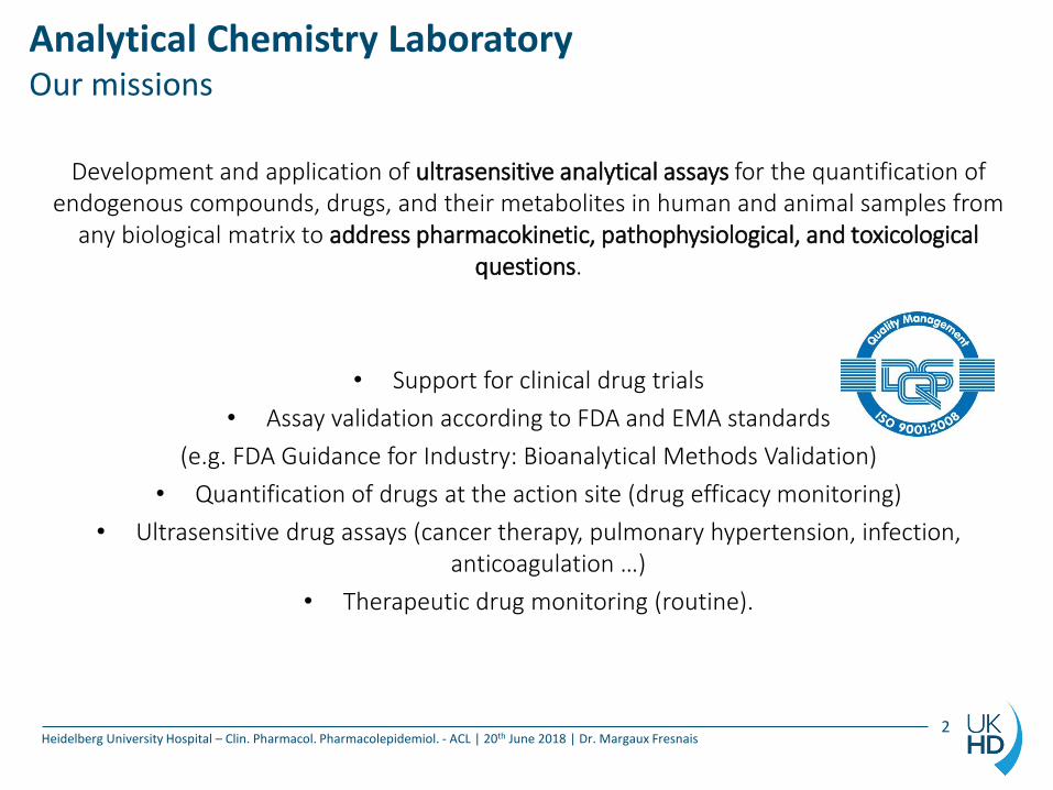
Development and application of ultrasensitive analytical assays for the quantification of endogenous compounds, drugs, and their metabolites in human and animal samples from
any biological matrix to address pharmacokinetic, pathophysiological, and toxicological questions.
• Support for clinical drug trials
• Assay validation according to FDA and EMA standards
(e.g. FDA Guidance for Industry: Bioanalytical Methods Validation)
• Quantification of drugs at the action site (drug efficacy monitoring)
• Ultrasensitive drug assays (cancer therapy, pulmonary hypertension, infection, anticoagulation …)
• Therapeutic drug monitoring (routine).
2
Analytical Chemistry Laboratory Matrices and targets
Heidelberg University Hospital – Clin. Pharmacol. Pharmacolepidemiol. - ACL | 20th June 2018 | Dr. Margaux Fresnais
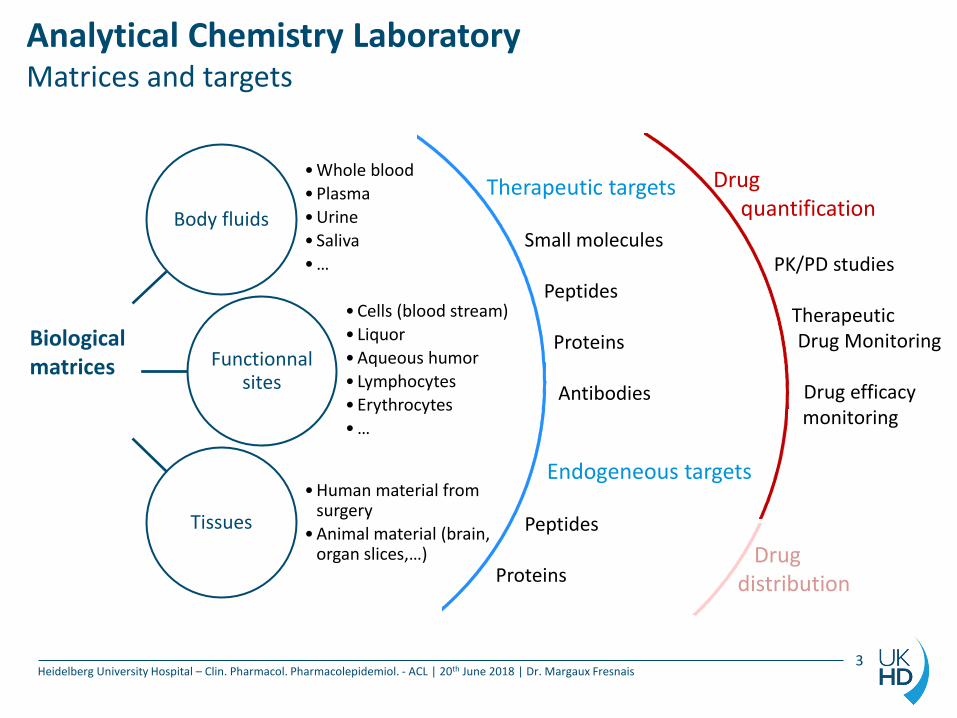
Drug quantification
PK/PD studies
Therapeutic Drug Monitoring
Drug efficacy monitoring
Drug distribution
Body fluids
Functionnal sites
Tissues
Biological matrices
•Whole blood
•Plasma
•Urine
• Saliva
•…
•Cells (blood stream)
• Liquor
•Aqueous humor
• Lymphocytes
•Erythrocytes
•…
•Human material from surgery
•Animal material (brain, organ slices,…)
Therapeutic targets Small molecules Peptides Proteins Antibodies
Endogeneous targets Peptides Proteins
3
Analytical Chemistry Laboratory Matrices and targets
Heidelberg University Hospital – Clin. Pharmacol. Pharmacolepidemiol. - ACL | 20th June 2018 | Dr. Margaux Fresnais
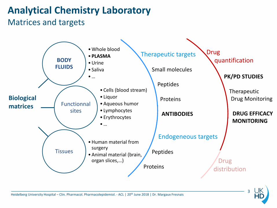
Drug quantification
PK/PD STUDIES
Therapeutic Drug Monitoring
DRUG EFFICACY MONITORING
Drug distribution
BODY FLUIDS
Functionnal sites
Tissues
Biological matrices
•Whole blood
•PLASMA
•Urine
• Saliva
•…
•Cells (blood stream)
• Liquor
•Aqueous humor
• Lymphocytes
•Erythrocytes
•…
•Human material from surgery
•Animal material (brain, organ slices,…)
Therapeutic targets Small molecules Peptides Proteins ANTIBODIES
Endogeneous targets Peptides Proteins
3
Antibody quantification in plasma using MS For what purpose ?
Heidelberg University Hospital – Clin. Pharmacol. Pharmacolepidemiol. - ACL | 20th June 2018 | Dr. Margaux Fresnais
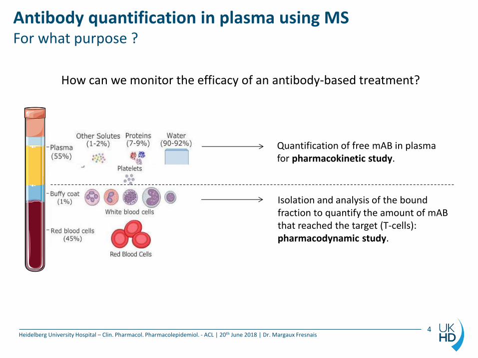
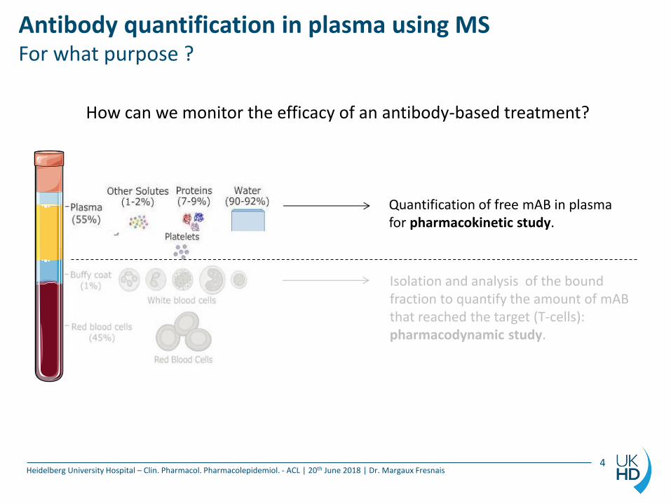
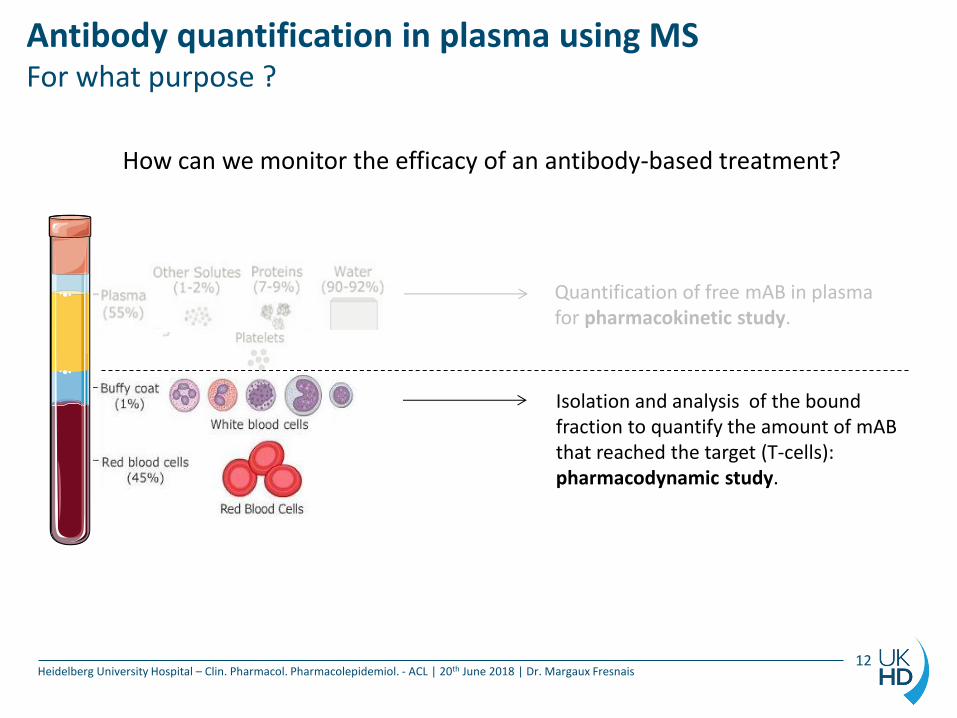
How can we monitor the efficacy of an antibody-based treatment?
Isolation and analysis of the bound fraction to quantify the amount of mAB that reached the target (T-cells): pharmacodynamic study.
Quantification of free mAB in plasma for pharmacokinetic study.
4
Antibody quantification in plasma using MS For what purpose ?
Heidelberg University Hospital – Clin. Pharmacol. Pharmacolepidemiol. - ACL | 20th June 2018 | Dr. Margaux Fresnais
How can we monitor the efficacy of an antibody-based treatment?
Isolation and analysis of the bound fraction to quantify the amount of mAB that reached the target (T-cells): pharmacodynamic study.
Quantification of free mAB in plasma for pharmacokinetic study.
4
Antibody quantification in plasma using MS Surrogate peptide approach for mABs
Heidelberg University Hospital – Clin. Pharmacol. Pharmacolepidemiol. - ACL | 20th June 2018 | Dr. Margaux Fresnais
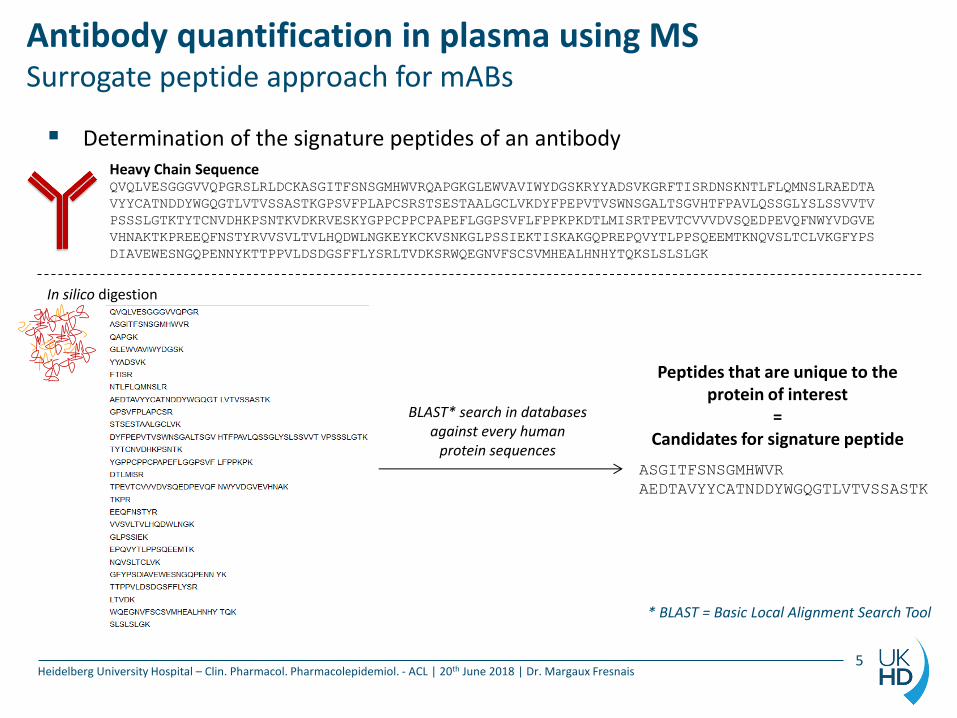
Determination of the signature peptides of an antibody
In silico digestion
Heavy Chain Sequence QVQLVESGGGVVQPGRSLRLDCKASGITFSNSGMHWVRQAPGKGLEWVAVIWYDGSKRYYADSVKGRFTISRDNSKNTLFLQMNSLRAEDTA
VYYCATNDDYWGQGTLVTVSSASTKGPSVFPLAPCSRSTSESTAALGCLVKDYFPEPVTVSWNSGALTSGVHTFPAVLQSSGLYSLSSVVTV
PSSSLGTKTYTCNVDHKPSNTKVDKRVESKYGPPCPPCPAPEFLGGPSVFLFPPKPKDTLMISRTPEVTCVVVDVSQEDPEVQFNWYVDGVE
VHNAKTKPREEQFNSTYRVVSVLTVLHQDWLNGKEYKCKVSNKGLPSSIEKTISKAKGQPREPQVYTLPPSQEEMTKNQVSLTCLVKGFYPS
DIAVEWESNGQPENNYKTTPPVLDSDGSFFLYSRLTVDKSRWQEGNVFSCSVMHEALHNHYTQKSLSLSLGK
ASGITFSNSGMHWVR
AEDTAVYYCATNDDYWGQGTLVTVSSASTK
BLAST* search in databases against every human
protein sequences
* BLAST = Basic Local Alignment Search Tool
Peptides that are unique to the protein of interest
= Candidates for signature peptide
5
Antibody quantification in plasma using MS Surrogate peptide approach for mABs
Heidelberg University Hospital – Clin. Pharmacol. Pharmacolepidemiol. - ACL | 20th June 2018 | Dr. Margaux Fresnais
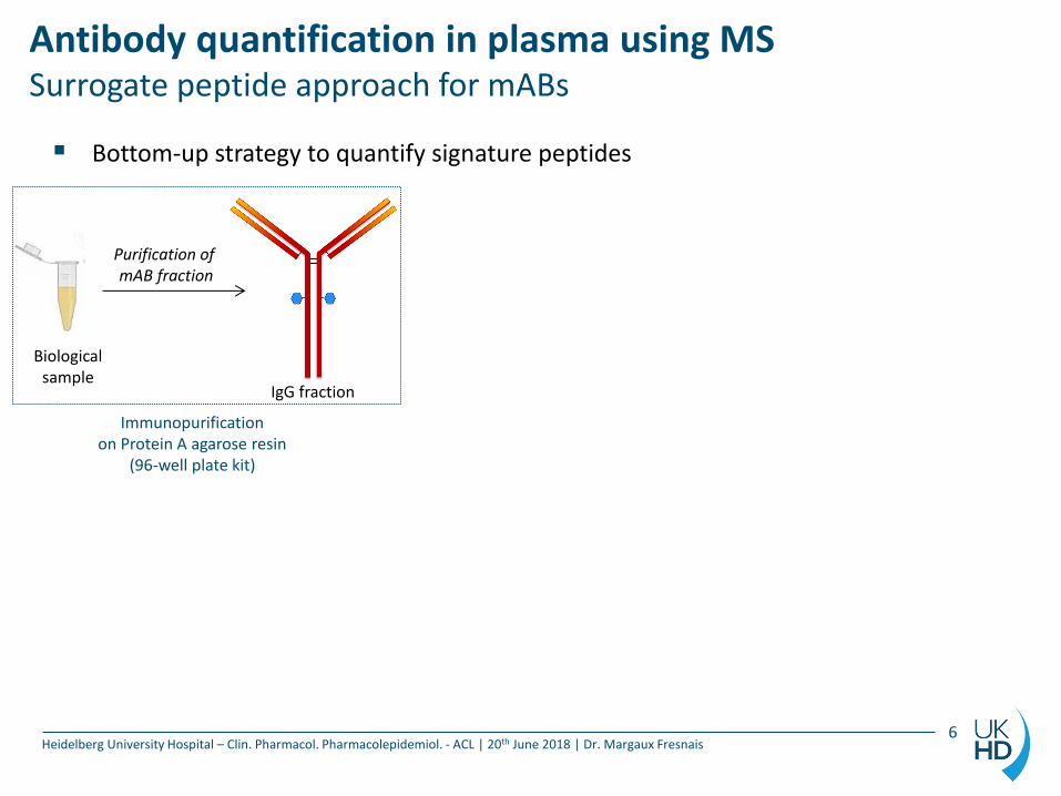
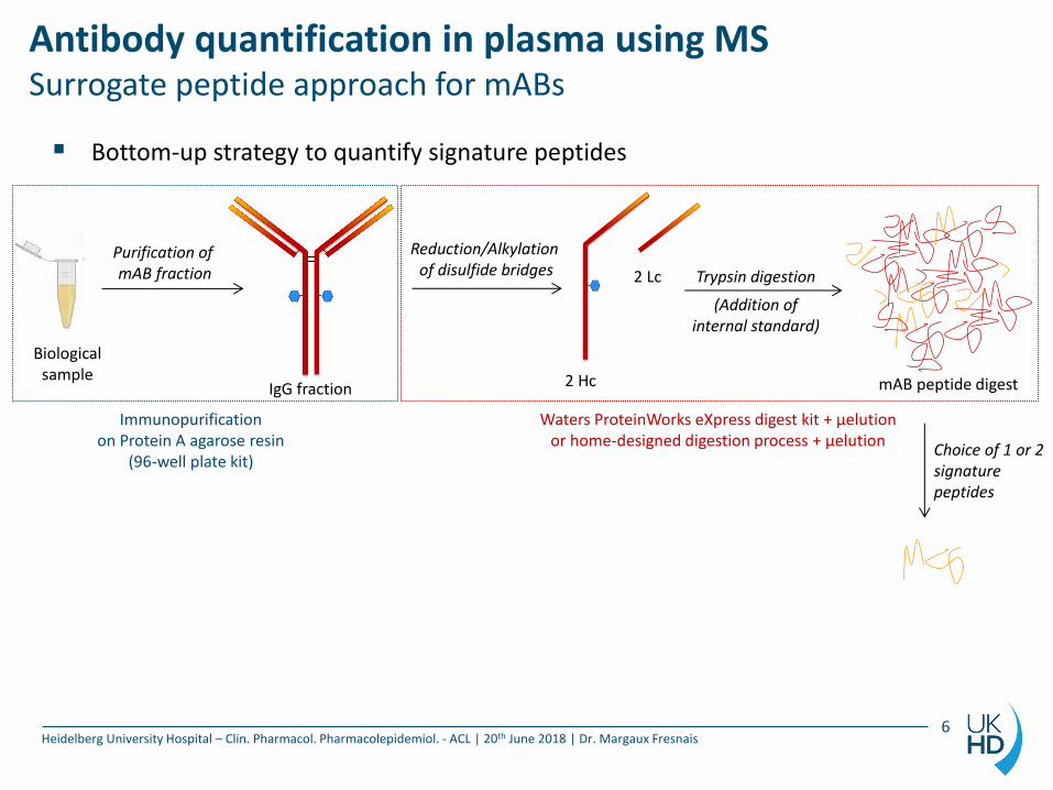
Bottom-up strategy to quantify signature peptides
Purification of mAB fraction
Biological sample
Immunopurification on Protein A agarose resin
(96-well plate kit)
IgG fraction
6
Antibody quantification in plasma using MS Surrogate peptide approach for mABs
Heidelberg University Hospital – Clin. Pharmacol. Pharmacolepidemiol. - ACL | 20th June 2018 | Dr. Margaux Fresnais
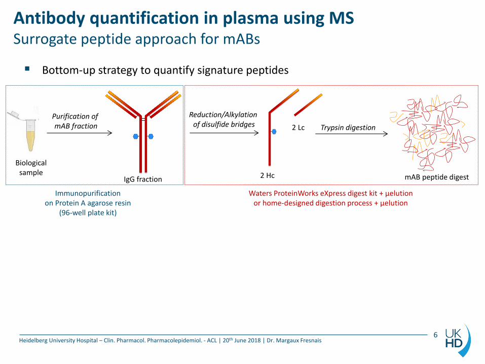
Bottom-up strategy to quantify signature peptides
Purification of mAB fraction
Biological sample
Immunopurification on Protein A agarose resin
(96-well plate kit)
IgG fraction
Reduction/Alkylation of disulfide bridges
2 Hc
2 Lc Trypsin digestion
mAB peptide digest
Waters ProteinWorks eXpress digest kit + µelution or home-designed digestion process + µelution
6
Antibody quantification in plasma using MS Surrogate peptide approach for mABs
Heidelberg University Hospital – Clin. Pharmacol. Pharmacolepidemiol. - ACL | 20th June 2018 | Dr. Margaux Fresnais
Bottom-up strategy to quantify signature peptides
Choice of 1 or 2 signature peptides
Purification of mAB fraction
Biological sample
Immunopurification on Protein A agarose resin
(96-well plate kit)
IgG fraction
Reduction/Alkylation of disulfide bridges
2 Hc
2 Lc Trypsin digestion
(Addition of internal standard)
mAB peptide digest
Waters ProteinWorks eXpress digest kit + µelution or home-designed digestion process + µelution
6
Antibody quantification in plasma using MS Surrogate peptide approach for mABs
Heidelberg University Hospital – Clin. Pharmacol. Pharmacolepidemiol. - ACL | 20th June 2018 | Dr. Margaux Fresnais
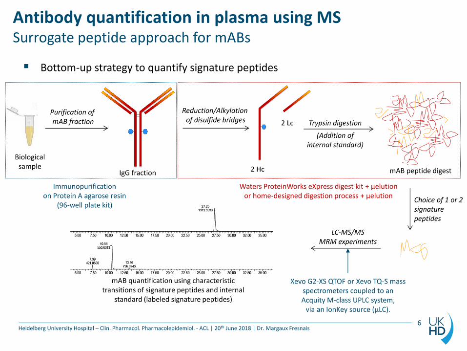
Bottom-up strategy to quantify signature peptides
Choice of 1 or 2 signature peptides
LC-MS/MS MRM experiments
mAB quantification using characteristic transitions of signature peptides and internal
standard (labeled signature peptides)
Purification of mAB fraction
Biological sample
Immunopurification on Protein A agarose resin
(96-well plate kit)
IgG fraction
Reduction/Alkylation of disulfide bridges
2 Hc
2 Lc Trypsin digestion
(Addition of internal standard)
mAB peptide digest
Waters ProteinWorks eXpress digest kit + µelution or home-designed digestion process + µelution
Xevo G2-XS QTOF or Xevo TQ-S mass spectrometers coupled to an Acquity M-class UPLC system,
via an IonKey source (µLC).
6
Antibody quantification in plasma using MS Case study : Nivolumab, an immune check-point inhibitor
Heidelberg University Hospital – Clin. Pharmacol. Pharmacolepidemiol. - ACL | 20th June 2018 | Dr. Margaux Fresnais 7
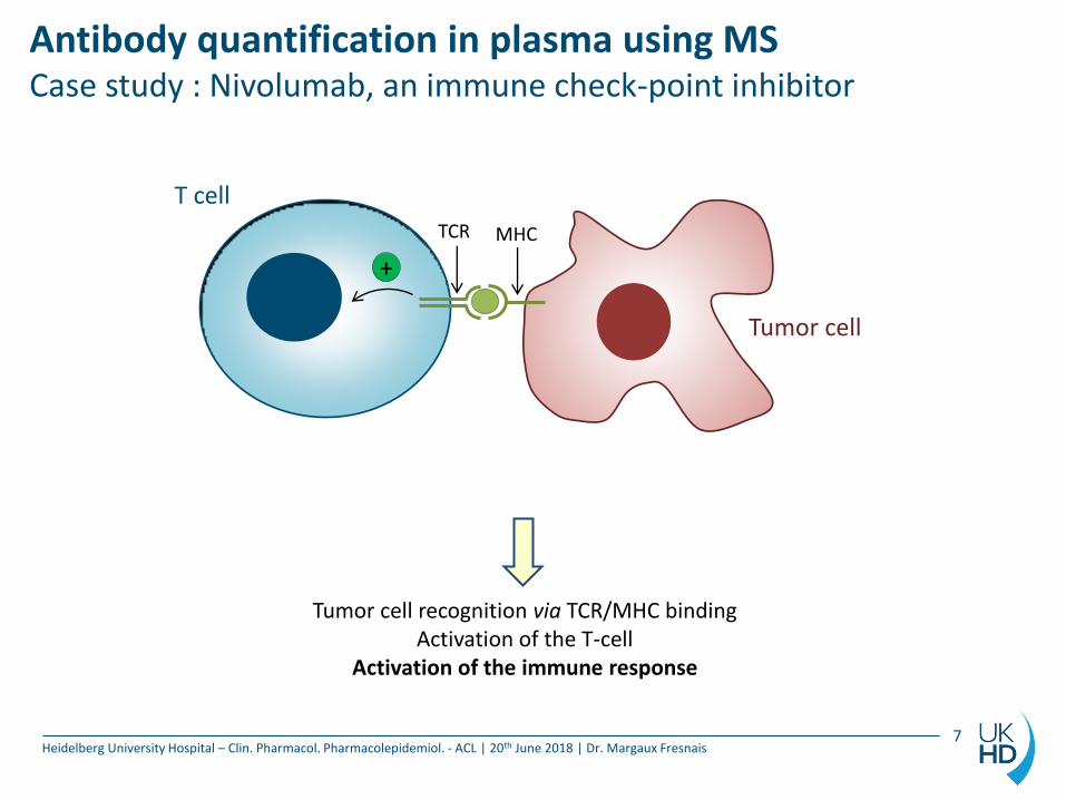
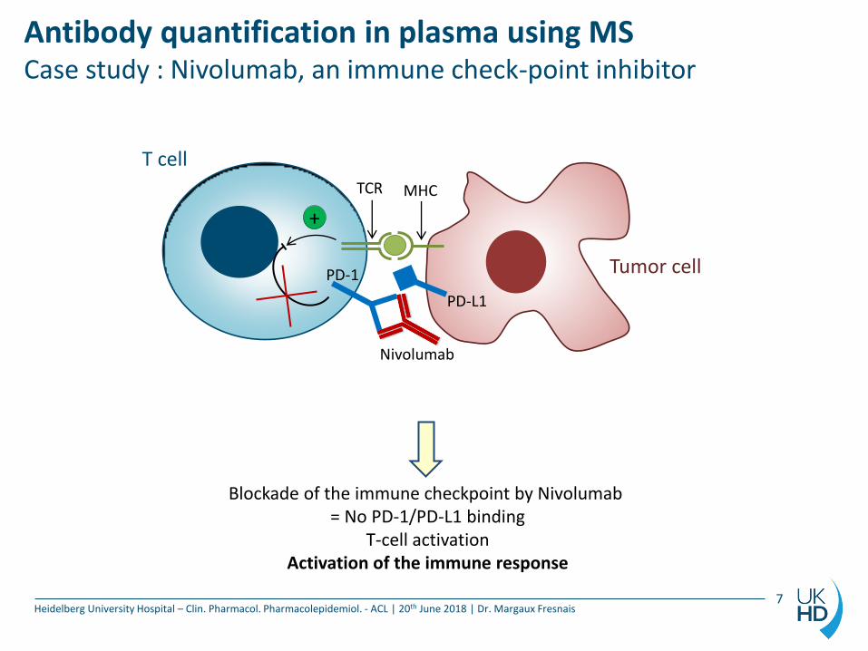
Tumor cell recognition via TCR/MHC binding Activation of the T-cell
Activation of the immune response
+
T cell
Tumor cell
TCR MHC
Antibody quantification in plasma using MS Case study : Nivolumab, an immune check-point inhibitor
Heidelberg University Hospital – Clin. Pharmacol. Pharmacolepidemiol. - ACL | 20th June 2018 | Dr. Margaux Fresnais 7
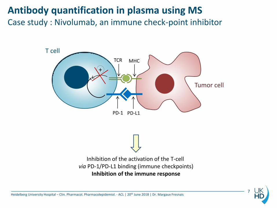
Inhibition of the activation of the T-cell via PD-1/PD-L1 binding (immune checkpoints)
Inhibition of the immune response
+
TCR MHC
T cell
Tumor cell
PD-1 PD-L1
Antibody quantification in plasma using MS Case study : Nivolumab, an immune check-point inhibitor
Heidelberg University Hospital – Clin. Pharmacol. Pharmacolepidemiol. - ACL | 20th June 2018 | Dr. Margaux Fresnais 7
Blockade of the immune checkpoint by Nivolumab = No PD-1/PD-L1 binding
T-cell activation Activation of the immune response
+
TCR MHC
T cell
Tumor cell PD-1
PD-L1
Nivolumab
Antibody quantification in plasma using MS Case study : Nivolumab, an immune check-point inhibitor
Heidelberg University Hospital – Clin. Pharmacol. Pharmacolepidemiol. - ACL | 20th June 2018 | Dr. Margaux Fresnais 8
In silico digestion
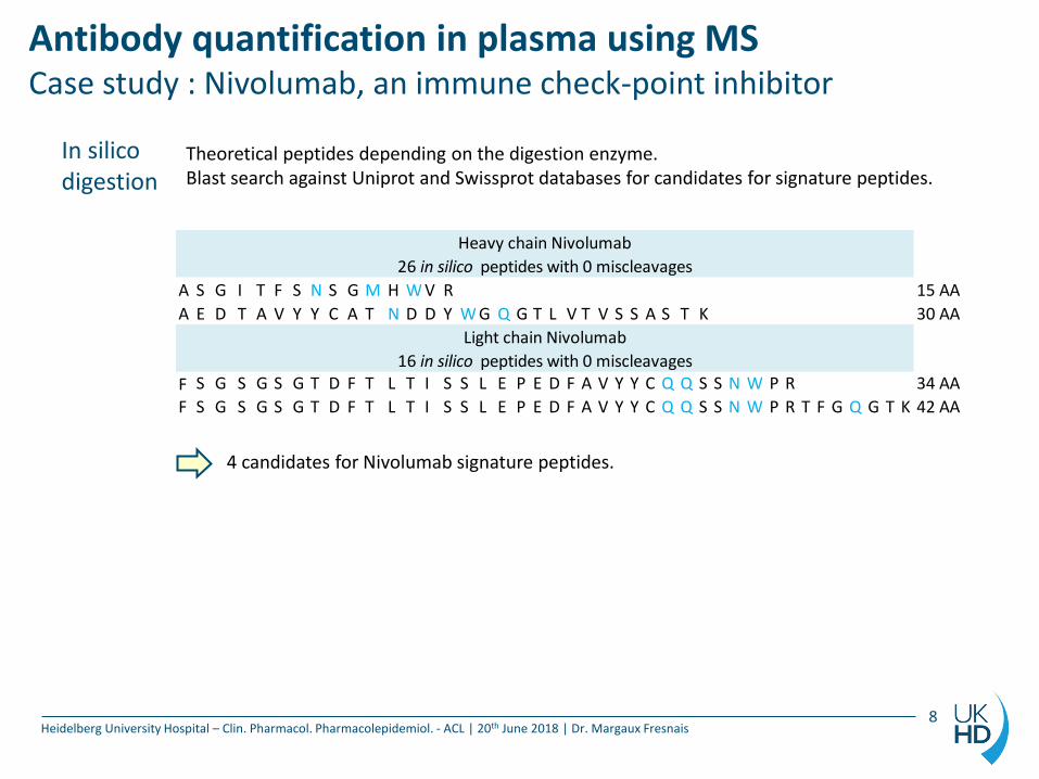
A S G I T F S N S G M H W V R 15 AA
A E D T A V Y Y C A T N D D Y W G Q G T L V T V S S A S T K 30 AA
F S G S G S G T D F T L T I S S L E P E D F A V Y Y C Q Q S S N W P R 34 AA
F S G S G S G T D F T L T I S S L E P E D F A V Y Y C Q Q S S N W P R T F G Q G T K 42 AA
Heavy chain Nivolumab
Light chain Nivolumab
26 in silico peptides with 0 miscleavages
16 in silico peptides with 0 miscleavages
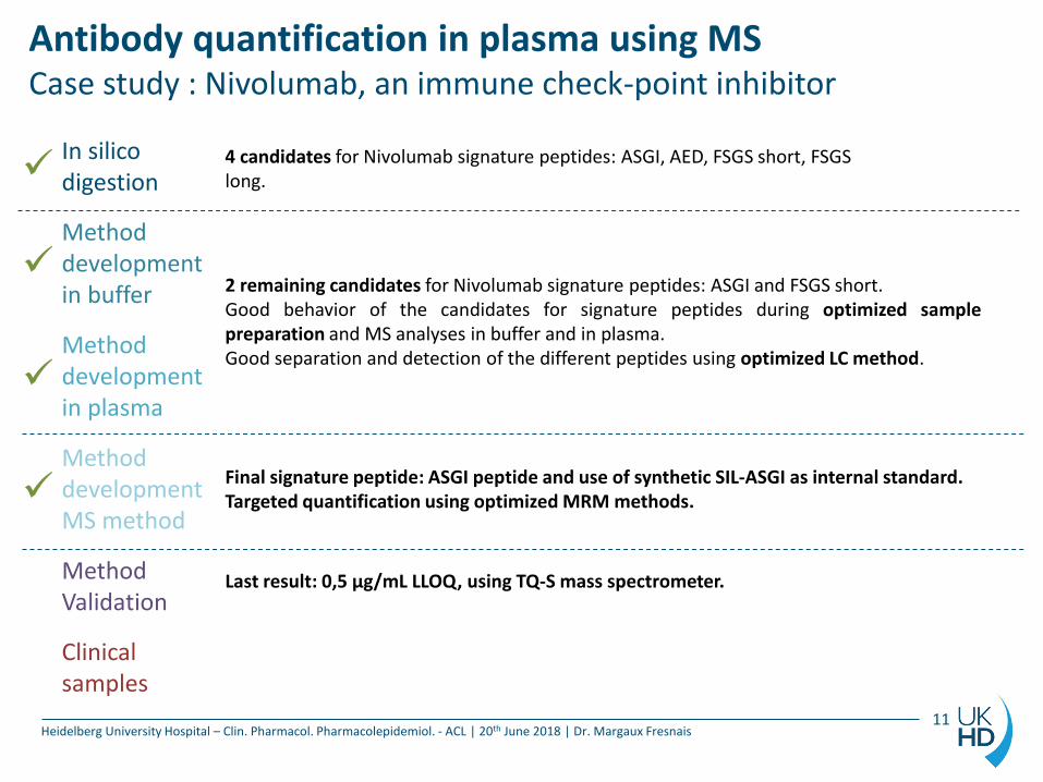
4 candidates for Nivolumab signature peptides.
Theoretical peptides depending on the digestion enzyme. Blast search against Uniprot and Swissprot databases for candidates for signature peptides.
Antibody quantification in plasma using MS Case study : Nivolumab, an immune check-point inhibitor
Heidelberg University Hospital – Clin. Pharmacol. Pharmacolepidemiol. - ACL | 20th June 2018 | Dr. Margaux Fresnais 9
In silico digestion
Method development in buffer
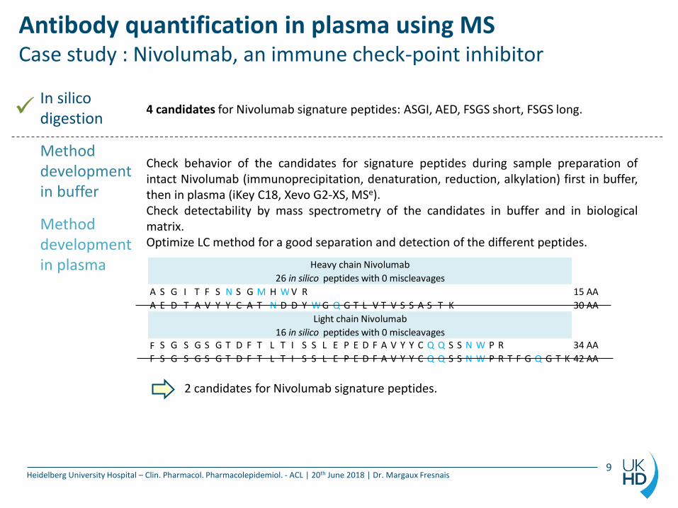
Check behavior of the candidates for signature peptides during sample preparation of intact Nivolumab (immunoprecipitation, denaturation, reduction, alkylation) first in buffer, then in plasma (iKey C18, Xevo G2-XS, MSe). Check detectability by mass spectrometry of the candidates in buffer and in biological matrix. Optimize LC method for a good separation and detection of the different peptides.
A S G I T F S N S G M H W V R 15 AA
A E D T A V Y Y C A T N D D Y W G Q G T L V T V S S A S T K 30 AA
F S G S G S G T D F T L T I S S L E P E D F A V Y Y C Q Q S S N W P R 34 AA
F S G S G S G T D F T L T I S S L E P E D F A V Y Y C Q Q S S N W P R T F G Q G T K 42 AA
Heavy chain Nivolumab
Light chain Nivolumab
26 in silico peptides with 0 miscleavages
16 in silico peptides with 0 miscleavages
2 candidates for Nivolumab signature peptides.
Method development in plasma
4 candidates for Nivolumab signature peptides: ASGI, AED, FSGS short, FSGS long.
Antibody quantification in plasma using MS Case study : Nivolumab, an immune check-point inhibitor
Heidelberg University Hospital – Clin. Pharmacol. Pharmacolepidemiol. - ACL | 20th June 2018 | Dr. Margaux Fresnais 10
In silico digestion
Method development in buffer
Method development in plasma
Method development MS method
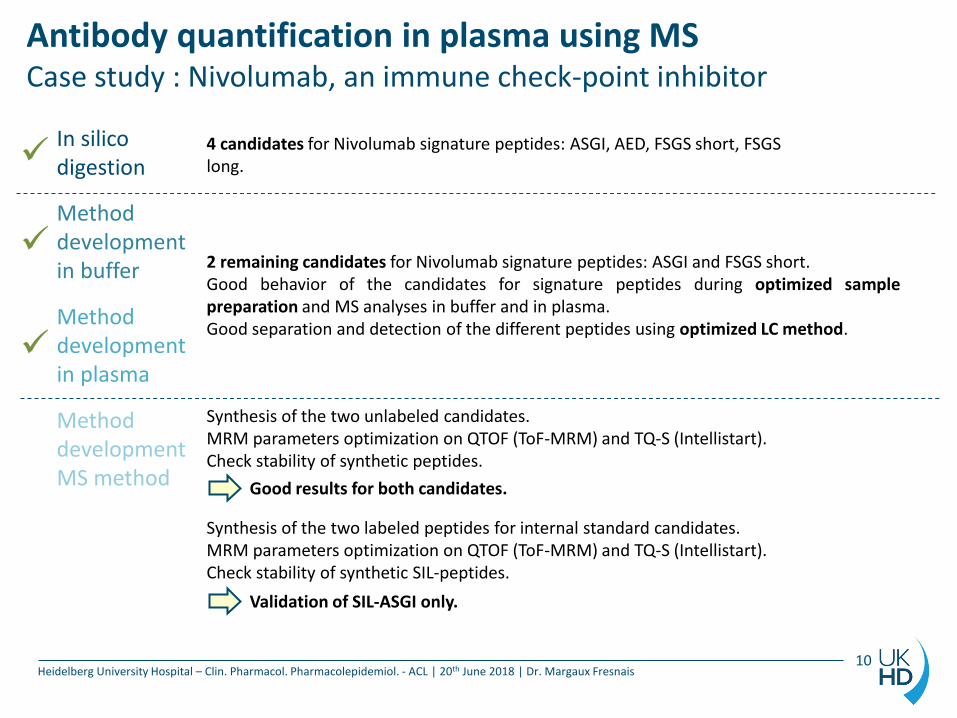
Synthesis of the two unlabeled candidates. MRM parameters optimization on QTOF (ToF-MRM) and TQ-S (Intellistart). Check stability of synthetic peptides. Synthesis of the two labeled peptides for internal standard candidates. MRM parameters optimization on QTOF (ToF-MRM) and TQ-S (Intellistart). Check stability of synthetic SIL-peptides.
Good results for both candidates.
Validation of SIL-ASGI only.
4 candidates for Nivolumab signature peptides: ASGI, AED, FSGS short, FSGS long.
2 remaining candidates for Nivolumab signature peptides: ASGI and FSGS short. Good behavior of the candidates for signature peptides during optimized sample preparation and MS analyses in buffer and in plasma. Good separation and detection of the different peptides using optimized LC method.
Antibody quantification in plasma using MS Case study : Nivolumab, an immune check-point inhibitor
Heidelberg University Hospital – Clin. Pharmacol. Pharmacolepidemiol. - ACL | 20th June 2018 | Dr. Margaux Fresnais 11
In silico digestion
Method development in buffer
Method development in plasma
Method development MS method
Method Validation
Clinical samples
4 candidates for Nivolumab signature peptides: ASGI, AED, FSGS short, FSGS long.
2 remaining candidates for Nivolumab signature peptides: ASGI and FSGS short. Good behavior of the candidates for signature peptides during optimized sample preparation and MS analyses in buffer and in plasma. Good separation and detection of the different peptides using optimized LC method.
Final signature peptide: ASGI peptide and use of synthetic SIL-ASGI as internal standard. Targeted quantification using optimized MRM methods.
Last result: 0,5 µg/mL LLOQ, using TQ-S mass spectrometer.
Antibody quantification in plasma using MS For what purpose ?
Heidelberg University Hospital – Clin. Pharmacol. Pharmacolepidemiol. - ACL | 20th June 2018 | Dr. Margaux Fresnais
How can we monitor the efficacy of an antibody-based treatment?
Isolation and analysis of the bound fraction to quantify the amount of mAB that reached the target (T-cells): pharmacodynamic study.
Quantification of free mAB in plasma for pharmacokinetic study.
12
Quantification of the cell-bound fraction by MS Case study : Nivolumab, an immune check-point inhibitor
Heidelberg University Hospital – Clin. Pharmacol. Pharmacolepidemiol. - ACL | 20th June 2018 | Dr. Margaux Fresnais 13
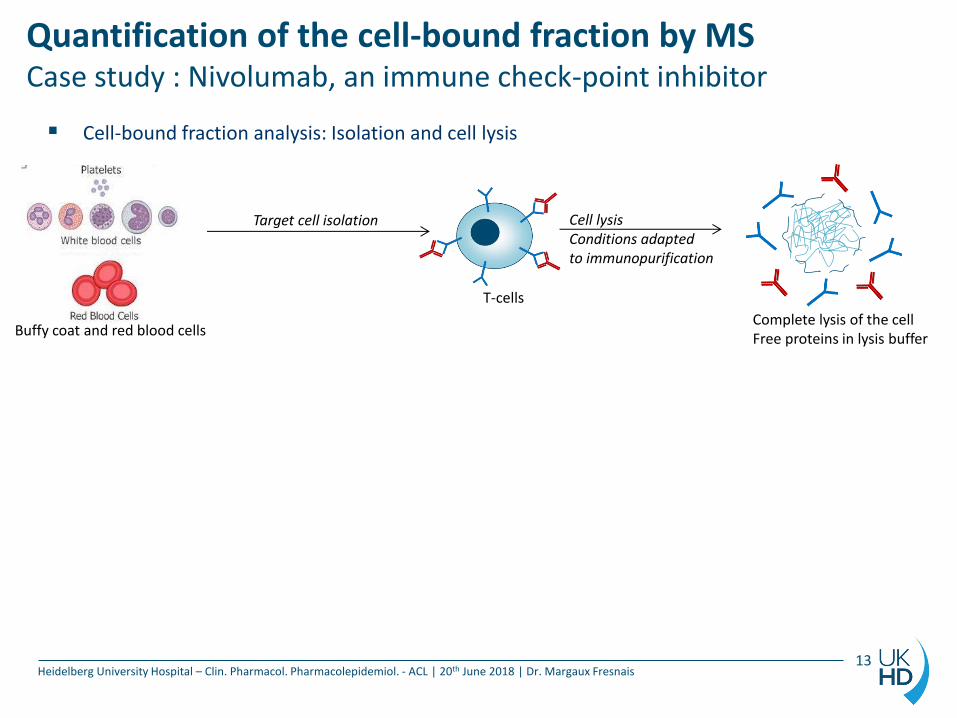
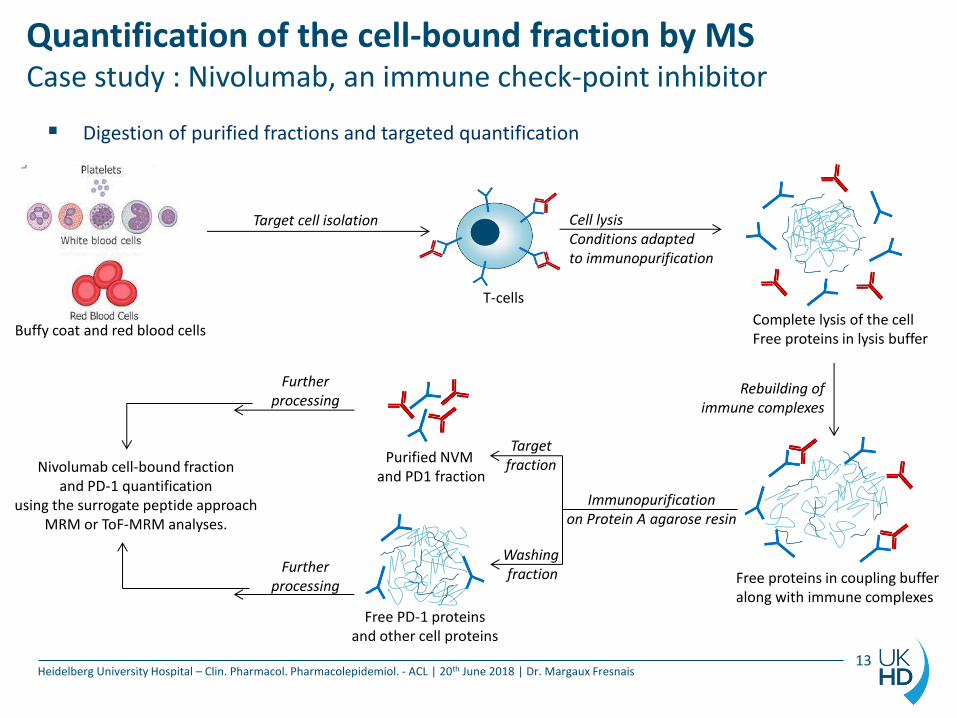
Cell-bound fraction analysis: Isolation and cell lysis
Cell lysis Conditions adapted to immunopurification
Target cell isolation
Buffy coat and red blood cells
T-cells
Complete lysis of the cell Free proteins in lysis buffer
Washing fraction
Target fraction
Immunopurification on Protein A agarose resin
Quantification of the cell-bound fraction by MS Case study : Nivolumab, an immune check-point inhibitor
Heidelberg University Hospital – Clin. Pharmacol. Pharmacolepidemiol. - ACL | 20th June 2018 | Dr. Margaux Fresnais 13
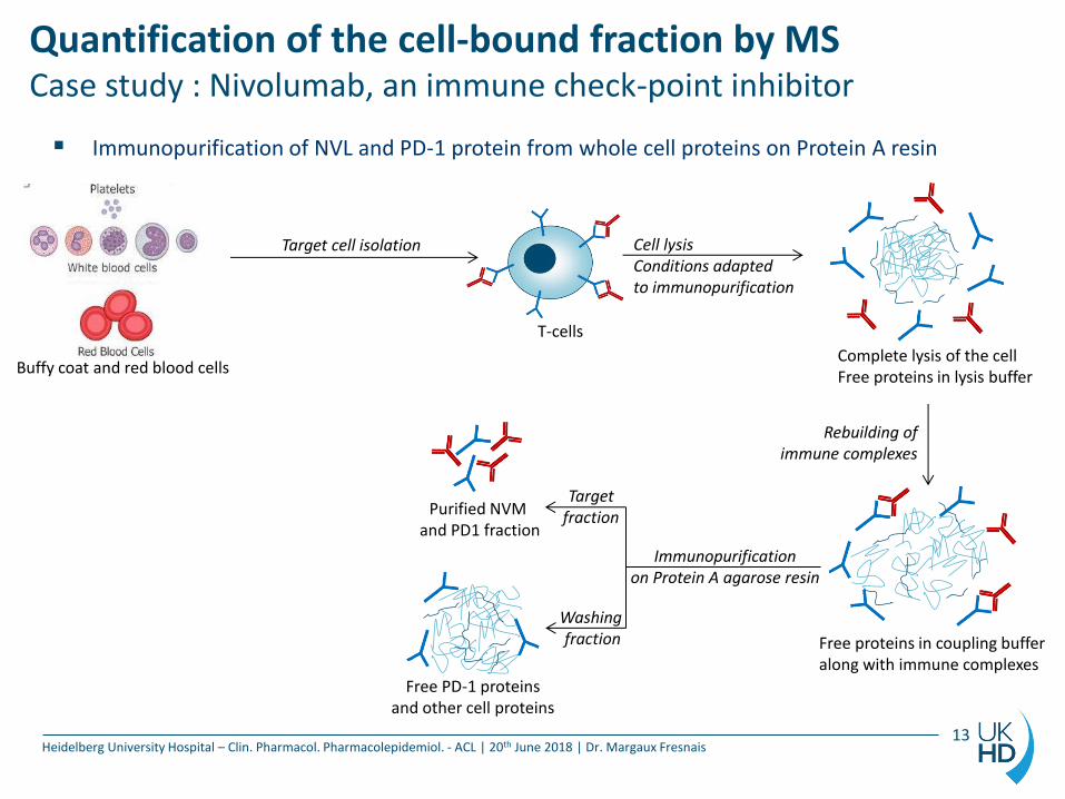
Immunopurification of NVL and PD-1 protein from whole cell proteins on Protein A resin
Rebuilding of immune complexes
Free proteins in coupling buffer along with immune complexes
Free PD-1 proteins and other cell proteins
Purified NVM and PD1 fraction
Cell lysis Conditions adapted to immunopurification
Target cell isolation
Buffy coat and red blood cells
T-cells
Complete lysis of the cell Free proteins in lysis buffer
Further processing
Further processing
Immunopurification on Protein A agarose resin
Quantification of the cell-bound fraction by MS Case study : Nivolumab, an immune check-point inhibitor
Heidelberg University Hospital – Clin. Pharmacol. Pharmacolepidemiol. - ACL | 20th June 2018 | Dr. Margaux Fresnais 13
Digestion of purified fractions and targeted quantification
Rebuilding of immune complexes
Free proteins in coupling buffer along with immune complexes
Free PD-1 proteins and other cell proteins
Purified NVM and PD1 fraction
Washing fraction
Target fraction
Cell lysis Conditions adapted to immunopurification
Target cell isolation
Buffy coat and red blood cells
T-cells
Complete lysis of the cell Free proteins in lysis buffer
Nivolumab cell-bound fraction and PD-1 quantification
using the surrogate peptide approach MRM or ToF-MRM analyses.
Quantification of the cell-bound fraction by MS Case study : Nivolumab, an immune check-point inhibitor
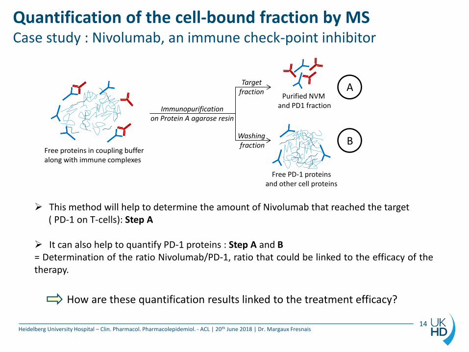
Heidelberg University Hospital – Clin. Pharmacol. Pharmacolepidemiol. - ACL | 20th June 2018 | Dr. Margaux Fresnais 14
Free proteins in coupling buffer along with immune complexes
Free PD-1 proteins and other cell proteins
Purified NVM and PD1 fraction Immunopurification
on Protein A agarose resin
Washing fraction
Target fraction A
B
This method will help to determine the amount of Nivolumab that reached the target ( PD-1 on T-cells): Step A It can also help to quantify PD-1 proteins : Step A and B = Determination of the ratio Nivolumab/PD-1, ratio that could be linked to the efficacy of the therapy.
How are these quantification results linked to the treatment efficacy?
Summary
Heidelberg University Hospital – Clin. Pharmacol. Pharmacolepidemiol. - ACL | 20th June 2018 | Dr. Margaux Fresnais 15
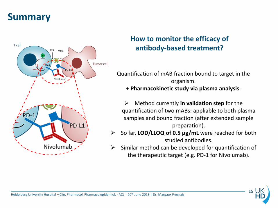
How to monitor the efficacy of antibody-based treatment?
Quantification of mAB fraction bound to target in the organism.
+ Pharmacokinetic study via plasma analysis.
Method currently in validation step for the quantification of two mABs: appliable to both plasma samples and bound fraction (after extended sample
preparation). So far, LOD/LLOQ of 0.5 µg/mL were reached for both
studied antibodies. Similar method can be developed for quantification of
the therapeutic target (e.g. PD-1 for Nivolumab).
Summary
Heidelberg University Hospital – Clin. Pharmacol. Pharmacolepidemiol. - ACL | 20th June 2018 | Dr. Margaux Fresnais 15
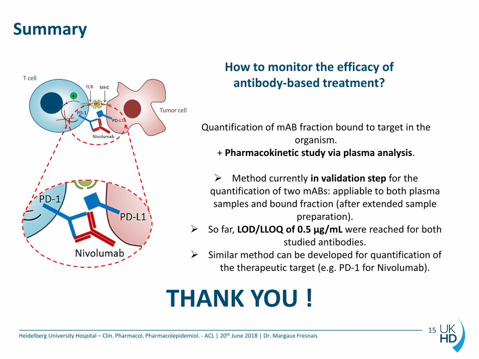
How to monitor the efficacy of antibody-based treatment?
Quantification of mAB fraction bound to target in the organism.
+ Pharmacokinetic study via plasma analysis.
Method currently in validation step for the quantification of two mABs: appliable to both plasma samples and bound fraction (after extended sample
preparation). So far, LOD/LLOQ of 0.5 µg/mL were reached for both
studied antibodies. Similar method can be developed for quantification of
the therapeutic target (e.g. PD-1 for Nivolumab).
THANK YOU !
Acknowledgements
Heidelberg University Hospital – Clin. Pharmacol. Pharmacolepidemiol. - ACL | 20th June 2018 | Dr. Margaux Fresnais
Clinical pharmacology and pharmacoepidemiology dept.
Walter-Emil Haefeli
Analytical chemistry laboratory
Jürgen Burhenne
Marius Roos-Majewski
Max Sauter
Philip Uhl
Andrea Deschlmayr
Magdalena Longo
Kevin Jansen
Kathrin Foerster
Manuela Vay
Molecular Biology Laboratory
Johanna Weiß
Dirk Theile
DKFZ and DKTK DKFZ - Division of Pediatric Neurooncology Stefan Pfister
Heidelberg DKTK coordinators Katja Engelmann Franziska Hasslinger-Pajtler
Heidelberg Immunotherapeutics Michaela Arndt
Radiopharmaceutical chemistry Walter Mier