Embed Size (px)
Citation preview

Targeted mutagenesis using CRISPR/Cas system Satoshi Ansai1 and Masato Kinoshita2
Genome editing using targetable nucleases has become a versatile and powerful tool for
genetic manipulation. These nucleases can introduce site-specific double-strand DNA breaks
that are repaired by either of two major pathways: non-homologous end joining (NHEJ) and
homology-directed repair (HDR) (Urnov et al., 2010). NHEJ joins the end of the broken DNA
strands without any templates, frequently inducing targeted gene disruption by a small
insertion and/or deletion (indel) around the cleaving site or site-specific addition of tags such
as GFP. HDR in the presence of DNA templates with homologous sequences can repair the
DSBs through the replacement of flanking homology arms, resulting in precise integrations of
a DNA fragment or targeted gene correction. To date, these nucleases have been effective
tools for targeted genomic engineering in a wide range of organisms (Peng et al., 2014).
Clustered regularly interspaced short palindromic repeats (CRISPR)/CRISPR-associated
(Cas) system is the most widely used nuclease system for genome editing, which is known as
an adaptive immune system in bacteria and archaea to detect and silence foreign genetic
elements such as viruses and plasmids (Wiedenheft et al., 2012). In type II CRISPR systems, a
Cas9 endonuclease forms complex with each CRISPR RNAs (crRNAs) hybridized with a trans-
activating crRNA (tracrRNA) and then cleaves target DNA sequences that are complement to
the crRNAs and adjacent to short sequences known as protospacer adjacent motifs (PAMs)
(Gasiunas et al., 2012; Jinek et al., 2012). A synthetic single-guide RNA (sgRNA) consisting of a
fusion of crRNA and tracrRNA can also program the recognition specificity of the Cas9
nuclease from Streptococcus pyogenes. This indicates the Cas9 nuclease act as an RNA-guided
endonuclease (RGEN) whose cleavage site can be easily altered only by engineering the 5’
sequence of the sgRNA.
In this protocol, we describe a method for targeted genome editing in medaka using
CRISPR/Cas-based RNA-guided endonucleases (RGENs) in medaka. Followed by describing
how the targetable nucleases are prepared for endogenous genomic loci of medaka, we also
describe an effective method, called heteroduplex mobility assay (HMA), to estimate the
efficacy of their genome editing activities in the injected embryos and to identify the induced
mutations in F1 or later generations.
1 Division of Ecological Genetics, Department of Population Genetics, National Institute of Genetics. (Email: [email protected]) 2 Division of Applied Biosciences, Graduate School of Agriculture, Kyoto University.

1. Preparation of RNAs for CRISPR/Cas genome editing
This section describes how a Cas9 nuclease and engineered sgRNAs are prepared for
microinjection into medaka fertilized eggs. Capped RNA encoding for a Cas9 nuclease is
transcribed from an expression vector constructed for fish. The sgRNAs are engineered by
cloning of a pair of custom-ordered oligonucleotides and therefore transcribed using a RNA
polymerase.
1.1. Materials
Plasmids
n pCS2+hSpCas9 (Addgene Plasmid #51815): A Cas9 expression plasmid is used to create a
capped RNA by in vitro transcription using a SP6 RNA polymerase for microinjection into
fish embryos (Ansai & Kinoshita, 2014). This vector contains a human codon-optimized Cas9
gene derived from Streptococcus pyogenes (Cong et al., 2013) and an ampicillin resistant
gene.
n pDR274 (Addgene Plasmid #42250): An empty gRNA plasmid is used to create a target-
specific gRNA by in vitro transcription using a T7 RNA polymerase (Hwang et al., 2013). This
vector contains a kanamycin resistant gene.
PCR primers for colony PCR checking
n M13 forward primer: 5’- GTAAAACGACGGCCAGT -3’.
Molecular biology reagents
n Restriction enzymes–BsaI, DraI, and NotI: The restriction enzymes with lower star
activity are preferable. For example, BsaI-HF (High-Fidelity version of BsaI; NEB, Cat. No.
R3535) is more suitable for overnight digestion than the BsaI without “HF” (NEB, Cat. No.
R0535).
n DNA ligase: Ligation reagents with high efficiency (e.g. Ligation high Ver.2, Toyobo, Cat.
No. LGK-201) are recommended.
n PCR enzyme: High-fidelity polymerase is not necessarily required. (e.g. HybriPol DNA
Polymerase, Bioline, Cat. No. BIO-21080; TaKaRa Ex Taq, Takara Bio, Cat. No. RR001A are
available.)
n T7 RNA polymerase: The high-yield in vitro transcription kit with T7 RNA polymerase (e.g.
AmpliScribe T7-Flash Transcription Kit, Epicentre, Cat. No. ASF3507) is recommended for
stable transcription of sgRNAs. However, the standard T7 RNA polymerase (e.g. Promega,

Cat. No. P2075) is also available.
n Capped RNA transcription kit with SP6 RNA polymerase: mMessage mMachine SP6
Transcription Kit (Life Technologies, Cat. No. AM1340) is recommended for high-yield
production of Cas9 RNA.
n Proteinase K: Prepare a 20 mg/mL of stock solution.
n Plasmid purification kit: The plasmid purification kit (spin column or higher grade) should
be used (e.g. NucleoSpin Plasmid QuickPure; MACHEREY-NAGEL, Cat. No. 740615).
n Gel extraction kit: The gel extraction kit (spin column type) should be used (e.g.
NucleoSpin Gel and PCR Clean-up; MACHEREY-NAGEL, Cat. No. 740609).
n RNA extraction kit: The RNA extraction kit with spin columns (e.g. RNeasy Mini Kit,
Qiagen, Cat. No. 74104) should be used for purification of transcribed RNAs.
n 10X annealing buffer: 400 mM Tris-HCl (pH 8.0) containig 200 mM MgCl2 and 500 mM
NaCl.
n 10% SDS
n Phenol-chloroform mixture: Mixture of equal volume of phenol saturated by TE buffer
and chloroform.
n Chloroform
n 3 M sodium acetate (CH3COONa) [pH5.2]
n 0.5 M EDTA [pH8.0]
n Co-precipitated reagent: Glycogen (e.g. Roche, Cat. No. 10901393001) or other co-
precipitated reagent is used for high-yield recovery of DNA in ethanol precipitation.
n Ethanol (100% and 70%)
n Competent E. coli: DH5α and any other competent cells can be used.
n LB broth: Both LB plates and liquid medium containing appropriate antibiotics at the
concentrations indicated below should be prepared. For liquid culture, higher nutrient
medium such as 2X YT and Plusgrow II (Nacalai Tesque, Cat. No. 08202-75) is more suitable
for plasmid DNA recovery with high yield.
n Ampicillin (amp): A stock solution is 50 mg/mL ampicilin sodium salt in H2O or 70%
ethanol. The final concentration is 50 µg/mL in the medium.
n Kanamycin (kan): A stock solution (20 mg/mL kanamycin sulfate in H2O) should be at a
final concentration of 20 µg/mL of medium according to standard protocols.

1.2. Production of custom-designed sgRNA 1.2.1. Preparation of the BsaI-digested sgRNA backbone
1. E. coli cotaining the pDR274 vector is spread on the LB plate containing 20 µg/mL
kanamycin and is incubated at 37˚C overnight.
2. Pick an isolated colony from the plate, inoculate into the LB or any liquid medium with 20
µg/mL kanamycin, and grow the cultures overnight at 37˚C. Prepare the amount of
medium that is enough to recovery more than 5 µg of plasmid DNA.
3. Purify the plasmid from the cultures using the plasmid purification kit with spin columns
and then quantify the concentration of each elution with the spectrophotometer or any
other methods for DNA quantification.
4. Purified plasmid is digested by the restriction enzyme BsaI-HF. Assemble the reaction as
described below and then incubate at 37˚C overnight.
pDR274 (5 µg) X µL
CutSmart Buffer (10X) 5 µL
BsaI-HF (NEB) 2 µL
H2O up to 50 µL
Total 50 µL
5. All the 50 µL of reaction are individually loaded into 1% agarose gels and separated by
electrophoresis at 135V for 25 min or the same separation condition.
6. Excise each DNA fragment (ca. 2.1 kb) with minimized UV exposure and then recover DNA
using the spin column gel extraction kit. Elute the DNA with 30 µL of the elution buffer
from each column whose residual ethanol is removed by longer duration of centrifuge
and incubating the columns for several minutes at 70˚C prior to elution.
7. Eluted DNAs are stored at -20˚C.
1.2.2. Design and production of customized sgRNA
Day 0: Design sgRNA for medaka endogenous loci and order oligonucleotides
1. Find the “5´-N21GG-3´” or “5´-CCN21-3´” sequence in the target locus because sgRNA can
recognize the sequence followed by a PAM (5´-NGG-3´) in both strands.
2. Choose 2 or more target sites in a target locus with reference to (a) potential off-target
sequences and/or (b) micro homology sequences.
(a) Off-target alterations are sometimes identified in the genomic sequences harboring up

to several base pairs of mismatches adjacent to a NGG PAM (Ansai & Kinoshita, 2014).
Target sequences with less off-target candidates in the medaka genome can be
predicted using the CRISPR/Cas9 target online predictor (CCTop, http://crispr.cos.uni-
heidelberg.de). Potential off-target candidates can also be found using the pattern
match tool for CRISPR/Cas (http://viewer.shigen.info/medakavw/crisprtool/).
(b) We previously found that microhomologous sequences striding over the DSB point
frequently induce specific patterns of deletion between the homologous sequences
(Ansai et al., 2014). Target candidates with microhomologies whose deletion pattern
frequently observed in the RNA-injected fish could be predicted are identified using the
program to search for CRISPR target sites with microhomology sequences
(http://viewer.shigen.info/cgi-bin/crispr/crispr.cgi).
3. Enter the selected target sequences as “5´-N21GG-3´” into the file “sgRNA_design.xls” for
Microsoft Excel or “sgRNA_design.ods” for Apache OpenOffice Calc, which produces each
pair of 22-mer oligonucleotide sequences for subcloning into the pDR274 vector. These
files can be downloaded from a website (http://satoshi-ansai.github.io/en/misc.html).
4. Order the designed oligonucleotides to a supplier. Purification by the PCR primer grade is
enough for the following experiments.
Day 1: Clone the annealed oligonucleotides into the sgRNA expression vector
1. Anneal each pair of the ordered oligonucleatides. Prepare the reaction solution described
as below. The mixture is heated to 95˚C for 2 minutes and then cooled slowly to 25˚C in 1
hour using a thermal cycler.
Oligonucleotide Sense (100 µM) 1 µL
Oligonucleotide Anti-Sense (100 µM) 1 µL
10X Annealing buffer 1 µL
H2O 7 µL
Total 10 µL
2. Assemble the ligation mixture using annealed oligonucleotides as below.
pDR274 (BsaI digested) 1 µL
Annealed oligonucleotides 1 µL
Ligation High Ver.2 (Toyobo) 2 µL
Total 4 µL
3. Incubate at 16˚C for 30 min
4. Transform 2 µL of the ligation solution into competent E. coli cells according to standard
protocols (e.g. on ice for 5 minutes, 42˚C for 45 seconds, and on ice for 5 minutes).

5. Add 100 µL of SOC medium into the transformed E. coli solution and then incubate at 37˚C
for 30–40 minutes with shaking.
6. Spread E. coli cells on the LB plate containing 20 µg/mL of kanamycin.
7. Incubate at 37˚C overnight.
Day 2: Colony PCR and mini culture of the sgRNA vector
1. Make a large master mix by mixing together the following reagents and aliquot 10 µL in
each PCR tube. Prepare 4–8 reactions for each sgRNA vector. The sense strand
oligonucleotide used for construction of each sgRNA vector is added as a primer.
DW 5.85 µL
10X Reaction Buffer 1 µL
100 mM dNTP mix 0.8 µL
50 mM MgCl2 0.3 µL
Oligonucleotide Sense (2 µM) 1 µL
M13 forward primer (2 µM) 1 µL
HybriPol DNA Polymerase 0.05 µL
Total 10 µL
2. Pick up a small amount of white colony, poke to a replica LB plate with ampicillin, and then
add to each PCR reaction. Do not pick a large amount of colony that could inhibit the PCR
reaction. Replica plates are incubated at 37˚C until getting a clear view of the colonies.
3. Run PCR with the following thermal cycle condition: 95˚C for 2 minute; 30 cycles of 95˚C
for 20 seconds, 55˚C for 30 seconds, and 72˚C for 30 seconds.
4. 5 µL of each PCR reaction with an appropriate volume of loading dye is subjected to
electrophoresis (135V, 25 min) on a 2% agarose gel.
5. Identify the colony successfully cloned the annealed oligonucleotides, which makes a 296
bp of PCR product.
6. Pick up the identified colony from the replica, inoculate into 2–5 mL of the LB or any liquid
medium with 20 µg/mL kanamycin, and then grow the cultures overnight at 37˚C.
Day 3: Mini preparation of the sgRNA vector and linearization for RNA sythesis
1. Purify the plasmid from each overnight culture using a plasmid purification kit with spin
columns.
2. Prepare the reaction solution for digestion with DraI as below.
sgRNA plasmid (5 µg) X µL
10X M buffer (Takara Bio) 10 µL
DraI (Takara Bio) 1 µL
H2O up to 100 µL
Total 100 µL
3. Incubate at 37˚C for overnight.
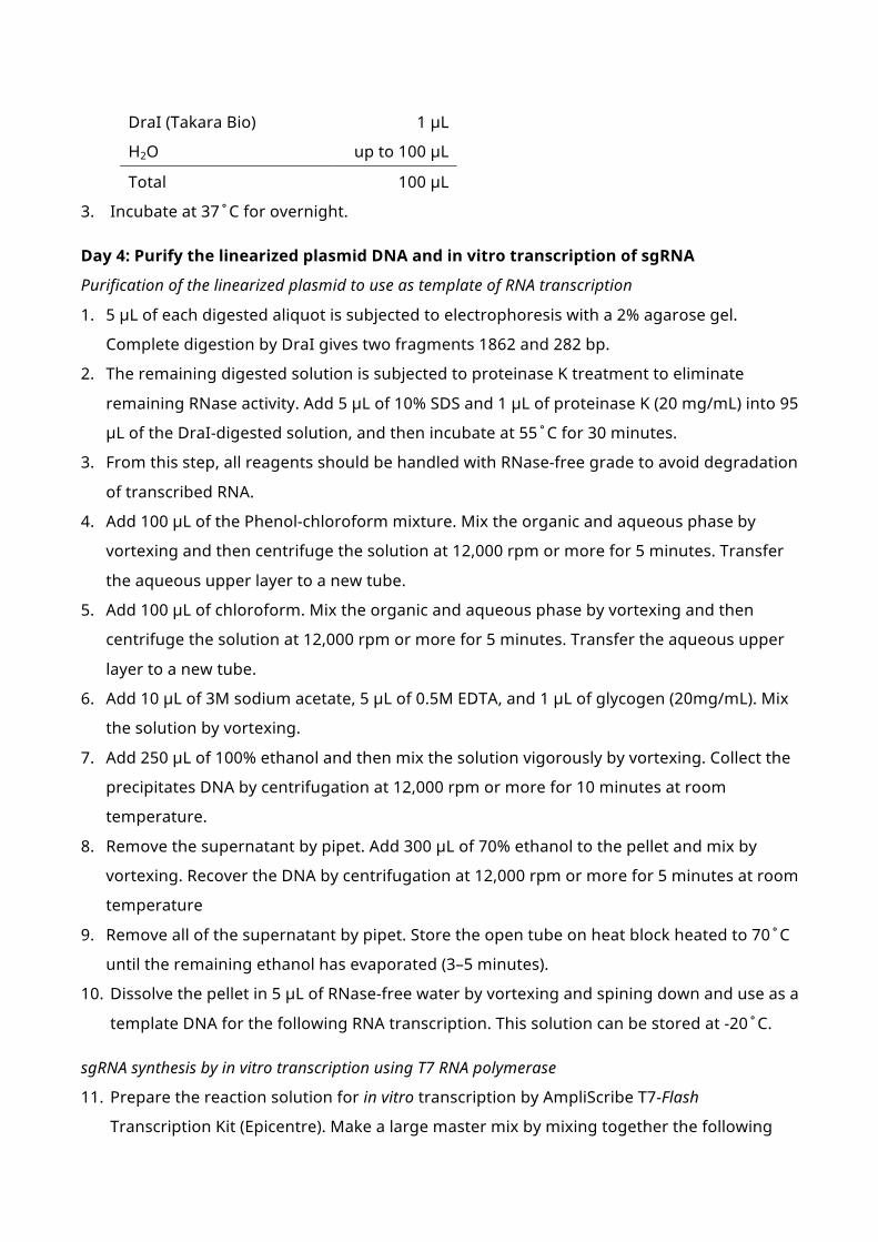
Day 4: Purify the linearized plasmid DNA and in vitro transcription of sgRNA
Purification of the linearized plasmid to use as template of RNA transcription
1. 5 µL of each digested aliquot is subjected to electrophoresis with a 2% agarose gel.
Complete digestion by DraI gives two fragments 1862 and 282 bp.
2. The remaining digested solution is subjected to proteinase K treatment to eliminate
remaining RNase activity. Add 5 µL of 10% SDS and 1 µL of proteinase K (20 mg/mL) into 95
µL of the DraI-digested solution, and then incubate at 55˚C for 30 minutes.
3. From this step, all reagents should be handled with RNase-free grade to avoid degradation
of transcribed RNA.
4. Add 100 µL of the Phenol-chloroform mixture. Mix the organic and aqueous phase by
vortexing and then centrifuge the solution at 12,000 rpm or more for 5 minutes. Transfer
the aqueous upper layer to a new tube.
5. Add 100 µL of chloroform. Mix the organic and aqueous phase by vortexing and then
centrifuge the solution at 12,000 rpm or more for 5 minutes. Transfer the aqueous upper
layer to a new tube.
6. Add 10 µL of 3M sodium acetate, 5 µL of 0.5M EDTA, and 1 µL of glycogen (20mg/mL). Mix
the solution by vortexing.
7. Add 250 µL of 100% ethanol and then mix the solution vigorously by vortexing. Collect the
precipitates DNA by centrifugation at 12,000 rpm or more for 10 minutes at room
temperature.
8. Remove the supernatant by pipet. Add 300 µL of 70% ethanol to the pellet and mix by
vortexing. Recover the DNA by centrifugation at 12,000 rpm or more for 5 minutes at room
temperature
9. Remove all of the supernatant by pipet. Store the open tube on heat block heated to 70˚C
until the remaining ethanol has evaporated (3–5 minutes).
10. Dissolve the pellet in 5 µL of RNase-free water by vortexing and spining down and use as a
template DNA for the following RNA transcription. This solution can be stored at -20˚C.
sgRNA synthesis by in vitro transcription using T7 RNA polymerase
11. Prepare the reaction solution for in vitro transcription by AmpliScribe T7-Flash
Transcription Kit (Epicentre). Make a large master mix by mixing together the following

reagents and aliquot 9 µL in each 0.2 mL PCR tube. DO NOT assemble the solution on ice.
RNase-free water 2.15 µL
10X Reaction Buffer 1 µL
100 mM ATP 0.9 µL
100 mM CTP 0.9 µL
100 mM GTP 0.9 µL
100 mM UTP 0.9 µL
100 mM DTT 1 µL
RiboGuard RNase Inhibitor 0.25 µL
AmpliScribe T7-Flash Enzyme Solution 1 µL
Total 9 µL
12. Add 1 µL of template DNA in each tube and then mix the solution by pipetting. Incubate at
37˚C for 3–4 hours using a thermal cycler.
13. Add 1 µL of RNase-free DNase I (or “TURBO DNase” included in the mMessage mMachine
Kit) and mix the solution by pipetting. Incubate at 37˚C for 15 minutes using the thermal
cycler.
Purification of transcribed sgRNA using spin column
14. For purification using RNeasy Mini Kit according to the RNA Cleanup protocol, adjust to a
volume of 100 µL by adding 89 µL of RNase-free water.
15. Add 350 µL of Buffer RLT and mix by vortexing.
16. Add 250 µL of 100% ethanol and then mix well by pipetting. Immediately transfer the
sample (700 µL) to an RNeasy Mini spin column.
17. Centrifuge at 8,000 x g for 15 seconds. Discard the flow-through.
18. Add 500 µL of Buffer RPE to the spin column, and centrifuge at 8,000 x g for 15 seconds.
Discard the flow-through.
19. Add 500 µL of Buffer RPE to the spin column again. Centrifuge at 8,000 x g for 2 minutes,
and discard the flow-through.
20. Place the RNeasy spin column in a new 2 mL collection tube, and centriguge at 10,000 x g
or more for 5 minutes.
21. Remove the remaining Buffer RPE on the upper edge of the column membrane by pipet.
Place the spin column in a new 1.5 mL tube, and store the open column on heat block
heated to 70˚C for 2 minutes to eliminate any possible carryover of Buffer RPE.
22. Place the spin column in a new 1.5 mL tube. Add 30 µL of RNase-free water directly to
center of the spin column membrane. Centrifuge at 8,000 x g for 2 minutes to elute RNA.

23. Eluted solution is immediately stored at -80˚C.
Confirmation of synthesized RNA by electrophoresis and quantification
24. 1 µL aliquot of the eluted RNA is subjected to electrophoresis using a 2% agarose gel
(Non-denaturing gel is available for simple identification of successful sgRNA synthesis.).
Successfully synthesized sgRNA shows a dense and broad band around a 100 bp band of
the ladder marker DNA.
25. Quantify the eluted RNA solution by a spectrophotometer or other methods. Adjust to an
appropriate concentration using the RNase-free water, and store at -80˚C until
microinjection.
1.3. Production of capped RNA encoding a Cas9 nuclease
1. E. coli harboring the pCS2+hSpCas9 vector is cultured in the liquid medium containing 50
µg/mL of ampicillin. Purify the plasmid using a plasmid purification kit. If the CRISPR/Cas
experiment is performed constantly, purification using a midiprep kit (e.g. NucleoBond
Xtra Midi, MACHEREY-NAGEL, cat. no. 740410.50) is recommended for high-yield recovery
of the plasmid.
2. Prepare the reaction solution for digestion with NotI as below.
pCS2+hSpCas9 (10 µg) X µL
10X M buffer (Takara Bio) 10 µL
Triton X-100 (0.1%) 10 µL
BSA (0.1%) 10 µL
NotI (Takara Bio) 1 µL
H2O up to 100 µL
Total 100 µL
3. Incubate at 37˚C for overnight.
4. 5 µL of each digested aliquot is subjected to electrophoresis with a 1% agarose gel.
Complete digestion by NotI shows a single band of 8343 bp.
5. The remaining digested solution is subjected to proteinase K treatment to eliminate
remaining RNase activity. Add 5 µL of 10% SDS and 1 µL of proteinase K (20 mg/mL) into
95 µL of the NotI-digested solution, and then incubate at 55˚C for 30 minutes.
6. From this step, all reagents should be handled with RNase-free grade to avoid
degradation of transcribed RNA.
7. Add 100 µL of the Phenol-chloroform mixture. Mix the organic and aqueous phase by
vortexing and then centrifuge the solution at 12,000 rpm or more for 5 minutes. Transfer
the aqueous upper layer to a new tube.
8. Add 100 µL of chloroform. Mix the organic and aqueous phase by vortexing and then
centrifuge the solution at 12,000 rpm or more for 5 minutes. Transfer the aqueous upper
layer to a new tube.
9. Add 10 µL of 3M sodium acetate, 5 µL of 0.5M EDTA, and 1 µL of Glycogen (20mg/mL). Mix
the solution by vortexing.
10. Add 250 µL of 100% ethanol and then mix the solution vigorously by vortexing. Collect the
precipitates DNA by centrifugation at 12,000 rpm or more for 10 minutes at room
temperature.
11. Remove the supernatant by pipette. Add 300 µL of 70% ethanol to the pellet and mix by

vortexing. Recover the DNA by centrifugation at 12,000 rpm or more for 5 minutes at
room temperature
12. Remove all of the supernatant by pipette. Store the open tube on heat block heated to
70˚C until the remaining ethanol has evaporated (3–5 minutes).
13. Dissolve the pellet in 10 µL of RNase-free water by vortexing and spining down and use as
a template DNA for the following RNA transcription. This solution can be stored at -20˚C.
14. Prepare the reaction solution for in vitro transcription by mMessage mMachine SP6
Transcription Kit (Life Technologies). Make a mixture in a 0.2 mL PCR tube as described
below. DO NOT assemble the solution on ice.
RNase-free water 2 µL
2X NTP/CAP 5 µL
10X Reaction Buffer 1 µL
Template DNA 1 µL
Enzyme Mix 1 µL
Total 10 µL
15. Incubate at 37˚C for 3–4 hours using a thermal cycler.
16. Add 1 µL of TURBO DNase and mix the solution by pipetting. Incubate at 37˚C for 15
minutes using the thermal cycler.
17. For purification using RNeasy Mini Kit according to the RNA Cleanup protocol, adjust to a
volume of 100 µL by adding 89 µL of RNase-free water.
18. Add 350 µL of Buffer RLT and mix by vortexing.
19. Add 250 µL of 100% ethanol and then mix well by pipetting. Immediately transfer the
sample (700 µL) to an RNeasy Mini spin column.
20. Centrifuge at 8,000 x g for 15 seconds. Discard the flow-through.
21. Add 500 µL of Buffer RPE to the spin column, and centrifuge at 8,000 x g for 15 seconds.
Discard the flow-through.
22. Add 500 µL of Buffer RPE to the spin column again. Centrifuge at 8,000 x g for 2 minutes,
and discard the flow-through.
23. Place the RNeasy spin column in a new 2 mL collection tube, and centriguge at 10,000 x g
or more for 5 minutes.
24. Remove the remaining Buffer RPE on the upper edge of the column membrane by pipet.
Place the spin column in a new 1.5 mL tube, and store the open column on heat block
heated to 70˚C for 2 minutes to eliminate any possible carryover of Buffer RPE.
25. Place the spin column in a new 1.5 mL tube. Add 30 µL of RNase-free water directly to
center of the spin column membrane. Centrifuge at 8,000 x g for 2 minutes to elute RNA.

Eluted solution is immediately stored at -80˚C.
26. 1 µL aliquot of the eluted RNA is subjected to electrophoresis using a 1% agarose gel (not
necessary to use a denaturing agarose gel).
27. Quantify the eluted RNA solution by a spectrophotometer or other methods. Adjust to an
appropriate concentration using the RNase-free water, and store at -80˚C until
microinjection.

2. Heteroduplex mobility assay – A simple method to detect targeted genome modification
Simple and rapid detection of indels is important for efficient targeted gene disruption using
targetable nucleases. Thus far, a number of methods, such as restriction length fragment
polymorphism (RFLP) analysis (Huang et al., 2011; Ansai et al., 2013), DNA-cleaving assay with
mismatch sensitive nucleases (Hwang et al., 2013), high-resolution melting analysis (HRMA)
(Dahlem et al., 2012), and LacZ disruption/recovery assay (Hisano et al., 2013), have been used
to detect indels induced by targetable nuculeases; however, each method has advantages and
disadvantages.
In this section, we describe estimation of the efficiency of targeted genome modifications
and identification and mutant individuals using heteroduplex mobility assay (HMA), one of the
simplest way to detect nuclease-induced indels only with PCR amplification followed by gel
electrophoresis (Chen et al., 2012; Ota et al., 2013; Ansai et al., 2014). The principal of HMA is
described as follows and Figure 1. In gel electrophoresis, the mobility of completely
complementary double-stranded DNA (homoduplex) depends on its length. Heteroduplex
DNA, which contains some mismatched nucleotides, however, moves more slowly than
homoduplex DNA because of its opened single-strand structure. Therefore, PCR products
amplified with a template containing both wild-type and mutated sequences show multiple
banding pattern, while PCR products with either wild-type or modified sequence show single
banding pattern.
2.1. Materials
Equipment
n Polyacrylamide gel electrophoresis (PAGE) apparatus: Prepare mini gel PAGE apparatus,
power supply, and 15% polyacrylamide gels (e.g. SuperSep DNA, 15%, 17 wells; Wako, Cat.
No. 190-15481). Electrophoresis is performed according to standard DNA-PAGE protocols.
n Automated electrophoresis system: A microchip electrophoresis system MCE-202 with
DNA-500 reagent kit (Shimadzu) can be used as a substitute for PAGE apparatus.
Solutions
n PCR enzymes: Any enzymes are available.
n PCR primers: Design and order pairs of oligonucleotides to amprify 80–250 bp of genomic
region containing each target site of the custom-designed nucleases.

2.2. Procedure
1. Amplify the target genomic region containing the target sequence by PCR according to a
standard method.
2. Apply the PCR amplicons to 15% polyacrylamide gel electrophoresis or automatic
electrophoresis system.
3. Analyze the HMA profiles by checking the electrophoretic results.
In case of discrimination among wild type, heterozygotes, and homozygotes, while wild
type and homozygous mutants show a single band with small differences in their mobility,
heterozygous mutants exhibit four bands including two bands of homoduplexes consisting
of either the wild type or the homozygous product and two bands of heteroduplexes
consisting of both the wild type and the mutated sequence (Figure 1A).
In case of evaluation of the efficiency of targeted genome modification, fish injected with
active targetable nucleases show a number of bands with different mobility with the wild
type product, which are derived from many types of homo- and heteroduplexes containing
wild type and/or various types of indel sequences induced by the nucleases (Figure 1B).
PCR
Wild type
Homoduplex(wild type)
PCR
Homozygousmutant
Homoduplex(mutant)
PCR
Heterozygous mutant
Electrophoresis
Homoduplex(wild & mutant)
Heteroduplex
A
PCR
Wild type
Homoduplex(wild type)
PCR
Nuclease-injected fish
Electrophoresis
Many types of homo- and heteroduplexes
B
…
WT Homo Hetero Nuclease-injected fishWT
Figure 1. Schematic illustration of the principle of heteroduplex mobility assay (HMA). (A) Completely complementary double-stranded DNA (homoduplex), which is amplified from wild-type or homozygous mutant, is segregated on the basis of its molecular weight and results in a single band in electrophoresis. On the other hand, heteroduplex DNA containing some mismatched nucleotides usually moves more slowly than homoduplex DNA, resulting in multiple band pattern of PCR products amplified from heterozygous mutants. (B) Fish injected with the targetable nucleases usually have various types of insertions and/or deletions, which exhibit a number of bands with different mobility from the wild type product derived from many types of homo- and heteroduplexes.

3. How to establish gene knockout strains
In this section, we describe how to establish gene knockout (KO) medaka strains using the
CRISPR/Cas system. Figure 2 shows a graphical abstract of a workflow.
3.1. Design and synthesis of Cas9 RNA and sgRNA(s)
For details, see the section 1.
3.2. Evaluation of sgRNA activity in fertilized eggs
One of the keys to establish genome-edited strains is selection of efficient sgRNA. Actually,
you should evaluate several sgRNA sequences targeted in a gene of interest and select more
efficient one(s).
Procedure
1. Introduce a sgRNA (10–50 ng/µL) and Cas9 RNA (100–200 ng/µL) into cytoplasm of 10 or
more fertilized medaka eggs at 1-cell stage by microinjection method.
2. Incubate these eggs at 26–28˚C for 3–5 days.
3. Prepare crude genome DNA from each embryo according to another protocol.
MicroinjectionCas9 RNA/sgRNA
G0 founder Wild-type
F1 heterozygote(+/m)
RNA-injected fish(1 dpf ~ hatching)
F1 fish (1 dpf ~ hatching)
F1 heterozygote(+/m)
F2 homozygote(m/m)
F2 heterozygote(+/m)
F2 wild-type(+/+): :
||||||||||||||||||||
1. Design and synthesis of Cas9 RNA and sgRNA(s)
2. Evaluation of sgRNA activity in fertilized eggs
3. Microinjection of the selected sgRNA(s) with Cas9 RNA
4. Selection of founder fish by genotyping F1 embryos
5. Selection of F1 fish carrying the same mutations and mating each other
6. Selection of homozygous mutant fish in F2 family
Figure 2. Schematic illustration of a workflow to establish gene knockout strains using the CRISPR/Cas system.

4. Perform PCR to amplify the target sequence. The size of amplicon should be designed less
than 250 bp because the resolution of HMA becomes lower with longer amplicon.
5. Apply amplicons on 15% polyacrylamide gel or automatic electrophoresis system. Figure 3
shows HMA estimation of the genome modifying activity of a sgRNA using automatic
electrophoresis system.
3.3. Microinjection of the selected sgRNA(s) with Cas9 RNA
Microinject approximately 2–4 nL of the selected RNA(s) into cytoplasm of 1-cell stage embryo
at the following concentration, sgRNA (10–50 ng/µL) and Cas9 RNA (100–200 ng/µL). Details of
the microinjection experiment are provided as an another protocol. Usually ~30 adult fish are
enough to establish several KO strains. Therefore, when you use standard strains (Cab and d-
rR) as background, enough number of injected egg is 30–50.
If unwanted off-target activity is observed, it is recommended to decrease the
concentration of each RNA to diminish off-target activity (Ansai & Kinoshita, 2014). If bi-allelic
disruption of target gene has lethal effects on the injected fish, it is also recommended to
decrease the concentration of each genome-editing tool to obtain single allele mutated fish.
3.4. Selection of founder fish by genotyping F1 embryos
Even if genome-editing tools are introduced into 1-cell stage of eggs, all of cells in adult are
not mutated. Therefore, we have to screen the individuals harboring mutation in germ cell.
Procedure
1. Breed the eggs injected genome editing tool up to adults and mates each adult fish with
WT
gDNA extraction from each embryo
Evaluation of sgRNA efficiency by HMA
sgRNA + Cas9 RNA
Figure 3. Evaluation of genome-editing efficiency of a sgRNA in the injected embryos. Genomic DNA is extracted from each injected embryo, and the target genomic region is analyzed by PCR amplification followed by heteroduplex mobility assay (HMA). If the injected sgRNA has genome-editing activity, each injected embryo shows multiple bands in HMA whereas wild-type embryos show a single band (red arrowhed).

wild-type counterpart.
2. Extract gDNA from 8 to 16 of their offsprings (F1 embryo at 3–5dpf).
3. Perform HMA as described in the section 2.
4. Identify the mutant DNA sequence of which the PCR product shows heteroduplex in HMA
by direct-sequencing. Because the PCR product is a mixture of wild and mutant DNA
sequence, the raw data contains two peaks in each position, one from wild-type and the
other from mutant (Figure 4). By subtracting wild-type date from this mixed data, mutant
sequence can be identified.
5. Select the founder fish that produces mutant off-springs desired (Figure 5). In case that
loss-of-function mutant is required, select founder fish that produce off-springs carrying
frame-shift mutations, generating non-sense amino acid sequence and/or stop codon
prior to normal one.
RGEN-injected Wild-type
#1 #2 #3 #4 #5
HMA profiles
CCCTCCAACAGGGAGTAAAACGCCTCAAGATCCCACCCCTCCAAC---------------TCAAGATCCCACCCCTCCAACAGGGAG-------CCTCAAGATCCCACCCCTCCAACAGGGAG--------------ATCCCAC
WT (#3, #4)#1 (Δ15)#2 (Δ7)#5 (Δ14)
Figure 5. Screening of founder fish by genotyping their F1 embryos. Each fish injected with targetable nucleases mates with wild-type fish and then their F1 embryos are individually subjected to heteroduplex mobility assay (HMA) followed by direct sequencing. This step is important to identify preferable founders, which produce F1 progeny harboring desirable types of mutations with high efficiencies.
C T T T G A T G
Raw data:
WT:
Mutant:
C T T T G A T G A G G A C A T G G C G G A G G A A G G TG C G G A G G A A G G T C T T C C A A C
C T T T G A T G
A G G A C A T G G C G G A G G A A G G T…
G C G G A G G A A G G T C T T C C A A C…
C T T T G A T G
C T T T G A T G 1 2 3 4 5 6 7 8 G C G G A G G A A G G T…8 bp deletion
Figure 4. Direct sequencing in heterozygous mutant that carries a wild-type allele and an 8-bp deletion allele. In case of sanger sequencing, the heterozygous mutant shows double waves from the deletion point in a raw data file. The deletion sequence can be identified by subtracting wild-type sequence from the double waves.
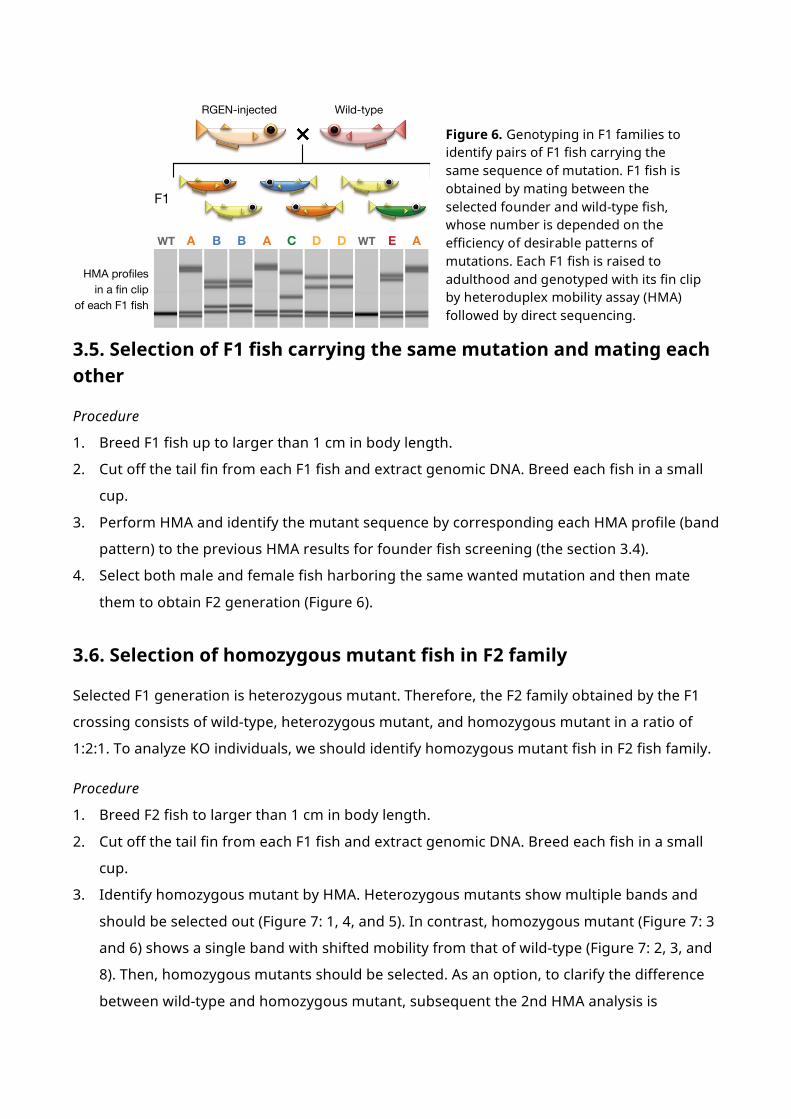
3.5. Selection of F1 fish carrying the same mutation and mating each other
Procedure
1. Breed F1 fish up to larger than 1 cm in body length.
2. Cut off the tail fin from each F1 fish and extract genomic DNA. Breed each fish in a small
cup.
3. Perform HMA and identify the mutant sequence by corresponding each HMA profile (band
pattern) to the previous HMA results for founder fish screening (the section 3.4).
4. Select both male and female fish harboring the same wanted mutation and then mate
them to obtain F2 generation (Figure 6).
3.6. Selection of homozygous mutant fish in F2 family
Selected F1 generation is heterozygous mutant. Therefore, the F2 family obtained by the F1
crossing consists of wild-type, heterozygous mutant, and homozygous mutant in a ratio of
1:2:1. To analyze KO individuals, we should identify homozygous mutant fish in F2 fish family.
Procedure
1. Breed F2 fish to larger than 1 cm in body length.
2. Cut off the tail fin from each F1 fish and extract genomic DNA. Breed each fish in a small
cup.
3. Identify homozygous mutant by HMA. Heterozygous mutants show multiple bands and
should be selected out (Figure 7: 1, 4, and 5). In contrast, homozygous mutant (Figure 7: 3
and 6) shows a single band with shifted mobility from that of wild-type (Figure 7: 2, 3, and
8). Then, homozygous mutants should be selected. As an option, to clarify the difference
between wild-type and homozygous mutant, subsequent the 2nd HMA analysis is
RGEN-injected Wild-type
F1
WT WTA B DB CA
HMA profilesin a fin clip
of each F1 fish
D AE
Figure 6. Genotyping in F1 families to identify pairs of F1 fish carrying the same sequence of mutation. F1 fish is obtained by mating between the selected founder and wild-type fish, whose number is depended on the efficiency of desirable patterns of mutations. Each F1 fish is raised to adulthood and genotyped with its fin clip by heteroduplex mobility assay (HMA) followed by direct sequencing.

effective. Separately prepared amplicon from the wild-type individual is added to the
amplicons of both possible wild-type and heterozygous mutant embryos. After re-
annealing, the solution is subjected to HMA again. In the 2nd HMA, the homozygous
mutant shows multiple bands (Figure 7: 3´ and 6´) whereas the wild-type shows a single
band (Figure 7: 2´, 7´, and 6´).
1 2 3 4 5 6 7 8 2´ 3´ 6´ 7´ 8´
m/+ m/+
m/+ m/+m/m +/+
2nd HMA[Re-annealed]
1st HMA[Normal]
F2 mix
HMA profilesin F2 fin clips
Incross between F1 fish harboring the same mutation
Identify fish harboring the mutation in both alleles (#3 and #6)
Figure 7. Genotyping in F2 family to identify homozygous mutant fish. F2 fish family obtained by crossing between F1 fish harboring the same pattern of mutation usually consists of wild-type, heterozygous mutant, and homozygous mutant in a ratio of 1:2:1. Heterozygous mutant (Lane No. 1, 4, and 5) can be easily distinguished by multiple band patterns in heteroduplex mobility assay (HMA), while both wild-type (Lane No. 2, 7, and 8) and homozygous mutant (Lane No. 3 and 6) shows a single band and therefore they are sometimes associated with difficulty in their distinction (“1st HMA” in the panel). To clearly distinguish between wild-type and homozygous mutant, the aliquot of PCR product subjected to the HMA is mixed with PCR product amplified from wild-type genome, and subsequently, the mixture is heat-denatured and annealed using a thermal cycler and is subjected to HMA again (“2nd HMA” in the panel). Homozygous mutants (Lane No. 3´ and 6´) show multiple bands similar to heterozygous mutant in the 1st HMA whereas wild-type fish (Lane No. 2´, 7´, and 8´) show a single band.

References
Ansai, S., Inohaya, K., Yoshiura, Y., Schartl, M., Uemura, N., Takahashi, R., & Kinoshita, M.
(2014) Design, evaluation, and screening methods for efficient targeted mutagenesis with
transcription activator-like effector nucleases in medaka. Dev. Growth Differ., 56, 98–107.
Ansai, S. & Kinoshita, M. (2014) Targeted mutagenesis using CRISPR/Cas system in medaka.
Biol. Open, 3, 362–371.
Ansai, S., Sakuma, T., Yamamoto, T., Ariga, H., Uemura, N., Takahashi, R., & Kinoshita, M.
(2013) Efficient targeted mutagenesis in medaka using custom-designed transcription
activator-like effector nucleases. Genetics, 193, 739–749.
Chen, J., Zhang, X., Wang, T., Li, Z., Guan, G., & Hong, Y. (2012) Efficient detection,
quantification and enrichment of subtle allelic alterations. DNA Res., 19, 423–433.
Dahlem, T.J., Hoshijima, K., Jurynec, M.J., Gunther, D., Starker, C.G., Locke, A.S., Weis, A.M.,
Voytas, D.F., & Grunwald, D.J. (2012) Simple Methods for Generating and Detecting Locus-
Specific Mutations Induced with TALENs in the Zebrafish Genome. PLoS Genet., 8,
e1002861.
Gasiunas, G., Barrangou, R., Horvath, P., & Siksnys, V. (2012) Cas9-crRNA ribonucleoprotein
complex mediates specific DNA cleavage for adaptive immunity in bacteria. Proc. Natl.
Acad. Sci. U. S. A., 109, E2579–E2586.
Hisano, Y., Ota, S., Arakawa, K., Muraki, M., Kono, N., Oshita, K., Sakuma, T., Tomita, M.,
Yamamoto, T., Okada, Y., & Kawahara, A. (2013) Quantitative assay for TALEN activity at
endogenous genomic loci. Biol. Open, 2, 363–367.
Huang, P., Xiao, A., Zhou, M., Zhu, Z., Lin, S., & Zhang, B. (2011) Heritable gene targeting in
zebrafish using customized TALENs. Nat. Biotechnol., 29, 699–700.
Hwang, W.Y., Fu, Y., Reyon, D., Maeder, M.L., Tsai, S.Q., Sander, J.D., Peterson, R.T., Yeh, J.-R.J.,
& Joung, J.K. (2013) Efficient genome editing in zebrafish using a CRISPR-Cas system. Nat.
Biotechnol., 31, 227–229.
Jinek, M., Chylinski, K., Fonfara, I., Hauer, M., Doudna, J.A., & Charpentier, E. (2012) A
programmable dual-RNA-guided DNA endonuclease in adaptive bacterial immunity.
Science, 337, 816–821.
Ota, S., Hisano, Y., Muraki, M., Hoshijima, K., Dahlem, T.J., Grunwald, D.J., Okada, Y., &
Kawahara, A. (2013) Efficient identification of TALEN-mediated genome modifications
using heteroduplex mobility assays. Genes to Cells, 18, 450–458.
Peng, Y., Clark, K.J., Campbell, J.M., Panetta, M.R., Guo, Y., & Ekker, S.C. (2014) Making designer
mutants in model organisms. Development, 141, 4042–4054.

Urnov, F.D., Rebar, E.J., Holmes, M.C., Zhang, H.S., & Gregory, P.D. (2010) Genome editing with
engineered zinc finger nucleases. Nat. Rev. Genet., 11, 636–646.
Wiedenheft, B., Sternberg, S.H., & Doudna, J. a. (2012) RNA-guided genetic silencing systems in
bacteria and archaea. Nature, 482, 331–338.