Embed Size (px)
Citation preview
© 7990 Oxford University Press Nucleic Acids Research, Vol. 18, No. 11 3203
Target Detection Assay (TDA): a versatile procedure todetermine DNA binding sites as demonstrated on SP1protein
Hans-Jurgen Thiesen and Christian BachBasel Institute for Immunology*, GrenzacherstraGe 487, CH-4005 Basel, Switzerland
Received March 14, 1990, Revised and Accepted May 11, 1990
ABSTRACT
We developed a rapid method designated TargetDetection Assay (TDA) to determine DNA binding sitesfor putative DNA binding proteins. A purified,functionally active DNA binding protein and a pool ofrandom double-stranded oligonucleotides harbouringPCR primer sites at each end are included the TDAcycle which consists of four separate steps: a DNAprotein incubation step, a protein DNA complexseparation step , a DNA elutfon step and a polymerasechain reaction (PCR) DNA amplification step. Thestringency of selection can be increased in consecutiveTDA cycles. Since tiny amounts of retained DNA canbe rescued by PCR, buffer systems, salt concentrationsand competitor DNA contents can be varied In orderto determine high affinity binding sites for the proteinof choice. To test the efficiency of the TDA procedurepotential DNA binding sites were selected by the DNAbinding protein SP1 from a pool of oligonucleotideswith random nucleotides at 12 positions. Target sitesselected by recomblnant SP1 closely matched the SP1consensus site. If DNA recognition sites have to bedetermined for known, mutated or putative DNAbinding proteins, the Target Detection Assay (TDA) isa versatile and rapid technique for consideration.
INTRODUCTION
Differentiation is thought to be a process determined by geneticprogramming of individual cells through the differential regulationof transcription initiation. With the analysis of DNA bindingproteins it has become clear that the transcription of genes isregulated by DNA-protein interactions and depends on theinterplay of common and cell type/ lineage specific DNA bindingproteins. In recent years a large number of DNA binding proteinshave been cloned based on evolutionary conserved homologiesin their DNA binding domains, e.g. homeo domains(l —6), pou-boxes (7-9), paired-boxes (10—12) and zinc (II) finger regions(13-19). However, the DNA sequences to which the variousdomains bind have been determined for only a few proteins(20-26). Recently, we cloned more than thirty genes encodingzinc finger domains from human T cell cDNA libraries. Some
of these genes named Kox 1 -30 are thought to be involved inregulating tissue specific gene expression (27).
We designed the Target Detection Assay (TDA) in order todetermine DNA recognition sites for these zinc finger proteins.The method requires a pool of randomized oligonucleotides anda functionally active DNA binding protein. To test theapplicability of the TDA procedure, we used FPLC purified,recombinant SP1 as a model protein. Template oligonucleotideswere chemically synthesized harbouring random nucleotides atN positions surrounded by specific sequences with desiredrestriction sites on each end. Two primers ( 5 ' , primer A and3', primer B) complementary to sequences at the ends of thetemplate oligonucleotides were used for PCR amplification (28).
Here, we describe how these double-stranded oligonucleotidescontaining random nucleotides can be used for determiningbinding sites of human transcription factor SP1.
MATERIAL AND METHODSExpression and purification of SP1 proteinThe Bam Hl-Bgl II fragment derived from pP^SPl-Sloc(29) encoding three zinc fingers of the human transcription factorSP1 (30) were cloned into the Bam HI site of pAR 3039 (31)to yield pAR Sp 1-516c. Expression of SP1 was induced by theaddition of IPTG (0.125mg/ml) to exponentially growing cellsfollowed by a 3 hrs incubation shaking at 37°C. The SP1 E.colipellet obtained from 800 ml cultures was resuspended in 50 mlbuffer I (50 mM NaCl, 20 mM Tris-HCl pH 8.0, 1% TntionX-100), sonicated for 10 min, pelleted (10000 RPM/10min/4°C)and washed consecutively with 20ml buffer Q ( 1 M NaCl, 20mM Tris-HCl), with 20 ml buffer IH (3 M NaCl, 20 mM Tris-HCl) and with 20 ml HjO. The remaining pellet was dissolvedin buffer IV(8M Urea, 0.5 M NaCl, 20 mM Tris-HCl pH 8.0),centrifuged (10000 RPM/10 min/4cC) and the supernatant frozenat -70°C degrees (20ml of the supernatant was analysed on 15 %polyacrylamide-SDS gel). Supernatant (5 ml aliquots) was filtered(0.22 micron), diluted 1:10 in buffer A (8 M Urea, 20 mM MESpH 6.0, 3 mM DTT, 2 mM EDTA) and applied to FPLC MonoS. Bound proteins were eluted by a linear gradient (0%—40%)of buffer B (1 M NaCL, 8 M Urea, 20 mM MES pH 6.0, 3mM DTT, 2 mM EDTA). Peak fractions were collected and
* The Basel Institute for Immunology was founded and is supported by Hoffinann-La Roche & Co Ltd, CH t̂OO5 Basel, Switzerland
Downloaded from https://academic.oup.com/nar/article-abstract/18/11/3203/1049096by gueston 21 March 2018
3204 Nucleic Acids Research, Vol. 18, No. 11
analysed on 15% polyacrylamide-SDS gel. Fractions containingSpl protein were pooled and the protein concentration wasdetermined to be 500 ng//*l by the method of Lowry.
PCR oligonucleotidesTemplate oligonucleotides and corresponding primers weresynthesized on 38OA Applied Biosystems DNA Synthesizer. Twotemplate oligonucleotides were designed and synthesized:template A that contains two consensus binding sites for humantranscription factor SP1 and template B that contains Nrandomized oligonucleotides at 12 positions flanked by specificsequences of 22 nucleotides at each end. In addition, two primerof 22 nucleotides (5' primer and 3'primer ) were synthesizedfor generating and rescuing double-stranded oligonucleotidemixes by PCR amplification. PCR amplification was done usingamplification kit (Perkin Elmer Cetus) with denaturation 93°C,30 sec; annealing 45°C, 2 min; temperature shift 45°C-67°C,lmin; extension 3 min, 67°C; cycles 25x. The oligonucleotidemix was extracted by phenol/chloroform , purified from a 6%polyacrylamide gel using ElutipD (Schleicher&Schull).Oligonucleotides were dissolved in 30 /tl T.E. (10 fd are usedin one filter binding assay). Filter eluted oligonucleotides wereamplified by using 1 /J of eluted material (200 /xl).
FirterbindingDouble stranded oligonucleotide mixes (10 /il) were incubatedwith recombinant SP1 (lmg) in Tns-HCl binding buffer (200til, 9mM Tns-HCl pH 8.0, 90 mM KCl, 90 /tM ZnSO4) for 30min. Nitrocellulose filters (BA85, Schleicher&Schull) wereinserted in a suction chamber (32) and prewet with 600 ml Tris-HC1 binding buffer. SP1 protein-oligonucleotide and albumin-oligonucleotide mixes were applied to nitrocellulose filters withgentle suction and the filters washed with 4 ml Tris-HCl bindingbuffer. In the first three TDA cycles, randomized oligonucleotidesretained on the filter were eluted with 200 /il Tris-HCl bindingbuffer substituted with 1 mM 1 —10-o-phenanthrohne. In thefourth TDA cycle the DNA was eluted as before, then with lmM1 - 10-o-phenanthroline and 500 mM KCl and finally with 10mM phenanthroline and 500 mM KCl. The DNA of the lastfraction was subjected to a fifth TDA cycle in the presence of500 mM KCl and eluted with 10 mM phenanthroline and 500mM KCl.
Band shift elect rophoresisBandshift gels containing 0.25% TBE and 6% polyacrylamide(30% acrylamide/0.8% bisacrylamide) were prerun for 30 min.Optimal protein concentrations were determined titrating SP1protein. Good gel-shift activities were obtained when the SP1protein was diluted in MES buffer, substituted with 200 mM NaClin the absence of urea. In a standard band shift, lOng proteinwere added to the band shift mix (30 /tl) containing 100 mMNaCl, 10 mM Tris-HCl pH 8.0, 1 mM EDTA, 100 iM ZnS04,10% glycerol, labelled oligonucleotides (40000-100000 cpiriCerenkoff) and competitor DNA (poly dA/dT or poly dl/dC).The gels were run at 10 V/cm for 3 hrs, dried onto Whatmanpaper and exposed to Kodak XAR between 3hrs and 24hrs.Oligonucleotides derived from the fifth TDA cycle were labelledand subjected to band shift electrophoresis. Retardedoligonucleotides were rescued by PCR and cloned.
CloningThe oligonucleotides derived from nitrocellulose filter bindingor from band shift electrophoresis gels were eluted, amplified,
TARGET DETECTION ASSAY (TDA)
Purified Protein ds Random Oligonucleotides
TDA Cycle (Filter Binding)
1. Incubation (protein and DNA)2. Separation of bound complexes3. Elution or bound DNA4. Amplification of eluted DNA
TDA Cycle (Band Shift Assay)
1. Incubation (protein and DNA)2. Separation of bound complexes3. Elution of bound DNA4. Amplification of eluted DNA
1 ICloning of DNA eluted from shifted band
Band shift of Individual ds oligonucleotides
ISequencing of shifting oligonucleotides
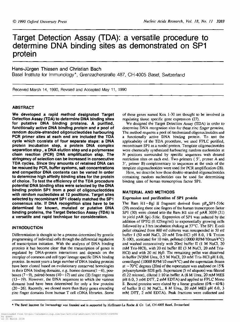
Figure 1. Target Detection Assay (TDA) The target detection assay (TDA)requires a purified functionally active DNA binding protein and a pool of randomoligonucleotides The TDA cycle describes the core procedure of this techniqueFilter binding is performed during the first TDA cycles Selected ohgonucleotidescan be cloned or subjected to further TDA cycles using band shift electrophoresisFinally, the TDA selected oligonucleotides are cloned, analysed and sequenced
phosphorylated with T4 polynucleotide kinase and cloned intothe dephosphorylated Eco RV site of Bluescript plasmid. Thetransformed host strain JM-103 was plated on LB/ampicillineplates containing IPTG and x-gal. White colonies were pickedand inserted oligonucleotides obtained using PCR conditions fromabove. The oligonucleotides were isolated from 6%polyacrylamide gels and concentrated by Elutip D. Thenucleotides were resuspended in 20 /tl T.E. For band shift assays,1 ill of the oligonucleotides were kmased with 100 /tCi yil?ATP or internally labelled using 20 /tCi a*1? dCTP during onePCR cycle with an extension time of 10 min.
SequencingOligonucleotides which were selected by positive band shift weresequenced using double-stranded miniprep DNA according themanufacturer's protocol (deaza-Kit, Pharmacia).
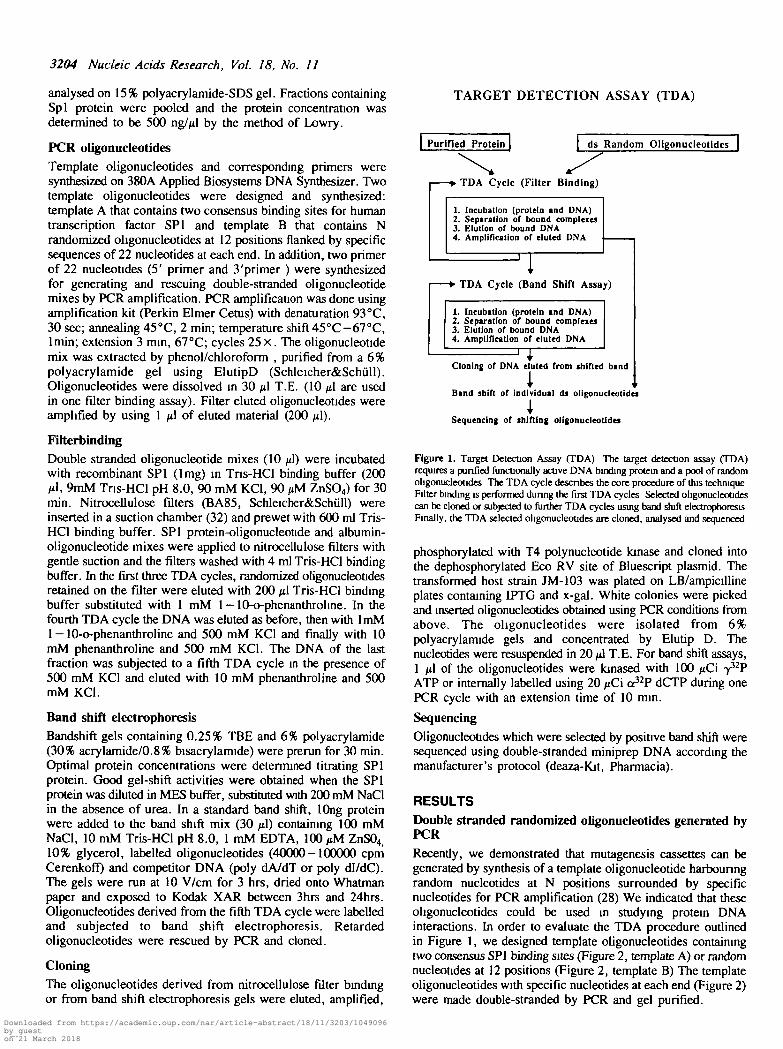
RESULTSDouble stranded randomized oligonucleotides generated byPCRRecently, we demonstrated that mutagenesis cassettes can begenerated by synthesis of a template oligonucleotide harbouringrandom nucleotides at N positions surrounded by specificnucleotides for PCR amplification (28) We indicated that theseoligonucleotides could be used in studying protein DNAinteractions. In order to evaluate the TDA procedure outlinedin Figure 1, we designed template oligonucleotides containingtwo consensus SP1 binding sites (Figure 2, template A) or randomnucleotides at 12 positions (Figure 2, template B) The templateoligonucleotides with specific nucleotides at each end (Figure 2)were made double-stranded by PCR and gel purified.
Downloaded from https://academic.oup.com/nar/article-abstract/18/11/3203/1049096by gueston 21 March 2018
Nucleic Acids Research, Vol. 18, No. 11 3205
TEMPLATE OLIGONUCLEOTIDES
5' Primer AGTCGGATCCTGTCTGAGGTGAG-
GTCGGATCCTGTCTGAGGTGAG
2x SPl concensus
GG GGGC GG GGGCGGCG GGCG
TA TAAT TA TAATGTCTTCCGACGTCGAATTCGCG
CAGAAGGCTGCAGCTTAAGCGC
31 Primer B
B5' Primer AGTCGGATCCTGTCTGAGGTGAGGTCGGATCCTGTCTGAGGTGAGTemplate (N=12)
N N N N N N N N N N N N GTCTTCCGACGTCGAATTCGCGCAGAAGGCTGCAGCTTAAGCGC
3' Primer B
Figure 2. PCR ohgonucleotides Four oligonucleoudes, two template ohgonucleotide and two pnmers, were synthesized on Applied Biosystems DNA SynthesizerThe template A oligonucleotide was designed to contain two consecutive SPl consensus binding sites and template B to contain random oligonucleoudes at 12 positionsboth flanked by specific sequences of 22 nucleotidcs at each end Pnmers of 22 nucleotides (5' primer A and 3'pnmer B) complementary to these ends are usedfor generation of double-stranded ohgonucleotide mixes by PCR amplification
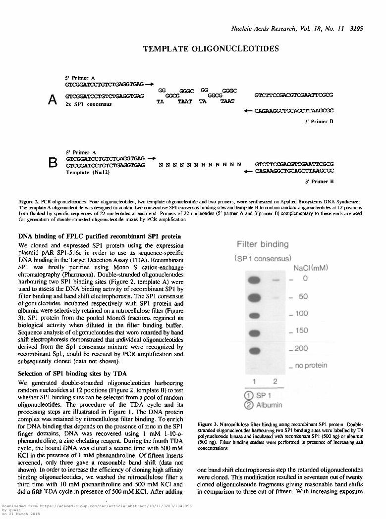
DNA binding of FPLC purified recombinant SPl proteinWe cloned and expressed SPl protein using the expressionplasmid pAR SPl-516c in order to use its sequence-specificDNA binding in the Target Detection Assay (TDA). RecombinantSPl was finally purified using Mono S cation-exchangechromatography (Pharmacia). Double-stranded oligonucleotidesharbouring two SPl binding sites (Figure 2, template A) wereused to assess the DNA binding activity of recombinant SPl byfilter binding and band shift electrophoresis. The SPl consensusoligonucleotides incubated respectively with SPl protein andalbumin were selectively retained on a nitrocellulose filter (Figure3). SPl protein from the pooled MonoS fractions regained itsbiological activity when diluted in the filter binding buffer.Sequence analysis of oligonucleotides that were retarded by bandshift electrophoresis demonstrated that individual ohgonucleotidesderived from the Spl consensus mixture were recognized byrecombinant Spl, could be rescued by PCR amplification andsubsequently cloned (data not shown).
Selection of SPl binding sites by TDAWe generated double-stranded oligonucleotides harbouringrandom nucleotides at 12 positions (Figure 2, template B) to testwhether SPl binding sites can be selected from a pool of randomoligonucleotides. The procedure of the TDA cycle and itsprocessing steps are illustrated in Figure 1. The DNA proteincomplex was retained by nitrocellulose filter binding. To enrichfor DNA binding that depends on the presence of zinc in the SPlfinger domains, DNA was recovered using 1 mM l-10-o-phenanthroline, a zinc-chelating reagent. During the fourth TDAcycle, the bound DNA was eluted a second time with 500 mMKC1 in the presence of 1 mM phenanthroline. Of fifteen insertsscreened, only three gave a reasonable band shift (data notshown). In order to increase the efficiency of cloning high affinitybinding oligonucleotides, we washed the nitrocellulose filter athird time with 10 mM phenanthroline and 500 mM KC1 anddid a fifth TDA cycle in presence of 500 mMXCl. After adding
Filter binding
(SP1 consensus)
m
—
1 2
(T)SPI(2) Albumin
NaCI(mM)0
50
100
150
200
no protein
Figure 3. Nitrocellulose filter binding using recombinant SPl protein Double-stranded oligonucleoudes harbouring two SPl binding sites were labelled by T4polynucleoude kinase and incubated with recombinant SPl (500 ng) or albumin(500 ng). Filter binding studies were performed in presence of increasing saltconcentrations
one band shift electrophoresis step the retarded oligonucleotideswere cloned. This modification resulted in seventeen out of twentycloned oligonucleotide fragments giving reasonable band shiftsin comparison to three out of fifteen. With increasing exposure
Downloaded from https://academic.oup.com/nar/article-abstract/18/11/3203/1049096by gueston 21 March 2018
3206 Nucleic Acids Research, Vol. 18, No. 11
time even nineteen out of twenty double-stranded oligonucleotidesshowed binding to SPl protein (data not shown).
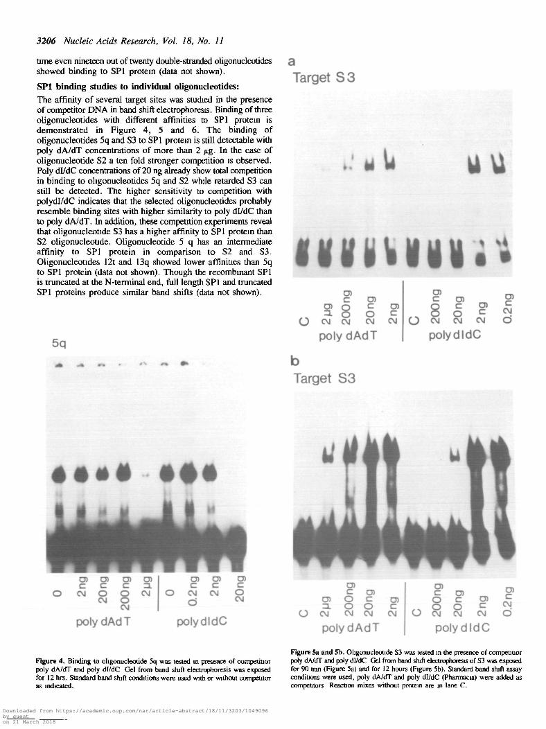
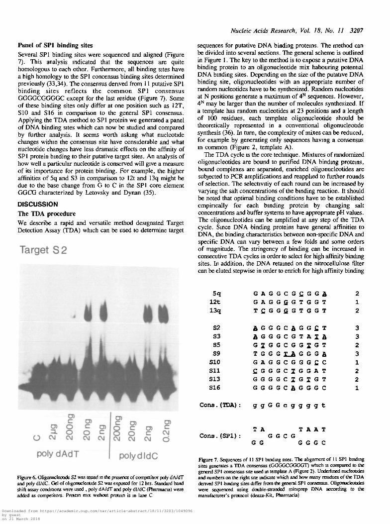
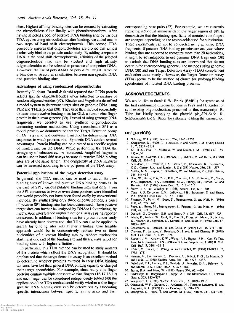
SPl binding studies to individual oligonucleotides:The affinity of several target sites was studied in the presenceof competitor DNA in band shift electrophoresis. Binding of threeoligonucleotides with different affinities to SPl protein isdemonstrated in Figure 4, 5 and 6. The binding ofoligonucleotides 5q and S3 to SPl protein is still detectable withpoly dA/dT concentrations of more than 2 jig. In the case ofoligonucleotide S2 a ten fold stronger competition is observed.Poly dl/dC concentrations of 20 ng already show total competitionin binding to oligonucleotides 5q and S2 while retarded S3 canstill be detected. The higher sensitivity to competition withpolydl/dC indicates that the selected oligonucleotides probablyresemble binding sites with higher similarity to poly dl/dC thanto poly dA/dT. In addition, these competition experiments revealthat oligonucleotide S3 has a higher affinity to SPl protein thanS2 oligonucleotide. Oligonucleotide 5 q has an intermediateaffinity to SPl protein in comparison to S2 and S3.Oligonucleotides 12t and 13q showed lower affinities than 5qto SPl protein (data not shown). Though the recombtnant SPlis truncated at the N-terminal end, full length SPl and truncatedSPl proteins produce similar band shifts (data not shown).
5q
Target S3
uu
ivmmC O)
0 ) 0 c o )3. O O c
O CM CM CM CM
poly dAdT
bTarget S3
O)C O) O)
cO CM CM CM
polydldC
O)
CM
* • » # • # •
O)
CM
O)coCM
O)c
8CM
poly dAdT
O)
CM
O) O)C C
O CM CM
polydldC
o>coCM
O)c
o) o3. OO CM CM
O)coCM
poly dAdT
O)c
CM
O) „C O ) 2O C O) =
, . O O c ^O CM CM CM O
polydldC
Figure 4. Binding to ohgonuckotide 5q was tested in presence of competitiorpoly dA/dT and poly di/dC Gel from band shjft electrophoresis was exposedfor 12 hrs. Standard band shift conditions were used with or without competitoras indicated.
Figure 5a and 5b. Oligonucleotide S3 was tested in the presence of competitiorpoty dA/dT and poly dl/dC Gel from band shift electrophoresis of S3 was exposedfor 90 mm (Figure 5a) and for 12 hours (Figure 5b). Standard band shift assayconditions were used, poly dA/dT and poly dl/dC (Pharmacia) were added ascompetitors Reaction mixes without protein are in lane C.
Downloaded from https://academic.oup.com/nar/article-abstract/18/11/3203/1049096by gueston 21 March 2018
Nucleic Acids Research, Vol. 18, No. 11 3207
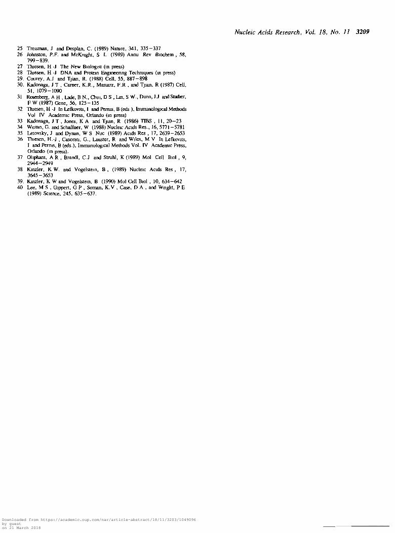
Panel of SP1 binding sitesSeveral SP1 binding sites were sequenced and aligned (Figure7). This analysis indicated that the sequences are quitehomologous to each other. Furthermore, all binding sites havea high homology to the SP1 concensus binding sites determinedpreviously (33,34). The consensus derived from 11 putative SP1binding sites reflects the common SP1 consensusGGGGCGGGGC except for the last residue (Figure 7). Someof these binding sites only differ at one position such as 12T,S10 and S16 in comparison to the general SP1 consensus.Applying the TDA method to SP1 protein we generated a panelof DNA binding sites which can now be studied and comparedby further analysis. It seems worth asking what nucleotidechanges within the consensus site have considerable and whatnucleotide changes have less dramatic effects on the affinity ofSP1 protein binding to their putative target sites. An analysis ofhow well a particular nucleotide is conserved will give a measureof its importance for protein binding. For example, the higheraffinities of 5q and S3 in comparison to 12t and 13q might bedue to the base change from G to C in the SP1 core elementGGCG characterized by Letovsky and Dynan (35).
DISCUSSIONThe TDA procedureWe describe a rapid and versatile method designated TargetDetection Assay (TDA) which can be used to determine target
Target S2
sequences for putative DNA binding proteins. The method canbe divided into several sections. The general scheme is outlinedin Figure 1. The key to the method is to expose a putative DNAbinding protein to an oligonucleotide mix habouring potentialDNA binding sites. Depending on the size of the putative DNAbinding site, oligonucleotides with an appropriate number ofrandom nucleotides have to be synthesized. Random nucleotidesat N positions generate a maximum of 4N sequences. However,4N may be larger than the number of molecules synthesized. Ifa template has random nucleotides at 23 positions and a lengthof 100 residues, each template oligonucleotide should betheoretically represented in a conventional oligonucleoudesynthesis (36). In turn, the complexity of mixes can be reduced,for example by generating only sequences having a consensusin common (Figure 2, template A).
The TDA cycle is the core technique. Mixtures of randomizedoligonucleotides are bound to purified DNA binding proteins,bound complexes are separated, enriched oligonucleotides aresubjected to PCR amplifications and reapphed to further roundsof selection. The selectivitiy of each round can be increased byvarying the salt concentrations of the binding reaction. It shouldbe noted that optimal binding conditions have to be establishedempirically for each binding protein by changing saltconcentrations and buffer systems to have appropriate pH values.The oligonucleotides can be amplified at any step of the TDAcycle. Since DNA binding proteins have general affinities toDNA, the binding characteristics between non-specific DNA andspecific DNA can vary between a few folds and some ordersof magnitude. The stringency of binding can be increased inconsecutive TDA cycles in order to select for high affinity bindingsites. In addition, the DNA retained on the nitrocellulose filtercan be eluted stepwise in order to enrich for high affinity binding
a II
en
O c\j
COcoo
enc coO cC\J CvJ
polydAdT
COc co§ c
oCvJ C\J
CO
CvJ
CO
cCvJ(D
polydldC
Figure 6. Oligonucleotide S2 was tested in the presence of competitior poly dA/dTand poly dl/dC. Gel of oligonucleoude S2 was exposed for 12 hrs. Standard bandshift assay conditions were used , poly dA/dT and poly dl/dC (Pharmacia) wereadded as competitors. Protein rrux without protein is in lane C
5q G A G G C G £ G G & 212t G A G G f i G T G G T 113q T C . G G G . G T G G T 2
52 J G G G C J G G £ T 353 J G G G C G T A 1 J 3S5 G I G G C G G J G T 2S9 T G G G X_A G G G A. 3
510 G A G G C G G G f i C 1511 fiGGGCIGGAT 2S13 G G G G C I G I G T 2S16 G G G G C A . G G G C 1
Cons.(TDA) : g g G G c g g g g t
T A T A A TCons. (SP1): G G C G
G G G G G C
Figure 7. Sequences of 11 SP1 binding sites. The alignment of 11 SP1 bindingsites generates a TDA consensus (GGGGCGGGGT) which is compared to thegeneral SP1 consensus site used in template A (Figure 2). Underlined nucleoudesand numbers on the right site indicate which and how many residues of the TDAderived SP1 binding sites differ from the general SP1 consensus. Oligonucleotideswere sequenced using double-stranded miruprep DNA according to themanufacturer's protocol (deaza-Kit, Pharmacia)
Downloaded from https://academic.oup.com/nar/article-abstract/18/11/3203/1049096by gueston 21 March 2018
3208 Nucleic Acids Research, Vol. 18, No. 11
sites. Highest affinity binding sites can be rescued by extractingthe nitrocellulose filter finally with phenol/chloroform. Afterhaving selected a pool of putative DNA binding sites by variousTDA cycles using nitrocellulose filter binding, we added one ortwo steps of band shift electrophoresis. This second TDAprocedure ensures that oligonucleotides are cloned that almostexclusively bind to the protein under study. By adding competitorDNA in the band shift electrophoresis, affinities of the selectedoligonucleotide mix can be studied and high affinityoligonucleotides can be selected in presence of competitor DNA.However, the use of poly dA/dT or poly dl/dC might introducea bias due to structural similarities between non-specific DNAand putative binding sites.
Advantages of using randomized oligonucleotidesRecently Oliphant, Brandl & Struhl reported that GCN4 proteinselects specific oligonucleotides when subjected to mixture ofrandom oligonucleotides (37). Kinzler and Vogelstein describeda model system to determine target sites on genomic DNA usingPCR and TFIHa protein (38). They used their method successfullyto determine putative binding sites for GLI, a human zinc fingerprotein in the human genome (39). Instead of using genomic DNAfragments, we decided to use synthetic oligonucleotidescontaining random nucleotides. Using recombinant SP1 as amodel protein we demonstrated that the Target Detection Assay(TDA) is a rapid and convenient method for determining DNAsequences to which proteins bind. Synthetic DNA confers severaladvantages. Protein binding can be directed to a specific regionof limited size on the DNA. While performing the TDA thestringency of selection can be monitored. Amplified fragmentscan be used in band shift assays because all putative DNA bindingsites are of the same length. The complexity of DNA mixturescan be assessed according to the purposes of the TDA assay.
Potential applications of the target detection assayIn general, the TDA method can be used to search for newbinding sites of known and putative DNA binding proteins. Inthe case of SP1, various putative binding sites that differ fromthe SP1 consensus in two or even three positons were identifiedthat would probably not have been found easily by conventionalmethods. By synthesizing only three oligonucleotides, a panelof putative SP1 binding sites has been determined. These putativetarget sites can further be analysed by DNAse I footprinting, bymethylation interference and/or functional assays using reporterconstructs. In additon, if binding sites for a protein under studyhave already been determined, the TDA can also be applied tosearch for binding sites with higher affinities. One feasibleapproach would be to consecutively replace two or threenucleotides of a known binding site by random nucleotidesstarting at one end of the binding site and then always select forbinding sites with higher affinities.
In particular, this TDA method can be used to study mutantsof the protein which effect the DNA recognition. It should beemphasized that the target detection assay is an excellent methodto determine whether proteins mutated in their DNA bindingdomains have lost their general DNA binding capacity or changedtheir target specificities. For example, since many zinc fingerproteins contain multiple consecutive zinc fingers (16,17,18,19)and each finger can be considered independently folded (40) theapplication of the TDA method could verify whether a zinc fingerspecific DNA binding code can be determined by associatingspecific interactions between specific amino acids and their
corresponding base pairs (27). For example, we are currentlyreplacing individual amino acids in the finger region of SP1 todemonstrate that the binding specificity of mutated zinc fingersare changed depending on the amino acids used for substitution.These experiments can not be conducted using genomic DNAfragments. If putative DNA binding proteins are analysed whosebinding sites are expected to recognize more than 20 nucleotides,it might be advantageous to use genomic DNA fragments (38)to exclude that DNA binding sites are determined that do notoccur in the corresponding genome. The methods using genomicDNA (38) and our Target Detection Assay (TDA) complementeach other quite nicely . However, the Target Detection Assay(TDA) seems to be the method of choice for studying bindingspecificities of mutated DNA binding proteins.
ACKNOWLEDGEMENTS
We would like to thank R.W. Frank (EMBL) for synthesis ofthe first randomized oligonucleotides in 1987 and H. Kiefer forsynthesizing oligonucleotides presented in this manuscript, R.Tjian for kindly supplying the plasmid pPacSPl-Slfjc, R.Scheuermann and S. Bauer for critically reading the manuscript.
REFERENCES1 Gehnng, W J (1987) Science , 236, 1245-12522 Kongsuwan, K , Webb, E , Housiaux, P and Adams, J M (1988) EMBO
J , 7, 2131-21383 Ko, H -S , Fast, P , McBnde, W and Staudt, L M (1988) Cell , 55,
135-1444 Bodncr, M , Castnllo, J -L , Deennck, T , Ellisman, M and Kann, M (1988)
Cell, 55, 505-5185 Schcidereit, C , Cromlish, J A , Gerster, T , Kawakami, K , Balmaceda,
C G , Cume, R.A and Roeder, R G (1988) Nature, 336, 551 -5576 Muller, M M , Rupert, S., Schaffner, W and Matthias, P (1988) Nature,
336, 544-5517 Herr, W , Sturm, R A, Clerc, R G , Corcoran, L M , Baltimore, D , Sharp,
P A., Ingraham, H A , Rosenfeld, M G , Finney, M , Ruvkin, G andHorvitz, H R (1988) Genes Dev , 2, 1513-1516
8 Sturm, R A and Winship, H. (1988) Nature, 336, 601-6049 Clerc, R G, Corcoran, L.M , LeBowitz, J H , Baltimore, D and Sharp,
PA (1988) Genes Dev , 2, 1570-158110. Fngeno, G , Bum, M., Bopp, D , Baumgartner, S. and Noll, M (1986)
Cell, 47, 735-746.11. Bopp, D , Bum, M , Baumgartner, S., Fngeno, G. and Noll, M (1986)
Cell, 47, 1033-1040.12 Deutsch, U , Dressier, G.R and Gruss, P (1988) Cell, 53, 617-62513 Schuh, R , Archer, W , Gaul, U., Cote, S , Preiss, A., Maier, D , Seifert,
E., Nauber, U , Schroder, C , Kernier, R and Jackie, H (1986) Cell, 47,1025-1032 .
14. Chowdhury, K., Deutsch, U and Gruss, P (1987) Cell ,48, 771-77815 Chavner, P , Lemaire, P., Revelant, O , Bravo, R and Charnay, P (1988)
Mol Cell. Biol, 8, 1319-132616 Ruppert, J M , Kinzler, K W , Wong, A J , Bigner, S H., Kao, Fa-Ten,
Law, M L , Seuanez, H.N , O'Brien, S J. and Vogelstrcin, (1988) B. Mol.Cell Biol. 8, 3104-3113
17 Kdster, M , Pieler, T., Poting, A and KnSchel, W (1988) EMBO J , 7,1735-1741
18. Pannuti, A , Lanfrancone, L , Pascucci, A , Pehcci, P -G , La Mantia, Gand Lania, L.(1988) Nucleic Acids Res , 16, 4227-4237.
19 Bellefroid, E J., Lecocq, PJ. , Benhida, A. Poncelet, D.A., Belayew, Aand Mamal, J A. (1989) DNA, 8, 377-387.
20. Sturm, R.A and Herr, W. (1988) Nature 336, 601-60421 Biedenkapp, H , Borgmeyer, U , Sippel, A.E. and Kkmpnauer, K-H.(1988)
Nature, 335, 835-83722. Wmgcnder, E (1988) Nucleic Acids Res., 16, 1879-190223. Odenwald, W F , Garbem, J , Arnheiter, H , Toumier-Lasserve, E and
Lazzarini, R A (1989) Genes Develop., 3, 158-172.24. Stanojevic, D., Hoey, T. and Levine, M (1989) Nature, 341, 331-335.
Downloaded from https://academic.oup.com/nar/article-abstract/18/11/3203/1049096by gueston 21 March 2018
Nucleic Acids Research, Vol. 18, No. 11 3209
25 Treisman, J and Desplan, C. (1989) Nature, 341, 335-33726 Johnston, P.F. and McKnight, S L (1989) Annu Rev Biochem , 58,
799-839.27 Thiesen, H -J The New Biologist (in press)28 Thiesen, H -J DNA and Protein Engineering Techniques (in press)29. Courey, AJ and Tjian, R. (1988) Cell, 55, 887-89830. Kadonaga, J T , Camer, K.R., Masiarz, F.R , and Tjian, R (1987) CeU,
51, 1079-109031 Rosenberg, A H , Lade, B N., Chui, D S , Lm, S W., Dunn, J J and Studter,
F W (1987) Gene, 56, 125-13532 Thiesen, H -J In Lefkovits, I and Penus, B (eds ), ImmunologKal Methods
Vol IV Academic Press, Orlando (in press)33 Kadonaga, J T , Jones, K A and Tjian, R (1986) TIBS , 11, 20-2334 Westin, G. and Schafrner, W (1988) Nucleic Acids Res., 16, 5771-578135 Letovsky, J and Dynan, W S Nuc (1989) Acids Res , 17, 2639-265336 Thiesen, H.-J , Casorati, G., Lauster, R and Wiles, M V In Lefkovits,
I and Perms, B (eds.), Immunological Methods Vol. IV Academic Press,Orlando (in press).
37 Ohphant, A R , Brandl, C.J and Struhl, K (1989) Mol Cell Biol , 9,2944-2949
38 Kinzler, K W. and Vogelstein, B , (1989) Nucleic Acids Res , 17,3645-3653
39. Kinzler, K W and Vogelstein, B (1990) Mol CeU Biol , 10, 634-64240 Lee, M S , Gippert, G P , Soman, K.V , Case, D A , and Wnght, P E
(1989) Science, 245, 635-637.
Downloaded from https://academic.oup.com/nar/article-abstract/18/11/3203/1049096by gueston 21 March 2018