Embed Size (px)
Citation preview
TANGO1/cTAGE5 receptor as a polyvalent template forassembly of large COPII coatsWenfu Maa,b and Jonathan Goldberga,b,1
aHoward Hughes Medical Institute, Memorial Sloan Kettering Cancer Center, New York, NY 10065; and bStructural Biology Program, MemorialSloan Kettering Cancer Center, New York, NY 10065
Edited by David J. Owen, University of Cambridge, Cambridge, United Kingdom, and accepted by Editorial Board Member Pietro De Camilli July 9, 2016(received for review April 12, 2016)
The supramolecular cargo procollagen is loaded into coat proteincomplex II (COPII)-coated carriers at endoplasmic reticulum (ER) exitsites by the receptor molecule TANGO1/cTAGE5. Electron micros-copy studies have identified a tubular carrier of suitable dimensionsthat is molded by a distinctive helical array of the COPII inner coatprotein Sec23/24•Sar1; the helical arrangement is absent from ca-nonical COPII-coated small vesicles. In this study, we combined X-raycrystallographic and biochemical analysis to characterize the associ-ation of TANGO1/cTAGE5 with COPII proteins. The affinity for Sec23is concentrated in the proline-rich domains (PRDs) of TANGO1 andcTAGE5, but Sec23 recognizes merely a PPP motif. The PRDs containrepeated PPP motifs separated by proline-rich linkers, so a singleTANGO1/cTAGE5 receptor can bind multiple copies of coat proteinin a close-packed array. We propose that TANGO1/cTAGE5 pro-motes the accretion of inner coat proteins to the helical lattice.Furthermore, we show that PPP motifs in the outer coat proteinSec31 also bind to Sec23, suggesting that stepwise COPII coat as-sembly will ultimately displace TANGO1/cTAGE5 and compartmen-talize its operation to the base of the growing COPII tubule.
vesicle traffic | coat protein | procollagen
The coat protein complex II (COPII)-coated vesicles trans-port secretory and plasma membrane proteins from the
endoplasmic reticulum (ER). COPII vesicle budding involves astepwise assembly reaction: Membrane-bound Sar1-GTP re-cruits Sec23/24 to form the inner coat complex that, in turn, re-cruits the Sec13/31 outer coat protein (1). Sec13/31 self-assemblesinto a polyhedron, and in the process, it sculpts the ER membraneinto a bud, producing a COPII-coated vesicle with a diameter of∼60 nm (2–4).Small cargo molecules are captured during the budding re-
action through mechanisms that are well established (5). How-ever, the formation of canonical 60-nm COPII vesicles cannotexplain the packaging of large cargos, such as the procollagen fi-bril with a length of 300–400 nm. Procollagen follows the con-ventional route of secretion taken by other extracellular proteins,and its ER export evidently depends on COPII carriers becausemutations in genes encoding COPII proteins result in collagensecretion blockade, impaired extracellular matrix deposition, andabnormal craniofacial development (6–8). A key discovery wasthe identification of the receptor TANGO1 and its coreceptorcTAGE5 as ER-localized machinery for loading procollagen intoCOPII carriers (9, 10). TANGO1 has a luminal SH3 domainshown to interact with collagen VII and a cytosolic domain thatinteracts with Sec23/24. Both TANGO1 and cTAGE5 are requiredfor collagen export from the ER (9, 10). The TANGO1 knockoutmouse has generalized defects in extracellular matrix formation,owing to a block in ER export of multiple collagen types (11).Recent research has implicated post-ER membranes in
TANGO1-mediated carrier formation (12, 13); however, themechanism of action of TANGO1/cTAGE5 in procollagen ex-port remains unclear. The possibility that it acts as a receptorlinking luminal procollagen cargo to cytosolic COPII is chal-lenged by the observation that TANGO1 does not depart in the
COPII carrier (9). Alternatively, TANGO1/cTAGE5 might con-stitute machinery that switches COPII coat assembly from smallspherical vesicles to large carriers, but such a mechanism has notbeen explored to date. Important in this respect is the observationof tubular COPII carriers with dimensions commensurate withprocollagen cargo; such tubules form, along with small vesicles, inbudding reactions in vivo and in vitro (14–16). COPII-coated tu-bules are observed as straight-sided tubes (17), or with a beads-on-a-string appearance in which regular constrictions may arise fromaborted attempts at membrane fission (14, 16). Whereas vesiclescontain a Sec23/24 inner coat and a self-assembled Sec13/31 cagewith isometric (cubic) symmetry, tubules are coated with a dis-tinctive, close-packed Sec23/24 helical lattice and a rhomboidalSec13/31 cage (17). The realization that COPII coat proteins canform two distinct and mutually incompatible lattices suggests thehypothesis that COPII carrier formation involves two self-assem-bly reactions that compete to determine the shape of the COPIIcarrier—either the isometric small vesicle or the helical tubule—with the outcome of budding influenced by regulatory proteinsthat promote one or other lattice.In this study, we have characterized the interaction of TANGO1/
cTAGE5 with COPII proteins. We report that TANGO1/cTAGE5binds to the inner coat protein Sec23 via a simple PPP motif. Theflexible cytosolic regions of TANGO1 and cTAGE5 moleculescontain repeated PPP motifs separated by proline-rich linkers, so asingle TANGO1/cTAGE5 receptor can bind multiple copies ofcoat protein in a close-packed array. From these results, we pro-pose a mechanism for TANGO1/cTAGE5-assisted assembly oflarge COPII carriers.
Significance
Proteins destined for secretion from cells enter the secretorypathway in coat protein complex II (COPII)-coated vesiclesbudding from the endoplasmic reticulum (ER). Most cargoproteins are small and exit the ER in 60-nm vesicles. However,some secretory cargos are too large to enter such carriers; inparticular, the procollagen precursor of the extracellular matrixexits the ER as a 300- to 400-nm fibril. Recent research suggeststhat procollagen may be packaged into large COPII-coated tu-bules, guided by the receptor molecule TANGO1/cTAGE5. Weshow that each TANGO1/cTAGE5 receptor protein has a mul-tiplicity of binding sites to recruit and concentrate COPII pro-teins. We propose the model that TANGO1/cTAGE5 instructsthe COPII coat to form large tubular carriers.
Author contributions: J.G. designed research; W.M. performed research; W.M. and J.G. an-alyzed data; and J.G. wrote the paper.
The authors declare no conflict of interest.
This article is a PNAS Direct Submission. D.J.O. is a Guest Editor invited by the EditorialBoard.
Data deposition: The atomic coordinates have been deposited in the Protein Data Bank,www.pdb.org (PDB ID codes 5KYN, 5KYX, 5KYU, 5KYW, and 5KYY).1To whom correspondence should be addressed. Email: [email protected].
This article contains supporting information online at www.pnas.org/lookup/suppl/doi:10.1073/pnas.1605916113/-/DCSupplemental.
www.pnas.org/cgi/doi/10.1073/pnas.1605916113 PNAS | September 6, 2016 | vol. 113 | no. 36 | 10061–10066
BIOCH
EMISTR
Y

ResultsInteraction of TANGO1 and cTAGE5 with COPII Proteins. Interactionsbetween TANGO1/cTAGE5 and the COPII inner coat proteinSec23/24 were identified by yeast two-hybrid analysis in two pub-lished studies; the homologous proline-rich domains (PRDs) ofTANGO1 and cTAGE5 were both shown to interact with humanSec23a (9, 10). The PRD of TANGO1 is also required for ER exitsite localization, presumably by virtue of its interaction with Sec23/24(9). The schematic in Fig. 1A indicates the position of the PRD atthe C terminus of the TANGO1 and cTAGE5 polypeptides.To map the Sec23/24 binding site to a circumscribed region of
TANGO1, we prepared a series of polypeptides (Fig. 1A, Lower)and tested these for binding to purified Sec23 and Sec24 proteins(Materials and Methods). TANGO1 polypeptides, expressed asC-terminal fusions to maltose binding protein (MBP), wereimmobilized on amylose resin beads and probed for binding tohuman Sec23a. Sec23a bound to a construct encompassing theentire PRD plus coiled coil 2 of TANGO1, residues 1444–1907(Fig. 1B, lane 3). No appreciable binding was observed betweenSec23a and MBP alone (lane 2). The affinity for Sec23 wascontained in the C-terminal half of the TANGO1 PRD (lane 5),and no significant binding was observed to the N-terminal half(lane 4). We repeated this analysis with cTAGE5 (Fig. 1C), andfound that cTAGE5 residues 651–804 bound to Sec23a (Fig. 1C,lane 4), as did shorter cTAGE5 fragments (lanes 5 and 6). Thesedata are consistent with two-hybrid interactions reported (9, 10).In addition to Sec23a, the paralog Sec23b bound to TANGO1and cTAGE5 (Fig. 1D). Finally, we tested for interactions withthe inner coat protein Sec24c; neither TANGO1 nor cTAGE5PRD bound to Sec24c, because binding was at background levels(Fig. 1E). This finding contrasts with experiments that reported atwo-hybrid interaction with Sec24c, albeit significantly weakerthan Sec23a interactions with TANGO1 and cTAGE5 (10).Taken together, we suggest that TANGO1/cTAGE5 interacts
specifically with the Sec23 component of the COPII innercoat complex.Next, we tested the series of short TANGO1 polypeptides (Fig.
1A) to pinpoint the interaction with Sec23a and to define a com-plex for crystallographic analysis. The tight-binding TANGO1construct 1751–1907 was used as a starting point (Fig. 1B, lane 5).N-terminal truncation of this construct reduced binding to Sec23a(Fig. 1B, compare, for example, lanes 7 and 8), in particular beyondresidue 1780 (compare lanes 10 and 12). Likewise, C-terminal trun-cations also reduced binding (Fig. 1B, compare, for example, lanes6 and 12). Because the TANGO1 PRD is an intrinsically disor-dered polypeptide, a clear cutoff effect might not be expectedfrom these experiments; nevertheless, it was puzzling to find thataffinity for Sec23a depends on TANGO1 residues that are dis-persed along 157 residues of polypeptide.The biochemical analysis indicated that TANGO1 residues 1780–
1840 retained appreciable binding to Sec23a (Fig. 1B, lane 11),albeit weaker than longer forms, so we chose this polypeptide for acrystallographic analysis of the complex with Sec23.
Molecular Recognition of TANGO1 by Sec23.The complex comprisingfull-length human Sec23a and TANGO1 residues 1780–1840 wascocrystallized, and the structure was determined and refined to2.6 Å resolution (Table S1). Despite the fact that 61 residuesof TANGO1 are present in the crystals, difference electron densityreveals just a three-residue peptide element bound to a site on thegelsolin-like domain of Sec23a (Fig. 2 A and B) [Sec23 domainnomenclature was defined previously (18)]. The tripeptide can bebest modeled as three proline residues in the polyproline type II(PPII) helical conformation. The central proline in particular ispositioned between Sec23a residues Trp667 and Tyr678 (Fig. S1,Left). Indeed, the entire binding site is formed from aromaticresidues, and a striking similarity is observed with the polyproline-binding sites of evolutionarily unrelated domains. Fig. S1 compares
Fig. 1. Dissection of TANGO1 and identification ofSec23-binding regions. (A) Upper diagram shows thedomain organization of the homologous humancTAGE5 and TANGO1 proteins. Cytosolic regions aredrawn to the right of the transmembrane domain.The PRDs of TANGO1 and cTAGE5 are colored green,and the repeated PPP sequences that our experimentsidentify as the Sec23-binding motif are indicated inred. TANGO1 and cTAGE5 interact via their second(C-terminal) coiled coil domains (10). SH3 denotes theluminal Src homology 3 domain of TANGO1. In addi-tion to the single transmembrane domain, TANGO1has an adjacent membrane-binding sequence thatpenetrates but does not cross the bilayer (9). Thelower diagram shows the series of constructs ofthe TANGO1 PRD that were prepared to explore theinteraction with Sec23/24. (B) TANGO1 polypeptidesfused to MBP were immobilized on amylose resinbeads, which were then incubated with a mixturecontaining 0.8 mg/mL human Sec23a. Proteins wereanalyzed by 4–20% (wt/vol) SDS/PAGE and Coomassieblue staining. (C ) Sec23a also binds to the PRD ofcTAGE5. (D) TANGO1 and cTAGE5 both bind to thealternative isoform Sec23b. (E) No binding is de-tected between human Sec24c and the PRDs ofTANGO1 or cTAGE5.
10062 | www.pnas.org/cgi/doi/10.1073/pnas.1605916113 Ma and Goldberg

Sec23 to the SH3 and GYF domains (WW and EVH1 domainsoffer additional examples). The domain topologies are unrelated,but all of the polyproline-binding sites are formed from aromaticresidues positioned to form a series of ridges and grooves thataccommodate the multiple proline side chains (19, 20).The unexpected crystallographic result of a minimal PPP se-
quence of TANGO1 bound to Sec23a suggests a model ofTANGO1 binding to Sec23 via a 3-aa motif that repeats throughthe PRD, thus providing a plausible explanation for our inabilityto pinpoint the binding sequence of TANGO1 in biochemicalexperiments: Seven copies of the PPP motif are present in thehuman TANGO1 PRD (cTAGE5 PRD contains five PPP motifs),and the crystallized fragment of TANGO1 (1780–1840) has threePPP motifs (Fig. 1A). However, the crystallographic result doesnot definitively assign the binding motif as being uniquely PPP,because the electron density is almost certainly a composite ofmultiple proline-rich tripeptide sequences from TANGO1 resi-dues 1780–1840 and may include XPP or PPX sequences. Thus,we proceeded to test the preference of Sec23 for PPP and related
motifs, and also to verify that the gelsolin-like domain is thebinding site for TANGO1.
Individual PPP Motifs Bind to Sec23. To confirm that TANGO1binds to the gelsolin-like domain of Sec23, we generated twoSec23a mutants (a double mutant Y672K/Y678A and a doublemutant F628A/F681A), thus altering key aromatic residues (Fig. S1,Left). Consistent with the crystallographic observation, both mu-tants showed impaired ability to bind TANGO1 (Fig. 3 A and B).The tyrosine double-mutant Sec23a lost rather more affinity, pos-sibly because of the critical role of residue Tyr678 in sandwichingthe central proline residue of PPP (Fig. S1).Next, we tested the binding of individual PPP motifs to Sec23a.
A single PPP motif binds to Sec23 too weakly to measure by usingconventional methods (e.g., isothermal calorimetry), because thebinding signal cannot be distinguished from background withconfidence. Our solution to this problem was to use crystallogra-phy to provide spatial information that confirms specific binding.We grew a crystal form of Sec23a/24d (described in ref. 21) inwhich the gelsolin-binding domain is unobstructed by crystal con-tacts, so that short proline-rich peptides could be soaked into, andequilibrated with, the binding site. Residual electron density at thegelsolin-binding domain was then assessed for evidence of specificbinding. Four TANGO1 peptides, 8–10 residues in length, weresynthesized. The sequences are partially overlapping (encom-passing TANGO1 residues 1790–1816) and were chosen such thattwo peptides contain a PPP motif, one peptide has a PP motif,and the final peptide contains only three isolated proline residues(Fig. 3 D–G). Peptides were soaked at high concentrations (6 mM)into crystals of Sec23a/24d. The crystals diffract to only mediumresolution (3.2–3.5 Å) (Table S1); nevertheless, specific bindingcould be established straightforwardly by the appearance of residualelectron density. TANGO1 peptides GPRPLPPP and LPPPFGPGM(Fig. 3 E and F) show clear binding to Sec23a (compare the electrondensity with the location of TANGO1 PPP in Fig. 3C, which is ori-ented similarly). By contrast, TANGO1 peptides lacking the PPPmotif—QLCGPFGPRP and GMRPPLGLRE in Fig. 3 D and G—
do not bind to Sec23a.These data indicate that TANGO1 binds to the gelsolin-like
domain of Sec23a via a PPP motif that repeats through theTANGO1 and cTAGE5 cytosolic regions. In Fig. 2C, we sum-marize these findings in a composite atomic model to indicate thelocation of the bound PPP motif on the COPII inner coat com-plex, near to Sar1-GTP and in proximity to the “active fragment”(the Sec23•Sar1-binding element) of Sec31.
Repeated PPPMotifs in TANGO1 and cTAGE5.A region of the TANGO1PRD containing seven PPP motifs, residues 1751–1907, seems to beresponsible for all of the Sec23a-binding affinity, and forms of thispolypeptide lacking the full complement of motifs bind less Sec23a(Fig. 1B). These results conform to our model of TANGO1 bindingto Sec23 through an array of distributed PPP motifs; however, theexperiment involves TANGO1 constructs of diverse sizes and com-positions, so binding cannot easily be compared quantitatively. Wemeasured, on a more systematic basis, how changes in the number ofPPP repeats affect binding of TANGO1, using a series of TANGO1polypeptides (residues 1751–1907) in which sets of proline residueswere mutated sequentially to glycine or serine (Fig. S2; see Fig. S3 forsequences of the mutants, named M1 to M6).A form of TANGO1 1751–1907 with all 46 of its proline resi-
dues mutated retains only residual affinity for Sec23a (mutant M6in Fig. S2). Weak binding is observed between Sec23a andTANGO1 mutant M5, which has just two PPP motifs. Inclusion ofan additional repeat (mutant M4 with three PPP motifs) increasesbinding further; each addition of PPP motifs increments thebinding to Sec23a. These results show a clear correlation betweenthe number of PPP repeats and the amount of Sec23a bound.
Fig. 2. TANGO1 binding site located by X-ray crystallography. (A) Crystalstructure of Sec23a bound to TANGO1 (residues 1780–1840) is drawn in ribbonrepresentation. Green contour lines show residual electron density (Fo-Fc map;2.6 Å resolution, 2.3 σ contour level) following refinement of Sec23a coordi-nates, in the vicinity of the gelsolin-like domain. (B) Close-up view of the dif-ference electron density for the bound TANGO1 peptide, with the densitycalculated as in A. A tripeptide PPP sequence is modeled in a PPII helicalconformation. (C) Composite model to indicate the location of the TANGO1-binding site in relation to the complete COPII inner coat complex. This is a viewtoward the membrane-distal surface of the complex. Sec23a (colored gold)and TANGO1 PPP motif (magenta) are drawn in the same orientation as A. Inaddition, we have included the structure of Sec24d, colored cyan (data fromref. 21; PDB ID code 3EG9), as well as Sar1 (colored red with bound GppNHpnucleotide in blue) and the 50-residue binding element of Sec31, drawn as adark blue worm (from the yeast crystal structure of Sec23•Sar1 complexedwith Sec31, PDB ID code 2QTV) (22).
Ma and Goldberg PNAS | September 6, 2016 | vol. 113 | no. 36 | 10063
BIOCH
EMISTR
Y
PPP Motifs in the Sec31 Outer Coat Protein Bind to Sec23. The COPIIouter coat protein Sec31 contains an intrinsically disordered PRDthat includes the active fragment for binding Sec23•Sar1 (22).Besides the repeated PPP motifs in TANGO1 and cTAGE5,we also noted the presence of conserved PPP motifs in thisstrategically important region of the Sec31 polypeptide. Spe-cifically, in human Sec31a, PPP motifs are located at residues838PPPP841 and 944PPPP947; the active fragment is located atresidues 980–1015.To test whether PPP motifs on Sec31 can bind to the gelsolin-like
domain of Sec23, we generated a form of human Sec31a containingboth PPP motifs plus the active fragment (in total, residues 835–1026), and expressed this as a C-terminal fusion to maltose-bindingprotein. Sec31a bound to Sec23a (Fig. 4A, lane 7); binding is weakbecause Sec31a has only two PPP motifs (compare, for example,with mutant M5 in Fig. S2). Importantly, Sec31a did not bind theSec23a mutants Y672K/Y678A or F628A/F681A, establishing thatthe interaction was via the gelsolin-like domain (Fig. 4 A and B).The background binding to the tyrosine double mutant Sec23a waslow, in concordance with the binding behavior of the two mutantstoward TANGO1 (compare Fig. 4B with Fig. 3B).It is important to note that, in this experiment, there is no
evidence for Sec31a binding to Sec23a via the active fragment;this interaction depends on the presence of GTP-bound Sar1(22). We verified this point by testing the binding of Sec31a(residues 835–1026) to Sec23a in the presence of Sar1 com-plexed with the nonhydrolyzable GTP analog GppNHp (we usedsoluble human Sar1a, residues 25–198, lacking the N-terminalmembrane anchor). Indeed, the interaction highly depended onSar1-GppNHp, which increased ∼fivefold the quantity of Sec23abound to Sec31a (Fig. 4C, lanes 5 and 6). By contrast, binding ofSec23a to TANGO1 is independent of Sar1-GppNHp (lanes 7and 8). Note that Sec23a binding to TANGO1 is appreciable inthis experiment because we used construct 1751–1907 containingseven PPP motifs, whereas the Sec31 polypeptide has just two
PPP motifs. If we adopt the data in Fig. S2 as a metric, it is clearthat the interaction of Sec31a with Sec23a•Sar1 is significantlytighter than the binding of a single PPP motif to Sec23a.Finally, we tested whether Sec31 can compete with a TANGO1
polypeptide for binding to Sec23 in vitro (Fig. S4). A TANGO1polypeptide containing three PPP motifs (residues 1800–1907,fused to MBP) was immobilized on amylose resin beads andprobed for binding to Sec23a in the presence (Fig. S4, lane 9)and absence (lane 8) of Sec31a (residues 835–1026) (SI Materialsand Methods). A twofold molar excess of Sec31a relative to Sec23ais sufficient to markedly reduce the binding of Sec23a to TANGO1.Note the reduction in binding of both the Sar1-GppNHp andSec23a proteins. The data support our model of a competitionbetween the TANGO1 and Sec31 polypeptides for bindingto Sec23.These results, regarding Sec31 interaction with Sec23•Sar1,
are summarized in the composite molecular model in Fig. 4D.
DiscussionIn this study, we have characterized the association betweenCOPII coat proteins and the TANGO1/cTAGE5 receptor forprocollagen packaging. We report that an unexpectedly shortPPP motif in the TANGO1 polypeptide binds to Sec23a, andthat TANGO1 and cTAGE5 contain repeated PPP motifs dis-tributed across their flexible cytosolic PRD regions. This unusualarrangement may enable TANGO1/cTAGE5 to engage multiplecopies of Sec23/24•Sar1 at ER exit sites, leading to the specu-lative model that the role of the receptor is to propagate theCOPII inner coat lattice of a growing tubular carrier.
Architecture of the COPII Inner Coat Complex. The picture in Fig. 2Cis a view toward the membrane-distal surface of the inner coatcomplex, to which the PPP motif and the Sec31 active fragmentbind. The gelsolin-like domain of Sec23 extends radially away fromthe membrane, such that the PPP motif will reside ∼45 Å from the
Fig. 3. Examination of the TANGO1-binding site onSec23a, and of the Sec23-binding motif on TANGO1.(A) Mutations to Sec23a were introduced intothe binding site defined crystallographically, andthese are found to substantially reduce binding toTANGO1 PRD residues 1751–1907. Two mutants ofSec23a were tested, both double mutants (Y672K/Y678A and F628A/F681A); the location of theseresidues is shown in Fig. S1. (B) The reduction inbinding to TANGO1 caused by mutation of Sec23awas quantified by densitometry of bands in A.(C) All-atom representation of the Sec23a/24d crystalstructure with bound TANGO PPP peptide (peptide se-quence GPRPLPPP) (Table S1). (D) Residual electrondensity in the vicinity of the gelsolin-like domain, fol-lowing refinement of Sec23a/Sec24d crystals soakedwith the TANGO1 peptide 1790QLCGPFGPRP1799 (greencontour lines show Fo–Fc difference electron density atthe 2.3 σ contour level, calculated at 3.4 Å resolu-tion). Note that the picture is oriented as in C. Thecrystallographic result indicates no binding for thispeptide. (E) Residual electron density in the vicin-ity of the gelsolin-like domain indicates boundTANGO1 peptide 1796GPRPLPPP1803 (Fo–Fc map; 3.5 Åresolution, 2.3 σ contour level). (F) Electron densityshows bound TANGO1 peptide 1800LPPPFGPGM1808
(Fo–Fc map; 3.3 Å resolution, 2.3 σ contour level).(G) Absence of bound peptide at the gelsolin-likedomain in crystals soaked with TANGO1 peptide1807GMRPPLGLRE1816 (Fo–Fc map; 3.2 Å resolution,2.3 σ contour level).
10064 | www.pnas.org/cgi/doi/10.1073/pnas.1605916113 Ma and Goldberg
bilayer surface (estimated from the tomography images presentedin ref. 17).The observation that TANGO1 and Sec31 both contain a
PRD led to the suggestion that TANGO1 may mimic the bindingmode of the Sec31 active fragment and, thereby, stall Sec13/31-catalyzed GTP hydrolysis on Sar1 (9). Although our data in-dicate interplay between the protein molecules (Fig. 4 A–C), theTANGO1 PPP motif clearly binds to a unique site on Sec23a.The idea that rapid GTP hydrolysis will mediate against assemblyof large COPII coats is countered by the alternative model thatSec12 (ER-localized Sar1 nucleotide exchange factor) activitymaintains sufficient steady-state levels of Sar1-GTP, and that theCOPII coat remains stably bound to membrane regardless ofdynamic Sar1 cycling (23, 24).
A Consideration of COPII Lattice Adaptability. Structural studieshave established that the adaptability of the COPII coat (i.e., thecapacity to adopt vesicular and tubular forms) depends on twomolecular features: variable angles at the vertex, and centrallyalong the edge, of the Sec13/31 cage; and a flexible (unstructured)polypeptide connection between the inner and outer coat proteins(17, 22, 25). The variable angles enable the Sec13/31 assembly unitto adapt to cages of different curvature (in two dimensions), andthe flexible link between Sec23/24•Sar1 and Sec13/31 allows forvariation in the geometric relationship between the inner andouter coat proteins (17).
Implications for the Role of TANGO1/cTAGE5 in COPII Coat Assembly.Through these mechanisms the COPII coat is inherently adapt-able, but what switches coat assembly from small spherical ves-icles to large tubular carriers? The essential difference betweenthe COPII coat on vesicles and tubules is the helical lattice ofSec23/24•Sar1 observed on the latter, which self-assembles to aclose-packed structure, to yield a coat that contains supernu-merary inner complexes (i.e., the stoichiometry of the coat layersis 2:1 of Sec23/24:Sec13/31, such that only half of all of theSec23/24 proteins bind a Sec13/31 partner). The helical sym-metry is incompatible with an isometric structure, thus the Sec23/24•Sar1 array is absent from COPII-coated vesicles, which havea more open and random arrangement of inner coat protein(17). In view of this finding, we propose that TANGO1/cTAGE5stimulates the growth of COPII-coated tubules by promoting theaddition of assembly units to the helical Sec23/24•Sar1 array(Fig. 4E). The repeat distance between gelsolin-like domains ona COPII-coated tubule (78 Å along the helical lattice line) (see,for example, ref. 17) and the distances between PPP motifs in thePRDs would allow TANGO1 and cTAGE5 to bridge as many asfour copies of Sec23/24•Sar1 (Fig. 4E). Although TANGO1/cTAGE5 PRD sequences are rather divergent across species, thehigh proline content and the PPP motifs are present throughout(note that we cannot rule out the possibility that XPP or PPX arebinding motifs, where X is a residue that favors the PPII helicalconformation, such as glycine or alanine). According to ourmodel, the arrangement of multiple weak-binding motifs spread
Fig. 4. Binding sites for Sec31 and TANGO1 on Sec23a•Sar1. (A) The polypeptide region of human Sec31 encompassing two PPP motifs and the activefragment (in total, residues 835–1026) binds to the PPP-binding site on Sec23a. Binding observed between Sec31 and wild-type Sec23a (lane 7) is substantiallyreduced by mutation of the PPP-binding site (lanes 8 and 9). (B) The reduction in binding to Sec31 caused by mutation to Sec23a was quantified by densi-tometry of bands in A. This figure can be usefully compared with Fig. 3B. (C) Binding of proteins to Sec23a was tested in the presence of Sar1 (human Sar1a,residues 25–198) complexed with the nonhydrolyzable GTP analog, GppNHp. Tight binding of Sec31 (residues 835–1026) to Sec23a requires Sar1-GppNHp(compare lanes 5 and 6). By contrast, TANGO1 (residues 1751–1907) binding to Sec23a is independent of Sar1 (lanes 7 and 8). (D) Surface representation of aCOPII complex in which we model the bivalent interaction of Sec31 to Sec23a, as indicated by the results of binding analysis. Residues 945PPP947 of humanSec31a are bound to the gelsolin-like domain of Sec23a, followed by a 32-residue linker (dotted line) and the active fragment (residues 980–1015). This is acomposite model of Sec23a/24d/PPP taken from the present analysis, plus Sar1-GppNHp and the active fragment of Sec31 (from the yeast crystal structure ofSec23•Sar1 complexed with Sec31 active fragment; PDB ID code 2QTV). Coloring is the same as Fig. 2C. (E) Model showing how the TANGO1/cTAGE5 proteinmight promote the growth of the Sec23/24•Sar1 helical lattice on a COPII tubule. The dimensions and symmetry of the COPII-coated tubule are replicatedfrom the published tomography images in ref. 17. The locations of the repeat PPP motifs on the TANGO1 and cTAGE5 molecules are indicated by black dots.In actuality, TANGO1 has seven PPP motifs and cTAGE5 five PPP motifs, but the spacing of motifs suggests that both proteins could bind to four copies ofSec23/24•Sar1 in array. This assumes a general PPII helical character for the bound PRD regions, induced by the ∼30% proline content.
Ma and Goldberg PNAS | September 6, 2016 | vol. 113 | no. 36 | 10065
BIOCH
EMISTR
Y
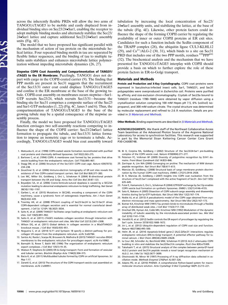
across the inherently flexible PRDs will allow the two arms ofTANGO1/cTAGE5 to be mobile and easily displaced from in-dividual binding sites on Sec23/24•Sar1, enabling the receptor toadopt multiple binding modes and alternately stabilize the Sec23/24•Sar1 lattice and capture additional Sec23/24•Sar1 assemblyunits (Fig. 4E).The model that we have proposed has significant parallel with
the mechanism of action of tau protein on the microtubule he-lical lattice. Four repeated binding motifs in tau are separated byflexible linker sequences, and the binding of tau to multiple tu-bulin units stabilizes and enhances microtubule lattice polymer-ization without impeding microtubule dynamics (26, 27).
Stepwise COPII Coat Assembly and Compartmentation of TANGO1/cTAGE5 to the ER Membrane. Puzzlingly, TANGO1 does not de-part with cargo in the COPII-coated carrier (9). The finding thatPPP motifs are present in Sec31 suggests that the recruitmentof the Sec13/31 outer coat could displace TANGO1/cTAGE5and confine it the ER membrane at the base of the growing tu-bule. COPII-coat assembly on membranes occurs stepwise—Sar1-GTP recruits Sec23/24, which recruits Sec13/31—because thebinding site for Sec31 comprises a composite surface of the Sec23and Sar1-GTPmolecules (1, 22) (Fig. 4C, lanes 5 and 6). Thus, thecompartmentation of TANGO1/cTAGE5 to the base of thegrowing tubule may be a spatial consequence of the stepwise as-sembly process.Finally, the model we have proposed for TANGO1/cTAGE5
function envisages two self-assembly reactions competing to in-fluence the shape of the COPII carrier: Sec23/24•Sar1 latticeformation to propagate the tubule, and Sec13/31 lattice forma-tion to impose an isometric cage or to terminate a tubule. Ac-cordingly, TANGO1/cTAGE5 would bias coat assembly toward
tubulation by increasing the local concentration of Sec23/24•Sar1 assembly units, and stabilizing the lattice, at the base ofthe tubule (Fig. 4E). Likewise, other protein factors could in-fluence the shape of the forming COPII carrier by regulating theavailability of inner or outer COPII proteins at ER exit sites.Candidates for such a function include the Sedlin component ofthe TRAPP complex (28), the ubiquitin ligase CUL3-KLHL12(29), and Ca2+/ALG-2 (30, 31), which binds to a site on Sec31PRD that includes one of the two PPP motifs, residues 838PPP841
(32). The biochemical analysis and the mechanism that we havepresented for TANGO1/cTAGE5 interplay with COPII shouldprovide a basis on which to further explore the role of theseprotein factors in ER-to-Golgi transport.
Materials and MethodsProtein and Production and X-Ray Crystallography. COPII coat proteins wereexpressed in baculovirus-infected insect cells. Sar1, TANGO1, and Sec31polypeptides were overproduced in Escherichia coli. Proteins were purifiedby affinity and size-exclusion chromatography. Crystals of Sec23a bound toTANGO1 (residues 1780–1840) were obtained by vapor diffusion using acrystallization solution comprising 100 mM Hepes pH 7.5, 6% (vol/vol) iso-propanol, and 300 mM sodium citrate. The crystal structure was determinedby molecular replacement and refined to 2.6 Å resolution. Details are pro-vided in SI Materials and Methods.
Other Methods. Binding experiments are described in SIMaterials andMethods.
ACKNOWLEDGMENTS. We thank staff of the Northeast Collaborative AccessTeam beamlines at the Advanced Photon Source of the Argonne NationalLaboratory for access to synchrotron facilities. This research was supportedin part by the Memorial Sloan Kettering Cancer Center Core GrantP30-CA008748.
1. Matsuoka K, et al. (1998) COPII-coated vesicle formation reconstituted with purifiedcoat proteins and chemically defined liposomes. Cell 93(2):263–275.
2. Barlowe C, et al. (1994) COPII: A membrane coat formed by Sec proteins that drivevesicle budding from the endoplasmic reticulum. Cell 77(6):895–907.
3. Stagg SM, et al. (2006) Structure of the Sec13/31 COPII coat cage. Nature 439(7073):234–238.
4. Zeuschner D, et al. (2006) Immuno-electron tomography of ER exit sites reveals theexistence of free COPII-coated transport carriers. Nat Cell Biol 8(4):377–383.
5. Lee MC, Miller EA, Goldberg J, Orci L, Schekman R (2004) Bi-directional proteintransport between the ER and Golgi. Annu Rev Cell Dev Biol 20:87–123.
6. Boyadjiev SA, et al. (2006) Cranio-lenticulo-sutural dysplasia is caused by a SEC23Amutation leading to abnormal endoplasmic-reticulum-to-Golgi trafficking. Nat Genet38(10):1192–1197.
7. Garbes L, et al. (2015) Mutations in SEC24D, encoding a component of the COPIImachinery, cause a syndromic form of osteogenesis imperfecta. Am J Hum Genet96(3):432–439.
8. Townley AK, et al. (2008) Efficient coupling of Sec23-Sec24 to Sec13-Sec31 drivesCOPII-dependent collagen secretion and is essential for normal craniofacial devel-opment. J Cell Sci 121(Pt 18):3025–3034.
9. Saito K, et al. (2009) TANGO1 facilitates cargo loading at endoplasmic reticulum exitsites. Cell 136(5):891–902.
10. Saito K, et al. (2011) cTAGE5 mediates collagen secretion through interaction withTANGO1 at endoplasmic reticulum exit sites. Mol Biol Cell 22(13):2301–2308.
11. Wilson DG, et al. (2011) Global defects in collagen secretion in a Mia3/TANGO1knockout mouse. J Cell Biol 193(5):935–951.
12. Nogueira C, et al. (2014) SLY1 and Syntaxin 18 specify a distinct pathway for pro-collagen VII export from the endoplasmic reticulum. eLife 3:e02784.
13. Santos AJ, Raote I, Scarpa M, Brouwers N, Malhotra B (2015) TANGO1 recruites ERGICmembranes to the endoplasmic reticulum for procollagen export. eLife 4:e10982.
14. Bannykh SI, Rowe T, Balch WE (1996) The organization of endoplasmic reticulumexport complexes. J Cell Biol 135(1):19–35.
15. Watson P, Stephens DJ (2005) ER-to-Golgi transport: Form and formation of vesicularand tubular carriers. Biochim Biophys Acta 1744(3):304–315.
16. Bacia K, et al. (2011) Multibudded tubules formed by COPII on artificial liposomes. SciRep 1:17.
17. Zanetti G, et al. (2013) The structure of the COPII transport-vesicle coat assembled onmembranes. eLife 2:e00951.
18. Bi X, Corpina RA, Goldberg J (2002) Structure of the Sec23/24-Sar1 pre-buddingcomplex of the COPII vesicle coat. Nature 419(6904):271–277.
19. Peterson FC, Volkman BF (2009) Diversity of polyproline recognition by EVH1 do-mains. Front Biosci (Landmark Ed) 14:833–846.
20. Zarrinpar A, Lim WA (2000) Converging on proline: The mechanism of WW domainpeptide recognition. Nat Struct Biol 7(8):611–613.
21. Mancias JD, Goldberg J (2008) Structural basis of cargo membrane protein discrimi-nation by the human COPII coat machinery. EMBO J 27(21):2918–2928.
22. Bi X, Mancias JD, Goldberg J (2007) Insights into COPII coat nucleation from thestructure of Sec23.Sar1 complexed with the active fragment of Sec31. Dev Cell 13(5):635–645.
23. Futai E, Hamamoto S, Orci L, Schekman R (2004) GTP/GDP exchange by Sec12p enablesCOPII vesicle bud formation on synthetic liposomes. EMBO J 23(21):4146–4155.
24. Sato K, Nakano A (2005) Dissection of COPII subunit-cargo assembly and disassemblykinetics during Sar1p-GTP hydrolysis. Nat Struct Mol Biol 12(2):167–174.
25. Noble AJ, et al. (2013) A pseudoatomic model of the COPII cage obtained from cryo-electron microscopy and mass spectrometry. Nat Struct Mol Biol 20(2):167–173.
26. Butner KA, Kirschner MW (1991) Tau protein binds to microtubules through a flexiblearray of distributed weak sites. J Cell Biol 115(3):717–730.
27. Drechsel DN, Hyman AA, Cobb MH, Kirschner MW (1992) Modulation of the dynamicinstability of tubulin assembly by the microtubule-associated protein tau. Mol BiolCell 3(10):1141–1154.
28. Venditti R, et al. (2012) Sedlin controls the ER export of procollagen by regulating theSar1 cycle. Science 337(6102):1668–1672.
29. Jin L, et al. (2012) Ubiquitin-dependent regulation of COPII coat size and function.Nature 482(7386):495–500.
30. Helm JR, et al. (2014) Apoptosis-linked gene-2 (ALG-2)/Sec31 interactions regulateendoplasmic reticulum (ER)-to-Golgi transport: A potential effector pathway for lu-minal calcium. J Biol Chem 289(34):23609–23628.
31. la Cour JM, Schindler AJ, Berchtold MW, Schekman R (2013) ALG-2 attenuates COPIIbudding in vitro and stabilizes the Sec23/Sec31A complex. PLoS One 8(9):e75309.
32. Takahashi T, et al. (2015) Structural analysis of the complex between penta-EF-handALG-2 protein and Sec31A peptide reveals a novel target recognition mechanism ofALG-2. Int J Mol Sci 16(2):3677–3699.
33. Otwinowski W, Minor W (1997) Processing of X-ray diffraction data collected in os-cillation mode. Methods Enzymol 276(Part A):307–326.
34. Adams PD, et al. (2010) PHENIX: A comprehensive Python-based system for macro-molecular structure solution. Acta Crystallogr D Biol Crystallogr 66(Pt 2):213–221.
10066 | www.pnas.org/cgi/doi/10.1073/pnas.1605916113 Ma and Goldberg