Embed Size (px)
Citation preview
JANUARY - MARCH 2017M E D I C A L D I G E S T
MCI (P) 094/08/2016
Improving InpatientDiabetes Care –Our Ongoing Journey
TTSH Research News
High-Sensitive Troponin Assays
The Case forPersonalised Medicine
Transitional Care –What happens duringand after PACH?
A New Class Of HeartFailure Medication
Hypoglycaemia-InducedCardiovascular Events InPatients With Diabetes:What Is The Evidence?
Quiz
11 Jalan Tan Tock SengSingapore 308433
Tel: 6256 6011Fax: 6252 7282
www.ttsh.com.sg
TAN TOCK SENG HOSPITAL
Medical Digest is a quarterly publication of Tan Tock Seng Hospital written by healthcare providers for healthcare providers, as a service to the medical community.
MEDICAL DIGEST1
EDITORLeong Khai Pang
ASSOCIATE EDITORS:Charmaine ManauisRaymond NgMelissa TienMohd Fadil Muhammad Farhan
MEMBERS:Tang Yee Lin Rafael Saclolo Ivan Huang Kuang Hsin Noorhazlina bte AliDeshan KumarTan Choon ChiehShirley BangChong Yaw KhianKalaiyarasi KaliyaperumalAdeline TehYew Min SenShirlene Ho Shu Min Gavin Lim Hock Tai Binedell Trevor Brian
EDITORIAL ASSISTANTAngie Theonis
COPY EDITORSafiyya Mohamed Ali
DESIGNERClinton Low
We value your feedback. Please email your questions, comments or suggestions to: [email protected] also contact us for notification of change of postal address or requests of additional copies.
While every endeavour is made to ensure that information herein is accurate at the time of publication, Tan Tock Seng Hospital shall not be held liable for any inaccuracies. The opinions expressed in this publication do not necessrily reflect those of Tan Tock Seng Hospital. The contents of this publication may not be reproduced without written permission from the publisher.
D I G E S TM E D I C A L
What is a professional? The Oxford English Dictionary lists the different shades of meaning. It describes a professional as one ‘that engages in a specified occupation or activity for money or as a means of earning a living, rather than as a pastime’ (this was first used in 1606). Another meaning is of a person engaged in work that requires ‘special skill or training’ (1779). Combining these, a professional is someone who possesses certain ability that others pay to benefit from. Kipling declared that prostitution is the oldest profession in the world in the short story On the City Wall collected in In Black and White (1888). Beauchamp and Childress (Principles of Biomedical Ethics, 5th edition, Oxford University Press 2001) felt that a profession must necessarily benefit society, so prostitution doesn’t really count but they concede that this point is debatable! Another requirement of a profession pointed out by these authors, but not found in the Dictionary, is self-regulation by practitioners.
So a professional works at a societally beneficial paid job within the bounds established by his or her colleagues. Professionalism is a slightly different matter. I am persuaded that it is the sum of the rules and expectations articulated and demanded by the practitioners. It deals with how we treat ourselves (competence, addictions), people (patients, staffs, and colleagues), entities (pharmaceutical companies, employer) and society (fairness, appropriateness). Isn’t a code of behaviour devised by a group of people to ensure that individual members toe the line also known as culture? Indeed, medical professionalism is but medical culture, albeit of the utmost standard. If you accept my argument, then professionalism is teachable (not inherent in the doctor), changeable, and susceptible to tensions in the health care system).
The Medical Digest wishes all Professional Readers a Happy New Year!
Dr Leong Khai PangEDITORMedical Digest
FROM THE
EDITOR
Every year, TTSH clinicians publish about 300 scientific papers. In this section, we selected a few reports and asked one of the authors of each to summarise and discuss the clinical relevance of their research. Our theme in this issue is ambulatory care and internal medicine.
TTSH RESEARCH NEWS
MEDICAL DIGEST3
his point-prevalence study was undertaken to investigate the frequency, indications and appropriateness of use of proton pump inhibitors (PPIs) in hospitalised patients on a randomly chosen day. Inpatient hospital ward census records were
analysed for the total number of inpatients seen from June to July 2011 across all disciplines in Tan Tock Seng Hospital, Singapore and the number of inpatients who were on PPIs were identified with the hospital electronic medical records. Details including the patients’ presenting complaint, duration of symptoms, salient features of the social history, medical history, family history, records of previous admissions, physical examination findings, investigation results, treatment administered and records of previous medications prescribed by the study hospital and its affiliates (other government/restructured hospitals and healthcare facilities, excluding private institutions and general practitioners) were analysed. Indications for maintaining these patients on PPIs were then recorded and cross-referenced against a list of accepted indications adapted from the US Food and Drug Administration (FDA)-approved list and also other accepted/off-label indications adapted from peer-reviewed clinical studies and guidelines. These indications include the treatment of peptic ulcer disease, erosive oesophagitis, Helicobacter pylori infection, gastro-oesophageal reflux disease, Zollinger-Ellison syndrome,
stress ulcer prophylaxis, risk reduction of NSAID-associated peptic ulcer disease with risk factors, oesophageal stricture, Barrett’s oesophagus and cystic fibrosis (to improve pancreatic enzyme absorption). Of the 477 (46.5%) inpatients receiving PPIs, only 219 (45.9%) fulfilled the FDA-approved indications, while the majority (n = 258, 54.1%) did not. Fifty-two (10.9%) had borderline indications based on expert
consensus/guidelines other than FDA criteria. Overall, PPIs were not strictly indicated for use in 206 (43.2%) inpatients, according to FDA criteria.
RESEARCH EXCERPT 1
This summary was prepared by Dr Christopher Chia
Tze Wei, a consultant in the Department of Gastroenterology and
Hepatology, Tan Tock Seng Hospital
randomised controlled trial of a Return-to-work coordinator (RTWC) model of care was conducted in Tan Tock Seng Hospital from September 2010 to November 2012. One hundred and sixty injured workers recruited from the Emergency Department
were randomly allocated to either control (receive usual hospital standard care) or intervention (assigned a RTWC) group. The RTWCs, who were occupational therapists by training, closely supported RTW arrangements and proactively liaised with employers and healthcare professionals on RTW solutions for the injured workers. At 3 months post injury, workers in the intervention group returned to work 10 days earlier than the control group, with a higher proportion of workers in the intervention group returning to modified jobs. There were no significant differences between the control and intervention groups in terms of the RTW rate at 3 months and 9 months post injury (3-month rate: 67% and 69%; 9-month rate: 75% and 78% respectively). It was concluded that the addition of a RTWC into the hospital model of care is effective in facilitating early RTW for injured workers.
RESEARCH EXCERPT 2
This summary was prepared by Ms Heidi Tan Siew Khoon, a senior principal occupational therapist in the Department of Occupational Therapy, Tan Tock Seng Hospital.
IMPORTANCE IN CLINICAL PRACTICEThe use of PPI is prevalent in our hospital, and likely in many others. Less than half of the hospitalised patients receiving PPIs in this study had evidence-based indications that supported such use. The overuse of PPIs has a negative impact on healthcare cost and may lead to adverse effects. Steps to curb the inappropriate use of PPIs should address measures such as appropriate indication for the initiation and continual assessment of the need for ongoing PPI use in the inpatient period, upon discharge and during outpatient review.
Chia CT, Lim WP, Vu CK. Singapore Med J 2014; 55:363-6.
MEDICAL DIGEST2
Inappropriate use of proton
pump inhibitors in a local setting
A randomised controlled trial of a Return-To-Work Coordinator model of care in a general hospital to facilitate return to work of injured workers
IMPORTANCE IN CLINICAL PRACTICEReturn-to-work programmes for injured workers, with RTW coordination as a key element in the programme, have been prevalent in Western countries with established work injury management policies for decades. This first local randomised controlled trial sought to determine the effectiveness of a Return-to-work coordinator model of care in facilitating early RTW for injured workers and to provide insights for developing a model of care for RTW that can be implemented in Singapore and other countries which do not have established work injury management policies. Results from the study showed that the addition of a RTWC into a hospital model of care is effective in facilitating earlier RTW of injured workers as well as improving the workers’ compliance to healthcare treatment, and employers’ compliance to legislative requirements of work injury notification. This could be a potential model of care that can be further explored in Singapore.
Tan HS, Yeo DS, Giam JY, Cheong FW, Chan KF. Work 2016; 54:209-22.
TTSH Research News is curated and edited by DR MELISSA TIEN, a consultant in the Department of Opthalmology, Tan Tock Seng Hospital.
IMPROVING INPATIENT DIABETES CARE – OUR ONGOING JOURNEY
HOT TOPIC
MEDICAL DIGEST5
Our war on diabetesuring the parliamentary debate on the Ministry of Health’s budget in April 2016, Minister for Health Mr Gan Kim Yong declared war on diabetes mellitus (DM).1
To this end, a new Diabetes Prevention and Care Taskforce will be set up, chaired by himself and the Minister for Education (Schools), Mr Ng Chee Meng.
Three key aims were outlined:1. Reducing the number of new DM cases by
promoting a healthy lifestyle and reducing obesity rates;
2. Strengthening early screening and intervention to identify those who are at risk and undiagnosed; and
3. Supporting better disease control to slow progression and reduce complications.
Tan Tock Seng Hospital (TTSH), being one of Singapore’s largest multi-disciplinary hospitals, has naturally become a battleground of this war. A new TTSH DM steering committee was set up to lead this fight against DM, chaired by the Head of the Department of Endocrinology, Adj. Assoc. Prof. Daniel Chew.
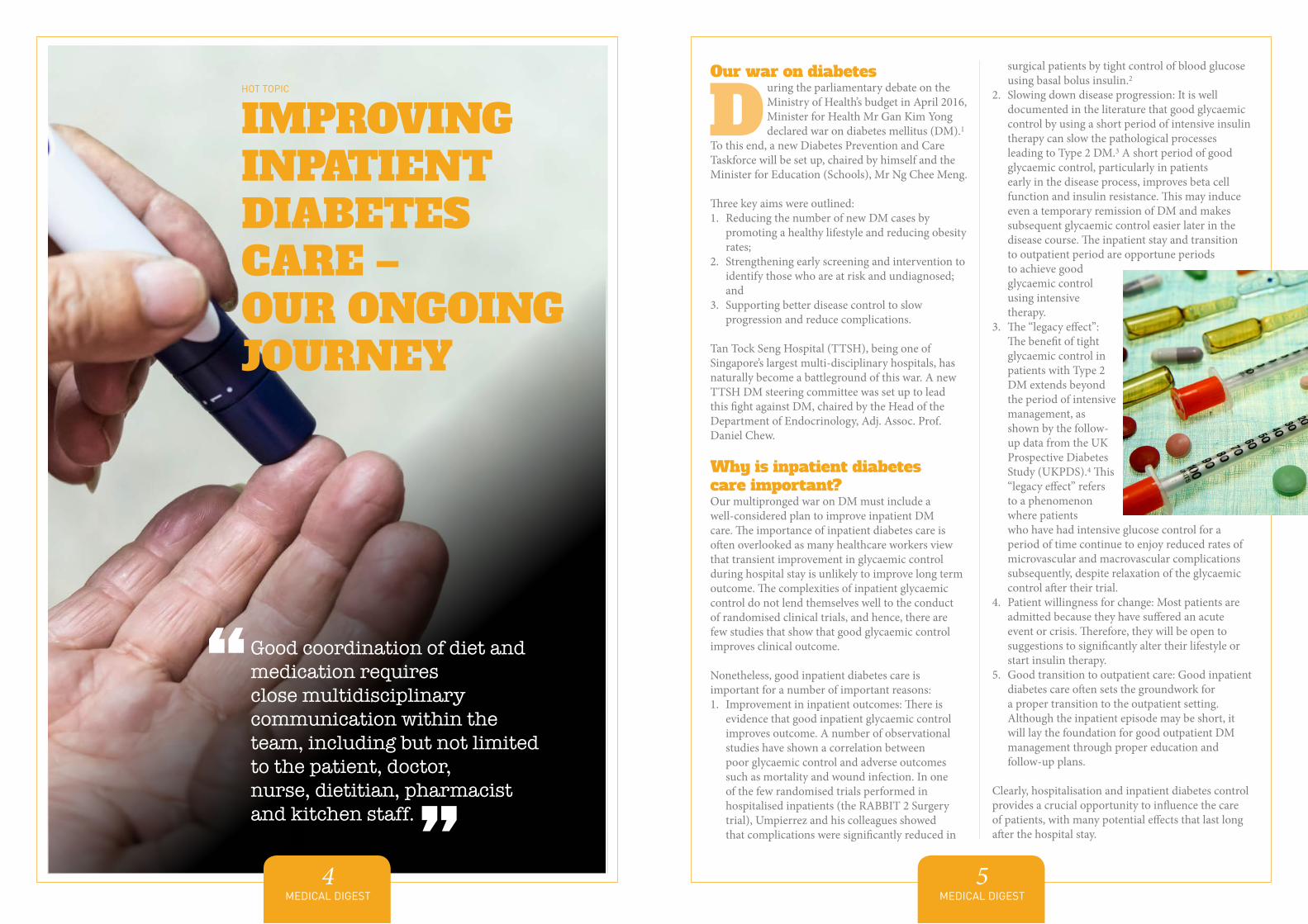
Why is inpatient diabetes care important?Our multipronged war on DM must include a well-considered plan to improve inpatient DM care. The importance of inpatient diabetes care is often overlooked as many healthcare workers view that transient improvement in glycaemic control during hospital stay is unlikely to improve long term outcome. The complexities of inpatient glycaemic control do not lend themselves well to the conduct of randomised clinical trials, and hence, there are few studies that show that good glycaemic control improves clinical outcome.
Nonetheless, good inpatient diabetes care is important for a number of important reasons:1. Improvement in inpatient outcomes: There is
evidence that good inpatient glycaemic control improves outcome. A number of observational studies have shown a correlation between poor glycaemic control and adverse outcomes such as mortality and wound infection. In one of the few randomised trials performed in hospitalised inpatients (the RABBIT 2 Surgery trial), Umpierrez and his colleagues showed that complications were significantly reduced in
surgical patients by tight control of blood glucose using basal bolus insulin.2
2. Slowing down disease progression: It is well documented in the literature that good glycaemic control by using a short period of intensive insulin therapy can slow the pathological processes leading to Type 2 DM.3 A short period of good glycaemic control, particularly in patients early in the disease process, improves beta cell function and insulin resistance. This may induce even a temporary remission of DM and makes subsequent glycaemic control easier later in the disease course. The inpatient stay and transition to outpatient period are opportune periods to achieve good glycaemic control using intensive therapy.
3. The “legacy effect”: The benefit of tight glycaemic control in patients with Type 2 DM extends beyond the period of intensive management, as shown by the follow-up data from the UK Prospective Diabetes Study (UKPDS).4 This “legacy effect” refers to a phenomenon where patients who have had intensive glucose control for a period of time continue to enjoy reduced rates of microvascular and macrovascular complications subsequently, despite relaxation of the glycaemic control after their trial.
4. Patient willingness for change: Most patients are admitted because they have suffered an acute event or crisis. Therefore, they will be open to suggestions to significantly alter their lifestyle or start insulin therapy.
5. Good transition to outpatient care: Good inpatient diabetes care often sets the groundwork for a proper transition to the outpatient setting. Although the inpatient episode may be short, it will lay the foundation for good outpatient DM management through proper education and follow-up plans.
Clearly, hospitalisation and inpatient diabetes control provides a crucial opportunity to influence the care of patients, with many potential effects that last long after the hospital stay.
MEDICAL DIGEST4
Good coordination of diet and medication requiresclose multidisciplinary communication within theteam, including but not limited to the patient, doctor,nurse, dietitian, pharmacist and kitchen staff.
““
Why is inpatient glycaemic control difficult?The most common acute side effect of many anti-diabetic medications is hypoglycaemia, which can lead to neuroglycopenic brain injury and mortality. The use of such medication is complex in the inpatient environment, where rapid changes in the patient’s condition and nutritional demands are the rule rather than the exception. Proper and timely use and cessation of such medications are therefore critical to ensure patient safety.
Good coordination of diet and medication requires close multidisciplinary communication within the team, including but not limited to the patient, doctor, nurse, dietitian, pharmacist and kitchen staff. However, there are many barriers to this, including stakeholders having differing but equally important priorities apart from glycaemic control, a rapid changeover of staff and an increasingly complex IT system.
Achieving good inpatient glycaemic control is a challenging task. Although there are available a number of international guidelines for inpatient diabetes control,5-6 indiscriminate adoption without consideration of the local context can potentially be dangerous.
How have we gone about improving inpatient diabetes care?TTSH is fortunate to have the support and vision of our senior leadership, who started laying the groundwork to improve inpatient diabetes control even before the declaration of the War on Diabetes.
In 2011, the Division of Surgery and the Department of Endocrinology launched a pilot project, the Inpatient Diabetes Care Team, to look after the glycaemic control and related medical needs of patients with poorly controlled DM admitted to surgical departments. This project was expanded to cover two levels of surgical patients in 2012, and has been continuing to the present.
In 2013, a DM Workgroup was set up to oversee hospital-wide DM-related multidisciplinary projects. This role was subsumed by the TTSH DM steering committee in 2016.
In addition, there have been numerous initiatives such as care paths for DM patients in the Emergency Department, management of inpatients with
hypoglycaemia, and a programme to improve DM medication safety, the Singapore Healthcare Improvement Network (SHINe). There is also an imminent Integrated Diabetes Care Programme (IDCP) under the MOH Health Services Development Programme (HSDP), which will utilise a new IT system and suitably trained staff to stratify all TTSH inpatients into risk groups to deliver appropriate intervention.
In the next section, we will describe the TTSH SHINe initiative to improve medication safety in greater detail as an example to illustrate the importance of multidisciplinary cooperation and the challenges in inpatient DM care.
SHINe initiative to improve DM medication safetyThe Singapore Healthcare Improvement Network (SHINe) is a group of healthcare institutions formed in 2014 by the Singapore Ministry of Health and facilitated by the US-based Institute for Healthcare Improvement, a leading innovator of healthcare improvement worldwide. TTSH is one of the 23 founding members of SHINe.
The SHINe group is collectively committed to provide better health, better care and lower cost to patients. It aims to accelerate the pace and scale of healthcare improvement, allowing members to collaborate with one another to improve patient safety and quality within their organisation, and influence the national quality improvement agenda. The initiative aims to focus on reducing patient harm by 30% in 3 years starting with areas of greatest risk across three
MEDICAL DIGEST6
MEDICAL DIGEST7
workstreams: medication safety, surgical safety and prevention of healthcare-associated infections.
In April 2014, one of the two key areas TTSH decided to focus on was medication safety surrounding the use of anti-diabetic drugs. We initially identified a core improvement team comprising an endocrinologist, pharmacist, diabetic nurse educator, nursing quality manager and hospital quality facilitator. A local improvement team comprising a general surgeon, ward nurse clinician, senior nurse manager, senior staff nurse and ward pharmacist was also formed at our pilot site to institute the changes. Kitchen staffs were also involved at the appropriate stages to ensure adequate buy-in and coordination.
Root cause analysis was performed to identify top factors that contribute to hypoglycaemia in the pilot ward. We identified the key steps to reduce hypoglycaemia in the ward: standardised prescribing and monitoring, matching nutritional intake to insulin dosing, maintaining dietary intake based on blood sugar levels and running staff educational programmes.
The team adopted the Institute for Healthcare Improvement methodology which improves quality, safety, and value in health care and emphasises innovation, rapid-cycle testing and spread. It is characterised by a combination of subject matter expert knowledge with improvement methods and tools.
A clear aim for improvement and data collection plan was formulated, followed by small rapid tests of the proposed changes. After refinement and successful implementation of these changes in the pilot site, scale-up and spread of the changes was carried out throughout the hospital. The emphasis was on starting improvement efforts on a small scale, and then leveraging the lessons learnt to plan for spread.
One of the first interventions tested in a general surgical ward was the implementation of the Nil-By-Mouth (NBM) protocol to ensure appropriate management of type 2 DM patients. This was followed by two other interventions: coordinating diet and medication administration, and the provision of bedtime snacks for patients with borderline low capillary blood glucose levels to reduce nocturnal and fasting hypoglycaemia. The second intervention resulted from a Clinical Practice Improvement Programme (CPIP) project carried out by our Endocrine Advanced Practice Nurse.
The testing of these interventions was done in close collaboration with ground nursing leads, with mass education sessions to nurses and doctors.
In our pilot surgical ward, we achieved more than 30% reduction in hypoglycaemia incidence compared with baseline. Thereafter, in December 2015, the same set of interventions was
tested and implemented in two other surgical wards within 6 months, achieving a similar reduction in hypoglycaemia.
The SHINe group is collectively committed to provide better
health, better care and lower cost to
patients.
MEDICAL DIGEST8
MEDICAL DIGEST9
In May 2016, senior nursing leadership planned for the spread of these DM-related interventions to all inpatient wards. By July 2016, 23 wards had adopted these measures. Currently, these interventions are being disseminated to the rest of the wards, with close monitoring of outcome and process measures.
We have also learnt that it is important to commemorate every milestone we reach, however small. Hence, celebratory activities are conducted for the participating wards in appreciation of their efforts in quality improvement. In November 2016, a senior leadership appreciation session
was held, in which Mr Yong Keng Kwang (Chief Nurse) and Adj. Assoc. Prof. Daniel Chew (Head of Department of Endocrinology) personally thanked all 23 wards for their hard work.
ConclusionReflecting on our journey to improve inpatient diabetes care, we learnt that to achieve and sustain change, a concerted multi-disciplinary effort by all stakeholders is required, together with strong support from senior leadership. We are proud that TTSH is committed to improve the care of diabetes patients and contribute to the “War on Diabetes”.
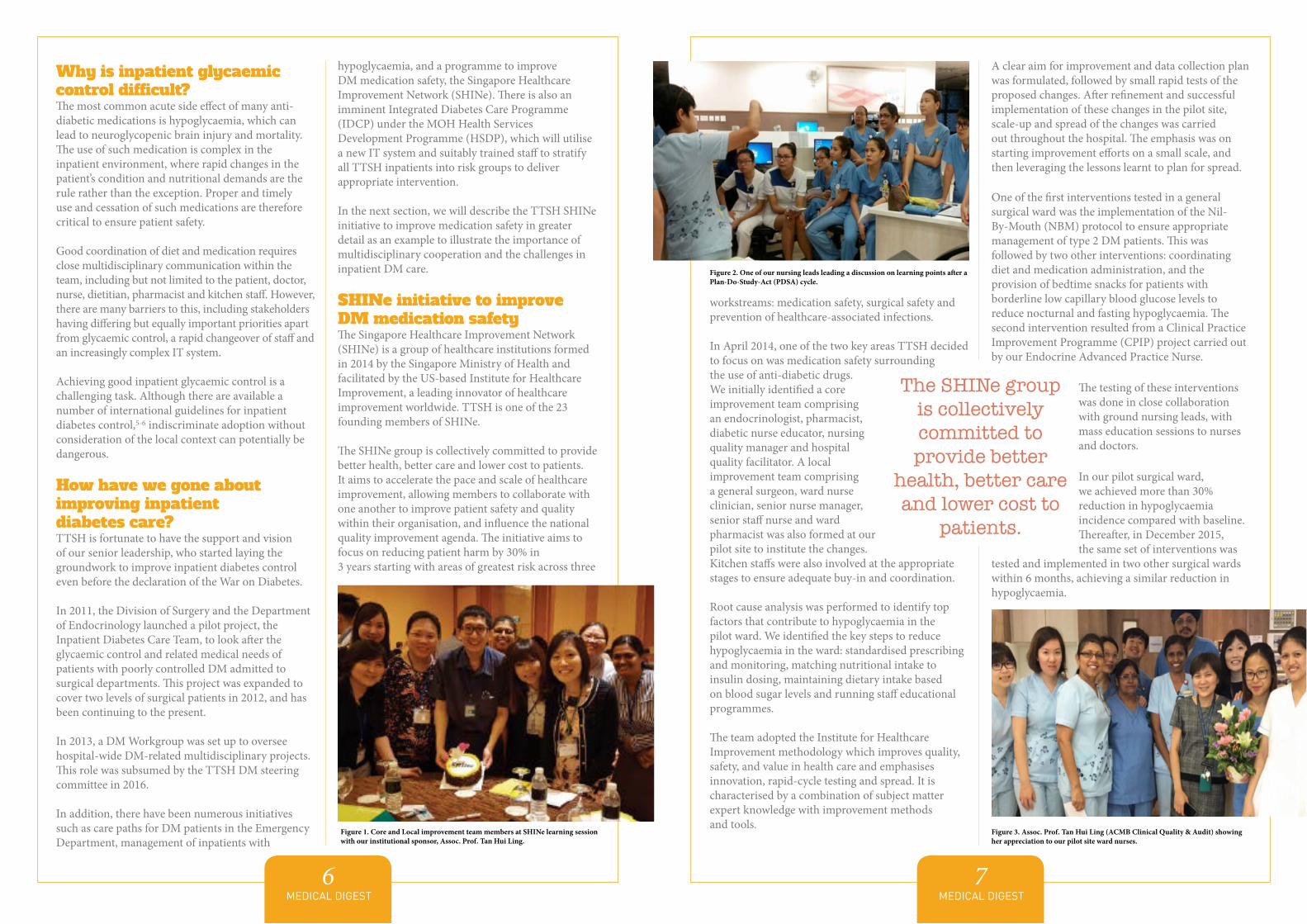
Figure 4. Assoc. Prof. Daniel Chew (Head of Department, Endocrinology) being updated on the pilot ward’s progress by nursing leaders.
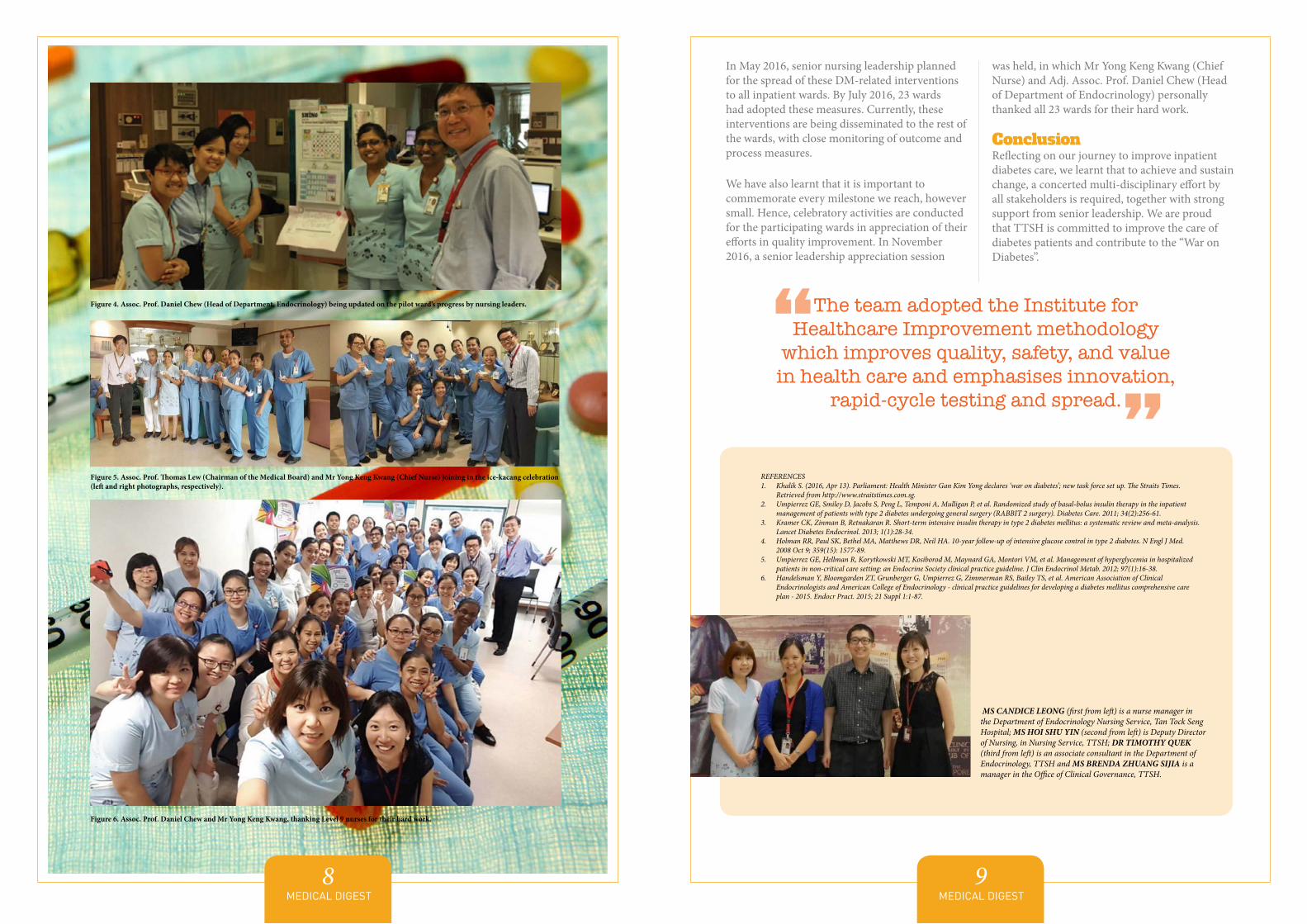
Figure 5. Assoc. Prof. Thomas Lew (Chairman of the Medical Board) and Mr Yong Keng Kwang (Chief Nurse) joining in the ice-kacang celebration (left and right photographs, respectively).
Figure 6. Assoc. Prof. Daniel Chew and Mr Yong Keng Kwang, thanking Level 9 nurses for their hard work.
REFERENCES1. Khalik S. (2016, Apr 13). Parliament: Health Minister Gan Kim Yong declares ‘war on diabetes’; new task force set up. The Straits Times.
Retrieved from http://www.straitstimes.com.sg.2. Umpierrez GE, Smiley D, Jacobs S, Peng L, Temponi A, Mulligan P, et al. Randomized study of basal-bolus insulin therapy in the inpatient
management of patients with type 2 diabetes undergoing general surgery (RABBIT 2 surgery). Diabetes Care. 2011; 34(2):256-61. 3. Kramer CK, Zinman B, Retnakaran R. Short-term intensive insulin therapy in type 2 diabetes mellitus: a systematic review and meta-analysis.
Lancet Diabetes Endocrinol. 2013; 1(1):28-34.4. Holman RR, Paul SK, Bethel MA, Matthews DR, Neil HA. 10-year follow-up of intensive glucose control in type 2 diabetes. N Engl J Med.
2008 Oct 9; 359(15): 1577-89.5. Umpierrez GE, Hellman R, Korytkowski MT, Kosiborod M, Maynard GA, Montori VM, et al. Management of hyperglycemia in hospitalized
patients in non-critical care setting: an Endocrine Society clinical practice guideline. J Clin Endocrinol Metab. 2012; 97(1):16-38.6. Handelsman Y, Bloomgarden ZT, Grunberger G, Umpierrez G, Zimmerman RS, Bailey TS, et al. American Association of Clinical
Endocrinologists and American College of Endocrinology - clinical practice guidelines for developing a diabetes mellitus comprehensive care plan - 2015. Endocr Pract. 2015; 21 Suppl 1:1-87.
MS CANDICE LEONG (first from left) is a nurse manager in the Department of Endocrinology Nursing Service, Tan Tock Seng Hospital; MS HOI SHU YIN (second from left) is Deputy Director of Nursing, in Nursing Service, TTSH; DR TIMOTHY QUEK (third from left) is an associate consultant in the Department of Endocrinology, TTSH and MS BRENDA ZHUANG SIJIA is a manager in the Office of Clinical Governance, TTSH.
The team adopted the Institute for Healthcare Improvement methodology
which improves quality, safety, and value in health care and emphasises innovation,
rapid-cycle testing and spread.
““
HIGH-SENSITIVE TROPONIN ASSAYS
FEATURE ARTICLE
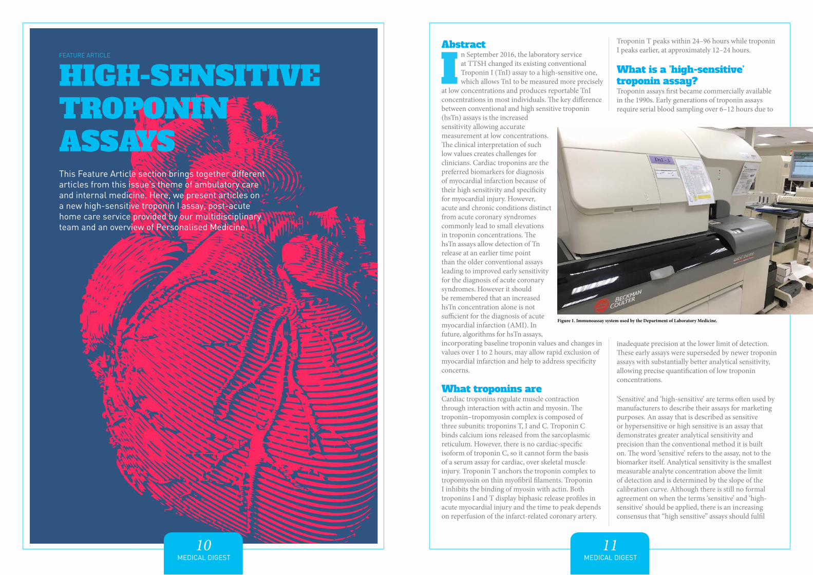
This Feature Article section brings together different articles from this issue’s theme of ambulatory care and internal medicine. Here, we present articles on a new high-sensitive troponin I assay, post-acute home care service provided by our multidisciplinary team and an overview of Personalised Medicine.
MEDICAL DIGEST11
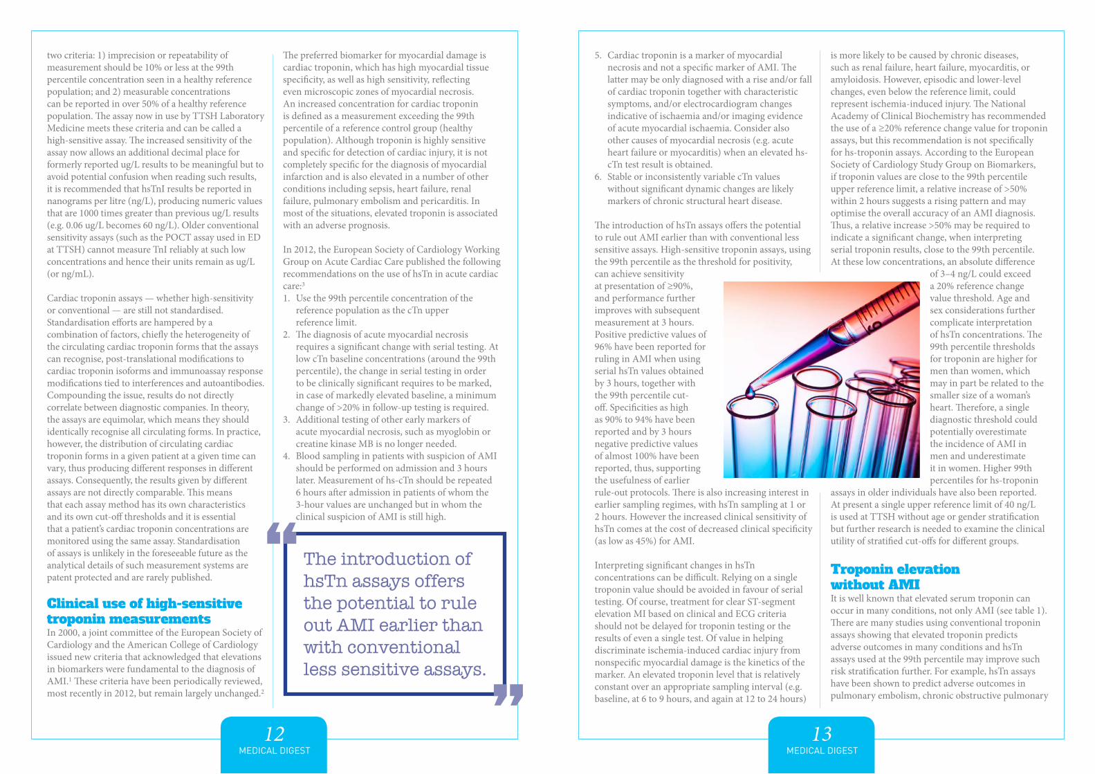
Abstractn September 2016, the laboratory service at TTSH changed its existing conventional Troponin I (TnI) assay to a high-sensitive one, which allows TnI to be measured more precisely
at low concentrations and produces reportable TnI concentrations in most individuals. The key difference between conventional and high sensitive troponin (hsTn) assays is the increased sensitivity allowing accurate measurement at low concentrations. The clinical interpretation of such low values creates challenges for clinicians. Cardiac troponins are the preferred biomarkers for diagnosis of myocardial infarction because of their high sensitivity and specificity for myocardial injury. However, acute and chronic conditions distinct from acute coronary syndromes commonly lead to small elevations in troponin concentrations. The hsTn assays allow detection of Tn release at an earlier time point than the older conventional assays leading to improved early sensitivity for the diagnosis of acute coronary syndromes. However it should be remembered that an increased hsTn concentration alone is not sufficient for the diagnosis of acute myocardial infarction (AMI). In future, algorithms for hsTn assays, incorporating baseline troponin values and changes in values over 1 to 2 hours, may allow rapid exclusion of myocardial infarction and help to address specificity concerns.
What troponins areCardiac troponins regulate muscle contraction through interaction with actin and myosin. The troponin–tropomyosin complex is composed of three subunits: troponins T, I and C. Troponin C binds calcium ions released from the sarcoplasmic reticulum. However, there is no cardiac-specific isoform of troponin C, so it cannot form the basis of a serum assay for cardiac, over skeletal muscle injury. Troponin T anchors the troponin complex to tropomyosin on thin myofibril filaments. Troponin I inhibits the binding of myosin with actin. Both troponins I and T display biphasic release profiles in acute myocardial injury and the time to peak depends on reperfusion of the infarct-related coronary artery.
Troponin T peaks within 24–96 hours while troponin I peaks earlier, at approximately 12–24 hours.
What is a ’high-sensitive’ troponin assay?Troponin assays first became commercially available in the 1990s. Early generations of troponin assays require serial blood sampling over 6–12 hours due to
inadequate precision at the lower limit of detection. These early assays were superseded by newer troponin assays with substantially better analytical sensitivity, allowing precise quantification of low troponin concentrations.
‘Sensitive’ and ‘high-sensitive’ are terms often used by manufacturers to describe their assays for marketing purposes. An assay that is described as sensitive or hypersensitive or high sensitive is an assay that demonstrates greater analytical sensitivity and precision than the conventional method it is built on. The word ‘sensitive’ refers to the assay, not to the biomarker itself. Analytical sensitivity is the smallest measurable analyte concentration above the limit of detection and is determined by the slope of the calibration curve. Although there is still no formal agreement on when the terms ‘sensitive’ and ‘high-sensitive’ should be applied, there is an increasing consensus that “high sensitive” assays should fulfil
MEDICAL DIGEST10
MEDICAL DIGEST12
two criteria: 1) imprecision or repeatability of measurement should be 10% or less at the 99th percentile concentration seen in a healthy reference population; and 2) measurable concentrations can be reported in over 50% of a healthy reference population. The assay now in use by TTSH Laboratory Medicine meets these criteria and can be called a high-sensitive assay. The increased sensitivity of the assay now allows an additional decimal place for formerly reported ug/L results to be meaningful but to avoid potential confusion when reading such results, it is recommended that hsTnI results be reported in nanograms per litre (ng/L), producing numeric values that are 1000 times greater than previous ug/L results (e.g. 0.06 ug/L becomes 60 ng/L). Older conventional sensitivity assays (such as the POCT assay used in ED at TTSH) cannot measure TnI reliably at such low concentrations and hence their units remain as ug/L (or ng/mL).
Cardiac troponin assays — whether high-sensitivity or conventional — are still not standardised. Standardisation efforts are hampered by a combination of factors, chiefly the heterogeneity of the circulating cardiac troponin forms that the assays can recognise, post-translational modifications to cardiac troponin isoforms and immunoassay response modifications tied to interferences and autoantibodies. Compounding the issue, results do not directly correlate between diagnostic companies. In theory, the assays are equimolar, which means they should identically recognise all circulating forms. In practice, however, the distribution of circulating cardiac troponin forms in a given patient at a given time can vary, thus producing different responses in different assays. Consequently, the results given by different assays are not directly comparable. This means that each assay method has its own characteristics and its own cut-off thresholds and it is essential that a patient’s cardiac troponin concentrations are monitored using the same assay. Standardisation of assays is unlikely in the foreseeable future as the analytical details of such measurement systems are patent protected and are rarely published.
Clinical use of high-sensitive troponin measurementsIn 2000, a joint committee of the European Society of Cardiology and the American College of Cardiology issued new criteria that acknowledged that elevations in biomarkers were fundamental to the diagnosis of AMI.1 These criteria have been periodically reviewed, most recently in 2012, but remain largely unchanged.2
The preferred biomarker for myocardial damage is cardiac troponin, which has high myocardial tissue specificity, as well as high sensitivity, reflecting even microscopic zones of myocardial necrosis. An increased concentration for cardiac troponin is defined as a measurement exceeding the 99th percentile of a reference control group (healthy population). Although troponin is highly sensitive and specific for detection of cardiac injury, it is not completely specific for the diagnosis of myocardial infarction and is also elevated in a number of other conditions including sepsis, heart failure, renal failure, pulmonary embolism and pericarditis. In most of the situations, elevated troponin is associated with an adverse prognosis.
In 2012, the European Society of Cardiology Working Group on Acute Cardiac Care published the following recommendations on the use of hsTn in acute cardiac care:3 1. Use the 99th percentile concentration of the
reference population as the cTn upper reference limit.
2. The diagnosis of acute myocardial necrosis requires a significant change with serial testing. At low cTn baseline concentrations (around the 99th percentile), the change in serial testing in order to be clinically significant requires to be marked, in case of markedly elevated baseline, a minimum change of >20% in follow-up testing is required.
3. Additional testing of other early markers of acute myocardial necrosis, such as myoglobin or creatine kinase MB is no longer needed.
4. Blood sampling in patients with suspicion of AMI should be performed on admission and 3 hours later. Measurement of hs-cTn should be repeated 6 hours after admission in patients of whom the 3-hour values are unchanged but in whom the clinical suspicion of AMI is still high.
MEDICAL DIGEST13
The introduction of hsTn assays offers the potential to rule out AMI earlier than with conventional less sensitive assays.
““
5. Cardiac troponin is a marker of myocardial necrosis and not a specific marker of AMI. The latter may be only diagnosed with a rise and/or fall of cardiac troponin together with characteristic symptoms, and/or electrocardiogram changes indicative of ischaemia and/or imaging evidence of acute myocardial ischaemia. Consider also other causes of myocardial necrosis (e.g. acute heart failure or myocarditis) when an elevated hs-cTn test result is obtained.
6. Stable or inconsistently variable cTn values without significant dynamic changes are likely markers of chronic structural heart disease.
The introduction of hsTn assays offers the potential to rule out AMI earlier than with conventional less sensitive assays. High-sensitive troponin assays, using the 99th percentile as the threshold for positivity, can achieve sensitivity at presentation of ≥90%, and performance further improves with subsequent measurement at 3 hours. Positive predictive values of 96% have been reported for ruling in AMI when using serial hsTn values obtained by 3 hours, together with the 99th percentile cut-off. Specificities as high as 90% to 94% have been reported and by 3 hours negative predictive values of almost 100% have been reported, thus, supporting the usefulness of earlier rule-out protocols. There is also increasing interest in earlier sampling regimes, with hsTn sampling at 1 or 2 hours. However the increased clinical sensitivity of hsTn comes at the cost of decreased clinical specificity (as low as 45%) for AMI.
Interpreting significant changes in hsTn concentrations can be difficult. Relying on a singletroponin value should be avoided in favour of serial testing. Of course, treatment for clear ST-segment elevation MI based on clinical and ECG criteria should not be delayed for troponin testing or the results of even a single test. Of value in helping discriminate ischemia-induced cardiac injury from nonspecific myocardial damage is the kinetics of the marker. An elevated troponin level that is relatively constant over an appropriate sampling interval (e.g. baseline, at 6 to 9 hours, and again at 12 to 24 hours)
is more likely to be caused by chronic diseases, such as renal failure, heart failure, myocarditis, or amyloidosis. However, episodic and lower-level changes, even below the reference limit, could represent ischemia-induced injury. The National Academy of Clinical Biochemistry has recommended the use of a ≥20% reference change value for troponin assays, but this recommendation is not specifically for hs-troponin assays. According to the European Society of Cardiology Study Group on Biomarkers, if troponin values are close to the 99th percentile upper reference limit, a relative increase of >50% within 2 hours suggests a rising pattern and may optimise the overall accuracy of an AMI diagnosis. Thus, a relative increase >50% may be required to indicate a significant change, when interpreting serial troponin results, close to the 99th percentile. At these low concentrations, an absolute difference
of 3–4 ng/L could exceed a 20% reference change value threshold. Age and sex considerations further complicate interpretation of hsTn concentrations. The 99th percentile thresholds for troponin are higher for men than women, which may in part be related to the smaller size of a woman’s heart. Therefore, a single diagnostic threshold could potentially overestimate the incidence of AMI in men and underestimate it in women. Higher 99th percentiles for hs-troponin
assays in older individuals have also been reported. At present a single upper reference limit of 40 ng/L is used at TTSH without age or gender stratification but further research is needed to examine the clinical utility of stratified cut-offs for different groups.
Troponin elevation without AMIIt is well known that elevated serum troponin can occur in many conditions, not only AMI (see table 1). There are many studies using conventional troponin assays showing that elevated troponin predicts adverse outcomes in many conditions and hsTn assays used at the 99th percentile may improve such risk stratification further. For example, hsTn assays have been shown to predict adverse outcomes in pulmonary embolism, chronic obstructive pulmonary
Table 1. Causes of elevated troponin, adapted from Chenevier-Gobeaux et al.4
Causes
Type 1 myocardial ischaemia
Type 2 myocardial ischaemia related to oxygen supply and demand imbalance
Injury not related to myocardial ischaemia
Multifactorial or indeterminate myocardial injury
Analytical interference
Examples
Plaque ruptureIntraluminal coronary artery thrombus formation
Tachyarrhythmias/bradyarrhythmiasCardiogenic, hypovolemic or septic shockAcute respiratory failureSevere anaemiaSevere hypertension with or without left ventricular hypertrophyHypertrophic cardiomyopathyAortic dissection or decompensated severe aortic valve disease (aortic insufficiency, aortic regurgitation)
Cardiac contusion, ablation, pacing or defibrillatorshockCardiotoxic agents (e.g. Herceptin®, anthracycline antibiotics)MyocarditisRhabdomyolysis with cardiac involvement
Heart failureStress (Takotsubo) cardiomyopathySevere pulmonary embolismSepsis and any distress syndrome justifying intensive careRenal insufficiencySevere acute neurological disease (e.g. stroke, subarachnoid haemorrhage)Infiltrative disease (e.g. amyloidosis)Strenuous exercise
E.g. heterophilic antibodies
MEDICAL DIGEST14
disease, chronic or acute heart failure, stable coronary artery disease, aortic stenosis, atrial fibrillation, chronic renal failure, diabetes mellitus, stroke, sepsis and even in healthy people without known coronary or cerebrovascular disease. Troponin can also be elevated in patients with myocarditis or in patients with pericarditis with myocardial involvement.
Several explanations for chronic troponin ‘leakage’ in non-AMI conditions have been suggested. These include impaired cell membrane integrity due to systemic inflammation, apoptosis with subsequent spillage of macrophage contents into the circulation, myocardial stretch resulting in increased membrane permeability mediated by stretch responsive integrins, shortening of diastole during tachycardia with subsequent ischaemia and clinically silent
coronary plaque rupture. The extensive list of non-coronary causes of elevated hs-Tn emphasises the point that indiscriminate testing of hsTn has the potential to find ‘false positives’, while capturing relatively few additional AMI cases. Therefore, broad testing of hsTn in patients in whom the suspicion of AMI is low is likely to be counterproductive and should be resisted.
Future prospectsFurther work is needed to establish the optimal reference change values and timing for serial hsTn measurements. Unfortunately, these reference change values may need to be age- and sex-, as well as vendor-specific, complicating interpretation for the clinician. At TTSH, we anticipated a three-fold increase in the rate of positivity across the hospital
MEDICAL DIGEST15
with the shift to the new hsTn assay. Discussion with the appropriate key stake holders (such as Cardiology and Emergency Medicine) regarding the clinical and health resource implications of such a change is essential to ensure a smooth transition to the new assay.
ConclusionTroponin is a highly sensitive test, and a negative result in the ED is very useful to ‘rule out’ acute coronary syndrome. It is also a very useful test to a cardiologist because of its relatively high specificity and most often a positive test can be used to ‘rule in’ AMI. The introduction of hsTn assays offers the possibility of performing rapidly repeated measurements to accelerate triage and identify patients with ischemic events who previously
would have gone unrecognised. However, it is clear given the large number of non-ischaemic causes of elevations in hsTn concentration that there is considerable potential for confusion when careful attention is not paid to the reasons for requesting hsTn measurement. If hsTn (or any other laboratory test) is applied indiscriminately in broad populations with a low pre-test probability of atherothrombotic disease, given its high sensitivity but low specificity for AMI among these patients, the positive predictive value for non–ST-segment elevation AMI is greatly diminished. The major challenge of troponin testing in clinical practice is often an inappropriate request and improper interpretation of the results. Troponin evaluation should be performed only if clinically indicated and an elevated troponin concentration should be interpreted in the clinical context.
REFERENCES1. Alpert JS, Thygesen K, Antman E, Bassand JP. Myocardial infarction redefined--a consensus document of
The Joint European Society of Cardiology/American College of Cardiology Committee for the redefinition of myocardial infarction. J Am Coll Cardiol 2000; 36:959-69. Erratum in: J Am Coll Cardiol 2001; 37:973.
2. Bonaca MP, Wiviott SD, Braunwald E, Murphy SA, Ruff CT, Antman EM, Morrow DA. American College of Cardiology/American Heart Association/European Society of Cardiology/World Heart Federation universal definition of myocardial infarction classification system and the risk of cardiovascular death: observations from the TRITON-TIMI 38 trial (Trial to Assess Improvement in Therapeutic Outcomes by Optimizing Platelet Inhibition With Prasugrel-Thrombolysis in Myocardial Infarction 38). Circulation 2012; 125:577-83.
3. Thygesen K, Mair J, Giannitsis E, Mueller C, Lindahl B, Blankenberg S, et al. How to use high-sensitivity cardiac troponins in acute cardiac care. Eur Heart J 2012; 33:2252-7.
4. Chenevier-Gobeaux C, Bonnefoy-Cudraz É, Charpentier S, Dehoux M, Lefevre G, Meune C, et al. High-sensitivity cardiac troponin assays: answers to frequently asked questions. Arch Cardiovasc Dis 2015; 108:132-49.
ASSOCIATE PROFESSOR ROBERT HAWKINS is a senior consultant in the Department of Laboratory Medicine, Tan Tock Seng Hospital.
Interpreting significant changes in hsTn concentrations can be difficult.
Relying on a single troponin value should be avoided in favour of serial testing. Of course, treatment for clear ST-segment elevation MI based on clinical and ECG criteria should not be delayed for troponin testing or the results
of even a single test.
““
MEDICAL DIGEST16
MEDICAL DIGEST17
found that a high percentage of CYP450 variants in our local population result in one or more CPIC actionable variants. Thus, implementing a pre-emptive genotyping approach could potentially reduce the clinical implications of drug toxicity.
Another area in which Personalised Medicine is already being widely used in is in cancer genetics. Genetic testing of tumour tissue allows more accurate diagnosis and classification, stratification of cancer types, and prediction of prognosis. Genotyping also allows tailor-made treatment to maximise success in a shorter period of time.
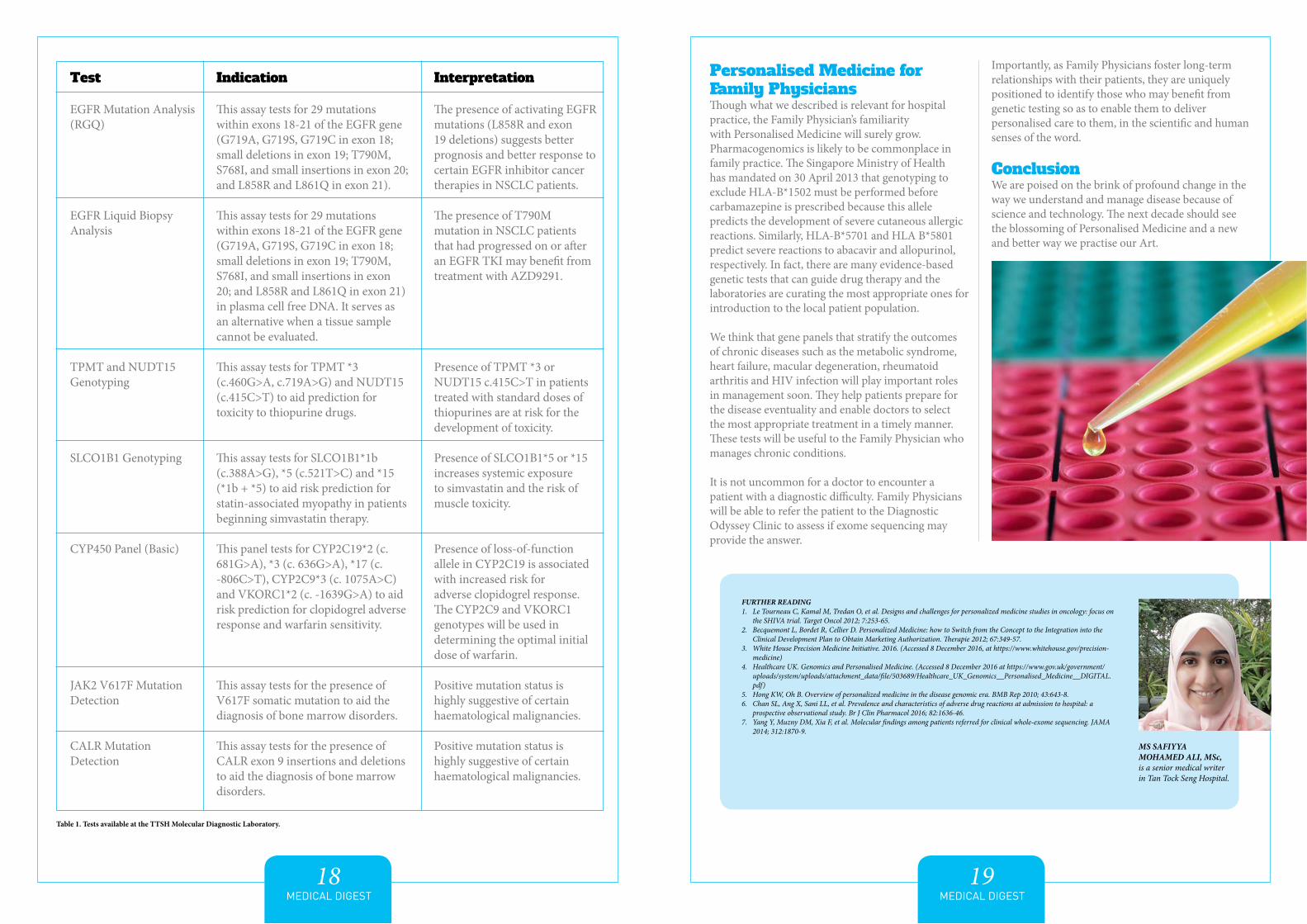
Some of us may also have encountered patients who have not received a diagnosis despite a long series of investigations and referrals. These patients may have early-onset disease or a syndromic disease with an unknown aetiology. Whole-exome sequencing (using Next Generation Sequencing or NGS) enables the diagnosis to be made in 25 to 30% of these patients.7 An ideal workflow of how Personalised Medicine can be utilised in the clinic is illustrated in figure 1.We acknowledge that Personalised Medicine at present is not a panacea. There are unresolved issues such as the limited amount of currently interpretable genetic contributions, increased costs, ethical issues like confidentiality, return of incidental findings and access, and clinicians’ lack of knowledge in utilising the results of the genetic tests. Nevertheless, we need to have a starting point and understand and use Personalised Medicine as it continues to develop.Personalised Medicine makes tailor-made medicine a reality for mankind. We believe that the impact of Personalised Medicine will be felt in the near future
as more is known about the genetic contributions to disease phenotype and response to treatment.
Personalised Medicine Services in TTSHTTSH is committed to develop Personalised Medicine to serve her patients. In October 2016, the TTSH Molecular Diagnostic Laboratory (MDL) launched a series of tests available to clinicians hospital-wide (table 1). Our services currently focus on two areas – pharmacogenetics and clinical genomics. In future, we intend to incorporate a ‘diagnostic odyssey’ component whereby patients with a long history of uncharacterised disease are able to receive a diagnosis with the help of genetic sequencing.
Physicians in TTSH and JHS are able to order genomic tests on blood, body fluids and tissues that are vital for disease diagnosis, disease monitoring/progression and drug response/efficacy. The laboratory participates in proficiency testing surveys administered by the College of American Pathologists (CAP).
What is Personalised Medicine?
ersonalised Medicine steps away from a “one-size-fits-all” management of patients. One definition of Personalised Medicine is “a form of medicine that uses
information about a person’s genes, proteins, and environment to prevent, diagnose, and treat disease”.1 The three aims of Personalised Medicine are: “to refine diagnosis by identifying early diagnostic markers and subpopulations of patients with different
natural evolution and prognosis; to rationalize treatment by switching from a mass concept where treatments are indistinctly applied to all patients to an individualized concept where treatments are determined on a case-by-case basis to optimize the benefit/risk ratio; to engage patients in a preventive approach
by increasing patient adherence and compliance while adapting prevention programs to patient profiles”.2
Many individuals and organisations throughout the world recognise that Personalised Medicine is the way forward in Medicine. In the US, President Obama announced the Precision Medicine Initiative in 2015 which aims to “enable a new era of medicine through research, technology, and policies that empower patients, researchers, and providers to work together toward development of individualized care”.3 Part of this budget has been allocated to USA’s National Institutes of Health to build a national, large-scale cohort of 1 million persons. In the UK, the 100,000 Genomes Project aims to sequence 100,000 genomes of National Healthcare System (NHS) patients,
with the long-term plan to integrate Personalised Medicine into day-to-day delivery of healthcare.4 With technological and scientific advances already available in Singapore, we must consider Personalised Medicine seriously so that we can respond and adapt to the needs of our population in line with modern capability.
How can Personalised Medicine be implemented in Healthcare?The concept of Personalised Medicine is not new. Physicians have long noticed differences between individuals in terms of disease manifestation and response to therapy. Today, we are able to predict how different individuals respond to specific interventions or identify people who are at risk of developing certain illnesses. Technologies such as whole genome sequencing, biobanking, bioinformatics, and wearable devices have made these possibilities attainable. The application of Personalised Medicine ranges from education of healthcare providers (doctors, nurses, pharmacists, therapists and others), health risk assessment utilising laboratory and epidemiological information, together with computerised clinician decision support, to patient education by Genetic Counsellors.5
One of the major benefits of Personalised Medicine in the clinical setting is the potential to reduce adverse drug reactions and maximise drug efficacy based on the genetic profile of individual patients. Based on the reported adverse drug reactions collated by the HSA in 2014, there were 316 and 60 medication-related incidents in TTSH and IMH, respectively. These numbers indicate that hospitals face a large number of medication-related clinical problems. A recent local study found that at least 30% of hospital admissions due to adverse drug reactions were caused by at least one drug with a clinical annotation in the Pharmacogenomics KnowledgeBase (PharmGKB).6 This implies that pharmacogenetic testing may have predicted, and thus potentially prevent, some of these adverse drug reactions. In TTSH, we also
The Case for Personalised Medicine
Figure 1. Components of a Personalised Medicine Service which are built around patients in the clinics and wards
MEDICAL DIGEST18
MEDICAL DIGEST19
Personalised Medicine for Family PhysiciansThough what we described is relevant for hospital practice, the Family Physician’s familiarity with Personalised Medicine will surely grow. Pharmacogenomics is likely to be commonplace in family practice. The Singapore Ministry of Health has mandated on 30 April 2013 that genotyping to exclude HLA-B*1502 must be performed before carbamazepine is prescribed because this allele predicts the development of severe cutaneous allergic reactions. Similarly, HLA-B*5701 and HLA B*5801 predict severe reactions to abacavir and allopurinol, respectively. In fact, there are many evidence-based genetic tests that can guide drug therapy and the laboratories are curating the most appropriate ones for introduction to the local patient population.
We think that gene panels that stratify the outcomes of chronic diseases such as the metabolic syndrome, heart failure, macular degeneration, rheumatoid arthritis and HIV infection will play important roles in management soon. They help patients prepare for the disease eventuality and enable doctors to select the most appropriate treatment in a timely manner. These tests will be useful to the Family Physician who manages chronic conditions.
It is not uncommon for a doctor to encounter a patient with a diagnostic difficulty. Family Physicians will be able to refer the patient to the Diagnostic Odyssey Clinic to assess if exome sequencing may provide the answer.
Importantly, as Family Physicians foster long-term relationships with their patients, they are uniquely positioned to identify those who may benefit from genetic testing so as to enable them to deliver personalised care to them, in the scientific and human senses of the word.
ConclusionWe are poised on the brink of profound change in the way we understand and manage disease because of science and technology. The next decade should see the blossoming of Personalised Medicine and a new and better way we practise our Art.
FURTHER READING 1. Le Tourneau C, Kamal M, Tredan O, et al. Designs and challenges for personalized medicine studies in oncology: focus on
the SHIVA trial. Target Oncol 2012; 7:253-65.2. Becquemont L, Bordet R, Cellier D. Personalized Medicine: how to Switch from the Concept to the Integration into the
Clinical Development Plan to Obtain Marketing Authorization. Therapie 2012; 67:349-57.3. White House Precision Medicine Initiative. 2016. (Accessed 8 December 2016, at https://www.whitehouse.gov/precision-
medicine)4. Healthcare UK. Genomics and Personalised Medicine. (Accessed 8 December 2016 at https://www.gov.uk/government/
uploads/system/uploads/attachment_data/file/503689/Healthcare_UK_Genomics__Personalised_Medicine__DIGITAL.pdf)
5. Hong KW, Oh B. Overview of personalized medicine in the disease genomic era. BMB Rep 2010; 43:643-8.6. Chan SL, Ang X, Sani LL, et al. Prevalence and characteristics of adverse drug reactions at admission to hospital: a
prospective observational study. Br J Clin Pharmacol 2016; 82:1636-46.7. Yang Y, Muzny DM, Xia F, et al. Molecular findings among patients referred for clinical whole-exome sequencing. JAMA
2014; 312:1870-9.
MS SAFIYYA MOHAMED ALI, MSc, is a senior medical writer in Tan Tock Seng Hospital.
Test
EGFR Mutation Analysis (RGQ)
EGFR Liquid Biopsy Analysis
TPMT and NUDT15 Genotyping
SLCO1B1 Genotyping
CYP450 Panel (Basic)
JAK2 V617F Mutation Detection
CALR Mutation Detection
Indication
This assay tests for 29 mutations within exons 18-21 of the EGFR gene (G719A, G719S, G719C in exon 18; small deletions in exon 19; T790M, S768I, and small insertions in exon 20; and L858R and L861Q in exon 21).
This assay tests for 29 mutations within exons 18-21 of the EGFR gene (G719A, G719S, G719C in exon 18; small deletions in exon 19; T790M, S768I, and small insertions in exon 20; and L858R and L861Q in exon 21) in plasma cell free DNA. It serves as an alternative when a tissue sample cannot be evaluated.
This assay tests for TPMT *3 (c.460G>A, c.719A>G) and NUDT15 (c.415C>T) to aid prediction for toxicity to thiopurine drugs.
This assay tests for SLCO1B1*1b (c.388A>G), *5 (c.521T>C) and *15 (*1b + *5) to aid risk prediction for statin-associated myopathy in patients beginning simvastatin therapy.
This panel tests for CYP2C19*2 (c. 681G>A), *3 (c. 636G>A), *17 (c. -806C>T), CYP2C9*3 (c. 1075A>C) and VKORC1*2 (c. -1639G>A) to aid risk prediction for clopidogrel adverse response and warfarin sensitivity.
This assay tests for the presence of V617F somatic mutation to aid the diagnosis of bone marrow disorders.
This assay tests for the presence of CALR exon 9 insertions and deletions to aid the diagnosis of bone marrow disorders.
Interpretation
The presence of activating EGFR mutations (L858R and exon 19 deletions) suggests better prognosis and better response to certain EGFR inhibitor cancer therapies in NSCLC patients.
The presence of T790M mutation in NSCLC patients that had progressed on or after an EGFR TKI may benefit from treatment with AZD9291.
Presence of TPMT *3 or NUDT15 c.415C>T in patients treated with standard doses of thiopurines are at risk for the development of toxicity.
Presence of SLCO1B1*5 or *15 increases systemic exposure to simvastatin and the risk of muscle toxicity.
Presence of loss-of-function allele in CYP2C19 is associated with increased risk for adverse clopidogrel response. The CYP2C9 and VKORC1 genotypes will be used in determining the optimal initial dose of warfarin.
Positive mutation status is highly suggestive of certain haematological malignancies.
Positive mutation status is highly suggestive of certain haematological malignancies.
Table 1. Tests available at the TTSH Molecular Diagnostic Laboratory.
MEDICAL DIGEST20
he Post Acute Care at Home (PACH) service was first introduced in Tan Tock Seng Hospital (TTSH) in 2008. It provides time-limited multidisciplinary care, which
includes medical and nursing assessment, caregiver training and support, therapy and laboratory services. It caters to patients residing within the Central region, discharged from TTSH. PACH is part of the Continuing and Community Care Department (CCC) in TTSH. The various programmes run by the department are detailed below.
Continuing and Community Care DepartmentThe Community Health Engagement ProgrammeThe Community Health Engagement Programme (CHEP), established under TTSH’s Continuing and Community Care Department in 2009, is dedicated towards empowering seniors to adopt an active and fulfilling lifestyle. All programmes, managed by TTSH’s team of healthcare staff (including doctors, nurses, physiotherapists and occupational therapists), are suited for seniors from all walks of life, and they
are specially designed to be safe, simple and fun. As CHEP is committed to improving the quality of lives of seniors, the team takes these programmes to the community.
Programmes under CHEP:• Stepping Out Into Active Life • Chronic Disease Self-Management Programme
(CDSMP) • Engage In Life (EIL)
Transitional Care ProgrammeThe Translational Care (TC) programme (formerly known as the Virtual Hospital (VH) and the Aged Care Transition (ACTION) programmes) was started in July 2016 to ensure that every patient with complex care needs has a single point of contact to coordinate his care plan and support a safe, coordinated and timely transition from the hospital to the community and home. Patients enrolled into the TC programme usually suffer from multiple chronic conditions, have limited social support, and require close monitoring for some time to help them cope with and in turn, actively manage their illnesses at home. Patients may also be frequent admitters who seek care at the hospital’s Emergency Department for problems that can potentially be managed at home.
Enrolled patients are assigned a TC Specialist who will be their single point of contact. The TC Specialists, supported by a team of clinicians, nurses, allied health professionals and administrators, provide in-hospital care coordination and follow-up with patients post-discharge through telephonic reviews and home visits.
Together with the NHG Neighbours Care Coordinators, the TC team also collaborates with a network of community and primary care partners (e.g. GPs, Polyclinics and Family Medicine Centres) to co-manage patients where suitable and assist patients to get help in navigating the various services in the community.
Post Acute Care at Home PACH is a post-discharge supportive care service,
MEDICAL DIGEST21
Transitional Care – What happens during and after PACH?
formed by a team of doctors, nurses, therapists and medical social workers. The PACH team aims to stabilise and rehabilitate patients with the sub-acute phase of the illness at home, as well as to provide appropriate home care support to promote better self care. PACH also aims to help the caregivers to be competent in managing the homebound patient at home so as to reduce the need for institutionalised care.
The doctor and nurse will assess referred patients at home in terms of medical, psychological, social and rehabilitation needs. A multidisciplinary plan is drawn up, and the management plan will be coordinated by the nurses and be agreed upon by the team during a multidisciplinary meeting.
The targeted period of care is for about three months. Following that, provision of treatment of ongoing medical conditions and treatment of any functional impairment may be in collaboration with community healthcare service providers.
Project CAREPalliative care has become increasingly vital as the number of frail elderly in Singapore grows. Nursing homes face the challenges of stretched manpower and limited funds, resulting in limited End-of-Life (EOL) discussions and limited medical care support for sick residents. To help improve the quality of care provided to nursing home residents and allow them the dignity of spending their final days in their preferred environment and receiving their preferred treatment, TTSH introduced Project CARE in 2009.
The scheme allows a multi-disciplinary care team from the hospital to train and coach nursing home staff in performing Advanced Care Planning and providing EOL care. Nursing home staff are also able to contact the team should they need assistance in managing the symptoms of EOL residents.
Participating nursing homes:• Ren Ci Nursing Home (Moulmein)• Ling Kwang Home• Saint Theresa’s Home• All Saints Home (Hougang)• Society for the Aged Sick• Lions Home for the Elders (Bishan)
Enrolling in the PACH programmeUpon receiving referral from the ward, the PACH coordinator will go to the ward to triage the case. Majority of the referred patients have complex medical/nursing needs and are usually referred for the following reasons: chair/bed bound patients with multiple co-morbidities, complex wound dressing needs, dementia with behavioural and psychological symptoms of dementia (BPSD), patients with newly inserted nasogastric tubes (NGTs) or catheters and have new caregivers who need some form of support after patients are discharged from the hospital.
Per Capita Monthly Household Income$0 to $700
$701 to $1,100$1,101 to $1,600$1,601 to $1,800$1,801 to $2,600$2,601 and above
Table 1: Subsidies for home and community-based services (non-residential services)Source: https://www.moh.gov.sg/content/moh_web/home/costs_and_financing/schemes_subsidies/subsidies_for_government_funded_ILTC_services.html
Subsidy Rate Singapore Citizens Permanent Residents
80% 55%75% 50%60% 40%50% 30%30% 15%0% 0%
Centre-based
Day Rehab
Dementia Day Care
Day Care
SPICE
Centre-Based Nursing
Transitional Care Facility
Centre-based Weekend Service
Home Health Care
Home Medical
Home Nursing
Home Therapy
Home Personal Care
Meals on Wheels
Medical escort and transport
Personal hygiene care support
Eldersitter service (dementia)
Caregiver Support
Alzheimer’s Disease Association Caregiver Support Service
TOUCH Caregivers Support
AWWA Caregiver Service
Caregivers Alliance Limited
Caregiving Welfare Association
Financial Assistance
Social Service Office
Financing schemes (subsidy):a) Senior Mobility
and Enabling Fund
b) EASE Financing schemes
(insurance/payouts):
a) Silver Support Scheme
b) IDAPEc) ElderShieldd) FDW Grante) Caregiver
Training Grant
Case Management/
Others
Family Service Centres
Cluster Support
Community Intervention Team (“COMIT”)
Community Resource, Engagement and Support Team (“CREST”).
Community Befriending:- Befrienders- CAN- NHG
Neighbours
- Peacehaven Nursing Home
- Pearl’s Hill Care Home
- NTUC Silver Circle (Serangoon Central)
- SWAMI Home- Peacehaven
SPICE@Changi
End of Life Palliative
Care
Hospice(Inpatient/ Outpatient)
AIC-HOME (Palliative home care for end-stage organ failure)
MEDICAL DIGEST22
MEDICAL DIGEST23
The coordinator will also contact the next-of-kin to explain household means testing and charges for PACH services and the duration of care. The PACH programme follows the ILTC (intermediate and long term care) services household means test for non-residential services (table 1).
PACH home visit(s) and managementDuring the first home visit, the visiting doctor and nurse will conduct a comprehensive assessment
of the patient’s health and care needs as well as the competency of the caregiver. The subsequent frequency and intensity of follow-ups will be based on the initial assessment. Otherwise, a periodic phone review will be conducted. If needed, simple procedures such as blood tests, trial off catheters, and bladder scans may also be conducted.
Whenever needed, allied health team members such as an occupational therapist, physiotherapist, speech therapist, dietician or medical social worker will be
activated. The multidisciplinary team meets on a weekly basis to present and discuss cases.
Example 1: Prior to admission, Mr Tan has been bedbound for about one year. He was admitted for a bout of chest infection and while he was warded, he had a newly inserted NGT for feeding due to his poor oral intake. He was referred to PACH for sacral wound management and support for his caregiver who did not feel confident with the NGT regime though she was able to administer the steps correctly.
While he was at home, Mr Tan began to have bouts of phlegm issue. A physiotherapist was activated and assisted the family to purchase a suction machine using the Senior Mobility and Enabling Fund and also taught them how to use the equipment. Mr Tan’s condition stabilised after two months and the team then activated the ‘Home Medical’ and ‘Home Nursing’ services via the Agency for Integrated Care (AIC) to take over the care. These services were identified because Mr Tan’s family found it too challenging to transfer him for clinic appointments
AICare Link @ Maxwell 7 Maxwell Road #04-01MND Complex Annex BSingapore 069111Above Amoy Street Food Centre
Operating Hours:Mondays to Fridays8.30 am to 5.30 pmClosed on weekends and public holidays
AICare Link @ Changi General Hospital (CGH) 2 Simei Street 3Singapore 529889Discharge Lounge at Main Building, Level 1, Atrium (next to Pharmacy B)
Operating Hours:Mondays to Fridays9.30 am to 6.00 pmSaturdays9.00 am to 1.00 pmClosed on Sundays and public holidays
AICare Link @ Khoo Teck Puat Hospital (KTPH) 90 Yishun CentralSingapore 768828Patient Service Centre, Tower B, Level 1
Operating Hours:Mondays to Fridays9.30 am to 6.00 pmSaturdays8.30 am to 12.30 pmClosed on Sundays and public holidays
AICare Link @ National University Hospital (NUH) 5 Lower Kent Ridge RoadSingapore 119074Main Building Lobby B, Level 1 (opposite The Coffee Bean and Tea Leaf Cafe)
Operating Hours:Mondays to Fridays9.30 am to 6.00 pmSaturdays9.00 am to 1.00 pmClosed on Sundays and public holidays
AICare Link @ Ng Teng Fong General Hospital (NTFGH) 1 Jurong East Street 21Singapore 609606Tower B, Level 2 (near Visitor Self-registration Kiosk)
Operating Hours:Mondays to Fridays9.30 am to 6.00 pmSaturdays9.00 am to 1.00 pmClosed on Sundays and public holidays
AICare Link @ Tan Tock Seng Hospital (TTSH)11 Jalan Tan Tock SengSingapore 308433CareConnect, Level 1, Atrium
Operating Hours: Mondays to Fridays, 9.30 am to 6.00 pm Saturdays, 9.00 am to 1.00 pm Closed on Sundays and public holidays
Table 2. Agency for Integrated Care services for post-PACH patients
Table 3. Common community services for post-PACH patients
A NEW CLASS OF HEART FAILURE MEDICATION
PHARMACY
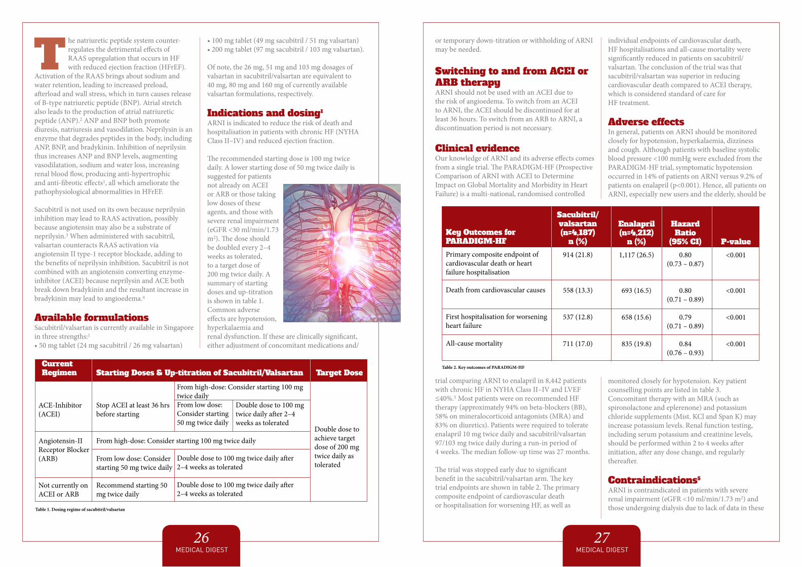
Sacubitril/valsartan (Entresto®), an angiotensin-neprilysin inhibitor (ARNI), is a recently approved agent for heart failure (HF), and the first to demonstrate mortality benefits versus standard therapy in many years.1 An ARNI combines two drugs, sacubitril, a neprilysin inhibitor, and valsartan, an angiotensin II receptor blocker (ARB) so that its dual action targets the natriuretic peptide and the renin-angiotensin-aldosterone systems (RAAS).
MEDICAL DIGEST25
MEDICAL DIGEST24
MS KAREN POH is a senior medical social worker at Tan Tock Seng Hospital and MS TNG TINA VERONIQUE is a nurse clinician at Tan Tock Seng Hospital.
and because his NGT needed to be changed regularly. Thus, they preferred to have a doctor review Mr Tan at home periodically. With the services in place, PACH exited.
Example 2: Mr Samy has dementia with BPSD. He can walk for a short distance at home but requires a wheelchair in the community. He was admitted after a fall from his bed. Pre-morbidly, his wife had been his primary caregiver, and during his admission, his family employed a foreign domestic worker as his full-time caregiver. PACH was activated to monitor his behaviours, titrate his medications and provide support to his family and new caregiver. The family was advised to join the support groups organised by the Alzheimer’s Disease Association of Singapore. Therapists were activated for home environment assessment and to provide home modification recommendations. The family was guided to apply for the HDB EASE programme to install grab bars around the house. When Mr Samy’s situation had stabilised, he was discharged from PACH to continue with his follow-ups with his geriatrician at the specialist outpatient clinic.
What’s after PACH? At an appropriate juncture, the team may explore and refer community services to come onboard in preparing for PACH to exit. For patients and caregivers in the community, there are several touch points that they can approach to discuss services and programmes available. The most accessible would be the AICare Links located in restructured hospitals where patients and/or caregivers can walk in to enquire with AICare Consultants (table 2). The landscape of community services is evolving. Table 3 presents some of the common community services for PACH patients post PACH.
ConclusionPACH is a service bridging the transit of patients with complex care from an acute hospital back home, providing support for patients and caregivers to sustain care of patients in the community and linking them with appropriate step-down community services thereafter.
The PACH team aims to stabilise and rehabilitate patients with the sub-acute phase of the illness at home, as well as to provide appropriate home care support to promote better self care.
““
MEDICAL DIGEST26
MEDICAL DIGEST27
he natriuretic peptide system counter-regulates the detrimental effects of RAAS upregulation that occurs in HF with reduced ejection fraction (HFrEF).
Activation of the RAAS brings about sodium and water retention, leading to increased preload, afterload and wall stress, which in turn causes release of B-type natriuretic peptide (BNP). Atrial stretch also leads to the production of atrial natriuretic peptide (ANP).2 ANP and BNP both promote diuresis, natriuresis and vasodilation. Neprilysin is an enzyme that degrades peptides in the body, including ANP, BNP, and bradykinin. Inhibition of neprilysin thus increases ANP and BNP levels, augmenting vasodilatation, sodium and water loss, increasing renal blood flow, producing anti-hypertrophic and anti-fibrotic effects1, all which ameliorate the pathophysiological abnormalities in HFrEF.
Sacubitril is not used on its own because neprilysin inhibition may lead to RAAS activation, possibly because angiotensin may also be a substrate of neprilysin.3 When administered with sacubitril, valsartan counteracts RAAS activation via angiotensin II type-1 receptor blockade, adding to the benefits of neprilysin inhibition. Sacubitril is not combined with an angiotensin converting enzyme-inhibitor (ACEI) because neprilysin and ACE both break down bradykinin and the resultant increase in bradykinin may lead to angioedema.4
Available formulationsSacubitril/valsartan is currently available in Singapore in three strengths:1 • 50 mg tablet (24 mg sacubitril / 26 mg valsartan)
• 100 mg tablet (49 mg sacubitril / 51 mg valsartan)• 200 mg tablet (97 mg sacubitril / 103 mg valsartan).
Of note, the 26 mg, 51 mg and 103 mg dosages of valsartan in sacubitril/valsartan are equivalent to 40 mg, 80 mg and 160 mg of currently available valsartan formulations, respectively.
Indications and dosing1
ARNI is indicated to reduce the risk of death and hospitalisation in patients with chronic HF (NYHA Class II–IV) and reduced ejection fraction.
The recommended starting dose is 100 mg twice daily. A lower starting dose of 50 mg twice daily is suggested for patients not already on ACEI or ARB or those taking low doses of these agents, and those with severe renal impairment (eGFR <30 ml/min/1.73 m2). The dose should be doubled every 2–4 weeks as tolerated, to a target dose of 200 mg twice daily. A summary of starting doses and up-titration is shown in table 1. Common adverse effects are hypotension, hyperkalaemia and renal dysfunction. If these are clinically significant, either adjustment of concomitant medications and/
or temporary down-titration or withholding of ARNI may be needed.
Switching to and from ACEI or ARB therapy ARNI should not be used with an ACEI due to the risk of angioedema. To switch from an ACEI to ARNI, the ACEI should be discontinued for at least 36 hours. To switch from an ARB to ARNI, a discontinuation period is not necessary.
Clinical evidenceOur knowledge of ARNI and its adverse effects comes from a single trial. The PARADIGM-HF (Prospective Comparison of ARNI with ACEI to Determine Impact on Global Mortality and Morbidity in Heart Failure) is a multi-national, randomised controlled
trial comparing ARNI to enalapril in 8,442 patients with chronic HF in NYHA Class II–IV and LVEF ≤40%.5 Most patients were on recommended HF therapy (approximately 94% on beta-blockers (BB), 58% on mineralocorticoid antagonists (MRA) and 83% on diuretics). Patients were required to tolerate enalapril 10 mg twice daily and sacubitril/valsartan 97/103 mg twice daily during a run-in period of 4 weeks. The median follow-up time was 27 months.
The trial was stopped early due to significant benefit in the sacubitril/valsartan arm. The key trial endpoints are shown in table 2. The primary composite endpoint of cardiovascular death or hospitalisation for worsening HF, as well as
individual endpoints of cardiovascular death, HF hospitalisations and all-cause mortality were significantly reduced in patients on sacubitril/valsartan. The conclusion of the trial was that sacubitril/valsartan was superior in reducing cardiovascular death compared to ACEI therapy, which is considered standard of care for HF treatment.
Adverse effects In general, patients on ARNI should be monitored closely for hypotension, hyperkalaemia, dizziness and cough. Although patients with baseline systolic blood pressure <100 mmHg were excluded from the PARADIGM-HF trial, symptomatic hypotension occurred in 14% of patients on ARNI versus 9.2% of patients on enalapril (p<0.001). Hence, all patients on ARNI, especially new users and the elderly, should be
monitored closely for hypotension. Key patient counselling points are listed in table 3.Concomitant therapy with an MRA (such as spironolactone and eplerenone) and potassium chloride supplements (Mist. KCl and Span K) may increase potassium levels. Renal function testing, including serum potassium and creatinine levels, should be performed within 2 to 4 weeks after initiation, after any dose change, and regularly thereafter.
Contraindications5
ARNI is contraindicated in patients with severe renal impairment (eGFR <10 ml/min/1.73 m2) and those undergoing dialysis due to lack of data in these Table 1. Dosing regime of sacubitril/valsartan
starting dose of 50 mg twice daily in patients who are naïve to ACEI or ARBs, PARADIGM-HF did not recruit any patients with newly diagnosed HFrEF or those naïve to RAAS blockade therapy. The utility of starting ARNI in these patients is hence unknown. Whether ACEI/ARB-naïve patients should be first started on an ACEI/ARB and later converted to ARNI or directly started on a lower dose of ARNI has also not been evaluated and should thus be based on clinician judgement and experience.
Patients should also be put on optimal dosages of other evidence-based drugs (BB, MRA and others) and devices (cardiac resynchronisation therapy or implantable cardioverter defibrillator) as appropriate. Further post-marketing surveillance and further research is essential to improve the quality of evidence supporting its use. A trial of ARNI versus valsartan (PARAGON-HF) in patients with HF with preserved ejection fraction is underway (NCT01920711).
ACEIs and ARBs are the current standard of care and widely available as generic, standard drugs made affordable by government subsidy. As with all new therapies, the cost versus benefit of ARNI should be discussed with the patient prior to starting.
populations. Patients with a history of idiopathic or hereditary angioedema or previous angioedema related to ACEI or ARBs should also not be given ARNI. ARNI is contraindicated in pregnancy; if a patient becomes pregnant whilst on ARNI, the drug should be discontinued as soon as possible. As animal studies show ARNI is present in breast milk, its use is not recommended during breastfeeding. ARNI must not be used concomitantly with aliskiren-containing products in patients with diabetes mellitus or renal impairment (eGFR <60 ml/min/1.73 m2). ARNI is not recommended in severe hepatic impairment.
Current guidelines Both the US and European Cardiology Societies recently updated their guidelines to include the role of ARNI in HF treatment.
The ACC/AHA/HFSA 2016 Focused Update of the 2013 HF Guideline provided a class I (strong) recommendation with B-R (randomised) level of evidence, for replacement of ARNI in chronic symptomatic HFrEF Class II or III patients who tolerate an ACEI or ARB, to further reduce morbidity and mortality.4
The European Society of Cardiology (ESC) Guidelines 2016 provides a class I, level of evidence B recommendation for ARNI as a replacement for ACEI to reduce the risk of HF hospitalisation and death in ambulatory patients with HFrEF who remain symptomatic despite optimal treatment with an ACEI, a BB and an MRA.6
Comments and conclusion Sacubitril/valsartan is a novel compound that has recently been approved for use in HFrEF due to its beneficial effect on morbidity and mortality compared with standard of care.
Although major society guidelines have incorporated it into their 2016 recommendations, it should be noted that this is based on a single trial, PARADIGM-HF. In addition, although local labelling suggests a
Table 3. Patient counselling for ARNI
General • Therapy with sacubitril/valsartan can improve symptoms, prevent disease progression or worsening of heart failure, reduce unplanned hospitalisation and increase survival.
• Symptom improvement may take place after weeks to months of initiation. • Even if the patient feels well, therapy should not be discontinued otherwise symptoms of heart failure may recur. • Consult a doctor or healthcare provider if any adverse effect becomes intolerable.
Hypotension • Light-headedness, giddiness or postural hypotension may occur especially during first few days of initiation or dosage increment.
• To minimise giddiness, rise slowly from a sitting, squatting or lying position. • Consult a doctor or healthcare provider if there is severe giddiness or tiredness.
Hyperkalaemia • Blood tests to check kidney function and potassium levels will be done regularly.and renal • If needed, the dose of ARNI may be reduced or stopped temporarily. dysfunction • Avoid using salt substitutes in cooking as these may lead to high potassium levels. • Avoid taking non-steroid anti-inflammatory drugs (NSAIDs) as painkillers as this may worsen kidney function.
Angioedema • Rarely, adverse reactions (angioedema) may occur. Get emergency medical care as soon as possible if swelling of the face, lips, tongue and throat occur after taking ARNI.
• If a reaction as described above has occurred while taking ARNI, inform your doctor and do not take it again.
COUNSELLING POINTS
MEDICAL DIGEST28
MEDICAL DIGEST29
REFERENCES1. Product Information for Entresto. Novartis Pharma AG, Basel, Switzerland. August 2015. SIN. .2. Jhund PS, McMurray JJ. The neprilysin pathway in heart failure: a review and guide on the use of sacubitril/valsartan. Heart. 2016;102(17):1342-7.3. Packer M, McMurray JJ, Desai AS, Gong J, Lefkowitz MP, Rizkala AR, et al. Angiotensin receptor neprilysin inhibition compared with enalapril on the risk
of clinical progression in surviving patients with heart failure. Circulation. 2015;131(1):54-61.4. Yancy CW, Jessup M, Bozkurt B, Butler J, Casey DE, Jr., Colvin MM, et al. 2016 ACC/AHA/HFSA Focused Update on New Pharmacological Therapy
for Heart Failure: An Update of the 2013 ACCF/AHA Guideline for the Management of Heart Failure: A Report of the American College of Cardiology/American Heart Association Task Force on Clinical Practice Guidelines and the Heart Failure Society of America. J Am Coll Cardiol. 2016;68(13):1476-88.
5. McMurray JJ, Packer M, Desai AS, Gong J, Lefkowitz MP, Rizkala AR, et al. Angiotensin-neprilysin inhibition versus enalapril in heart failure. N Engl J Med. 2014;371(11):993-1004.
6. Ponikowski P, Voors AA, Anker SD, Bueno H, Cleland JG, Coats AJ, et al. 2016 ESC Guidelines for the diagnosis and treatment of acute and chronic heart failure: The Task Force for the diagnosis and treatment of acute and chronic heart failure of the European Society of Cardiology (ESC). Developed with the special contribution of the Heart Failure Association (HFA) of the ESC. Eur J Heart Fail. 2016;18(8):891-975.
MS GRACE CHANG is a pharmacy resident (cardiology) and MS WONG YEE MAY is a principal pharmacist (clinical) in the Department of Pharmacy, Tan Tock Seng Hospital.
In general, patients on ARNI should be monitored closely for hypotension, hyperkalaemia, dizziness and cough.
““
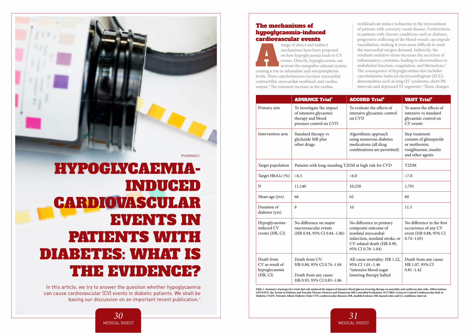
HYPOGLYCAEMIA-INDUCED
CARDIOVASCULAR EVENTS IN
PATIENTS WITH DIABETES: WHAT IS
THE EVIDENCE?In this article, we try to answer the question whether hypoglycaemia
can cause cardiovascular (CV) events in diabetic patients. We shall be basing our discussion on an important recent publication.1
PHARMACY
MEDICAL DIGEST30
MEDICAL DIGEST31
The mechanisms of hypoglycaemia-induced cardiovascular events
range of direct and indirect mechanisms have been proposed on how hypoglycaemia leads to CV events. Directly, hypoglycaemia can activate the sympatho-adrenal system,
causing a rise in adrenaline and norepinephrine levels. These catecholamines increase myocardial contractility, myocardial workload, and cardiac output.2 The transient increase in the cardiac
workload can induce ischaemia in the myocardium of patients with coronary vessel disease. Furthermore, in patients with chronic conditions such as diabetes, progressive stiffening of the blood vessels can impede vasodilation, making it even more difficult to meet the myocardial oxygen demand. Indirectly, the resultant oxidative stress increases the secretion of inflammatory cytokines, leading to abnormalities in endothelial function, coagulation, and fibrinolysis.3The consequence of hypoglycaemia also includes catecholamine-induced electrocardiogram (ECG) abnormalities such as long QT syndrome, short PR intervals and depressed ST segments.4 These changes
Primary aim
Intervention arm
Target population
Target HbA1c (%)
N
Mean age (yrs)
Duration of diabetes (yrs)
Hypoglycaemia- induced CV events (HR, CI)
Death from CV as result of hypoglycaemia (HR, CI)
ADVANCE Trial7
To investigate the impact of intensive glycaemic therapy and blood pressure control on CVD
Standard therapy vs gliclazide MR plus other drugs
Patients with long-standing T2DM at high risk for CVD
<6.5
11,140
66
8
No difference on major macrovascular events (HR 0.94, 95% CI 0.84–1.06)
Death from CV: HR 0.88, 95% CI 0.74–1.04
Death from any cause: HR 0.93, 95% CI 0.83–1.06
ACCORD Trial8
To evaluate the effects of intensive glycaemic control on CVD
Algorithmic approach using numerous diabetes medications (all drug combinations are permitted)
<6.0
10,250
62
10
No difference in primary composite outcome of nonfatal myocardial infarction, nonfatal stroke, or CV-related death (HR 0.90, 95% CI 0.78–1.04)
All-cause mortality: HR 1.22, 95% CI 1.01–1.46*intensive blood sugar lowering therapy halted
VADT Trial9
To assess the effects of intensive vs standard glycaemic control on CV events
Step treatment consists of glimepiride or metformin, rosiglitazone, insulin and other agents
T2DM
<7.0
1,791
60
11.5
No difference in the first occurrence of any CV event (HR 0.88, 95% CI 0.74–1.05)
Death from any cause: HR 1.07, 95% CI 0.81–1.42
Table 1. Summary of prospective trials that sub-analysed the impact of intensive blood glucose lowering therapy on mortality and cardiovascular risks. Abbreviations: ADVANCE, the Action in Diabetes and Vascular Disease: Preterax and Diamicron MR Controlled Evaluation; ACCORD, Action to Control Cardiovascular Risk in Diabetes; VADT, Veteran’s Affairs Diabetes Trial; CVD, cardiovascular diseases; MR, modified release; HR, hazard ratio; and CI, confidence interval.
in ECG can lead to ventricular tachycardia and sudden death.
What prompted the study?Despite the postulated mechanisms of hypoglycaemia-induced CV events, the association between hypoglycaemia and the CV system has not been well-established. In canine studies, depressed creatine phosphokinase activity and histological evidence of early myocardial necrosis were observed following an intentionally induced hypoglycaemia episode.5 In a human case study, a silent myocardial infarction as a result of coronary artery vasospasm was detected on ECG following hypoglycaemic coma secondary to acute insulin poisoning.6 Furthermore, a meta-analysis of six cohort studies (N=903,510) reported a two-fold increase in the risk of CV events among patients with type 2 diabetes (T2DM) who experienced severe hypoglycaemia (RR 2.05 [95% CI 1.74, 2.42]).7
However, conflicting results were found in larger prospective trials that sub-analysed the impact of intensive blood glucose lowering therapy on mortality and CV risks (table 1).8-10 While the ADVANCE Trial7 and the VADT Trial9 showed no differences in CV events among patients with T2DM, a 22% increase was observed in the ACCORD Trial which required a premature halting of the study arm that received intensive blood glucose lowering therapy.10
Description of the current studyA cohort study to determine the association between hypoglycaemia and CV-related events and
mortality in patients with type 1 diabetes (T1DM) and T2DM treated with insulin was conducted in 2015.1 This study was first in the literature to address the association of hypoglycaemia and CV events as a primary objective. In addition, the study also aimed to quantify CV risks separately for those with and without pre-existing CVD, and investigated if different oral anti-diabetic agents have differing effects on CV events.
The cohort study used the Clinical Practice Research Datalink database in the United Kingdom.10 All insulin-treated patients >30 years of age diagnosed between 2001 and 2007 were included. Patients with ≥1 episode of recorded hypoglycaemia before the initiation of insulin were excluded from the analysis. Patients with T1DM or T2DM were considered separately (table 2).
The numbers of CV events preceded by a hypoglycaemic episode were counted. The time-to-event period was calculated from the time of the first episode of hypoglycaemia. Stratified univariate and multivariate Cox regression models were used to estimate the risk of CV events and the mortality associated with hypoglycaemia.
The median duration of follow-up was 5.0 years and 4.8 years for T1DM and T2DM patients, respectively. The proportion of T1DM and T2DM patients who experienced hypoglycaemia was 18% (573 patients) and 14% (1463 patients) respectively. The median time from first hypoglycaemia event to first CV event was approximately 1.5 years for both T1DM and T2DM patients. The percentage of patients with a reported CV event or death was similar in both cohorts.
MEDICAL DIGEST32
MEDICAL DIGEST33
PATIENT CHARACTERISTICS
Age (yrs), mean ± SD
Male, n (%)
BMI (kg/m2), mean ± SD
HbA1c level (%), mean ± SD
History of CVD prior to insulin initiation, n (%)
Charlson comorbidity score, mean ± SD
T1DM (n=3,260)
60 ± 15
1,828 (56)
29.93 ± 6.35
8.69 ± 1.90
367 (11)
1.83 ± 1.23
T2DM (n=10,422)
63 ± 13
5,819 (56)
29.88 ± 6.44
8.89 ± 1.89
1,477 (14)
2.03 ± 1.41
Table 2. Baseline characteristics of patients with DM (N=13,682).
Regardless of CV history, T2DM patients who were prescribed metformin had approximately half the risk of death compared with patients who received sulfonylureas (HR 0.49 [95% CI 0.28, 0.87] in those patients with CVD and HR 0.54 [95% CI 0.42, 0.72] in those without).
In general, patients who had ≥1 episode of hypoglycaemia were at a higher risk of CV events with 92% increase in CV events observed among T1DM patients without history of CVD. In addition, there was an approximately two-fold increase in the risk of all-cause mortality in patients with T1DM or T2DM regardless of CVD history.
Applying this information in practiceThis study carries two important clinical implications. First, hypoglycaemia-related complications go beyond
the commonly known signs of fatigue, delirium and coma. A hypoglycaemia-induced CV event, a delayed adverse event, may seriously affect the patients’ cardiovascular health, especially in those who are treated with sulfonylurea and/or insulin. Second, this study highlights the need for individualised glycaemic targets for DM patients based on their age, CVD history and risk factors. Given that diabetes is also considered a coronary heart disease equivalent, it is important for the healthcare professional to strike a balance as overtreatment and under-treatment may lead to increased CV events.
Future studies should further investigate if the duration of insulin therapy and the frequency and severity of hypoglycaemia are associated with CV events. The impact of newer anti-diabetic agents such as the DPP-4 inhibitors and SGLT2 inhibitors on the association of hypoglycaemia and CV events should also be studied.
REFERENCES1. Khunti K, Davies M, Majeed A, Thorsted BL, Wolden ML, Paul SK. Hypoglycemia and risk of cardiovascular disease and all- cause mortality in insulin-
treated people with type 1 and type 2 diabetes: a cohort study. Diabetes Care 2015; 38:316–322.2. Egeli ES, Berkmen R. Action of hypoglycemia on coronary insufficiency and mechanism of ECG alterations. Am Heart J 1960; 59:527–40.3. Razavi Nematollahi L, Kitabchi AE, Kitabchi AE, Stentz FB, Wan JY, Larijani BA, et al. Proinflammatory cytokines in response to insulin-induced
hypoglycemic stress in healthy subjects. Metabolism 2009; 58:443–48.4. Laitinen T, Lyyra-Laitinen T, Huopio H, Vauhkonen I, Halonen T, Hartikainen J, et al. Electrocardiographic alterations during hyperinsulinemic
hypoglycemiahypoglycemia in healthy subjects. Ann Noninvasive Electrocardiol 2008; 13:97-105.5. Libby P, Maroko PR, Braunwald E. The effect of hypoglycemia on myocardial ischemic injury during acute experimental coronary artery occlusion.
Circulation 1975; 51:621-6.6. Meier M. Suicide attempt with insulin as a cause of myocardial infarct. Z Kardiol 2002; 91:178-81.7. Goto A, Arah OA, Goto M, Terauchi Y, Noda M. Severe hypoglycaemia and cardiovascular disease: systematic review and meta-analysis with bias analysis.
BMJ 2013; 347:f4533.8. Patel A, MacMahon S, Chalmers J, Neal B, Billot L, Woodward M, et al. Intensive blood glucose control and vascular outcomes in patients with type 2
diabetes. N Engl J Med 2008; 358:2560-72. 9. Duckworth W, Abraira C, Moritz T, Reda D, Emanuele N, Reaven PD, et al. Glucose control and vascular complications in veterans with type 2 diabetes. N
Engl J Med 2009; 360:129-39.10. Gerstein HC, Miller ME, Byington RP, Goff DC Jr, Bigger JT, Buse JB, et al. Effects of intensive glucose lowering in type 2 diabetes. N Engl J Med 2008;
358:2545-59.
DR JOYCE LEE is a principal pharmacist (clinical) in the Department of Pharmacy, Tan Tock Seng Hospital, and Associate Professor in the Department of Pharmacy, National University of Singapore.
MS ONG KAI XIN is a senior pharmacist (clinical) in the Department of Pharmacy, Tan Tock Seng Hospital.
MS YEO YAN TING is a PharmD candidate from the National University of Singapore undertaking her ambulatory care clinical rotation at the Department of Pharmacy, Tan Tock Seng Hospital.
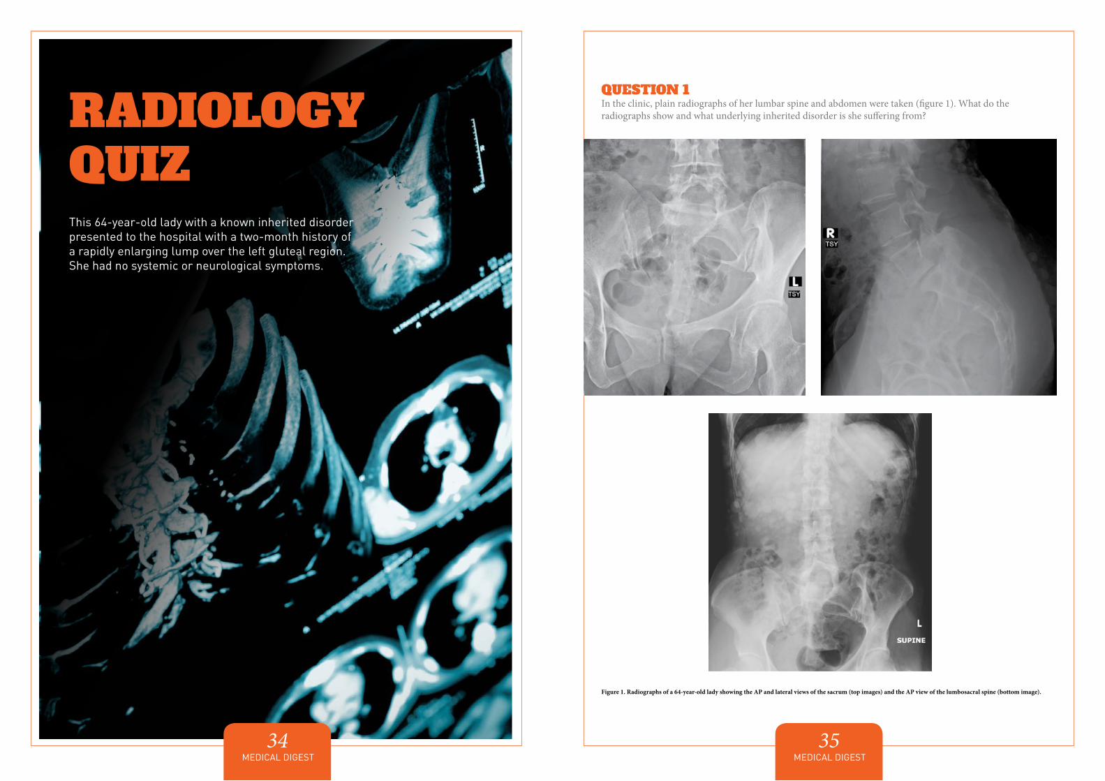
RADIOLOGY QUIZThis 64-year-old lady with a known inherited disorder presented to the hospital with a two-month history of a rapidly enlarging lump over the left gluteal region. She had no systemic or neurological symptoms.
MEDICAL DIGEST35
Figure 1. Radiographs of a 64-year-old lady showing the AP and lateral views of the sacrum (top images) and the AP view of the lumbosacral spine (bottom image).
MEDICAL DIGEST34
QUESTION 1In the clinic, plain radiographs of her lumbar spine and abdomen were taken (figure 1). What do the radiographs show and what underlying inherited disorder is she suffering from?
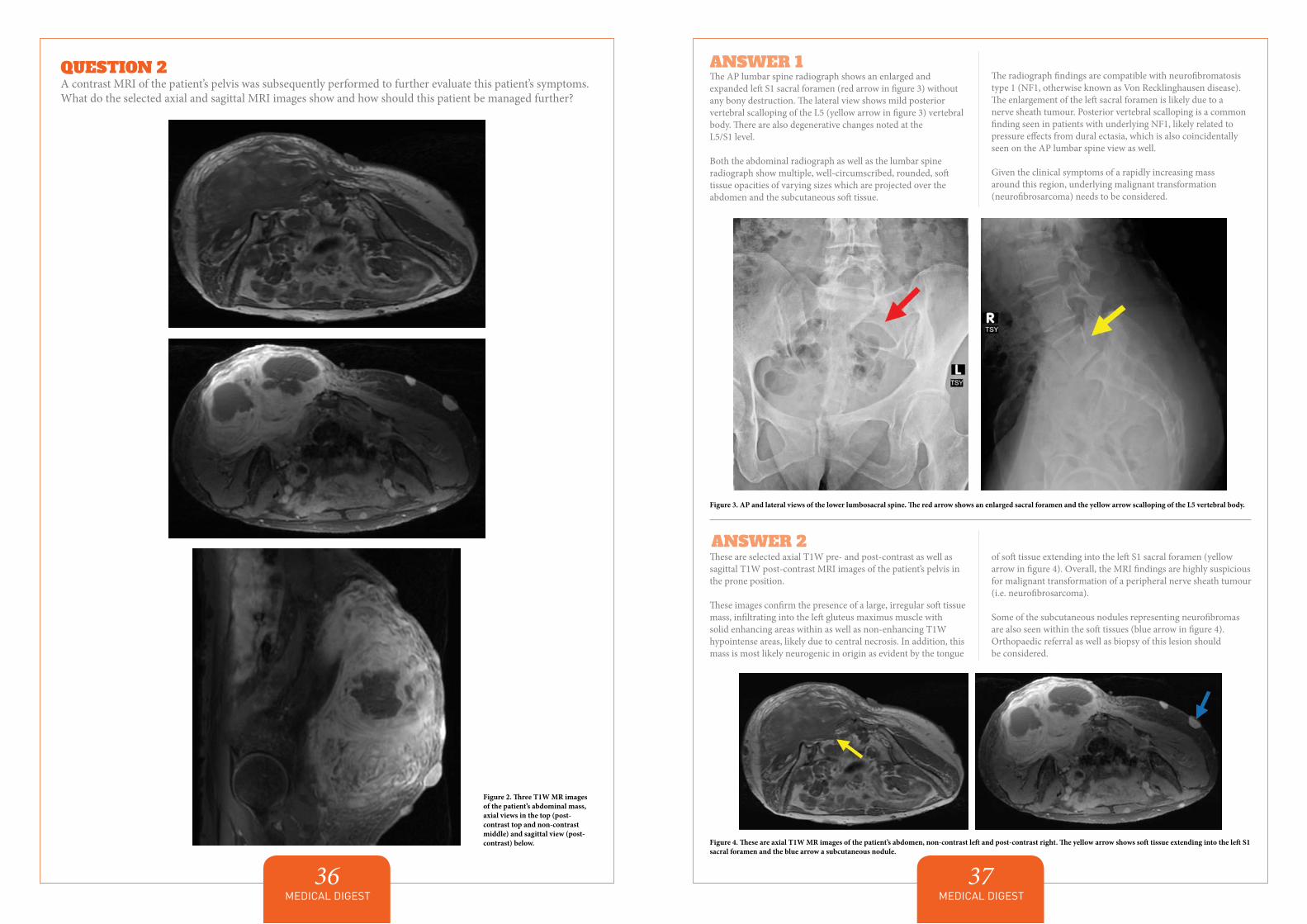
MEDICAL DIGEST37
MEDICAL DIGEST36
ANSWER 1 The AP lumbar spine radiograph shows an enlarged and expanded left S1 sacral foramen (red arrow in figure 3) without any bony destruction. The lateral view shows mild posterior vertebral scalloping of the L5 (yellow arrow in figure 3) vertebral body. There are also degenerative changes noted at the L5/S1 level.
Both the abdominal radiograph as well as the lumbar spine radiograph show multiple, well-circumscribed, rounded, soft tissue opacities of varying sizes which are projected over the abdomen and the subcutaneous soft tissue.
The radiograph findings are compatible with neurofibromatosis type 1 (NF1, otherwise known as Von Recklinghausen disease). The enlargement of the left sacral foramen is likely due to a nerve sheath tumour. Posterior vertebral scalloping is a common finding seen in patients with underlying NF1, likely related to pressure effects from dural ectasia, which is also coincidentally seen on the AP lumbar spine view as well.
Given the clinical symptoms of a rapidly increasing mass around this region, underlying malignant transformation (neurofibrosarcoma) needs to be considered.
Figure 3. AP and lateral views of the lower lumbosacral spine. The red arrow shows an enlarged sacral foramen and the yellow arrow scalloping of the L5 vertebral body.
These are selected axial T1W pre- and post-contrast as well as sagittal T1W post-contrast MRI images of the patient’s pelvis in the prone position.
These images confirm the presence of a large, irregular soft tissue mass, infiltrating into the left gluteus maximus muscle with solid enhancing areas within as well as non-enhancing T1W hypointense areas, likely due to central necrosis. In addition, this mass is most likely neurogenic in origin as evident by the tongue
of soft tissue extending into the left S1 sacral foramen (yellow arrow in figure 4). Overall, the MRI findings are highly suspicious for malignant transformation of a peripheral nerve sheath tumour (i.e. neurofibrosarcoma).
Some of the subcutaneous nodules representing neurofibromas are also seen within the soft tissues (blue arrow in figure 4). Orthopaedic referral as well as biopsy of this lesion should be considered.
ANSWER 2
Figure 2. Three T1W MR images of the patient’s abdominal mass, axial views in the top (post-contrast top and non-contrast middle) and sagittal view (post-contrast) below.
QUESTION 2A contrast MRI of the patient’s pelvis was subsequently performed to further evaluate this patient’s symptoms. What do the selected axial and sagittal MRI images show and how should this patient be managed further?
Figure 4. These are axial T1W MR images of the patient’s abdomen, non-contrast left and post-contrast right. The yellow arrow shows soft tissue extending into the left S1 sacral foramen and the blue arrow a subcutaneous nodule.
ECG QUIZA 50-year-old lady presented to her General Practitioner (GP) for the complaint of right-sided chest discomfort at rest for several months. She does not smoke and has no other cardiovascular risk factors. A resting 12 lead electrocardiogram (ECG) was performed at the GP clinic (figure 1).
MEDICAL DIGEST39
MEDICAL DIGEST38
Discussion NF1 is one of the most common autosomal dominant disorders, affecting approximately one in 3000 individuals.1,2 Diagnostic criteria for NF1 include two or more neurofibromas (NFs) of any type or one plexiform NF, café-au-lait macules, axillary or inguinal freckles, optic glioma, Lisch nodules, distinctive osseous lesions, and/or a first-degree relative with NF1.3 Patients with NF1 have an increased risk of developing malignant tumours, in particular a tendency for some NFs to undergo malignant transformation to a malignant peripheral nerve sheath tumour (MPNST).2,4
As the disease is due to inactivation of a tumour suppressor gene, it is also commonly associated with increased incidence of other tumours, such as pheochromocytoma, rhabdomyosarcoma, renal angiomyolipoma and gliomas.1-6 No single treatment exists, and a combination of supportive and surgical therapies are employed depending on the specific tumours and anomalies.
Although prognosis is very variable, overall patients with NF1 have a life expectancy approximately half that of non-affected individuals; they usually succumb to tumours or cardiovascular complications.7
Whilst imaging of the associated tumours can demonstrate a large range of appearances, invariably all patients with NF1 will demonstrate subcutaneous nodules on radiology which manifest as numerous, well-circumscribed soft tissue lesions with heterogeneous enhancement on CT or MRI. Enlargement and expansion of the exit foramen related to the presence of neurofibromas along the peripheral nerves is also a common finding. Any sudden, rapid enlargement or pain related to these lesions, in particular the plexiform neurofibromas, should raise the suspicion of malignant transformation and prompt orthopaedic referral should be made.
REFERENCES1. Karnes PS. Neurofibromatosis: a common neurocutaneous disorder. Mayo Clin Proc 1998; 73:1071-6.2. Zöller MET, Rembeck B, Odén A, Samuelsson M, Angervall L. Malignant and benign tumors in patients with
neurofibromatosis type 1 in a defined Swedish population. Cancer 1997; 79:2125-31.3. National Institutes of Health Consensus Development Conference: Neurofibromatosis Conference Statement. Arch
Neurol 1988; 45:575-8.4. Ducatman BS, Scheithauer BW, Piepgras DG, Reiman HM, Ilstrup DM. Malignant peripheral nerve sheath tumors.
A clinicopathologic study of 120 cases. Cancer 1986; 57:2006-21.5. Itoh T, Magnaldi S, White RM, Denckla MB, Hofman K, Naidu S, et al. Neurofibromatosis type 1: the evolution of
deep gray and white matter MR abnormalities. AJNR Am J Neuroradiol 1994; 15:1513-9.6. Albright AL, Adelson PD, Pollack IF. Principles and practice of pediatric neurosurgery. Thieme Medical Pub (2007)
ISBN: 1588903958. 7. Ruggieri M. Neurocutaneous Disorders, Phakomatoses & Hamartoneoplastic Syndromes. Springer Verlag. (2007)
ISBN: 3211213961.
DR JUSTINE KWAN is an associate consultant
in the Department of Diagnostic Radiology,
Tan Tock Seng Hospital.
Whilst imaging of the associated tumours can demonstrate a large range of appearances,
invariably all patients with NF1 will demonstrate subcutaneous nodules on radiology which manifest as numerous,
well-circumscribed soft tissue lesions with heterogeneous enhancement on CT or MRI.
““
Figure 1. ECG performed at the GP clinic
MEDICAL DIGEST41
DR YEW MIN SEN is an associate consultant in the Department of Cardiology, Tan Tock Seng Hospital.
ANSWERAt first glance, the ECG taken at the GP clinic revealed borderline ST segment elevations associated with pathological Q waves over the inferior leads. Accompanied by a history of chest pain, these changes are worrisome for an ST segment elevation myocardial infarction.
However, on closer inspection, one may notice some unusual ECG features. The P wave in leads II, III and aVF are inverted, unlike the typical upright P waves seen in normal sinus rhythm. The axis is also abnormal with positivity of lead aVR.
The above ‘abnormalities’ were not reproduced when the ECGs were repeated (figures 2 and 3). Of note, the P waves are now upright in the inferior leads, the axis is normal and aVR is predominantly negative. ST segment elevations are no longer seen.
The most likely diagnosis is ECG electrode misplacement, specifically a right arm (RA) and left leg (LL) electrode reversal.
DiscussionA 12-lead ECG is performed by the correct placement of six chest and four limb electrodes over standardised body regions. Deflections recorded on the ECG are dependent on the direction of depolarisation relative to the position of these electrodes. Since there are 10 electrodes, reversed placement of any two can give rise to a huge variety of different ECG patterns depending on the particular pair of erroneous leads. For example, a common misplacement error (not in this case) is the reversal of the left arm (LA) and right arm (RA) electrode, giving rise to a classical pattern of inverted P, QRS and T waves over leads I and aVL.
As illustrated, incorrect electrode placement can result in erroneous diagnosis and inappropriate treatment. A full discussion of the different ECG patterns seen in various electrode misplacements is beyond the scope of this article. The reader is encouraged to use the REVERSE mnemonic to rapidly screen ECGs for the most common electrode reversal patterns (Baranchuk A, Shaw C, Alanazi H, Campbell D, Bally K, Redfearn DP, et al. Electrocardiography pitfalls and artifacts: the 10 commandments. Crit Care Nurse 2009; 29:67-73).
MEDICAL DIGEST40
In view of the ECG abnormalities, the GP dialed for an emergency ambulance. An ECG was repeated by the civil defence paramedics en route to hospital (figure 2). An ECG was performed again at the Emergency Department on arrival (figure 3). The patient was pain-free and haemodynamically stable.
QUESTIONSQ1: What are the abnormalities seen in the GP’s ECG (figure 1)?Q2: What is the most likely diagnosis?
Figure 2. ECG performed on the ambulance en route to hospital
Figure 3. ECG performed at the hospital Emergency Department