Embed Size (px)
Citation preview

Vol. 4, 697-71/. Marc/i 1998 Clinical Cancer Research 697
Tamoxifen-resistant Fibroblast Growth Factor-transfected MCF-7
Cells Are Cross-Resistant in Vivo to the Antiestrogen
IC! 182,780 and Two Aromatase Inhibitors1
Sandra W. McLeskey, Lurong Zhang,
Dorraya El-Ashry, Bruce J. Trock,
Cecilia A. Lopez, Samir Kharbanda,
Christopher A. Tobias, Lori A. Lorant,
Rachel S. Hannum, Robert B. Dickson, and
Francis G. Kern2
Lombardi Cancer Center [S. W. M., L. Z., B. J. T., C. A. L., S. K..
C. A. T.. R. S. H.. D. E-A.. R. B. D.. F. G. K.l. Departments ofBiochemistry and Molecular Biology ID. E-A.. F. G. K.]. CellBiology [R. B. D.. L. Z.], Medicine [B. J. TI, and Pharmacology
[S. W. M.]. and the School of Nursing [S. W. M.], GeorgetownUniversity Medical Center, Washington. D. C. 20007
ABSTRACTAlthough the antiestrogen tamoxifen has been the
mainstay of therapy for estrogen receptor (ER)-positive
breast cancer, successful treatment of responsive tumors is
often followed by the acquisition of tamoxifen resistance.
Subsequently, only 30-40% of patients have a positive re-
sponse to second hormonal therapies. This lack of response
might be explained by mechanisms for tamoxifen resistance
that sensitize ER pathways to small amounts of estrogenicactivity present in tamoxifen or that bypass ER pathways
completely. To elucidate one possible mechanism of tamox-
ifen resistance, we treated ovariectomized tumor-bearing
mice injected with fibroblast growth factor (FGF)-trans-
fected MCF-7 breast carcinoma cells with the steroidal an-
tiestrogen ICI 182,780 or one of two aromatase inhibitors,
4-OHA or letrozole. These treatments did not slow estrogen-independent growth or prevent metastasis of tumors pro-
duced by FGF-transfected MCF-7 cells in ovariectomizednude mice. FGF-transfected cells had diminished responsesto ICI 182,780 in vitro, suggesting that autocrine activity of
the transfected FGF may be replacing estrogen as a mito-
Received 7/3/97: revised 1 1/26/97; accepted 12/10/97.
The costs of publication of this article were defrayed in part by the
payment of page charges. This article must therefore be hereby marked
advertiseinepit in accordance with 18 U.S.C. Section 1734 solely to
indicate this fact.
I This work was supported by NIH Grants CA50376 (to F. G. K.).
CA09218 (to F.G.K. and S.W.M.), CA53185 (to F.G.K. and
R. B. D.), CA66154 (to S. W. M.). CA71465 (to D. E-A.), and Cancer
Center Grant CA5IOO8; American Cancer Society Grant IRG-193 (to
S. W. M.): U.S. Army Medical Research and Material Command GrantsDAMD 17-94-4172 (to D. E-A.) and DAMD l7-94-J-4173 (to
S. W. M.): and a Susan Komen Foundation Fellowship (to L. Z.).2 To whom requests for reprints should be addressed, at SouthernResearch Institute, P. 0. Box 55305, 2000 Ninth Avenue South, Bir-
mingham. AL 35255-5305. Phone: (205)581-2480: Fax: (205)581-
2877: E-mail: [email protected].
genic stimulus for tumor growth. ER levels in FGF trans.
fectants were not down-regulated, and basal levels of tran-
scripts for estrogen-induced genes or of ER-mediated
transcription of estrogen response element (ERE) luciferase
reporter constructs in the FGF expressing cells were not
higher than parental cells, implying that altered hormonal
responses are not due to down-regulation of ER or to FGF-
mediated activation of ER. These studies indicate that estro-
gen independence may be achieved through FGF signaling
pathways independent of ER pathways. If so, therapies di-
rected at the operative mechanism might produce a thera-
peutic response or allow a response to a second course of
antiestrogen treatment.
INTRODUCTIONBecause conventional therapy is not usually curative in
clinical breast cancer, development of tamoxifen resistance. in
which breast tumors previously growth-inhibited by tamoxifen
become refractory, represents an important therapeutic di-
lemma. However, the development of tamoxifen resistance is
not necessarily associated with progression to an ER3-negative
phenotype. In many cases of clinical tamoxifen resistance, ER
expression may be retained ( 1-4), implying that the resistance is
due an alteration in activity of the tamoxifen/ER complex.
Tamoxifen resistance in such a case could result from three
possible mechanisms that, according to present knowledge,
would not preclude successful treatment with an alternative
hormonal therapy. First. alterations in the ER could arise, which
might diminish or extinguish inhibitory responses to tamoxifen,
leaving only its partial agonist effects to predominate (5-8).
Second, tamoxifen resistance arising in the setting of an intact
ER could be a result of altered intratumoral tamoxifen metab-
olism, which might produce more estrogenic metabolites locally
(7, 9-1 1). Third, available tamoxifen could be sequestered by an
increase in antiestrogen binding sites not associated with ERs
(12). As mentioned, in each of these three instances. substitution
of a hormonal therapy different from tamoxifen might result in
a clinical response. Two such alternative therapies used in this
report are steroidal estrogen antagonists, such as IC! 182,780,
which lack the partial agonist activity of tamoxifen, and aro-
matase inhibitors, which inhibit endogenous estrogen produc-
tion by all tissues, depriving the ER of its bigand.
Although the mechanisms of tamoxifen resistance de-
3 The abbreviations used are: ER. estrogen receptor; FGF. fibroblastgrowth factor: IMEM, improved minimal essential medium: X-gal,
5-bromo-4-chloro-3-indoyl-�3-D-galactopyranoside: FBS. fetal bovine
serum: 4-OHA. 4-hydroxyandrostenedione: NK, natural killer: CCS.
charcoal-stripped calf serum: ERE. estrogen response element: CAT.
chloramphenicol acetyltransferase: RT. reverse transcription.
Research. on June 15, 2018. © 1998 American Association for Cancerclincancerres.aacrjournals.org Downloaded from

698 ICI 182.780 Effects on FGF-transfected MCF-7 Cells
scribed above should be amenable to alternative hormonal ther-
apy, early results for small numbers of tamoxifen-resistant pa-
tients have shown that only about 30-40% of such patients have
a positive response to subsequent ICI 1 82,780 or aromatase
inhibitor therapy ( I 3-20). These data imply alternative mecha-
nisms for tamoxifen resistance. Constitutive production of au-
tocrine growth factor(s) or growth factor receptors by tumor
cells has been proposed as a mechanism for tamoxifen resist-
ance that may or may not involve ER pathways. Evidence
supporting this hypothesis is gained from the acquisition of
estrogen-independent growth in tumor models, including the
one used in this report. in which growth factors or growth factor
receptors have been overexpressed in estrogen-dependent breast
carcinoma cell lines (21-26). In addition, recent clinical data
showing decreased efficacy of tamoxifen in treating tumors
overexpressing c-erbB2 (27) supports a role for growth factor
signaling in clinical tamoxifen resistance. Because some growth
factor signaling pathways, including the ERB-B pathway. have
been shown to interact with ER signaling pathways (25, 28-32),
increased growth factor signaling could be one mechanism by
which cells could become sensitive to previously ineffective
amounts of estrogenic stimulation produced by the partial ago-
nist activity of tamoxifen itself or its estrogenic metabolites,
above. In cases in which such interactions have been demon-
strated, the growth factor and ER pathways may act collabora-
lively (25), making the final outcome susceptible to pharmaco-
logical manipulations of either pathway and implying that
second line hormonal therapies might have an effect. However,
increased autocrine or intracrine growth factor signaling might
also bypass the need for ER-mediated growth stimulation in
tumor cells or affect stromal components of the tumor, such as
endothelial or immune cells (33-36), to alter the tumor envi-
ronment in ways conducive to tumor growth. In either case,
alternative hormonal therapies might not be effective.
Recently, cell-specific coactivators and corepressors have
been identified for steroid hormone receptors, including the ER,
which may influence steroid receptor-induced transcription pos-
itively or negatively (37, 38). Thus, the activity of tamoxifen in
inhibiting or even stimulating tumor growth might depend on
the relative expression of various stimulatory or inhibitory co-
factors in a particular tumor (39, 40). However, transient trans-
fection experiments suggest that tamoxifen-resistant tumors pro-
duced by such mechanisms should still be sensitive to pure
antiestrogens (40).
FGFs and their receptors have been shown to be present
with high frequency in breast cancer specimens (41-50). Evi-
dence for a possible role for FGF signaling in the estrogen-
independent growth of breast tumors is gained from study of
clonab and polyclonal FGF-transfected MCF-7 cell lines, which
are capable of forming large, progressively growing tumors in
ovariectomized or tamoxifen-treated nude mice. Moreover, the
FGF-transfected cells are metastatic, forming micrometastases
in lymph nodes, lungs, and other organs (21 , 22, 5 1). The
estrogen-independent and tamoxifen-resistant growth of FGF-
transfected MCF-7 cells suggests an interaction between FGF
signaling pathways and ER-activated pathways that could occur
at the level of the ER itself or at the end point of both pathways,
where they impinge on growth mechanisms. If FGF-mediated
growth pathways bypass the ER pathway to affect growth di-
rectly, we would expect that growth would be unaffected by
hormonal treatments devoid of agonist activity. We therefore
sought to determine the sensitivity of the estrogen-independent
tumor growth of FGF-transfected MCF-7 cells to IC! 182,780 or
aromatase inhibitors. In contrast to what was seen with ERB-B
signaling pathways, we report that FGF-mediated pathways
appear to provide an alternative growth stimulatory signal that is
not dependent on ER activation.
MATERIALS AND METHODS
Cell Lines. FGF-transfected MCF-7 cell lines have been
described previously (2 1 , 22. 5 1 , 52). Briefly, the ML-20 clonal
cell line is a MCF-7-derived cell line that is stably transfected
with a lacZ expression vector. The in vitro and in viva growth
characteristics of ML-20 cells are indistinguishable from wild-
type MCF-7 cells (5 1 ). and >90% of the cells routinely stain
positive for 3-galactosidase expression by X-gal staining (52).
MKL-F (FGF-4-transfected; Ref. 52) and FGF- 1 clone 18 (FGF-
1-transfected) cells (22) resulted from the stable transfection of
the ML-20 cbonal cell line with expression vectors for FGF-4
(also known as hst-l/K-FGF) and FGF-l (also known as acidic
FGF or aFGF), respectively. Both cell lines continue to stably
express 3-galactosidase, allowing effects of FGF overexpres-
sion on metastatic capability to be assessed by X-gal staining of
organs and tissues of tumor-bearing mice. The MKL-4 cell line
was derived by transfecting wild-type MCF-7 cells (of similar
passage number used for the ML-20 transfection) with an ex-
pression vector for FGF-4, which produced the cbonal MKS-l
cells (2 1 ). These cells were then retransfected with an expres-
sion vector for lacZ, yielding MKL-4 cells (5 1 ). Cells were
maintained in IMEM (Biofluids, Rockville, MD) supplemented
with 5% FBS in a humidified, 37#{176}C,5% CO2 incubator in
routine culture until used for tumor cell injection.
Drugs. ICI 1 82,780 was kindly donated by Dr. Alan
Wakeling of Zeneca Pharmaceuticals (Macclesfield, England),
and was administered s.c. at a dose of 5 mg in 0. 1 ml of vehicle
every week. For the experiment depicted in Fig. 1 , powdered
drug was first dissolved in 100% ethanol and spiked into
warmed peanut oil (Eastman Kodak, Rochester, NY) to give a
final concentration of 50 mg/mi. For the experiments depicted in
Fig. 1 , B and C, 50 mg/ml preformulated drug in a vehicle of
10% ethanol, 15% benzyl benzoate. 10% benzyb alcohol,
brought to volume with castor oil, was supplied by B. M. Vose
(Zeneca Pharmaceuticals). 4-OHA was donated by Angela Bro-
die (University of Maryland, Baltimore, MD) and was admin-
istered s.c. at a dose of 1 mg/mouse/day 6 days of the week in
a vehicle of 0.3% hydroxypropylcelbuose. Letrozole was do-
nated by Dr. Ajay Bhatnagar (Novartis, Ltd., Basil, Switzerland)
and was administered via gavage at a dose of 1 mg/mouse/day
6 days of the week in a vehicle of 0.3% hydroxypropylceblulose.
Sustained-release (60 day) pellets containing 5 mg of tamoxifen
were obtained from Innovative Research of America (Sarasota,
FL) and implanted s.c. in the interscapular area at the time of
tumor cell injection.
Tumor Cell Injection. The procedure for tumor cellinjection has been described previously (21). Briefly, tumor
cells were scraped into their normal growth medium, and viable
cells were quantified using trypan blue exclusion. The cells were
Research. on June 15, 2018. © 1998 American Association for Cancerclincancerres.aacrjournals.org Downloaded from

Clinical Cancer Research 699
resuspended in their normal growth medium at a density of
66.7 X 10⁶ cells/mI and 0.15 ml (containing 10 million cells)
were used to inject ovariectomized mice (nude or beige/nude/
xid) into the mammary fat pad. For the experiment involving
MKL-4 cells and nude mice (Fig. 1A), each mouse was injected
bilaterally into the thoracic mammary fat pads (two injections
per mouse). There were seven mice in the vehicle group and five
mice in each treatment group. For the experiments involving
MKL-4 cells and beige/nudeixid mice (Fig. 2), four tumor cell
injections were given, two on each side in the thoracic mam-
mary fat pad and two in the inguinal mammary fat pad; treat-
ment groups consisted of four mice. For the experiments involv-
ing MKL-F and FGF-l, clone 18 cells (Fig. I, B and C). each
mouse was injected once in the right thoracic mammary fat pad.
There were seven mice in the each vehicle group, and treatment
groups consisted of five or six mice each. Tumors resulting from
the injections were measured twice weekly in three dimensions
using calipers. Tumor volume is the product of the largest
dimension, the orthogonal measurement, and the tumor depth, as
described previously (21). Because the FGF-l-transfected clone
1 8 cell line produces tumors that in some cases are surrounded
by a fluid-filled sac that confounds tumor measurements (22),
these tumors were measured postmortem by weighing them.
Determination of Metastasis. Organs were harvested
from tumor-bearing animals, fixed briefly, and stained with
X-gal as reported previously (51) and viewed through a dissect-
ing microscope (Olympus SZH). Clusters of blue-staining cells
were identified as micrometastases. In accordance with previous
results, no macrometastases were identified (21, 22, 51, 53).
Growth Assays. Anchorage-dependent and anchorage-
independent growth assays were performed as described (2 1).
Briefly, for anchorage-dependent growth, cells were plated in
24-well culture dishes at a density of 10,000 cells/well for the
time course experiments (Fig. 4) and 20,000 or 30,000 cells/well
for the concentration-response experiments (Fig. 5). For growth
in FBS, following overnight attachment, treatments were added
at the indicated concentrations, and cells were counted on the
indicated days. For growth assays under estrogen-depleted con-
ditions, cells were stripped of estrogens during a 24-h period the
day following plating by changing the medium four times to
phenol red-free IMEM supplemented with 5% CCS (21 ). We
have found that this stripping procedure allows complete re-
moval of estrogens without substantial proliferation of cells
before treatments are added. Following the stripping procedure,
on day 0, treatments were added, and counting of cells was done
as above.
Doubling times were determined according to the follow-
ing equation: doubling time = t2 - t1/3.32log(N,/N1), where N,
and N, are the number of cells at times t-, and t, . respectively. N1
and N, are the means of quadruplicate determinations.
Anchorage-independent assays in FBS-containing medium
were done as described previously (2 1 ). For experiments using
estrogen-depleted conditions, cells were stripped of estrogens
over a 24-h period as described above before being plated in soft
agar. Colonies greater than 60 p.m were counted using an
Omnicon 3600 Image Analysis system.
ER Assays. [3H]Estradiol binding has been described
previously (54, 55). Briefly, cells grown to 70% confluence
were stripped with twice daily medium changes over 4 days
with 5% CCS in phenol red-free IMEM. The prolonged strip-
ping method allows ERs to become up-regulated to maximal
levels. Cells were harvested, washed sequentially at 4#{176}Cwith
serum-free, phenol red-free IMEM followed by TEG (10 m�i
Tris, pH 7.4, 1 mM EDTA, 10% glycerol), and resuspended in 1
ml of TEG plus I mrvi DTT, 0.5 M NaCl and a cocktail of
protease inhibitors (I mg/ml leupeptin. 77 p.g/ml aprotinin. I
jig/mI pepstatin A). A whole-cell extract was prepared by ho-
mogenization with 40 strokes in a Teflon-glass Dounce homog-
enizer followed by centrifugation at 105,000 x g for 30 mm.
Protein content of the supernatant was determined by the
method of Bradford (56), and protein concentrations were ad-
justed to 2 mg/mb. Extracts were incubated with 10 nM [3H]173-
estradiol with or without a 100-fold excess of unlabeled estra-
diol for 16 h at 4/C. Unbound ligand was removed by absorption
with dextran-coated charcoal followed by centrifugation. Ali-
quots of the supernatant were counted in a Beckman liquid
scintillation counter.
Northern Blots. Cells were grown to 50% confluence in
IMEM supplemented with 5% FBS and then stripped of estro-
gens as described for the growth assays, above. Treatments of
0.1% ethanol (vehicle) or l0�� M 173-estradiol in the same
medium were added. Cultures were harvested after 3 days of
treatment, and RNA was extracted using RNAzoI B (Tel-Test.
Inc.) according to the manufacturer’s directions. Thirty p.g of
each RNA were subjected to electrophoresis in a I .2% formal-
dehyde/agarose gel and transferred to nylon (Hybond-N. Am-
ersham Corp., Arlington Heights, IL) by capillarity. 32P-labeled
antisense riboprobes for p5-2, GAPDH, and cathepsin D were
prepared and sequentially hybridized to the membrane overnight
at 65#{176}C[hybridization buffer was 50% formamide, 50 m�i
Na,HPO4, 0.8 M NaC1, 10 m�i EDTA, 2.5 X Denhardt’s solu-
tion (I X Denhardt’s = 0.02% polyvinylpyrrolidone, 0.02%
BSA), 0.2% SDS, 400 p.g/ml yeast tRNA. and 400 p.g/inl
sonicated salmon sperm DNA with 106 DPM/ml of the appro-
priate probe]. The membrane was washed three times in 0. 1%
SDS/0. I X SSC at 80#{176}Cfor the P5-2 and cathepsin D probes,
and 75#{176}Cfor the GAPDH probe. Autoradiograms and Phospho-
rlmager (Molecular Dynamics Model 445S1) quantitation of
individual hybridization signals were obtained between the se-
quential hybridizations. For the results depicted in Fig. 7, A and
B, Phosphorlmager values obtained for PS-2 or cathepsin were
normalized to those obtained for GAPDH.
Progesterone Receptor mRNA Determination by RT-
PCR. The primers for human progesterone receptor that pro-
duce a 205-bp PCR product have been described previously
(57). The human GAPDH primers that produce a 437-bp PCR
product are as follows: 5’-AAG GTC GGT GTG AAC GGA
TTf G-3’ (sense) and 5’-TGG TGC AGO ATO CAT TGC
TG-3’ (antisense). RT-PCR was performed with 0.1 p.g of test
RNAs, except T47D cells, where 0.02 p.g was used, using the
GeneAmp RNA PCR kit (PE Applied Biosystems. Foster City.
CA) according to the manufacturer’s instructions with the fol-
bowing modifications: the RT reaction was primed with 0.0625
p.M random hexamers in a volume of 40 p.1, with 2 p.1 each of
35S-labeled UTP and 35S-labeled ATP (each 3000 Ci/mmol, 10
p.Ci/p.l, Amersham Corp.) substituted for water in the reaction.
Then, 20 p.1 of each RT reaction were transferred into two tubes
for separate GAPDH and progesterone receptor PCR reactions.
Research. on June 15, 2018. © 1998 American Association for Cancerclincancerres.aacrjournals.org Downloaded from

700 ICI 182.780 Effects on FGF-transfected MCF-7 Cells
Cycle analyses using RNA from ML-20, estradiol-treated cells
(the highest expressors of progesterone receptor) revealed that
amplification remained logarithmic at 35 cycles for the GAPDH
reaction and 40 cycles for the progesterone receptor reaction,
making these assays semiquantitative. The GAPDH PCR reac-
tion was performed using standard reagent conditions recom-
mended by the manufacturer and cycles of 95#{176}Cfor 45 s and
50#{176}Cfor 45 5 for 35 cycles. For the progesterone receptor PCR
reaction, final MgC1, concentrations were adjusted to 1 .25 msi,
and 0.25 M acetamide was included. Cycles were of 95#{176}Cfor
45 5 and 50#{176}Cfor 45 s for 40 cycles. GAPDH and progesterone
receptor reaction products were first visualized by ethidium
bromide staining following ebectrophoresis in a 2% agarose gel.
Products were then electrophoresed on a 4-20% acrylamide gel
that was subjected to both autoradiography and Phosphorlmager
quantitation as described above.
Transient Transfection, Luciferase, and CAT Reporter
Assays. ML-20 and clone 18 cells were plated in 6-well
plates, allowed to attach overnight, and stripped of estrogens in
a procedure similar to that for the growth assays (see above).
Following stripping, cells were transfected by the calcium phos-
phate, low-CO2 method (58). The luciferase plasmids pGLB-
MERE or pGLB-MNON were obtained by inserting an approx-
imately 1 .48-kb fragment containing a glucocorticoid response
element-deleted mouse mammary tumor virus promoter with
either a substituted double consensus ERE (MERE) or the same
sequence with the ERE palindromes scrambled (MNON) (59)
into the HindIII site of pGLB (Promega, Madison, WI). Each
dish received 2.5 p.g of either pGLB-MERE or pGLB-MNON
and I .0 p.g pCMV-CAT. which directs constitutive expression
of CAT, cotransfected as a control for transfection efficiency.
Following transfection, each well was washed twice with PBS
and incubated for 48 h in medium containing vehicle (0.01%
ethanol). l0� M estradiol, l0� M IC! 182,780, a combination
of E, and ICI, 10 ng/ml FGF-l plus 10 p.g/ml heparin, or a
combination of FGF, heparin, and ICI I 82,780. (Duplicate sam-
pIes of each treatment were used.) Cells were lysed and assayed
for luciferase activity using the Luciferase Reporter Gene Assay
(Boehringer Mannheim, Indianapolis, IN) according to the man-
ufacturer’s instructions. Luciferase values, expressed as relative
light units, for each sample were corrected for background by
subtracting the value of lysates of untransfected cells prepared
in parallel. CAT expression was assayed using the CAT ELISA
(Boehringer Mannheim. Indianapolis, IN) according to the man-
ufacturer’s instructions. Protein content of the lysates was de-
termined using the BCA Protein Assay Reagent (Pierce, Rock-
ford, IL). Luciferase and CAT values, normalized for protein,
were used to calculate mean specific relative light units/ng CAT.
Statistical Analyses. Statistical methods used for tumor
growth have been described previously (53, 60). For Figs. 1 and
2. only mice surviving at the end of the experiment were
included in the analysis. When no tumor developed from a
particular injection, tumor volume was recorded as zero. The
repeated measures ANOVA (60) was used to compare tumor
volumes among the treatment groups using measurements taken
over the entire time course of the experiment. In addition, final
tumor volumes (or weights in the case of clone 1 8) were
compared among treatment groups at the end of each experi-
ment using ANOVA. For analysis of metastasis in Table 1 . for
each transfectant, analysis of covariance was used to compare
the effects of treatment on total metastases, total distant metas-
tases (lung metastases plus other metastases), lymph node me-
tastases, lung metastases, and other metastases. The analyses
were all conducted with final tumor volume (or weight for the
clone 1 8 cells) included in the model as a covariate. The
analyses considered the effects of all treatments simultaneously,
as well as the effects of individual treatment comparisons
(which were adjusted for multiple comparisons using Dunnett’s
method). For each transfectant, the effect of final tumor volume
(or weight for clone 1 8) on the number of metastases was
evaluated using linear regression (for each of the categories of
metastasis described above). In Fig. 3, paired t tests were per-
formed comparing control and transfected cells under different
conditions of treatment. For the anchorage-dependent growth
assays depicted in Fig. 4, we examined the effect of treatment on
the rate of cell growth, using linear regression with an interac-
tion between time and treatment. To compare cell growth rates
and doubling times among the cell lines under specific treatment
conditions, nested linear regression models were used. For Fig.
6, ANOVA was used to determine significant differences in ER
binding among cell lines.
RESULTS
Estrogen-independent Growth of Tumors Produced by
FGF-transfected MCF-7 Cells Is Not Inhibited by Treat-ment with a Pure Antiestrogen or with Aromatase Inhibi-tors. We have previously shown that both FGF-l- and FGF-
4-transfected MCF-7 cells form progressively growing tumors
in ovariectomized nude mice, as well as in similar mice
treated with tamoxifen (21, 22, 53). Although ovariectomized
mice could be expected to have substantially lower levels of
estrogenic compounds than reproductively intact mice, some
estrogens are synthesized at extraovarian sites, such as adre-
nab gland, liver, fat, or possibly the tumor itself. The trans-
fected cells evidently still possess ERs, because they respond
to estrogen and tamoxifen administered to the mice, as well
as to these compounds used in tissue culture (21, 22). To test
the hypothesis that growth of the FGF-transfected cells in
ovariectomized or tamoxifen-treated nude mice is due to
increased sensitivity to the small amounts of estrogens still
present in ovariectomized nude mice, we tested the ability of
a pure antiestrogen, IC! 182,780, and two aromatase inhibi-
tors, 4-OHA and letrozobe, to inhibit the estrogen-indepen-
dent tumor growth produced by these FGF-transfected cell
lines.
In a first experiment to test the above hypothesis, FGF-4-
transfected MKL-4 cells were injected as before, and the mice
were treated with vehicle, tamoxifen, or ICI 182,780. There
were no significant differences in tumor volume among the
treatment groups considered over the entire time course of the
experiment (P - 0.72) or at the final time point (Fig. 1A; P =
0.72). Treatment with IC! 182,780 did not inhibit tumor growth
below that achieved in vehicle-treated mice (P = 0.675). Thus,
the failure of IC! I 82,780 to inhibit the estrogen-independent
growth exhibited by this cell line supports the hypothesis that
such growth does not result from small amounts of estrogenic
Research. on June 15, 2018. © 1998 American Association for Cancerclincancerres.aacrjournals.org Downloaded from

2000
I500
1000
500
0
400
300
200
100
0’
B MKL-F(FGF-4 C FGF-l,clonel8
.
.
.
()()
1001
� 60 ba.�
�:‘�
0
F-
300
200
�l0O
0�
so
II53
27 48 61
Days after Injection
23 45 63
Days after Injection Treatment
Clinical Cancer Research 701
4 Unpublished results.
5)
0>
0
E
C
I)
VEH 4-OHA
- TAM LET
�j ICI
Fig. I Growth of FGF-transfected MCF-7 cells in ovariectomized nude mice is not inhibited by treatment with ICI 182,780, 4-OHA, or letrozole.
Ten million cells from the indicated cell lines were injected into the mammary fat pads of ovariectomized nude mice treated with vehicle (VEi-1): a
S-mg, 60-day-release tamoxifen pellet (TAM); ICI I 82,780. 5 mg s.c. every week (IC!): I mg of 4-OHA s.c. per day 6 days of the week (4-OHA):or I mg of letrozole per day via gavage 6 days of the week (LET). Columns, group mean; bars SE. Numbers above each column are the percentagesof injections resulting in measurable tumors at that time point. A. volumes of tumors produced by one clonal FGF-4-transfected MCF-7 cell line,
MKL-4, at the indicated number of days following tumor cell injection. B. volumes of tumors produced by a second clonal FGF-4-transfected MCF-7cell line. MKL-F. at the indicated number of days following tumor cell injection. C. weights of tumors produced by a clonal FGF- 1-transfected MCF-7cell line, FGF-l , clone 18. weighed after sacrifice of the animals 28 days after tumor cell injection. (Because the FGF-l producing MCF-7 cells mayform fluid-filled sacs around the tumor. confounding tumor measurements before sacrifice, only postmortem weights are presented here.)
growth stimulation achieved by extraovarian estrogen produc-
tion.
We wished to assess the effect of IC! 182,780 on metastasis
as well as on tumor growth. In spite of its retention of the
transfected lacZ expression plasmid, the MKL-4 cell line be-
comes heterogeneous over time with respect to 3-galactosidase
expression, such that a few cells have high expression, but most
are negative (52). We therefore used a second clonal FGF-4-
transfected MCF-7 cell line, MKL-F, the 3-galactosidase ex-
pression of which is stable, for a subsequent experiment involv-
ing FGF-4-transfected MCF-7 cells. Because FOF- 1 has also
been shown to produce estrogen-independent in vito growth
when transfected into MCF-7 cells (22), we also included a
clone of FGF-l-transfected cells designated clone 18, the 3-ga-
lactosidase expression of which is also stable. For these exper-
iments, two aromatase inhibitors, 4-OHA (61, 62) and letrozole
(63), were also used to inhibit extraovarian synthesis of estro-
gens.
In agreement with the experiment using MKL-4 cells de-
picted in Fig. 1A, when the FGF-4-transfected MKL-F cells
were used, there were no differences in tumor volume among
treatment groups over all time points (P = 0.382), and IC!
182,780 did not decrease tumor growth below that obtained in
vehicle-treated animals (Fig. lB; P 0.837 for the last time
point). In addition, neither 4-OHA nor letrozole decreased tu-
mor growth below vehicle-treated levels (P = 0.571 and 0.931
for the last time point, respectively).
FGF- 1 -transfected clone 18 cells form tumors that are
sometimes surrounded by a fluid-filled sac (22, 53), preventing
accurate tumor volume measurements during the course of the
experiment. Consequently, when these cells were used (Fig.
I C), only terminal tumor weights were analyzed with ANOVA.
As with the MKL-4 and MKL-F cells, IC! 182,780 did not
inhibit estrogen-independent tumor growth in the clone I 8 cells
(P = 0.977). Administration of ICI 1 82,780 to animals injected
with ML-20 cells, a clonal line of 3-galactosidase-transfected
wild-type MCF-7 cells (5 1 ), also produced no effect when
compared with vehicle-treated animals [i.e. , no progressively
growing tumors were obtained in either case (data not shown)].
In other, separate experiments. a polyclonal population of con-
trol vector-transfected ML-20 cells that forms progressively
growing tumors in estrogen-supplemented mice (22) did not
form tumors in either untreated or ICI 182,780-treated animals.4
Thus, the continued progressive in viva growth of FGF-trans-
fected cells in ovariectomized animals treated with either a pure
antiestrogen or aromatase inhibitors demonstrates that the estro-
gen-independent growth of these cells in untreated ovariecto-
mized nude mice is not due to estrogenic activity produced at
extraovarian sites.
Because ICI 182,780, 4-OHA, and betrozole were without
effect in the experiments described above, we injected repro-
ductively intact female mice for 2 weeks with these compounds
at the same doses used in the above experiments to observe for
activity in preventing effects of endogenous estrogens on the
Research. on June 15, 2018. © 1998 American Association for Cancerclincancerres.aacrjournals.org Downloaded from

702 ICI 182.780 Effects on FGF-transfected MCF-7 Cells
Table I Metastasis of FGF-transfected MCF-7 cells is not inhibited
by treatment with ICI 182,780 or aromatase inhibitors
Mice were sacrificed and tumors and organs were subjected to
X-gal staining as described previously (51). Mice bearing tumors pro-duced by injection of MKL-4 cells were sacrificed at 61 days; for
MKL-F tumors, mice were sacrificed after 64 days: and for FGF-1 clone
I 8 tumors, mice were sacrificed after 28 days.
Metastatic site
Positive lymph nodes/
Injected celbs/ No. of tumor- lymph nodes
treatment bearing mice examined Lung Other
MKL-4Vehicle 3 3/10 3 7
TAM� 4 5/18 2 2ICI S 4/23 3 4
182,780
MKL-F
Vehicle 6 0/27 3 1
TAM 5 4/20 3 0
ICI 3 0/14 1 0
182,780
4-OHA 3 0/13 0 0LET 3 1/12 0 0
FGF-l clone18
Vehicle 6 5/24 2 0
TAM 6 3/23 3 3ICI 4 2/13 3 1182.780
4-OHA 5 5/18 2 1
LET 5 4/22 3 0
Li TAM. tamoxifen; LET. letrozole.
endometrium. Uteri harvested from mice injected with either ICI
182,780, 4-OHA, and betrozole weighed less than those from
control mice and exhibited a complete lack of endometriab
glandular structures (data not shown). Thus, these compounds
retained activity, although they had no effect on tumor growth in
our experiments.
Metastatic Frequency of Tumors Produced by FGF-
transfected MCF-7 Cells in Mice Treated with ICI 182,780
or Aromatase Inhibitors Is Not Affected by Treatment. Be-
cause the FGF-4-transfected MKL-F cells and the FGF-l-trans-
fected clone 18 cells stably express bacterial 3-galactosidase by
virtue of lacZ transfection, we were able to detect distant mi-
crometastases from tumors produced by these cells. Although
the MKL-4 cells become heterogeneous over time with respect
to 3-galactosidase expression, some high-expressing cells do
remain (52), so animals from the experiment depicted in Fig. 1A
were also analyzed. Table 1 shows the frequency of metastasis
detected for each organ examined. However, there were no
significant effects of treatment on reduction of total metastases,
distant metastases, lymph node, lung, or other metastases for
tumors produced by any of the cell lines. Because we have
previously shown that the degree of metastasis in this tumor
system is correlated with tumor size in tumors produced by both
the MKL-4 and MKL-F cells (51, 53), we evaluated the corre-
lation of individual tumor size with frequency of metastasis in
individual mice for the clone 18 cells and found that tumor size
and incidence of metastasis were indeed significantly correlated
(P = 0.014).
Effects of FGF and/or Estrogen on the Residual Im-
mune System of Nude Mice Is Not Responsible for theEstrogen-independent Growth of FGF-transfected MCF-7
Cells. Although nude mice have a T-cell defect, they retain
NK cell activity. It has been postulated that the residual NK
activity in nude mice is responsible for some xenograft rejection
and poor metastatic ability of xenografts (35). Estrogen and
tamoxifen have been shown to decrease NK cell activity in nude
mice (36), but estrogen increases the ability of NK cells to kill
MCF-7 cells in cytotoxicity assays (64). In addition, transform-
ing growth factor 3,which might be secreted by MCF-7 cells in
response to tamoxifen treatment (65), can decrease NK cell
activity (33, 34). FGF-2 (also known as basic FGF or bFGF) has
been shown to negatively affect NK activity by decreasing
endothelial cell adhesion molecule expression (66), raising the
possibility that FGF-1 and/or FGF-4 might have the same effect.
In addition, B-cell maturation of nude mice is defective because
it lacks appropriate help from T-cells (35). Because of the
complexity of possible interactions of estrogen, FGFs, and
MCF-7 cells with the immune system, and because of the
possibility that the estrogen-independent or tamoxifen-stimu-
lated in vivo phenotype of the MKL-4 cells is due to an effect of
the transfected FGF and/or estrogen on the remaining immuno-
competence in the nude mouse, we injected the MKL-4 clonal
line of FGF-4-transfected MCF-7 cells into triply deficient
beige/nude/xid mice. These mice exhibit intermediate NK ac-
tivity coupled with defects in maturation of both B and T
lymphocytes (35). Tumor growth in this host was somewhat
slower than in the athymic nude mice because tumors measured
at 74 days (Fig. 2A) were smaller than tumors using the same
cells in nude mice measured at 61 days (Fig. 1A). However,
estrogen-independent and tamoxifen-resistant growth was again
seen in these animals. Pulmonary and lymphatic micrometasta-
ses were present in two of two tumor-bearing mice examined
(data not shown). Injection of the cbonal MCF-7 cell line ML-20
into this host produced much smaller tumors in estrogen-treated
animals, as depicted in Fig. 2B. Tumor nodules produced by
ML-20 cells in animals treated with tamoxifen were quite small
and static, and ultimately they regressed, as has been previously
shown in nude mice (21, 67). Although tumor growth was
slower and the differences between treatment groups did not
reach significance in this host for either cell line, the tumori-
genic, tamoxifen-resistant, metastatic phenotype of MKL-4 cells
was not altered in this host, and estrogen-independent growth of
control ML-20 cells did not occur. We therefore conclude that
the residual immunocompetence remaining in nude mice is not
important in the estrogen-independent, tamoxifen-resistant in
vivo growth of these transfectants.
FGF-transfected MCF-7 Clonal Cell Lines Have Dimin-
ished in Vitro Responses to ICI 182,780. Because IC!182,780 did not affect the estrogen-independent growth of the
FGF-transfected MCF-7 cells in viva and because we have
previously shown that the FGF transfectants do not respond to
4-hydroxytamoxifen in estrogen-containing medium to the same
extent as the parental cells (21, 22), we determined their growth
responses to IC! 182,780 in vitro.
In anchorage-independent growth assays using FBS-con-
Research. on June 15, 2018. © 1998 American Association for Cancerclincancerres.aacrjournals.org Downloaded from
1000
A
750
MKL-4
5000
0>
0
01-
250
70
60
� 50
I> 30
j 20
10
00
B
IaSi
<� �, /�
‘� ,�.�-
Treatment ..�0 Treatment
300
250
250 -
200
C0
0
C_) 1500
I50
Vehicle A
- IC! 182,780
II
II �iI
EI� Vehicle B
Estradiol
- ICI 182,780 -
� IC! 182,780+E2
�-I � I nih I50 -
0ML-20 MKL-4 MKL-F Clone I 8 ML-20 MKL-4 MKL-F Clone I 8
Cell Line Cell Line
Fig. 3 Effect of ICI 182,780 on anchorage-independent growth of FGF-transfected cells. A. for assay of anchorage-independent growth in
FBS-containing soft agar, 20,000 cells from each cell line were plated in top agar in 35-mm dishes as described (2 1). After 8 days of growth, coloniesgreater than 60 p.m in diameter were quantitated using an Omnicon Image Analysis system. Columns, mean of triplicate dishes; bars, SE. B, for assayof anchorage-independent growth in estrogen-depleted medium. cells from each cell line were subjected to a 24-h stripping procedure using 5% CCS
in IMEM as described. Twenty thousand stripped cells were plated into top agar in 35-mm dishes, and after 14 days of growth, colonies were
quantitated as above. Columns, mean of triplicate dishes; bars, SE.
Clinical Cancer Research 703
Fig. 2 Tumorigenicity of FGF-4-transfected MCF-7 cells is not
increased by injection into beige!
nudeixid mice. Ten millionMKL-4 cells were injected into
the mammary fat pads of ovari-
ectomized beige/nude/xid mice
(four sites per mouse). Tumorswere measured as in Fig. 1. The
measurements shown are from 74
days after tumor cell injection A,
volumes of tumors produced byFGF4-transfected MKL-4 cells;
B, volumes of tumors producedby ML-20 cells (a clonal line of
MCF-7 cells).
.� 2000
0
C.)
� 150
.0
0
z �
taming medium (Fig. 3A), as previously reported (21 , 22, 74),
the baseline colony formation of the FGF transfectants is higher
than that of the parental cells (P < 0.03). Moreover, when
l0� M IC! 182,780 was added to this medium, the control
ML-20 cells and the FGF transfectants were all growth inhib-
ited, but the FGF transfectants still exhibited a higher rate of
colony formation than the control ML-20 cells (P < 0.008).
Whereas colony formation by control transfected cells was
inhibited by 99% by ICI 182,780 treatment, the inhibition of
colony formation of the FGF-transfected cells ranged from 67 to
84%. For all cell lines tested, addition of l08 M estradiol to the
IC! 182,780-containing medium reversed the inhibition pro-
duced by IC! 182,780 (data not shown). Thus, in this assay, the
FGF transfectants retained an increased ability for anchorage-
independent growth in spite of treatment with IC! 182,780.
In agreement with what we have previously reported (21,
22, 74), in estrogen-depleted medium (Fig. 3B), FGF-trans-
fected clonal lines again had significantly greater baseline col-
ony formation than ML-20 cells (P < 0.05), with the exception
of the MKL-4 line, which just missed significance (P = 0.06).
As in FBS-containing medium, when ICI 182,780 was added to
the medium, the FGF transfectants had significantly increased
colony formation when compared with the control ML-20 cells
(P < 0.015), indicating that the increased colony formation in
estrogen-depleted medium is not due to increased sensitivity to
residual estrogens remaining after the stripping process. Colony
formation in the presence of IC! 182,780 was variably increased
by the addition of 10_8 M estradiol. Thus, in estrogen-depleted
medium, the FGF transfectants again had increased ability for
anchorage-independent growth in the presence of ICI 182,780.
Although the anchorage-dependent growth rate of the FGF
transfectants did not differ substantially from ML-20 cells in
Research. on June 15, 2018. © 1998 American Association for Cancerclincancerres.aacrjournals.org Downloaded from

�‘ 10’
5)
5-C
V
.0
EC
z10�
�) 10’V
C.)
0
V
Iz 10�
0 1 2 3 4 5 6 7 0 1 2 3 4 5 6 7
Cell Line
-.- Vehicle
U IC! 182,780-A-. ICI+E2
2 3 4 5 6 7 0 1 2 3 4 5 6 7Days Days
704 ICI 182,780 Effects on FGF-transfected MCF-7 Cells
Fig. 4 Effect of ICI-182,780 on anchorage-dependent growth of FGF-transfected MCF-7 cells in 10% FBS. A, ML-20 (parental) cells: B,
FGF-4-transfected MKL-4 cells: C, FGF-4-transfected MKL-F cells; D. FGF-l-transfected clone 18 cells. E. doubling times calculated from the
experiments depicted in A-D. Cells were plated in 24-well plates at 10,000 cells/well and allowed to attach overnight. The following day (day 0),
media were changed to the indicated treatments. Treatment concentrations were as follows: vehicle. 0. 1% ethanol: ICI 182,780, l0� M; estradiol,
lO’ M. Cells were harvested and counted at the indicated time points. Linear regression was performed on the data points for each treatment and
the lines obtained are shown as indicated. This is a representative experiment of two.
FBS-containing medium (doubling time for ML-20 cells was
36.3 h, versus 24.4-30.2 h for the FGF transfectants), in me-
dium supplemented with l0�� M ICI 182,780, their growth rate
was slowed to a much lesser extent than the control cells (Fig.
4). The doubling time for ML-20 cells in IC! 182,780-contain-
ing medium (83.6 h) was more than twice the doubling times for
the FGF transfectants (29.9-39.7 h; Fig. 4E), and all of the FGF
transfectants had significantly higher growth rates in the pres-
ence of IC! 182,780 than ML-20 cells (P = 0.001 for MKL-4,
0.007 for MKL-F, and 0.0001 for clone 18). The effect of ICI
182,780 was partially reversed by l0� M 173-estradiol in all
cell lines tested. Thus, in this assay, as in the anchorage-
independent growth assay, the FGF transfectants grew better in
the presence of IC! 182,780 than the control ML-20 cells.
Because others have shown that MCF-7 cells that have
acquired estrogen independence exhibit increased sensitivity to
estradiol or to the partial agonist properties of tamoxifen (68,
69), we determined the concentration-response relationships for
173-estradiol, ICI 1 82,780, and 4-hydroxytamoxifen for the
control ML-20 cells and the three FGF transfectants. In estro-
gen-depleted medium, estradiol stimulated growth with approx-
imately the same potency (Table 2 and Fig. 5A) in all four cell
Table 2 Potencies of 17�3-estradiol in stimulation of growth and IC!
182,780 in inhibition of estradiol stimulation of growth
Potencies were determined graphically from the concentration-
response relationships depicted in Fig. 5.
Cell line EC50, l7�3-Estradiol (M) IC50. ICI 182,780 (M)
ML-20 2 x l0� 2 X l0�
MKL-4 0.5 x 10_l � x 10#{176}
MKL-F 2 x l0�’ 8 x l0�
Clone 18 2 x l0� 10 X l0�
lines, in agreement with published results for MCF-7 cells (70).
As previously shown (Ref. 22 and this report), the maximal
effect of estradiol is diminished in stimulating growth of the
FGF transfectants, which had a maximal response about 70% of
that ofthe control ML-20 cells (Fig. 5A). When IC! 182,780 was
added to estrogen-depleted medium supplemented with l0 ‘#{176}M
estradiob, its potency was slightly lower for the FGF-transfected
cells than for the control cells, with IC51) values ranging from 2.5
to 5 times less than that of the parental cells (Table 2 and Fig.
SB). In accordance with our previous results for 4-hydroxyta-
Research. on June 15, 2018. © 1998 American Association for Cancerclincancerres.aacrjournals.org Downloaded from

B
.C
C
0.
A
F
0
�0
0.
100 -
75 -
50 -
25 -
0
io-’� l0-’� 1012 10’’ 1010 10’
1713-Estradiol Concentration (M)
10-I’ 10” 10’ 10’ 10’ 10’
Concentration oflCi 182,780 (M)
120 -
,g 100 -5)
05-0.
CaE 80-
0
�60-0
0�5)5)
5)� 40-C5)CO0
l� 20-
0-�Z5 535 �c,
4%,)
Clinical Cancer Research 705
800
700
600
500
400
300
200
100 -
0-
Fig. 5 Concentration-response relationships for l7�3-estradiol and ICI 182,780. A, 30,000 cells/well of the indicated cell lines were plated in 24-well
dishes. After overnight attachment, the cells were stripped with four changes of estrogen-depleted medium over 24 h, after which the indicated
concentrations of estradiob were added in fresh estrogen-depleted medium. Cells were harvested after S days of growth and counted on a Coulter
counter. Data points (#{149},ML-20; U, MKL-F; A, MKL-4; #{149},clone 18), mean of quadruplicate determinations; bars. SE. B, 20,000 cells/well were
plated and stripped as forA. Treatments consisted of 10 #{176}M 173-estradiol alone or with the addition ofthe indicated concentrations ofICI 182,780.
Cells were harvested and counted after S days. Data points (#{149},ML-20; #{149}.MKL-F; A. MKL-4: #{149},clone 18), mean of quadruplicate determinations:
bars, SE.
moxifen (2 1 , 22), the maximal growth-inhibitory effect of IC!
182,780 was less for the FGF transfectants than for the parental
cells. All four cell lines exhibited similar small growth stimu-
lation when treated with varying concentrations of 4-hydroxyta-
moxifen in estrogen-depleted medium (data not shown), in
agreement with published reports (68). We conclude that the
FGF transfectants do not exhibit substantially increased sensi-
tivity to ER agonists but may be slightly less sensitive to IC!
182,780 when compared with the control ML-20 cells.
FGF-transfected MCF-7 Cells Have Numbers of ERs
Similar to the Parental Cells. Others have shown that
heregulin-induced growth factor signaling in MCF-7 cells re-
sults in down-regulated ERs (25, 26). Because FOF-transfected
MCF-7 cells still respond to some extent to estrogen, tamoxifen,
and IC! 182,780 in vivo and in vitro (Figs. 2-5 and Refs. 21 and
22), it seems obvious that they still have ERs. Nonetheless, we
measured ER binding on the four cell lines used in these
experiments to see whether there were differences between cell
lines. Fig. 6 shows ER binding data observed for ML-20,
MKL-4, MKL-F, and clone 18 cell lines. ANOVA used with
these data revealed no significant differences among cell lines in
numbers of binding sites for [3H]estradiob (P 0.566). More-
over, each cell line contains ample numbers of ERs that are
functional, at least with respect to estrogen binding. Although it
is difficult to make a direct comparison of these results with
those obtained in other laboratories, it would seem that trans-
fections with FGF-l and FGF-4 produce a different result than
transfection of MCF-7 cells with heregulin, which down-regu-
lated ER number.
ERs Are Not Constitutively Activated in FGF-trans-fected MCF-7 Cells and Remain Capable of Inducing Tran-
scription When Activated with Estrogen. As mentioned,
others have reported that growth factor signaling by IGF, EGF,
and heregulin can activate ER (25, 71-73). We therefore sought
to determine whether ER was constitutively activated in FGF-
Fig. 6 ER levels by ligand binding assay. Ligand binding was per-
formed using [3H]l73-estradiol on cells stripped of estrogens prior to
the assay. C’olumns, means of four separate determinations; bars. SE.
transfected MCF-7 cells by determining whether basal levels of
expression of estrogen-inducible genes, encoding pS2, cathepsin
D, and progesterone receptor were elevated. We also evaluated
the capability of the ER expressed in these cells to induce
increased bevels of these genes when activated by estrogen (Fig.
7). Although basal levels of expression of pS2 and cathepsin D
(Fig. 7B) or progesterone receptor mRNA (Fig. 7D) varied
between the cell lines, the FGF-transfected lines did not have
consistently elevated levels of expression, which would be ex-
Research. on June 15, 2018. © 1998 American Association for Cancerclincancerres.aacrjournals.org Downloaded from

A B
200
� iso
� 00
�5o
Fold Induction, Cathepsin D I 4.3 1 4.2 I S3 I 7.4
GAPDH �
C0�’ �‘ ‘�‘ F
�5 � � ,-� �
E - +- + . +. +
FOR *
D300
� 200
� 00
706 IC! 182.780 Effects on FGF-transfected MCF-7 Cells
0 �
�- I_ I� �-
E - + - + - + - +
pS2 . O� O
Fold Induction, pS2 I Ii.9 I 7.3 I 4.4 I 5.7 0
Cathepsin D
Fold Induction, POR I it I 170 1 3.6 1 27
GAPDH *
Fig. 7 Basal and estrogen-induced levels of transcripts for estrogen-responsive genes. RNA (0. 1 rig) extracted from cells growing in phenol red-free
IMEM supplemented with 5% CCS and either 0.1% ethanol or l0� M l7�3-estradiol was subjected to Northern blot analysis for p52. cathepsin D.
and GAPDH transcripts using 30 jig of total RNA (A ) or semiquantitative RT-PCR for progesterone receptor (PGR) and GAPDH transcripts using
0.1 p.g of total RNA as template for RT (C). RNA from T47D cells (0.02 gig). which express high levels of progesterone receptor, was used as a
positive control. and 0. 1 �ig of RNA from MDA-MB 23 1 cells. which do not express progesterone receptor. was used as a negative control. Reactions
that contained no RNA or no reverse transcriptase yielded no amplified bands (data not shown). TranscriptlGAPDH ratios obtained by Phospho-
rlmager analysis were analyzed for fold induction produced by l7�-estradiol (A and C) or comparison of basal expression with that of control ML-20
cells (B and D).
pected if the ER were constitutiveby activated by virtue of the
FGF transfection. Similarly, the degree of induction for p52,
cathepsin D. and progesterone receptor (Fig. 7, A and C) at-
tamed by estrogen treatment was variable between cell lines, but
the transfected cells did not exhibit consistently increased or
decreased sensitivity to estrogen when compared with controls.
Thus, the differences in basal expression or degree of estrogen
induction for these estrogen-induced genes between the differ-
ent cell lines is probably due to cbonal or experimental varia-
bility. rather than being an effect of transfection.
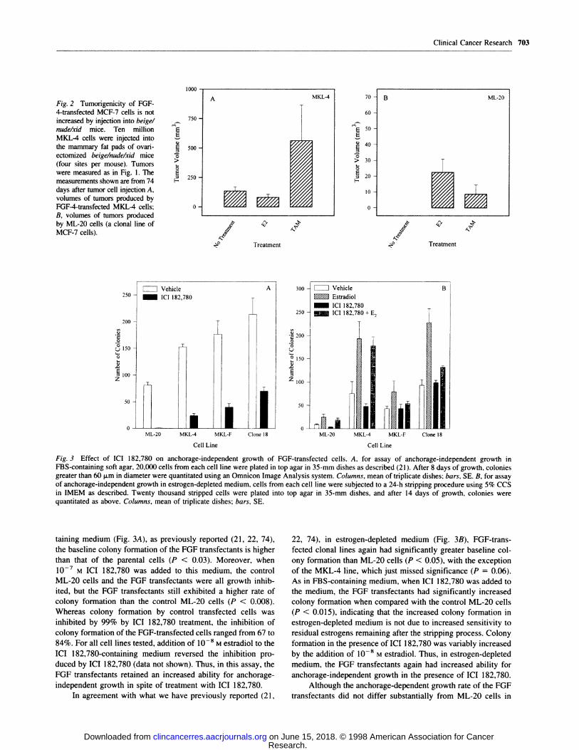
Transient transfection of control and an FGF-overexpress-
ing cell line with an ERE-containing luciferase reporter con-
struct also indicated that ER was not constitutiveby activated by
virtue of FGF transfection. We measured the ability of ERs
expressed by the control ML-20 and one FGF-transfected cell
line to direct transcription of the buciferase reporter gene driven
by an ERE-containing promoter under basal and estrogen or
FGF-l stimulated conditions (Fig. 8). Neither the clone 18 nor
the control ML-20 cells exhibited transcriptional activity greater
than background, as determined by a similar reporter plasmid in
which the ERE sequences were scrambled, under any experi-
mental conditions other than estrogen treatment. In particular,
transcription of the reporter was not elevated in the FGF-l-
transfected clone 1 8 cells under estrogen-depleted conditions or
decreased when treated with IC! 182,780. Moreover, treatment
of the control ML-2O cell line with FGF-l did not induce
transcription of the reporter. We conclude that ERs in the
FGF-transfected clone 1 8 line do not exhibit constitutive tran-
scriptional activity and that activation of FGF receptors of
untransfected cells by FGF-l does not induce ER-mediated
transcriptional activity. Thus, taken together. our results indicate
that the transfected FGFs are stimulating growth by a mecha-
nism that bypasses the ER-mediated growth-stimulatory path-
way.
DISCUSSION
In this report, we have shown that the estrogen-independent
in vivo growth of FGF-transfected MCF-7 cells is not affected
by ICI 182,780 or by either of two aromatase inhibitors. This
treatment failure cannot be attributed to an estrogen-, tamox-
ifen-, or FGF-induced decrease in the immunocompetence re-
maining in nude mice. The persistence of estrogen-independent
growth despite pharmacological strategies to abrogate all estro-
genie activity supports the hypothesis that the effect of FOF
transfection in promoting such growth is due to a direct effect of
the transfected FGF. These findings are supported by our data
showing normal numbers of ERs present in the FGF transfec-
tants, which are able to direct expression of known estrogen-
induced genes and interact with an ERE-containing promoter in
a reporter plasmid but which are not constitutively activated in
the FOF-transfected cell lines. The direct effect of FGF trans-
fection on tumor growth may be to promote mitogenesis of the
transfected cells by autocrine or intracrine FGF receptor activa-
tion. This viewpoint is supported by the generally increased
proliferation rate and colony-forming ability of the FGF trans-
fectants under estrogen-depleted tissue culture conditions. How-
ever, in addition to having a mitogenic effect on tumor cells. the
Research. on June 15, 2018. © 1998 American Association for Cancerclincancerres.aacrjournals.org Downloaded from
5000 -
i 4000 -
� 3000
CC
�. 2000S/C
1000
0
Clone 18
r-i#{149} fl#{149}n.
6000 -�-------�-�� -��-�---� �________� ML-20
5000 H
F- �;e: 4000 -��
05
C
� 30000�
�. 2000 -
000- �
0 � �
-� .� �- .�
�, ‘�-
Treatment
ni�7 .�. �
�vTreatment
Clinical Cancer Research 707
Fig. 8 ER is not constitutivelyactivated in FGF-l-overexpress-
ing or FGF-l-treated MCF-7
cells. Cells stripped of estrogens
were transiently transfected witha luciferase reporter plasmid(pGLB, Promega) driven by amouse mammary tumor virus
promoter that contained two
ERE sequences (pGLB-MERE)
or the same plasmid with the
ERE sequences scrambled(pGLB-MNON). Luciferase re-suIts were corrected for protein
content of lysates. transfectionefficiency by comparison with a
cotransfected constitutively ex-pressed CAT reporter plasmid
(pCMV-CAT), and background
luciferase activity.
transfected FGF may also stimulate tumor growth via effects on
stromal components of the tumor, such as fibroblasts or endo-
thebial cells. Investigations concerning the relative contributions
of autocrine and/or paracrine effects of the transfected FGF- 1 to
tumor growth of the clone 1 8 cells have revealed that autocrine
or intracrine effects are important for estrogen-independent tu-
mor growth but do not seem to be necessary for either estrogen-
stimulated or tamoxifen-resistant tumor growth (74). Moreover,
the insensitivity of the estrogen-independent in viva growth of
the FOF transfectants to IC! 182,780 or the aromatase inhibitors
implies that clinical tamoxifen resistance due to FGF receptor-
mediated signaling may not respond to a second hormonal
therapy.
The mechanism(s) determining whether a given clinical
case of antiestrogen resistance will be responsive to a second
hormonal manipulation has not been elucidated and may be
multifactorial. Because only 30-40% of patients with acquired
tamoxifen resistance have a positive response to a second hor-
monal therapy, with an additional 30% showing no immediate
disease progression after switching therapies ( 13, 14, 19, 20),
ER alterations that render the receptor unresponsive or differ-
entially responsive to hormones or utilization of alternative
pathways for growth that do not involve the ER may be respon-
sible for 30-70% of acquired tamoxifen resistance. ER muta-
tions and/or splice variants have been shown to be present in
only a low percentage of clinical breast cancer cases (2, 1 1 , 75,
76). Only a subset of these alterations results in receptors
capable of producing tamoxifen resistance, and one of these, an
exon S deletion. is not present more frequently in tamoxifen-
resistant patients (77). Therefore, this mechanism is probably
not a common mode of tamoxifen resistance.
The recent discovery of the ERI3 gene (78) has raised the
question of whether responses to antiestrogens for this gene
product differ from those of the previously studied ERa. To
date. transcription driven by ER�3 at a consensus ERE in re-
sponse to various antiestrogens does not seem to be different
from that of ERa (79). However, AP-l-mediated transcription
can be influenced by ER activation independent of ER-ERE
- p(uIi3-MERE�
- pGLB-MNONJ
interactions (71, 80). Both !CI 182,780 and tamoxifen were
shown to activate transfected ER�3-induced transcription at
AP- 1 sites in a transient transfection assay using MCF-7 cells
(79). However, native MCF-7 cells have not been shown to
express substantial levels of ER�3 (81). Therefore, if our ML-20
cells are representative of native MCF-7 cells described in the
literature, we would not expect that the effects of FGF trans-
fection on in vivo and in vitro growth are due to ER�3-mediated
stimulation of transcription at AP- I sites.
Ligand-independent activation of the ER by growth factors
has been shown to occur when activated mitogen-activated
protein kinase phosphorylates a serine residue within the AF1
domain of the ER (30-32). This phosphorylation also appears to
increase the agonistic effects of tamoxifen but does not alter the
antagonistic properties of pure antiestrogens, such as IC!
182,780 (30). Thus, growth factor pathways that result in acti-
vation of the ER might be expected to produce a situation in
which tumor growth becomes supersensitive to low concentra-
tions of hormonal agonists. Under such circumstances, the par-
tial agonist activity of tamoxifen in promoting growth might be
enhanced, whereas a pure antiestrogen would remain growth
inhibitory. In one such example. overexpression of ERB-B2 in
MCF-7 cells has been shown to result in estrogen-independent
and tamoxifen-insensitive growth in vitro and in vivo (23, 30).
Moreover, clinical studies show decreased benefit of tamoxifen
treatment in node-negative ERB-B2-overexpressing breast tu-
mors (27). In support of the possibility that activation of this
particular signaling pathway results in down-regulation of ERs,
similar to the effect of agonist activation, it has been found that
activation of ERB-B2 signaling pathways in MCF-7 cells by the
ligand, heregulin, activates the ER by phosphorybation (25) and
down-regulates ER number (25, 26). This implies that an inter-
action between the activated ERB-B2 signal transduction path-
way and the ER is responsible for the estrogen independence
and decreased tamoxifen sensitivity of the ERB-B2 transfec-
tants. This interpretation is further supported by the observation
that the effects of added heregulin on ER activation in parental
MCF-7 cells can be blocked with IC! 182,780, which also
Research. on June 15, 2018. © 1998 American Association for Cancerclincancerres.aacrjournals.org Downloaded from
708 ICI I 82.780 Effects on FGF-transfected MCF-7 Cells
blocks activation resulting from ERB-B2 overexpression (25).
The results using these transfected cell systems, therefore, sup-
port the view that interactions between these particular growth
factor pathways and the ER can produce tamoxifen resistance
but may still be at least partially sensitive to IC! 182,780. Our
data in this report suggest that this is not the case with FGF
signaling, further suggesting that there are alternative growth-
stimulating pathways that bypass the ER.
In vitro growth assays with the FGF transfectants demon-
strate an increased estrogen-independent growth and reduced
effectiveness of a pure antiestrogen, under both anchorage-
dependent and anchorage-independent conditions, and suggest
that increased growth is not due to increased potency of residual
low levels of estrogen. Because separate experiments using
pooled FGF- 1 transfectants, as compared with pooled control
transfectants, also demonstrate reduced sensitivity to IC!
182,780 (data not shown) similar to that seen with the clonal cell
lines used in this study, this effect is unlikely to be due to clonal
variation. Moreover, when autocrine FGF-l signaling in the
FGF- 1-transfected clone 18 cells is abrogated by subsequent
transfection with a dominant negative FOF receptor, sensitivity
to IC! 182,780 is restored (74), implying that the reduced
sensitivity seen in these experiments is due to FGF receptor
activation by the transfected FGF.
Despite the activation of endogenous FGF receptors (82)
by the transfected ligand, we did not observe a down-regulation
of ERs in these cells, as was reported for the ERB-B2 transfec-
tants, above. Although our data showing a slightly decreased
potency of IC! 182,780 in inhibiting estradiol-stimulated growth
could be interpreted as showing a slight effect of FGF receptor
pathway activation on the affinity of the ER for ICI 1 82,780, the
similar potency of l7�3-estradiol in all cell lines argues against
sensitization of the ER to small amounts of estradiol being
responsible for the estrogen-independent growth of these cells
and suggests that FGF overexpression does not alter the affinity
of ER for l7�3-estradiol. In addition, IC! 182,780 did not reduce
anchorage-independent growth to bevels of the parental cells, as
one would expect if such growth were due to bigand-indepen-
dent activation of the ER by the transfected FGF (Fig. 3).
Moreover, we do not observe enhanced levels of mRNA estro-
gen-responsive genes, such as pS2, cathepsin D, or progesterone
receptor under estrogen-depleted conditions in our transfectants.
Finally, transcriptional assays using an ERE-containing reporter
did not show high basal levels of transcriptional activity in the
FOF transfectants. When taken together, these data provide
evidence for a mechanism by which FGF-stimulated estrogen-
independent growth bypasses the ER signal transduction path-
way. Moreover, the algebraically additive effects of tamoxifen
and estrogen to the estrogen-independent in vivo growth of some
of the FGF transfectants (21, 22) and continued high frequency
of colony formation in ICI 182,780-containing medium argues
for an additive effect of ER signaling to that produced by the
FGF. Studies to further investigate interactions between ER and
FGF receptor signaling pathways in these transfectants are un-
der way in our laboratory.
Previous laboratory attempts to mimic tamoxifen resistance
have produced varied results with respect to cross-resistance to
steroidal antiestrogens and evidence of interaction of growth
factor receptor-activated and ER-activated growth pathways.
MCF-7 cells selected for growth in estrogen-depleted medium
have acquired supersensitivity to estrogen in vitro and in viva
and remain sensitive to steroidal antiestrogens (69). When the
LCC 1 cell line, derived from MCF-7 cells by progressive in vivo
and in vitro selection under estrogen-depleted conditions (83),
was subjected to a subsequent in vitro selection in tamoxifen,
the resulting LCC2 cell line remained sensitive to IC! 182,780
(84). However, a second cell line, designated LCC9, derived
from the same LCC1 parent but selected instead with IC!
182,780, is cross-resistant to tamoxifen (85). Other cell lines
selected for resistance to the steroidal pure antiestrogens IC!
164,384 or IC! 182,780 are cross-resistant to the other steroidal
antiestrogen but not to tamoxifen (86). Additionally, a MCF-7-
derived cell line selected for estrogen-independent growth in
nude mice exhibits decreased numbers of ERs, is growth stim-
ulated in vivo by tamoxifen, and exhibits increased AP-l-medi-
ated transcriptional activation independent of ER activation but
retains sensitivity to IC! 182,780 (87). However, tumors pro-
duced by MCF-7 cells selected in viva for resistance to IC!
182,780 have shown only weak responses to tamoxifen (88).
MCF-7 or T47D cells that inducibly overexpress cyclin D have
been found to exhibit resistance to both tamoxifen and steroidal
antiestrogens (89). Because cyclin D has been shown to be at the
convergence of growth factor and ER pathways that stimulate
growth (90), these results could be pertinent to our model
system. Together, these diverse data imply heterogeneity for the
mechanism of antiestrogen resistance and predict that clinical
response to a second hormonal therapy in a given case of breast
cancer will depend on the characteristics of that particular
tumor.
In summaiy, our studies implicate direct action by FGFs in the
estrogen-independent growth produced by transfection of either
FGF-4 or FGF-l into MCF-7 cells, and they rule out effects
resulting from increased sensitivity of the transfectants to small
amounts of extraovarian estrogen production. Our data also imply
that effects of the transfected FGFs do not involve a direct inter-
action with the ER itselfor ER signal transduction pathways, which
ultimately stimulate growth, although the two pathways may still
converge or interact at common downstream targets (90). We
demonstrate that FGF activity at its receptor is capable of produc-
ing an increased proliferation rate of the transfectants under estro-
gen-depleted conditions in vitro, and this effect may be partly
responsible for estrogen-independent growth in vivo. We and others
have found FGF family members to be expressed in breast tissue
and/or breast tumors (41-48). Moreover, FGF receptors are rather
ubiquitously expressed, have been shown to be present in clinical
breast cancer (49, 50), and can be activated by multiple FGF family
members as well as heparin, cell adhesion molecules, or activating
mutations (91). Thus, it is likely that FGF receptor-mediated sig-
naling is operative in a significant proportion of ER-positive breast
tumors. Therefore, the model described in this report might be
pertinent to a number of clinical cases of tumor growth that is
refractory to therapy with antiestrogens. In contrast to some of the
models mentioned above, which may mimic tamoxifen-resistant
breast tumors that would respond to a second hormonal therapy, we
predict that tumors in which FGF receptor-mediated signaling
drives autonomous growth would be refractory to alternative hor-
monab therapies, as well as to tamoxifen. Therapy of such tumors
with agents directed against the autocrine or paracrine effects of
Research. on June 15, 2018. © 1998 American Association for Cancerclincancerres.aacrjournals.org Downloaded from

Clinical Cancer Research 709
FGFs (53) might result in beneficial effects in such cases and might
result in the restoration of antiestrogen sensitivity.
ACKNOWLEDGMENTS
The authors are indebted to A. Brodie, A. Bhatnagar, and A.
Wakeling for providing 4-OHA, letrozole, and IC! 182,780, respec-
tively. B. M. Vose supplied formulated ICI 182,780. A. Murray and the
Lombardi Cancer Center Animal Research Resource assisted with ani-
mal experiments. P. Chambon and B. R. Westley provided the pS2 andcathepsin D riboprobes, respectively. S. Thomas and J. Alexander
provided technical assistance. We thank A. Wellstein and R. Clarke forthoughtfully critiquing the manuscript. Animal protocols for this workwere approved by the Georgetown University Animal Care and Use
Committee.
REFERENCES
1. Robertson, J. F. R. Oestrogen receptor: a stable phenotype in breastcancer. Br. J. Cancer, 73: 5-12. 1996.
2. Karnik, P. S., Kulkarni, S., Liu. X-P., Budd, G. T., and Bukowski,R. M. Estrogen receptor mutations in tamoxifen-resistant breast cancer.
Cancer Res., 54: 349-353, 1994.
3. Johnston, S. R. D., Saccani-Jotti, G., Smith, I. E., Salter, J., Newby,
J., Coppen. M., Ebbs, S. R., and Dowsett, M. Changes in estrogenreceptor, progesterone receptor. and p52 expression in tamoxifen-resist-ant human breast cancer. Cancer Res., 55: 3331-3338, 1995.
4. Johnston, S. R. D., Lu, B., Dowsett, M., Liang, X., Kaufman, M.,
Scott, G. K., Osborne, C. K., and Benz, C. C. Comparison of estrogenreceptor DNA binding in untreated and acquired antiestrogen-resistanthuman breast tumors. Cancer Res., 57: 3723-3727, 1997.
5. Jiang, S-Y., Langan-Fahey, S. M., Stella, A. L., McCague, R.. andJordan, V. C. Point mutation of estrogen receptor (ER) in the ligand-binding domain changes the pharmacology of antiestrogens in ER-
negative breast cancer cells stably expressing complementary DNAs forER. Mol. Endocrinol., 6: 2167-2174, 1992.
6. Catherino, W. H., Wolf, D. M., and Jordan, V. C. A naturallyoccurring estrogen receptor mutation results in increased estrogenicityof a tamoxifen analog. Mol. Endocrinol., 9: 1053-1063, 1995.
7. Wolf, D. M., and Fuqua. S. A. W. Mechanisms of action of anties-
trogens. Cancer Treat. Rev., 21: 247-271, 1995.
8. Mahfoudi, A., Roulet, E., Dauvois, S., Parker, M. G., and Wahli, W.
Specific mutations in the estrogen receptor change the properties of
antiestrogens to full agonists. Biochemistry, 92: 4206-4210, 1995.
9. Osborne, C. K.. Coronado, E., Allred, D. C., Wiebe, V., and DeGre-gorio. M. Acquired tamoxifen resistance: correlation with reducedbreast tumor levels of tamoxifen and isomerization of trans-4-hy-
droxytamoxifen. J. NatI. Cancer Inst., 83: 1477-1482, 1991.
10. Tonetti, D. A., and Jordan, V. C. Possible mechanisms in the
emergence of tamoxifen-resistant breast cancer. Anticancer Drugs. 6:
498-507. 1995.
1 1. Osborne, C. K., and Fuqua, S. A. W. Mechanisms of tamoxifenresistance. Breast Cancer Res. Treat., 32: 49-55, 1994.
12. Pavlik, E. J., Nelson, K., Srinivasan, S., Powell, D. E., Kenady,
D. E., DePriest, P., Gallion, H. H., and van Nagell, J. R., Jr. Resistanceto tamoxifen with persisting sensitivity to estrogen: possible mediationby excessive antiestrogen binding site activity. Cancer Res.. 52: 4106-4112, 1992.
13. Howell, A., DeFriend, D., Robertson, J., Blamey, R., and Walton, P.
Response to a specific antioestrogen (ICI 182780) in tamoxifen-resistantbreast cancer. Lancet, 345: 29 -30, 1995.
14. Howell, A., Downey. S.. and Anderson. E. New endocrine therapiesfor breast cancer. Eur. J. Cancer, 32A: 576-588, 1996.
15. Miller, W. R. Aromatase inhibitors: where are we now9 Br. J.
Cancer. 73: 415-417, 1996.
16. Buzdar, A., Jonat, W., Howell, A.. Jones, S. E., Blomqvist. C..Vogel, C. L., Eiermann, W., Wolter, J. M., Azab, M., Webster, A., and
Plourde, P. V. Anastrozole, a potent and selective aromatase inhibitor,
versus megestrol acetate in postmenopausal women with advanced
breast cancer: results ofoverview analysis oftwo phase III trials. J. Clin.
Oncol., 14: 2000-2011, 1996.
17. Bonnefoi, H. R., Smith, I. E.. Dowsett, M., Trunet. P. F., Houston.S. J., da Luz, R. J., Rubens, R. D., Coombes, R. C., and Powles, T. J.Therapeutic effects of the aromatase inhibitor fadrozole hydrochloride
in advanced breast cancer. Br. J. Cancer, 73: 539-542, 1996.
18. Buzdar, A. U., Smith, R., Vogel. C.. Bonomi, P., Keller, A. M.,Favis, G., Mulagha. M., and Cooper. J. Fadrozole HCL (CGS- l6949A)
versus megestrol acetate treatment of postmenopausal patients with
metastatic breast carcinoma. Cancer (Phila.), 77: 2503-2513, 1996.
19. Howell, A., DeFriend, D. J., Robertson, J. F. R., Blamey, R. W..Anderson, L., Anderson, E., Sutcliffe, F. A., and Walton, P. Pharmaco-
kinetics, pharmacological and anti-tumour effects of the specific anti-
oestrogen IC! I 82780 in women with advanced breast cancer. Br. J.
Cancer, 74: 300-308, 1996.
20. Castiglione-Gertsch. M. New aromatase inhibitors: more selectiv-
ity, less toxicity, unfortunately, the same activity. Eur. J. Cancer, 3:
393-395. 1996.
21. McLeskey. S. W., Kurebayashi, J.. Honig, S. F.. Zwiebel, J.,
Lippman. M. E., Dickson, R. B., and Kern, F. G. Fibroblast growth
factor 4 transfection of MCF-7 cells produces cell lines that are tumor-
igenic and metastatic in ovariectomized or tamoxifen-treated athymic
nude mice. Cancer Res., 53: 2168-2177, 1993.
22. Zhang, L.. Kharbanda, S., Chen, D., Bullocks, J., Miller, D. L.,Ding, I. Y. F., Hanfelt, J., McLeskey, S. W.. and Kern, F. G. MCF-7
breast carcinoma cells overexpressing FGF- 1 form vascularized meta-
static tumors in ovariectomized or tamoxifen-treated nude mice. Onco-
gene, 15: 2093-2108, 1997.
23. Liu, Y., El-Ashry, D., Chen, D., Ding. I. Y. F.. and Kern, F. G.MCF-7 breast cancer cells overexpressing transfected c-erbB-2 have an
in vitro growth advantage in estrogen-depleted conditions and reduced
estrogen dependence and tamoxifen sensitivity in t’ivo. Breast CancerRes. Treat., 34: 97-1 17, 1995.
24. Benz, C. C., Scott, G. K.. Sarup. J. C., Johnson, R. M., Tripathy. D.,Coronado, E.. Shepard, H. M., and Osborne. C. K. Estrogen-dependent.tamoxifen-resistant tumorigenic growth of MCF-7 cells transfected with
HER2/neu. Breast Cancer Res. Treat., 24: 85-95, 1992.
25. Pietras, R. J., Arboleda, J., Reese, D. M., Wongvipat, N., Pegram,M. D., Ramos, L., Gorman, C. M., Parker, M. G., Sliwkowski, M. X.,and Slamon, D. J. HER-2 tyrosine kinase pathway targets estrogen
receptor and promotes hormone-independent growth in human breastcancer cells. Oncogene, 10: 2435-2446, 1995.
26. Tang, C. K., Perez, C.. Grunt, T., Waibel, C., Cho, C., and Lupu. R.
Involvement of heregulin432 in the acquisition of the hormone-inde-pendent phenotype of breast cancer cells. Cancer Res., 56: 3350-3358,
1996.
27. Carlomagno, C., Perrone, F.. Gallo, C.. De Laurentiis, M., Lauria,R., Morabito, A., Pettinato, G., Panico, L., D’Antonio, A., Bianco,A. R., and De Placido, S. c-erbB2 overexpression decreases the benefitof adjuvant tamoxifen in early stage breast cancer without axillarylymph node metastases. J. Clin. Oncol., 14: 2702-2708, 1996.
28. Arnold, S. F., Obourn, J. D., Jaffe, H., and Notides, A. C. Phos-phorylation of human estrogen receptor by mitogen-activated protein
kinase and casein kinase I!: consequences on DNA binding. J. Steroid.Biochem. Mol. Biol., 55: 163-172, 1995.
29. Katzenellenbogen, B. S., Montano, M. M., Le Goff, P., Schodin,
D. J., Kraus, W. L., Bhardwaj. B., and Fujimoto, N. Antiestrogens.Mechanisms and actions in target cells. J. Steroid Biochem. Mol. Biol.,
53: 387-393, 1995.
30. Kato, S., Endoh, H., Masuhiro, Y., Kitamoto, T., Uchiyama, S..Sasaki, H., Masushige, S., Gotoh, Y., Nishida, E., Kawashima, H.,
Metzger. D., and Chambon. P. Activation of the estrogen receptorthrough phosphorylation by mitogen-activated protein kinase. Science(Washington DC). 270: 1491-1494. 1995.
31. Bunone, 0.. Briand, P. A., Miksicek, R. J., and Picard, D. Activa-tion of the unliganded estrogen receptor by EGF involves the MAP
Research. on June 15, 2018. © 1998 American Association for Cancerclincancerres.aacrjournals.org Downloaded from

710 ICI I 82.780 Effects on FGF-transfected MCF-7 Cells
kinase pathway and direct phosphorylation. EMBO J., 15: 2174-2 183,
1996.
32. Ignar-Trowbridge, D. M., Pimentel, M., Parker, M. G., McLachlan,
J. A., and Korach, K. S. Peptide growth factor cross-talk with theestrogen receptor requires the A/B domain and occurs independently of
protein kinase C or estradiol. Endocrinology, 137: 1735-1744, 1996.
33. Arteaga, C. L.. Carty-Dugger, T., Moses, H. L., Hurd, S. D., and
Pietenpol, J. A. Transforming growth factor � can induce estrogen-independent tumorigenicity of human breast cancer cells in athymicmice. Cell Growth Differ., 4: 193-201 , 1993.
34. Arteaga, C. L., Hurd, S. D., Winnier, A. R., Johnson, M. D., Fendly,
B. M., and Forbes, J. T. Anti-transforming growth factor (TGF)-�3antibodies inhibit breast cancer cell tumorigenicity and increase mouse
spleen natural killer cell activity. J. Clin. Invest., 92: 2569-2576, 1993.
35. Clarke, R. Human breast cancer cell line xenografts as models ofbreast cancer: the immunobiologies of recipient mice and the charac-teristics of several tumorigenic cell lines. Breast Cancer Res. Treat., 39:
69-86. 1996.
36. Gottardis, M. M., Wagner, R. J., Borden, E. C., and Jordan, V. C.
Differential ability of antiestrogens to stimulate breast cancer (MCF-7)
growth in vivo and in vitro. Cancer Res., 49: 4765-4769, 1989.
37. Katzenellenbogen, J. A., O’Malley, B. W., and Katzenellenbogen,
B. S. Tripartite steroid hormone receptor pharmacology: interaction withmultiple effector sites as a basis for the cell- and promoter-specific
action of these hormones. Mol. Endocrinol., 10: 119-131, 1996.
38. Horwitz, K. B., Jackson, T. A., Bain, D. L., Richer, J. K., Takimoto,
G. S.. and Tung, L. Nuclear receptor coactivators and corepressors. Mob.Endocrinol., 10: 1167-1177, 1996.
39. Smith, C. L., Nawaz, Z.. and O’Malley, B. W. Coactivator andcorepressor regulation of the agonistlantagonist activity of the mixed
antiestrogen, 4-hydroxytamoxifen. Mol. Endocrinol., II: 657-666,
1997.
40. Jackson, T. A., Richer, J. K., Bain, D. L., Takimoto, 0. S., Tung, L.,and Horwitz, K. B. The partial agonist activity of antagonist-occupied
steroid receptors is controlled by a novel hinge domain-binding coacti-
vator L7/SPA and the corepressors N-CoR or SMRT. Mol. Endocrinob.,Ii: 693-705, 1997.
41. Ding, I. Y. F., McLeskey, S. W., Chang, K., Fu, Y. M., Acol, J. C.,
Shou, M. T., Alitalo, K., and Kern, F. G. Expression of fibroblast growth
factors (FGFs) and receptors (FGFRs) in human breast carcinomas.Proc. Am. Assoc. Cancer Res., 33: 269, 1992.
42. Ulich, T. R., Yi, E. S., Cardiff, R., Yin, S., Bikhazi, N., Biltz, R.,
Moms, C. F., and Pierce, 0. F. Keratinocyte growth factor is a growth
factor for mammary epithelium in viva. Am. J. Pathol., 144: 862-868,
1994.
43. Visscher, D. W., DeMattia, F., Ottosen, S., Sarkar, F. H., and
Crissman, J. D. Biologic and clinical significance of basic fibrobbast
growth factor immunostaining in breast carcinoma. Mod. Pathol., 8:
665-670, 1995.
44. Koos, R. D., Banks, P. K., Inkster, S. E., Yue, W., and Brodie,A. M. H. Detection of aromatase and keratinocyte growth factor expres-sion in breast tumors using reverse transcription-polymerase chain re-
action. J. Steroid Biochem. Mol. Biol.. 45: 217-225, 1993.
45. Yiangou, C., Gomm, J. J., Coope, R. C., Law, M., Luqmani, Y. A.,
Shousha. S., Coombes, R. C., and Johnston, C. L. Fibrobbast growthfactor 2 in breast cancer: occurrence and prognostic significance. Br. J.
Cancer, 75: 28-33, 1997.
46. Gomm, J. J., Smith, J., Ryall, 0. K., Baillie, R., Turnbull, L., andCoombes, R. C. Localization of basic fibrobbast growth factor andtransforming growth factor �3 I in the human mammary gland. CancerRes.. 51: 4685-4692, 1991.
47. Bansai, G. S., Yiangou, C.. Gomm, J. J., Luqmani, Y. A., Coombes,
R. C., and Johnston, C. L. Expression of fibroblast growth factor 1 is
lower in breast cancer than in normal breast. Br. J. Cancer, 72: 1420-
1426, 1995.
48. Smith, J., Yelland, A., Baillie, R., and Coombes, R. C. Acidic and
basic fibroblast growth factors in human breast tissue. Eur. J. Cancer,
30A: 496-503, 1994.
49. Luqmani, Y. A., Mortimer, C., Yiangou, C., Johnston, C. L.,Bansab, G. S., Sinnett, D., Law, M., and Coombes, R. C. Expression
of two variant forms of fibroblast growth factor receptor type 1 in
human breast. Int. J. Cancer, 64: 274-279, 1995.
50. Penault-Llorca, F., Bertucci, F., AdelaIde, J., Parc, P., Coulier, F..Jacquemier, J., Birnbaum, D., and deLapeyriere, 0. Expression of FGFand FGF receptor genes in human breast cancer. Int. J. Cancer. 61:
170-176, 1995.
51. Kurebayashi, J., McLeskey, S. W., Johnson, M. D., Lippman, M. E.,Dickson, R. B., and Kern, F. 0. Quantitative demonstration of sponta-neous metastasis by MCF-7 human breast cancer cells cotransfectedwith fibroblast growth factor 4 and LacZ. Cancer Res., 53: 2178-2187,
1993.
52. McLeskey, S. W., Zhang, L., Kharbanda, S., Kurebayashi, J.,Lippman, M. E., Dickson, R. B., and Kern, F. G. Fibroblast growth
factor overexpressing breast carcinoma cells as models of angiogenesisand metastasis. Breast Cancer Res. Treat., 39: 103-117, 1996.
53. McLeskey, S. W., Zhang, L., Trock, B. J., Kharbanda, S., Liu, Y.,
Gottardis, M. M., Lippman, M. E., and Kern, F. 0. Effects of AGM-1470 and pentosan polysulphate on tumorigenicity and metastasis of
FGF-transfected MCF-7 cells. Br. J. Cancer. 73: 1053-1062, 1996.
54. Edwards, D. P., and McGuire, W. L. 1743-estradiol is a biologically
active estrogen in human breast cancer cells in tissue culture. Endocri-
nology, 107: 884-891, 1980.
55. Miller, D. L., El-Ashry, D., Cheville, A. L., Liu, Y., McLeskey,S. W., and Kern. F. G. Emergence of MCF-7 cells overexpressing atransfected epidermal growth factor receptor (EGFR) under estrogen-
depleted conditions: evidence for a role of EGFR in breast cancergrowth and progression. Cell Growth Differ., 5: 1263-1274, 1994.
56. Bradford, M. A rapid and sensitive method for quantitation ofmicrogram quantities of protein utilizing the principle of protein-dye
binding. Anal. Biochem., 72: 248-254, 1976.
57. Chevillard, S., MUller, A., Levalois, C., Lain#{233}-Bidron,C., Vielh, P.,and Magdel#{233}nat,H. Reverse transcnption-polymerase chain reaction(RT-PCR) assays of estrogen and progesterone receptors in breast
cancer. Breast Cancer Res. Treat., 41: 81-89, 1996.
58. Chen, C., and Okayama, H. High-efficiency transformation ofmammalian cells by plasmid DNA. Mol. Cell. Biol., 7: 2745-2752,
1987.
59. El-Ashry, D., Chrysolgelos, S. A., Lippman, M. E., and Kern, F. G.
Estrogen induction of TGF-a is mediated by an estrogen responseelement composed of two imperfect palindromes. J. Steroid Biochem.Mol. Biol., 59: 261-269, 1996.
60. Heitjan, D. F., Manni, A., and Santen, R. J. Statistical analysis of in
viva tumor growth experiments. Cancer Res., 53: 6042-6050, 1993.
61. Brodie, A. M. H., and Santen, R. J. Aromatase and its inhibitors in
breast cancer treatment: overview and perspective. Breast Cancer Res.Treat., 30: 1-6, 1994.
62. Dowsett, M., and Coombes, R. C. Second generation aromataseinhibitor: 4-hydroxyandrostenedione. Breast Cancer Res. Treat., 30:
81-87, 1994.
63. Demers, L. M., Lipton, A., Harvey, H. A., Kambic, K. B., Gross-berg. H., Brady, C., and Santen, R. J. The efficacy of CGS 20267 insuppressing estrogen biosynthesis in patients with advanced stage breast
cancer. J. Steroid Biochem. Mol. Biol., 44: 687-691, 1993.
64. Screpanti, I., Felli, M. P., Toniato, E., Meco, D., Martinotti, S.,Frati, L., Santoni, A., and Gulino, A. Enhancement of natural-killer-cell
susceptibility of human breast-cancer cells by estradiol and v-Ha-ras
oncogene. Int. J. Cancer, 47: 445-449, 1991.
65. Knabbe, C., Lippman, M. E., Wakefield, L. M., Flanders, K. C.,Kasid, A., Derynck. R., and Dickson, R. B. Evidence that transforminggrowth factor �3 is a hormonally regulated negative growth factor inhuman breast cancer cells. Cell, 48: 417-428, 1987.
66. Melder, R. J., Koenig, G. C., Witwer, B. P., Safabakhsh, N., Munn,L. L., and Jam, R. K. During angiogenesis, vascular endothelial growth
Research. on June 15, 2018. © 1998 American Association for Cancerclincancerres.aacrjournals.org Downloaded from

Clinical Cancer Research 711
factor and basic fibroblast growth factor regulate natural killer celladhesion to tumor endothelium. Nat. Med., 2: 992-997, 1996.
67. Osborne, C. K., Hobbs, K., and Clark, G. M. Effect ofestrogens andantiestrogens on growth of human breast cancer cells in athymic nude
mice. Cancer Res., 45: 584-590, 1985.
68. Katzenellenbogen, B. S., Kendra, K. L., Norman, M. J., andBerthois, Y. Proliferation, hormonal responsiveness, and estrogen re-
ceptor content of MCF-7 human breast cancer cells grown in theshort-term and long-term absence of estrogens. Cancer Res., 47: 4355-
4360, 1987.
69. Masamura, S., Santner, S. J., Heitjan, D. F., and Santen, R. J.Estrogen deprivation causes estradiol hypersensitivity in human breastcancer cells. J. Clin. Endocrinol. Metab., 80: 2918-2925, 1995.
70. Wakeling, A. E. Comparative studies on the effects of steroidal and
nonsteroidal oestrogen antagonists on the proliferation of human breastcancer cells. J. Steroid Biochem., 34: 183-188, 1989.
71. Umayahara, Y., Kawamon, R., Watada, H., Imano, E., Iwama. N..
Morishima, T., Yamasaki, Y., Kajimoto, Y., and Kamada, T. Estrogen
regulation of insulin-like growth factor I gene transcription involves anAP-l enhancer. J. Biol. Chem., 269: 16433-16442, 1994.
72. Ignar-Trowbridge, D. M., Teng, C. T., Ross, K. A., Parker, M. G.,
and Korach, K. S. Peptide growth factors elicit estrogen receptor-dependent transcriptional activation of an estrogen-response element.Mol. Endocnnol., 7: 992-998, 1993.
73. El-Tanani, M. K. K., and Green, C. D. Two separate mechanisms
for bigand-independent activation of the estrogen receptor. Mol. Endo-crinol., 11: 928-937, 1997.
74. Zhang, L., Kharbanda, S., Hanfelt, J., and Kern, F. 0. Both auto-
crine and paracrine effects of transfected acidic fibroblast growth factorare involved in the estrogen-independent and antiestrogen-resistantgrowth of MCF-7 breast cancer cells. Cancer Res., 58: 352-361, 1998.
75. Leygue, E., Huang, A., Murphy, L. C., and Watson, P. H. Preva-lence of estrogen receptor variant messenger RNAs in human breastcancer. Cancer Res., 56: 4324-4327. 1996.
76. Roodi, N., Bailey, L. R., Kao, W-Y., Vemer, C. S.. Yee, C. J..
Dupont, W. D., and Parl, F. F. Estrogen receptor gene analysis inestrogen receptor-positive and receptor-negative primary breast cancer.J. Natl. Cancer Inst., 87: 446-451, 1995.
77. Daffada, A. A. I., Johnston, S. R. D., Smith, I. E., Detre, S., King,N., and Dowsett, M. Exon 5 deletion variant estrogen receptor messen-ger RNA expression in relation to tamoxifen resistance and progesteronereceptor/pS2 status in human breast cancer. Cancer Res., 55: 288 -293,
1995.
78. Kuiper, G. 0. J. M., Enmark, E., Pelto-Huikko, M., Nilsson, S., andGustafsson, J. A. Cloning of a novel estrogen receptor expressed in ratprostate and ovary. Proc. Nail. Acad. Sci. USA, 93: 5925-5930, 1997.
79. Paech, K., Webb, P., Kuiper, G. G. J. M., Nilsson, S., Gustafsson,
J. A., Kushner, P. J., and Scanlon, T. S. Differential ligand activation ofestrogen receptors ERrs and ER[3 at APi sites. Science (WashingtonDC), 277: 1508-1510, 1997.
80. Gaub. M-P., Bellard, M., Scheuer, I., Chambon, P., and Sassone-Corsi, P. Activation of the ovalbumin gene by the estrogen receptorinvolves the fos-jun complex. Cell, 63: 1267-1276, 1990.
81. Kuiper, G. 0. J. M., Carlsson, B., Grandien, K., Enmark, E.,
Haggblad, J., Nilsson, S., and Gustafsson, J. A. Comparison of theligand binding specificity and transcript tissue distribution of estrogen
receptors a and �3. Endocrinology, /38: 863-870, 1997.
82. McLeskey, S. W., Ding, I. Y. F., Lippman, M. E., and Kern, F. G.
MDA-MB- I 34 breast carcinoma cells overexpress FGF receptors andare growth-inhibited by FGF ligands. Cancer Res., 54: 523-529, 1994.
83. Brtinner, N., Boulay, V., Fojo, A., Freter. C. E., Lippman. M. E..
and Clarke, R. Acquisition of hormone-independent growth in MCF-7cells is accompanied by increased expression of estrogen-regulated
genes, but without detectable DNA amplifications. Cancer Res., 53:
283-290, 1993.
84. Brunner, N., Frandsen, T. L., Holst-Hansen, C., Bei, M., Thompson.
E. W.. Wakeling, A. E., Lippman, M. E., and Clarke, R. MCF7ILCC2:a 4-hydroxytamoxifen resistant human breast cancer variant that retains
sensitivity to the steroidal antiestrogen ICI 182,780. Cancer Res.. 53:
3229-3232, 1993.
85. Brunner, N., Boysen. B., Jirus, S., Skaar, T. C., Holst-Hansen, C..
Lippman. J.. Frandsen, T., Spang-Thomsen, M., Fuqua, S. A. W., andClarke, R. MCF7ILCC9: an antiestrogen-resistant MCF-7 variant inwhich acquired resistance to the steroidal antiestrogen ICI I 82,780
confers an early cross-resistance to the nonsteroidal antiestrogen tamox-ifen. Cancer Res., 57: 3486-3493, 1997.
86. Lykkesfeldt, A. E., Larsen, S. S., and Briand, P. Human breast
cancer cell lines resistant to pure anti-estrogens are sensitive to tamox-ifen treatment. mt. J. Cancer, 61: 529-534, 1995.
87. Dumont, J. A.. Bitonti, A. J., Wallace, C. D., Baumann, R. J.,Cashman, E. A., and Gross-Doersen, D. E. Progression of MCF-7 breast
cancer cells to antiestrogen-resistant phenotype is accompanied by dc-
vated levels of AP-l DNA binding activity. Cell Growth Differ.. 7:351-359, 1996.
88. Osborne, C. K., Coronado-Heinsohn, E. B., Hilsenbeck, S. G.,
McCue, B. L., Wakeling, A. E., McClelland, R. A., Manning. D. L.. andNicholson, R. I. Comparison of the effects of a pure steroidal antiestro-
gen with those of tamoxifen in a model of human breast cancer. J. NatI.Cancer Inst., 87: 746-750, 1995.
89. Wilcken, N. R. C., Prall, 0. W. J., Musgrove, E. A., and Sutherland,
R. L. Inducible overexpression of cyclin Dl in breast cancer cellsreverses the growth-inhibitory effects of antiestrogens. Clin. CancerRes., 3: 849-854, 1997.
90. Lukas, J., Bartkova, J., and Bartek, J. Convergence of mitogenicsignalling cascades from diverse classes of receptors at the cyclin
D-cyclin-dependent kinase-pRb-controlled G1 checkpoint. Mol. Cell.
Biol., 16: 6917-6925, 1996.
91. Green, P. J., Walsh, F. S., and Doherty, P. Promiscuity of fibroblastgrowth factor receptors. BioEssays, 18: 639-646, 1996.
Research. on June 15, 2018. © 1998 American Association for Cancerclincancerres.aacrjournals.org Downloaded from

1998;4:697-711. Clin Cancer Res S W McLeskey, L Zhang, D El-Ashry, et al. and two aromatase inhibitors.cells are cross-resistant in vivo to the antiestrogen ICI 182,780 Tamoxifen-resistant fibroblast growth factor-transfected MCF-7
Updated version
http://clincancerres.aacrjournals.org/content/4/3/697
Access the most recent version of this article at:
E-mail alerts related to this article or journal.Sign up to receive free email-alerts
Subscriptions
Reprints and
To order reprints of this article or to subscribe to the journal, contact the AACR Publications
Permissions
Rightslink site. Click on "Request Permissions" which will take you to the Copyright Clearance Center's (CCC)
.http://clincancerres.aacrjournals.org/content/4/3/697To request permission to re-use all or part of this article, use this link
Research. on June 15, 2018. © 1998 American Association for Cancerclincancerres.aacrjournals.org Downloaded from