Embed Size (px)
Citation preview

18
Biomarkers for Melanoma Diagnosis and the Technologies Used to Identify Them
Takeshi Mori1 and Jeong-Hun Kang2 1Department of Applied Chemistry, Faculty of Engineering,
Kyushu University, 744 Motooka, Nishi-Ku, Fukuoka 819-0395, 2Department of Biomedical Engineering,
National Cerebral and Cardiovascular Center Research Institute, 5-7-1 Fujishiro-dai, Suita, Osaka 565-8565,
Japan
1. Introduction
Melanoma is a malignant tumor originating from melanocytes (pigment-producing cells).
Although the tumor is mainly detected in skin (cutaneous melanoma), it can also be
detected in the eye (uveal melanoma), gastrointestinal (GI) tract and oral mucosa and genital
tract (mucosal melanoma) (Landreville et al., 2008; Akaraviputh et al., 2010; Bakalian et al.,
2008; Rigel et al., 2010; Seetharamu et al., 2010). Melanoma can be classified as belonging to
one of four subtypes: superficial spreading, nodular, lentigo maligna, and acral lentiginous
melanoma. These subtypes are characterized based on prognosis, incidence of metastasis
and the frequency of gene mutations (e.g., BRAF and NRAS) (Saldanha et al., 2006; Jaeger et
al., 2007; Markovic et al., 2007; Jönsson et al., 2010). Superficial spreading melanoma is the
most common form of melanoma found in Caucasian populations, while the acral
lentiginous melanoma is frequently detected in Asian and African populations (Cress and
Holly, 1997; Weyers et al., 1999).
Several important risk factors that have been linked to the development of melanoma have
been identified. Of these risk factors, most can be considered to be either environmental
factors, such as exposure to ultraviolet (UV) radiation, especially in childhood, or other
host factors such as family history and melanocytic nevi (Markovic et al., 2007; Schulman
and Fisher, 2009), but other cancer risk factors such as smoking (Osterlind et al., 1988), diet
(Osterlind et al., 1988; Veierod et al., 1997) or hormone therapy (Naldi et al., 2005) have not
been found to be associated with an increased risk of melanoma. The risk of developing
melanoma is higher in Caucasian than in Asian or African populations. This is closely
related to skin pigmentation as melanin has been shown to have a protective function for
UV-induced melanoma and Caucasian populations show low levels of melanogenesis
(Lens and Dawes, 2004; Hu et al., 2008; Jemal et al., 2010). In general, the melanocortin-1
receptor (MC1R), which is a G-protein-coupled receptor (GPCR), stimulates melanogenesis
through the activation of adenylate cyclase and protein kinase A (PKA) (Jordan and
Jackson, 1998; Rouzaud et al., 2003). Its genetic variants are associated with melanoma
www.intechopen.com
Breakthroughs in Melanoma Research
390
incidence and sun sensitivity (Box et al., 2001; Markovic et al., 2007). Moreover, the risk of
developing melanoma is greater in males than in females over the age of 40, although the
opposite effect is observed in patients under 40 years old (Lens and Dawes, 2004; Jemal et
al., 2010).
The global incidence of melanoma has increased over the past decades (Markovic et al.,
2007; Jemal et al., 2010, Rigel et al., 2010). The 5-year survival rate for melanoma is higher
than for other prominent cancers such as tumors of the prostate, ovary, liver and bile duct,
lung and bronchus, colon and rectum, and stomach. Yet, the early diagnosis and treatment
of melanoma is crucial to increasing the survival rate (Jemal et al., 2010, Rigel et al., 2010).
An important early diagnostic methodology for melanoma is the ABCDE criteria, which is
defined by describing changes to the appearance of the suspected lesion based on the
following features: Asymmetry, Border (irregularity), Color (variegation), Diameter and
Evolution (over time). Other diagnostic strategies also typically utilized include histological
and/or molecular analysis (e.g., genes or proteins profiling) of biopsied material,
dermoscopy (also known as dermatoscopy or epiluminescent microscopy) using a light-
based magnification or digital (computer)-assisted device, ultrasound imaging and magnetic
resonance imaging (Abbasi et al., 2004; Rigel et al., 2005; Markovic et al., 2007; Psaty and
Halpern, 2009; Rigel et al., 2010).
Recently there has also been a move toward establishing biomarkers for malignant
melanoma. These types of biological markers are not only beneficial for the diagnosis of
melanoma, but also allow physicians to monitor the recurrence of melanoma after surgical
resection, or to monitor the effect of radiation or anticancer drug therapies. To identify
putative melanoma biomarkers in tissue samples or body fluids, a number of methodologies
can be utilized, including two-dimensional gel electrophoresis (2-DE) and high throughput
microarray technology.
2. Melanoma biomarkers
2.1 Cellular signals and tissue biomarkers (immunohistochemical biomarkers)
Signal transduction pathways are the mechanism through which cells respond to the
extracellular signals (ligands) required to regulate or modulate downstream gene
expression. These extracellular signals activate signal transduction pathways by either
penetrating the cellular membrane or binding to specific receptors. The activated receptors
are then able to change the quantity or intracellular distribution of the second messengers
through the use of effector molecules. Second messengers also activate protein targets,
which control downstream gene expression. In these cellular signal transduction pathways,
phosphorylation of the target proteins (by protein kinases), or dephosphorylation or
proteolytic cleavage (by proteases) play a key role in cell division and motility, apoptosis
and carcinogenesis.
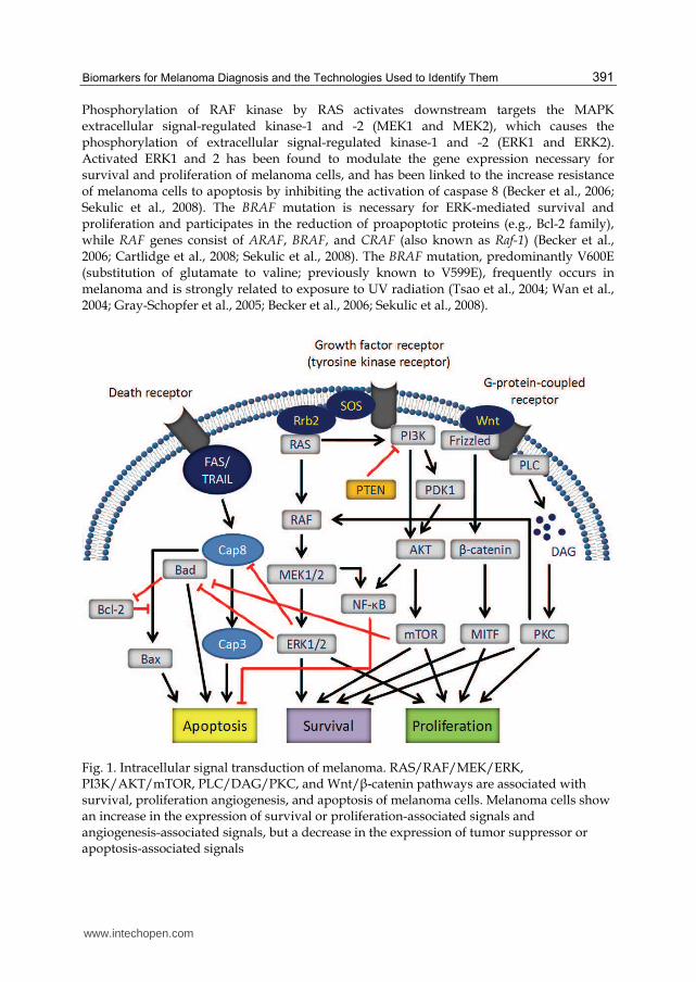
In melanoma cells, major signal transduction pathways are RAS/RAF/MEK/ERK and the PI3K/AKT (as known as protein kinase B)/mTOR pathway; however, other pathways such as PLC/DAG/PKC or Wnt/β-catenin pathway have also been identified (Figure 1). The interaction of a number of different ligands [e.g., fibroblast growth factor (FGF), vascular endothelial growth factor (VEGF), or epidermal growth factor (EGF)] with their respective receptors [e.g., growth factor receptors (GFRs) (tyrosine kinase receptors)] leads to the stimulation of the RAS target protein, which has three members (HRAS, KRAS, and NRAS).
www.intechopen.com
Biomarkers for Melanoma Diagnosis and the Technologies Used to Identify Them
391
Phosphorylation of RAF kinase by RAS activates downstream targets the MAPK extracellular signal-regulated kinase-1 and -2 (MEK1 and MEK2), which causes the phosphorylation of extracellular signal-regulated kinase-1 and -2 (ERK1 and ERK2). Activated ERK1 and 2 has been found to modulate the gene expression necessary for survival and proliferation of melanoma cells, and has been linked to the increase resistance of melanoma cells to apoptosis by inhibiting the activation of caspase 8 (Becker et al., 2006; Sekulic et al., 2008). The BRAF mutation is necessary for ERK-mediated survival and proliferation and participates in the reduction of proapoptotic proteins (e.g., Bcl-2 family), while RAF genes consist of ARAF, BRAF, and CRAF (also known as Raf-1) (Becker et al., 2006; Cartlidge et al., 2008; Sekulic et al., 2008). The BRAF mutation, predominantly V600E (substitution of glutamate to valine; previously known to V599E), frequently occurs in melanoma and is strongly related to exposure to UV radiation (Tsao et al., 2004; Wan et al., 2004; Gray-Schopfer et al., 2005; Becker et al., 2006; Sekulic et al., 2008).
Fig. 1. Intracellular signal transduction of melanoma. RAS/RAF/MEK/ERK, PI3K/AKT/mTOR, PLC/DAG/PKC, and Wnt/β-catenin pathways are associated with survival, proliferation angiogenesis, and apoptosis of melanoma cells. Melanoma cells show an increase in the expression of survival or proliferation-associated signals and angiogenesis-associated signals, but a decrease in the expression of tumor suppressor or apoptosis-associated signals
www.intechopen.com

B
rea
kth
rou
gh
s in
Me
lan
om
a R
ese
arc
h
392
Biomarkers Changes of expression References
Survival or proliferation-associated molecules GFRs (e.g., EGFR, VEGFR, FGFR, and PIGFR) 1) Increased Lacal et al., 2000; Ribatti et al., 2003;
Odorisio et al., 2006; Diaz et al., 2007 GPCRs (e.g., Wnt/fizzled receptor and chemokine receptors) 1)
Increased Lee et al., 2008
c-kit, one of GPCRs Increased, but decreased in metastatic melanoma
Janku et al., 2005
Activated PI3K Increased Becker et al., 2006; Sekulic et al., 2008 Activated AKT Increased Stahl et al., 2004 Activated ERK1/2 Increased Cohen et al., 2002
Activated protein kinase C (PKC)α Increased Lahn and Sundell, 2004; Kang et al., 2008
β-catenin 2) Increased Sanders et al., 1999; Widlund et al., 2002 Cytokines (e.g., IL-1, IL-6, IL-8, and IL-10) Increased Ciotti et al., 1995 Heat shock proteins (HSPs) (e.g., HSP 27 or 90) Increased McCarthy et al., 2008; Coupland et al., 2010 Microphthalmia transcription factor (MITF) Increased, but decreased
in metastatic melanoma Garraway et al., 2005; Fecker et al., 2006; Hoek et al., 2008
Apoptosis-associated molecules Antiapoptotic Bcl-2 family (Bcl-2, Bcl-XL, and Mcl-1) Increased 3) Leiter et al., 2000; Boisvert-Adamo et al., 2009 Proapoptotic Bcl-2 family (multidomain proteins; Bax and Bak)
Decreased Fecker et al., 2006; Tchernev and Orfanos, 2007
Proapoptotic Bcl-2 family (BH3-only proteins; Bad, Bid, Bim, PUMA, and NOXA)
Decreased Eisenmann et al., 2003; Karst et al., 2005; Ley et al., 2005; Qin et al., 2005; Zhang et al., 2006
TRAIL-R1 (DR4) and TRAIL-R2 (DR5) Decreased Zhang et al., 1999; Zhuang et al., 2006 Activated NF-κB Increased Ueda and Richmond, 2006 Tumor suppressor-associated molecules p4ARF Decreased Krimpenfort et al., 2001; Rizos et al., 2001 p16INK4A Decreased Krimpenfort et al., 2001 PTEN Decreased Stahl et al., 2003; Wu et al., 2003 Inhibitor of growth family member 3 (ING3) Decreased Wang et al., 2007 Angiogenesis-associated molecules 4) Chemokine receptors (CXCR1 and CXCR2); one family of GPCRs
Increased Scala et al., 2005; Varney et al., 2006; Richmond et al., 2009
Matrix metalloproteinases (MMPs) Increased Hofmann et al., 2000 Urokinase plasminogen activator receptor (uPAR) Increased de Vries et al., 1994; Ferrier et al., 2000 Metastasis-associated proteins Chemokine receptors (CXCR4, CCR7, and CCR10) 5) Increased Payne and Cornelius, 2002;
Scala et al., 2005; Richmond et al., 2009 Cell adhesion-associated molecules Cytoskeleton/structure proteins (e.g., vimentin) Increased Coupland et al., 2010; Li et al., 2010
1
) Th
ese recepto
rs also stim
ulate m
elano
ma an
gio
gen
esis. 2) T
he β
-catenin
fun
ction
s as a cell adh
esion
-asso
ciated m
olecu
le. 3) O
ther stu
dies su
gg
ested a d
ecrease in th
e exp
ression
of an
tiapo
pto
tic Bcl-2
in
metastatic m
elano
ma (F
ecker et al., 2006; Z
hu
ang
et al., 2007). 4) Th
e ang
iog
enesis-asso
ciated m
olecu
les take
part in
melan
om
a metastasis. 5) T
he ch
emo
kin
e recepto
rs also p
lay an
imp
ortan
t role in
melan
om
a gro
wth
.
Tab
le 1. (con
tinu
es on
nex
t pag
e) Tissu
e bio
mark
ers (imm
un
oh
istoch
emical b
iom
arkers) fo
r th
e diag
no
sis of m
elano
ma an
d ch
ang
es in th
eir exp
ression
levels
ww
w.intechopen.com

Bio
ma
rke
rs fo
r Me
lan
om
a D
iag
no
sis
an
d th
e T
ech
no
log
ies U
se
d to
Ide
ntify
Th
em
393
Biomarkers Changes of expression References
MUC18 Increased Lai et al., 2007
Integrin αvβ3 Increased Hieken et al., 1995; Natali et al., 1997
Integrin α6β4 Increased Nikolopoulos et al., 2004
N-cadherin Increased Li et al., 2001; Qi et al., 2005 P-cadherin Decreased Sanders et al., 1999 E-cadherin 6) Decreased Sanders et al., 1999; Poser et al., 2001;
Molina-Ortiz et al., 2009 Antigens 7) Melanocyte lineage/differentiation antigens TRP1/gp75 Increased Thomson et al., 1985; Winder et al., 1994;
Rad et al., 2004 TRP2 Increased Winder et al., 1994; Rad et al., 2004 Melan-A/MART-1 Increased Chen et al., 1998; Murer et al., 2004 Tyrosinase Increased Sonesson et al., 1995; Stevens et al., 1996 gp100/pmel-17 Increased Pardo et al., 2007 S100 proteins (e.g., S100B) Increased Henze et al., 1997; Schlagenhauff et al., 2000 Melanoma inhibitory activity (MIA) Increased Bosserhoff et al., 1999;
Schmidt and Bosserhoff, 2009 Cancer/testis antigens BAGE family Increased Simpson et al., 2005; Barrow et al., 2006 GAGE family Increased Simpson et al., 2005; Barrow et al., 2006 MAGE family Increased Simpson et al., 2005; Barrow et al., 2006 NY-ESO-1 Increased Chen et al., 1998; Simpson et al., 2005;
Barrow et al., 2006 Melanoma-associated antigens A 90-kDa glycoprotein (e.g., TA-90 and periostin) Increased Rote et al., 1980; Kelley et al., 1998;
Paulitschke et al., 2009 Survivin (one of apoptosis inhibitors gene family) Increased Tas et al., 2004 Other antigens Cytotoxic T-lymphocyte antigen-4 (CTLA-4) (a negative regulator for T cells)
Increased O’Day et al., 2007; Robert and Ghiringhelli, 2009
Galectin-3 (a β-galactoside-binding protein) Increased Prieto et al., 2006; Vereecken et al., 2006 Preferentially expressed antigen of melanoma (PRAME) (a repressor of retinoic acid)
Increased Epping and Bernards, 2006
Multiple myeloma1 (MUM1) (melanoma associated antigen)
Increased Natkunam et al., 2001
Other tissue biomarkers Nodal/Cripto-1 (nodal coreceptor) Increased Topczewska et al., 2006; Strizzi et al., 2009
6) Th
e E-cad
herin
also fu
nctio
ns as a tu
mo
r sup
presso
r-associated
mo
lecule. 7
) Th
e antig
ens are fo
un
d
main
ly in
metastatic m
elano
ma.
Tab
le 1. (con
tinu
es) Tissu
e bio
mark
ers (imm
un
oh
istoch
emical b
iom
arkers) fo
r the d
iagn
osis
of m
elano
ma an
d ch
ang
es in th
eir exp
ression
levels
ww
w.intechopen.com
Breakthroughs in Melanoma Research
394
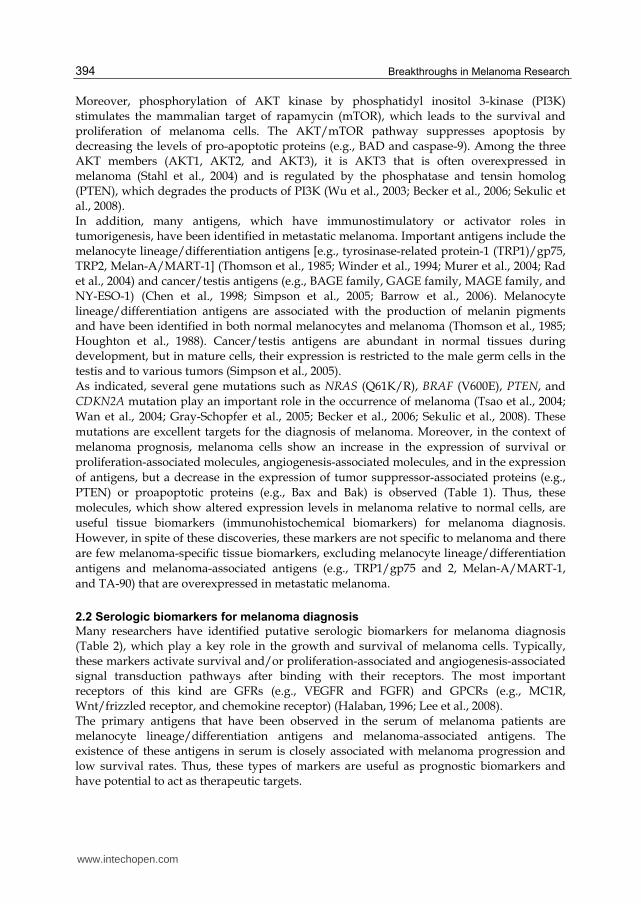
Moreover, phosphorylation of AKT kinase by phosphatidyl inositol 3-kinase (PI3K) stimulates the mammalian target of rapamycin (mTOR), which leads to the survival and proliferation of melanoma cells. The AKT/mTOR pathway suppresses apoptosis by decreasing the levels of pro-apoptotic proteins (e.g., BAD and caspase-9). Among the three AKT members (AKT1, AKT2, and AKT3), it is AKT3 that is often overexpressed in melanoma (Stahl et al., 2004) and is regulated by the phosphatase and tensin homolog (PTEN), which degrades the products of PI3K (Wu et al., 2003; Becker et al., 2006; Sekulic et al., 2008). In addition, many antigens, which have immunostimulatory or activator roles in tumorigenesis, have been identified in metastatic melanoma. Important antigens include the melanocyte lineage/differentiation antigens [e.g., tyrosinase-related protein-1 (TRP1)/gp75, TRP2, Melan-A/MART-1] (Thomson et al., 1985; Winder et al., 1994; Murer et al., 2004; Rad et al., 2004) and cancer/testis antigens (e.g., BAGE family, GAGE family, MAGE family, and NY-ESO-1) (Chen et al., 1998; Simpson et al., 2005; Barrow et al., 2006). Melanocyte lineage/differentiation antigens are associated with the production of melanin pigments and have been identified in both normal melanocytes and melanoma (Thomson et al., 1985; Houghton et al., 1988). Cancer/testis antigens are abundant in normal tissues during development, but in mature cells, their expression is restricted to the male germ cells in the testis and to various tumors (Simpson et al., 2005). As indicated, several gene mutations such as NRAS (Q61K/R), BRAF (V600E), PTEN, and
CDKN2A mutation play an important role in the occurrence of melanoma (Tsao et al., 2004;
Wan et al., 2004; Gray-Schopfer et al., 2005; Becker et al., 2006; Sekulic et al., 2008). These
mutations are excellent targets for the diagnosis of melanoma. Moreover, in the context of
melanoma prognosis, melanoma cells show an increase in the expression of survival or
proliferation-associated molecules, angiogenesis-associated molecules, and in the expression
of antigens, but a decrease in the expression of tumor suppressor-associated proteins (e.g.,
PTEN) or proapoptotic proteins (e.g., Bax and Bak) is observed (Table 1). Thus, these
molecules, which show altered expression levels in melanoma relative to normal cells, are
useful tissue biomarkers (immunohistochemical biomarkers) for melanoma diagnosis.
However, in spite of these discoveries, these markers are not specific to melanoma and there
are few melanoma-specific tissue biomarkers, excluding melanocyte lineage/differentiation
antigens and melanoma-associated antigens (e.g., TRP1/gp75 and 2, Melan-A/MART-1,
and TA-90) that are overexpressed in metastatic melanoma.
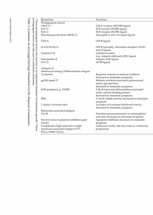
2.2 Serologic biomarkers for melanoma diagnosis
Many researchers have identified putative serologic biomarkers for melanoma diagnosis (Table 2), which play a key role in the growth and survival of melanoma cells. Typically, these markers activate survival and/or proliferation-associated and angiogenesis-associated signal transduction pathways after binding with their receptors. The most important receptors of this kind are GFRs (e.g., VEGFR and FGFR) and GPCRs (e.g., MC1R, Wnt/frizzled receptor, and chemokine receptor) (Halaban, 1996; Lee et al., 2008). The primary antigens that have been observed in the serum of melanoma patients are melanocyte lineage/differentiation antigens and melanoma-associated antigens. The existence of these antigens in serum is closely associated with melanoma progression and low survival rates. Thus, these types of markers are useful as prognostic biomarkers and have potential to act as therapeutic targets.
www.intechopen.com

Biomarkers for Melanoma Diagnosis and the Technologies Used to Identify Them
395
In the absence of vascularization, the growth of melanoma is limited to 0.2 – 0.3 cm due to the limited diffusion of oxygen and nutrients into the tumor. For additional growth, angiogenesis is essential for providing adequate blood supply to the growing lesion. Angiogenesis is regulated by proangiogenic factors, such as VEGF, FGF, tumor necrosis factor (TNF), and interleukin-8 (IL-8) and by antiangiogenic factors, such as interferons (IFNs) and angiostatin. An increase in vascular density provides a greater supply of oxygen and nutrients to cells, leading to melanoma growth (Folkman, 2007; Mahabeleshwar and Byzova, 2007). High levels of proangiogenic factors in the serum of melanoma patients can be used as an indicator of melanoma at diagnosis. Multiple cytokines (e.g., IL-1, 4, 6, 8, 10 and 14), which are correlated with melanoma growth, angiogenesis and metastasis, have been observed in the serum of melanoma patient at both the protein and/or mRNA level. Serum levels of these cytokines are increased in metastatic melanoma patients, suggesting that they can be used as an indicator of melanoma progression (Porter et al., 2001; Varney et al., 2006; Yurkovetsky et al., 2007). Moreover, the serum concentration of the soluble IL-2 receptor is elevated in patients with metastatic melanoma and elevated serum IL-2 receptor levels are associated with lowered survival rates (Boyano et al., 1997; Ottaiano et al., 2006). Interestingly, IFNs are soluble cytokines, but possess antiangiogenic and antitumor activities. An increase in the melanoma progression-associated cytokines leads to a reduction in IFNs levels, but there is a decrease in the serum levels of melanoma progression-associated cytokines in melanoma patients following
immunomodulatory therapy with IFNs (mainly IFN-α2b)(Singh and Varner, 1998; Jonasch and Haluska, 2001; Yurkovetsky et al., 2007; Dummer and Mangana, 2009; Hofmann et al., 2011). Thus, the analysis of a number of different serum cytokines may be a useful means of monitoring the efficacy of immunomodulatory therapy.
2.3 Urinary biomarkers for melanoma diagnosis
Urinary biomarkers for melanoma diagnosis have received much greater interest because of
the relative ease of sample collection and handling compared with the analysis of blood or
tissue samples, but this form of sample may lack the sensitivity required for a diagnostic
biomarker. Of the urinary biomarkers of melanoma already identified (Table 3), 5SCD and
6-hydroxy-5-methoxyindole-2-carboxylic acid (6H5MI2C), are intermediate metabolites in
melanin pigment formation, and have been the most extensively studied. An increase in
urine levels of these markers has been associated with tumor progression and low survival
rates (Kärnell et al., 1997; Bánfalvi et al., 2000; Wakamatsu et al., 2002). In healthy patients,
the urinary levels of these markers are influenced by age (Meyerhöffer et al., 1998), sex
(Morishima and Hanawa, 1981; Kågedal et al., 1992), skin color (Wirestrand et al., 1985) and
season (Ito et al., 1987), but not by pregnancy (Carstam et al., 1985). Although both 5SCD
and 6H5MI2C have been detected in the urine of melanoma patients, because of the higher
levels of 5SCD, this marker is considered a more reliable urinary biomarker for melanoma
than the 6H5MI2C (Kärnell et al., 1997, 2000; Wakamatsu et al., 2006). Moreover, the 90-kDa
glycoprotein (TA-90)(Rote et al., 1980; Euhus et al., 1989), S100A7 (Brouard et al., 2002) and
β-human chorionic gonadotropin (Carter et al., 1995) have also been identified in the urine
of patients with melanoma (Table 3).
Of these urinary biomarkers, 5SCD, 6H5MI2C, and S100A7 can be considered the most
melanoma-specific of the urinary biomarkers (Kärnell et al., 1997; Bánfalvi et al., 2000;
Brouard et al., 2002; Wakamatsu et al., 2002).
www.intechopen.com
B
rea
kth
rou
gh
s in
Me
lan
om
a R
ese
arc
h
396
Biomarkers Functions References
Proangiogenic factors VEGF 1) VEGF receptor (VEGFR) ligand Lacal et al., 2000 FGF 1) FGF receptor (FGFR) ligand Ribatti et al., 2003 EGF 1) EGF receptor (EGFR) ligand Hurks et al., 2000 Placental growth factor (PIGF) 1) Neuropilin-1 and -2 receptor ligand Lacal et al., 2000;
Odorisio et al., 2006 TNF 1) GPCR ligand Singh and Varner 1998;
Lie et al., 2005 IL-8 (CXCL8) 1) GPCR (specially, chemokine receptor CXCR1
and 2) ligand Singh and Varner 1998; Lie et al., 2005; Varney et al., 2006
Laminin-5 2) Laminin receptor (e.g., integrin α6β4 and α7β1) ligand
Ziober et al., 1999; Nikolopoulos et al., 2004
Osteopontin 2) Integrin αvβ3 ligand Zhou et al., 2005; Kadkol et al., 2006 uPA 2) uPAR ligand Delbaldo et al., 1994; de Vries et al.,
1994; Ferrier et al., 2000 Antigens 3) Melanocyte lineage/Differentiation antigens Tyrosinase Regulator enzyme in melanin synthesis;
Increased in metastatic prognosis Sonesson et al., 1995; Stevens et al., 1996
gp100/pmel-17 Melanin synthesis-associated melanosomal matrix glycoprotein; Increased in metastatic prognosis
Pardo et al., 2007
S100 proteins (e.g., S100B) Cell division and differentiation-associated acidic calcium-binding protein; Increased in metastatic prognosis
Henze et al., 1997; Schlagenhauff et al., 2000; Findeisen et al., 2009
MIA A small soluble protein; Increased in metastatic prognosis
Bosserhoff et al., 1999; Schmidt and Bosserhoff, 2009
L-dopa/L-tyrosine ratio An index of tyrosinase functional activity; Increased in metastatic prognosis
Letellier et al., 1999; Stoitchkov et al., 2002
Melanoma-associated antigens TA-90 Potential immunostimulator or antineoplastic
activator; Increased in metastatic prognosis Rote et al., 1980; Kelley et al., 1998
Survivin (one of apoptosis inhibitors gene family)
Apoptosis inhibition; Increased in metastatic prognosis
Tas et al., 2004
Cytoplasmic/high-molecular-weight melanoma-associated antigen (CYT-MAA/HMW-MAA)
Unknown exactly, but may relate to melanoma progression
Vergilis et al., 2005
1) T
hese p
roan
gio
gen
ic factors also
fun
ction
as an
imp
ortan
t stimu
lator fo
r melan
om
a gro
wth
. 2) T
hese
pro
ang
iog
enic fa
ctors also
take p
art in m
elano
ma m
etastasis.
Tab
le 2. (con
tinu
es on
nex
t pag
e) Sero
log
ic bio
mark
ers for th
e diag
no
sis of m
elano
ma
ww
w.intechopen.com
Bio
ma
rke
rs fo
r Me
lan
om
a D
iag
no
sis
an
d th
e T
ech
no
log
ies U
se
d to
Ide
ntify
Th
em
397
Biomarkers Functions References
Other antigens Galectin-3 A β-galactoside-binding protein;
Increased in metastatic prognosis Vereecken et al., 2006, 2009
Synovial sarcoma X breakpoint-2 (SSX-2) A family of highly homologous synovial sarcoma X (SSX) breakpoint proteins and repressive gene regulator
Kyyamova et al., 2006
Gangliosides (GM2, GD2, GM3, and GD3) Group of glycosphingolipids; Relate to interactions between melanoma cells
Ravindranath et al., 2003
Cytokines and cytokine receptors 4) IL-1 Survival or proliferation-associated factor Porter et al., 2001;
Yurkovetsky et al., 2007 IL-4 Survival or proliferation-associated factor Porter et al., 2001 IL-6 Survival or proliferation-associated factor Deichmann et al., 2000; Moretti et
al., 2001; Porter et al., 2001; Yurkovetsky et al., 2007
IL-10 Survival or proliferation-associated factor Moretti et al., 2001; Porter et al., 2001
IL-12 Survival or proliferation-associated factor Moretti et al., 2001; Yurkovetsky et al., 2007
Soluble IL-2 receptor Survival or proliferation-associated factor Boyano et al., 1997; Ottaiano et al., 2006
Other serologic biomarkers YKL-40 Unknown exactly, but may function
as a survival or proliferation-associated factor Johansen et al., 2006; Schmidt et al., 2006
C reactive protein (CRP) Unknown exactly, but may relate to tumor-associated inflammatory response
Deichmann et al., 2000; Findeisen et al., 2009
Lactate dehydrogenase (LDH) An indicator for liver metastasis; a prognostic indicator in metastatic melanoma
Deichmann et al., 2000; Findeisen et al., 2009; Zhuang et al., 2010
Glypican-3 (GPC3) Unknown exactly, but may function as a survival or proliferation-associated factor
Nakatsura et al., 2004; Ikuta et al., 2005
PKCα A survival or proliferation-associated protein Kang et al., 2009 5SCD A precursor of melanin;
Increased in metastatic prognosis Kärnell et al., 1997, 2000; Bánfalvi et al., 2000; Wakamatsu et al., 2002
6H5MI2C A precursor of melanin; Increased in metastatic prognosis
Hara et al., 1994; Kärnell et al., 1997, 2000
Serum amyloid A (SAA) A superfamily of acute-phase proteins and proinflammatory adipokine
Findeisen et al., 2009
Cystatin C A potent inhibitor of cysteine proteases; Increased primary and metastatic melanoma
Kos et al., 1997; Ervin and Cox, 2005
3) A
ntig
ens th
at are fo
un
d in
mela
no
ma tissu
es beco
me g
oo
d im
mu
no
histo
chem
ical bio
mark
ers. 4) T
he
cyto
kin
es also ta
ke p
art in m
elano
ma m
etastasis.
Tab
le 2. (con
tinu
es) Sero
log
ic bio
mark
ers for th
e diag
no
sis of m
elano
ma
ww
w.intechopen.com

Breakthroughs in Melanoma Research
398
Biomarkers References
5SCD Yamada et al., 1992; Kärnell et al., 1997; Bánfalvi et al., 2000; Wakamatsu et al., 2002
6H5MI2C Yamada et al., 1992; Kärnell et al., 1997 TA-90 Rote et al., 1980; Euhus et al., 1989 S100A7 Brouard et al., 2002 β-human chorionic gonadotropin Carter et al., 1995
Table 3. Urinal biomarker for the diagnosis of melanoma
2.4 Biomarkers for early melanoma diagnosis
The early diagnosis of melanoma is closely related to an increase in survival rate. Although
many prognostic biomarkers (mainly metastatic prognosis biomarkers) of melanoma have
been reported, there are very few capable of allowing an early diagnosis. Glypican-3 (GPC3)
is a membrane-bound heparin sulfate proteoglycan which is overexpressed in several
tumors. It has been suggested that GPC3 may be a useful early stage biomarker for patients
with the early stages of the disease (0 - II) (Nakatsura et al., 2004; Ikuta et al., 2005).
Moreover, cyclooxygenase-2 (COX-2) (Chwirot and Kuźbicki, 2007), serum amyloid A
(SAA) (Mian et al., 2005; Findeisen et al., 2009) and DNA methylation profiling (Conway et
al., 2011) can be used to distinguish between early melanomas and benign nevi.
3. Screening techniques of melanoma biomarkers
Before the development of high throughput proteomics techniques, biomarker candidates were identified based on known melanoma molecular pathways and validated by traditional techniques such as western blotting, ELISA and immunohistochemical analysis. However, the recent development of proteomics has enabled novel biomarkers to be screened from across a much larger section of the proteome. The most widely used technique for this form of screening is one whereby the samples are first separated by 2-DE and then each protein is identified by mass spectrometry (MS). Recently, simple gel-free techniques such as shotgun proteomics (Liu et al., 2002) and surface-enhanced laser desorption/ionization time-of-flight mass spectrometry (SELDI-TOF MS)(Petricoin et al., 2002) have been developed. These techniques employ simple separation procedures such as capillary chromatography and surface chromatography prior MS analysis.
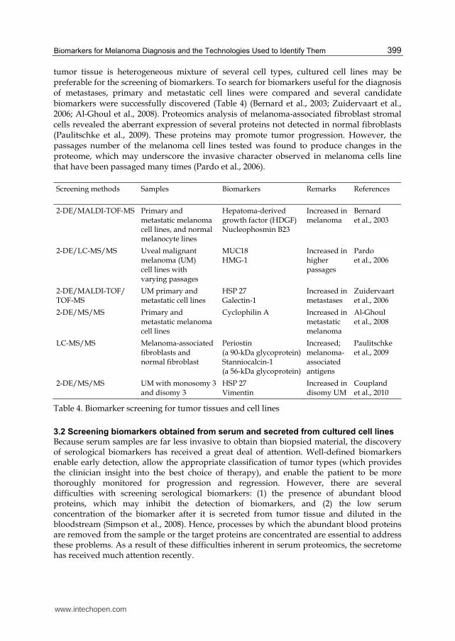
3.1 Screening for tumor tissues and cell lines
Biomarkers discovered from tissue samples and cultured cell lines also have utility for the development of diagnostic and prognostic assays. Tissue microarray is a high throughput technique in which many tissue samples can be screened simultaneously. This technique is suitable for the validation of candidate biomarkers, which are first obtained by other proteomics techniques. Several biomarkers of melanoma such as HSP 90 (McCarthy et al., 2008), ING3 (Wang et al., 2007), and the epidermal growth factor receptor family member HER3 (Reschke et al., 2008) were validated using this technique. For comprehensive screening of biomarkers from lysates collected from tumor tissue and cultured cells, 2-DE combining MS is used as a standard method. The representative attempts of biomarker screening of melanoma lysates are summarized in Table 4. Since
www.intechopen.com
Biomarkers for Melanoma Diagnosis and the Technologies Used to Identify Them
399
tumor tissue is heterogeneous mixture of several cell types, cultured cell lines may be preferable for the screening of biomarkers. To search for biomarkers useful for the diagnosis of metastases, primary and metastatic cell lines were compared and several candidate biomarkers were successfully discovered (Table 4) (Bernard et al., 2003; Zuidervaart et al., 2006; Al-Ghoul et al., 2008). Proteomics analysis of melanoma-associated fibroblast stromal cells revealed the aberrant expression of several proteins not detected in normal fibroblasts (Paulitschke et al., 2009). These proteins may promote tumor progression. However, the passages number of the melanoma cell lines tested was found to produce changes in the proteome, which may underscore the invasive character observed in melanoma cells line that have been passaged many times (Pardo et al., 2006).
Screening methods Samples Biomarkers Remarks References
2-DE/MALDI-TOF-MS Primary and metastatic melanoma cell lines, and normal melanocyte lines
Hepatoma-derived growth factor (HDGF) Nucleophosmin B23
Increased in melanoma
Bernard et al., 2003
2-DE/LC-MS/MS Uveal malignant melanoma (UM) cell lines with varying passages
MUC18 HMG-1
Increased in higher passages
Pardo et al., 2006
2-DE/MALDI-TOF/ TOF-MS
UM primary and metastatic cell lines
HSP 27 Galectin-1
Increased in metastases
Zuidervaart et al., 2006
2-DE/MS/MS Primary and metastatic melanoma cell lines
Cyclophilin A Increased in metastatic melanoma
Al-Ghoul et al., 2008
LC-MS/MS Melanoma-associated fibroblasts and normal fibroblast
Periostin (a 90-kDa glycoprotein)Stanniocalcin-1 (a 56-kDa glycoprotein)
Increased; melanoma-associated antigens
Paulitschke et al., 2009
2-DE/MS/MS UM with monosomy 3 and disomy 3
HSP 27 Vimentin
Increased in disomy UM
Coupland et al., 2010
Table 4. Biomarker screening for tumor tissues and cell lines
3.2 Screening biomarkers obtained from serum and secreted from cultured cell lines
Because serum samples are far less invasive to obtain than biopsied material, the discovery of serological biomarkers has received a great deal of attention. Well-defined biomarkers enable early detection, allow the appropriate classification of tumor types (which provides the clinician insight into the best choice of therapy), and enable the patient to be more thoroughly monitored for progression and regression. However, there are several difficulties with screening serological biomarkers: (1) the presence of abundant blood proteins, which may inhibit the detection of biomarkers, and (2) the low serum concentration of the biomarker after it is secreted from tumor tissue and diluted in the bloodstream (Simpson et al., 2008). Hence, processes by which the abundant blood proteins are removed from the sample or the target proteins are concentrated are essential to address these problems. As a result of these difficulties inherent in serum proteomics, the secretome has received much attention recently.
www.intechopen.com
Breakthroughs in Melanoma Research
400
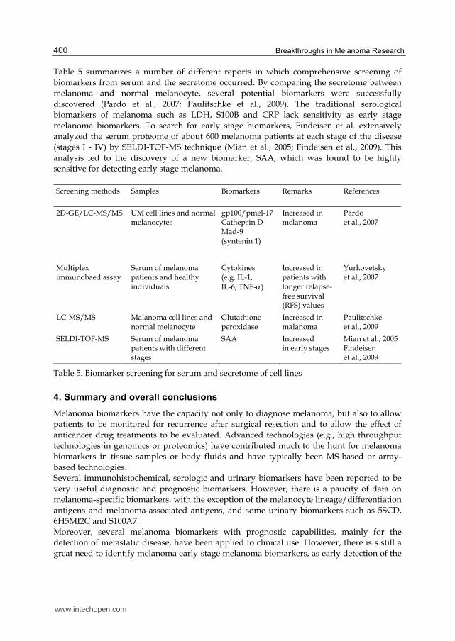
Table 5 summarizes a number of different reports in which comprehensive screening of
biomarkers from serum and the secretome occurred. By comparing the secretome between
melanoma and normal melanocyte, several potential biomarkers were successfully
discovered (Pardo et al., 2007; Paulitschke et al., 2009). The traditional serological
biomarkers of melanoma such as LDH, S100B and CRP lack sensitivity as early stage
melanoma biomarkers. To search for early stage biomarkers, Findeisen et al. extensively
analyzed the serum proteome of about 600 melanoma patients at each stage of the disease
(stages I - IV) by SELDI-TOF-MS technique (Mian et al., 2005; Findeisen et al., 2009). This
analysis led to the discovery of a new biomarker, SAA, which was found to be highly
sensitive for detecting early stage melanoma.
Screening methods Samples Biomarkers Remarks References
2D-GE/LC-MS/MS UM cell lines and normal melanocytes
gp100/pmel-17 Cathepsin D Mad-9 (syntenin 1)
Increased in melanoma
Pardo et al., 2007
Multiplex immunobaed assay
Serum of melanoma patients and healthy individuals
Cytokines (e.g. IL-1,
IL-6, TNF-α)
Increased in patients with longer relapse-free survival (RFS) values
Yurkovetsky et al., 2007
LC-MS/MS Malanoma cell lines and normal melanocyte
Glutathione peroxidase
Increased in malanoma
Paulitschke et al., 2009
SELDI-TOF-MS Serum of melanoma patients with different stages
SAA Increased in early stages
Mian et al., 2005 Findeisen et al., 2009
Table 5. Biomarker screening for serum and secretome of cell lines
4. Summary and overall conclusions
Melanoma biomarkers have the capacity not only to diagnose melanoma, but also to allow
patients to be monitored for recurrence after surgical resection and to allow the effect of
anticancer drug treatments to be evaluated. Advanced technologies (e.g., high throughput
technologies in genomics or proteomics) have contributed much to the hunt for melanoma
biomarkers in tissue samples or body fluids and have typically been MS-based or array-
based technologies.
Several immunohistochemical, serologic and urinary biomarkers have been reported to be
very useful diagnostic and prognostic biomarkers. However, there is a paucity of data on
melanoma-specific biomarkers, with the exception of the melanocyte lineage/differentiation
antigens and melanoma-associated antigens, and some urinary biomarkers such as 5SCD,
6H5MI2C and S100A7.
Moreover, several melanoma biomarkers with prognostic capabilities, mainly for the
detection of metastatic disease, have been applied to clinical use. However, there is s still a
great need to identify melanoma early-stage melanoma biomarkers, as early detection of the
www.intechopen.com
Biomarkers for Melanoma Diagnosis and the Technologies Used to Identify Them
401
disease is key to increasing the survival rate. Of several melanoma biomarkers identified,
GPC3, COX-2, SAA and DNA methylation profiling may hold promise for the diagnosis of
early melanoma.
5. References
Abbasi NR, Shaw HM, Rigel DS, Friedman RJ, McCarthy WH, Osman I, Kopf AW, Polsky D
(2004). Early diagnosis of cutaneous melanoma: revisiting the ABCD criteria.
JAMA, 292, 2771-2776.
Akaraviputh T, Arunakul S, Lohsiriwat V, Iramaneerat C, Trakarnsanga A (2010). Surgery
for gastrointestinal malignant melanoma: experience from surgical training center.
World J Gastroenterol, 16, 745-748
Al-Ghoul M, Brück TB, Lauer-Fields JL, Asirvatham VS, Zapata C, Kerr RG, Fields GB
(2008). Comparative proteomic analysis of matched primary and metastatic
melanoma cell lines. J Proteome Res 7, 4107-4118.
Bakalian S, Marshall JC, Logan P, Faingold D, Maloney S, Di Cesare S, Martins C, Fernandes
BF, Burnier MN Jr (2008). Molecular pathways mediating liver metastasis in
patients with uveal melanoma. Clin Cancer Res, 14, 951-956.
Bánfalvi T, Gilde K, Boldizsár M, Fejös Z, Horváth B, Liszkay G, Beczássy E, Kremmer T
(2000). Serum concentration of 5-S-cysteinyldopa in patients with melanoma. Eur J
Clin Invest, 30, 900-904.
Barrow C, Browning J, MacGregor D, Davis ID, Sturrock S, Jungbluth AA, Cebon J (2006).
Tumor antigen expression in melanoma varies according to antigen and stage. Clin
Cancer Res, 12, 764-771.
Becker JC, Kirkwood JM, Agarwala SS, Dummer R, Schrama D, Hauschild A (2006)
Molecularly targeted therapy for melanoma. Cancer, 106, 2317-2327.
Bernard K, Litman E, Fitzpatrick JL, Shellman YG, Argast G, Polvinen K, Everett AD,
Fukasawa K, Norris DA, Ahn NG, Resing KA (2003). Functional proteomic analysis
of melanoma progression. Cancer Res, 63, 6716-6725.
Boisvert-Adamo K, Longmate W, Abel EV, Aplin AE (2009). Mcl-1 is required for melanoma
cell resistance to anoikis. Mol Cancer Res, 7, 549-556.
Bosserhoff AK, Lederer M, Kaufmann M, Hein R, Stolz W, Apfel R, Bogdahn U, Buettner R
(1999). MIA, a novel serum marker for progression of malignant melanoma.
Anticancer Res, 19, 2691-2693.
Box NF, Duffy DL, Irving RE, Russell A, Chen W, Griffyths LR, Parsons PG, Green AC,
Sturm RA (2001). Melanocortin-1 receptor genotype is a risk factor for basal and
squamous cell carcinoma. J Invest Dermatol, 116, 224-229.
Boyano MD, García-Vázquez MD, Gardeazabal J, García de Galdeano A, Smith-Zubiaga I,
Cañavate ML, Raton JA, Bilbao I, Díaz-Pérez JL (1997). Serum-soluble IL-2 receptor
and IL-6 levels in patients with melanoma. Oncology, 54, 400-406.
Brouard MC, Saurat JH, Ghanem G, Siegenthaler G (2002). Urinary excretion of epidermal-
type fatty acid-binding protein and S100A7 protein in patients with cutaneous
melanoma. Melanoma Res, 12, 627-631.
www.intechopen.com
Breakthroughs in Melanoma Research
402
Carstam R, Hansson C, Rorsman H, Rosengren E, Sjöberg NO, Wirestrand LE (1985).
Urinary excretion of melanocytic metabolites in fertile women. Acta Derm Venereol,
65,543-545.
Carter PG, Iles RK, Neven P, Ind TE, Shepherd JH, Chard T (1995). Measurement of
urinary beta core fragment of human chorionic gonadotrophin in women with
vulvovaginal malignancy and its prognostic significance. Br J Cancer, 71, 350-
353.
Cartlidge RA, Thomas GR, Cagnol S, Jong KA, Molton SA, Finch AJ, McMahon M (2008).
Oncogenic BRAF(V600E) inhibits BIM expression to promote melanoma cell
survival. Pigment Cell Melanoma Res, 21, 534-544.
Chen YT, Güre AO, Tsang S, Stockert E, Jäger E, Knuth A, Old LR (1998). Identification of
multiple cancer/testis antigens by allogeneic antibody screening of a melanoma cell
line library. Proc Natl Acad Sci USA, 95, 6919-6923.
Chwirot BW, Kuźbicki Ł (2007). Cyclooxygenase-2 (COX-2): first immunohistochemical
marker distinguishing early cutaneous melanomas from benign melanocytic skin
tumours. Melanoma Res, 17, 139-145.
Ciotti P, Rainero ML, Nicolò G, Spina B, Garrè C, Casabona F, Santi PL, Bianchi-Scarrà G
(1995). Cytokine expression in human primary and metastatic melanoma cells:
analysis in fresh bioptic specimens. Melanoma Res, 5, 41-47.
Cohen C, Zavala-pompa A, Sequeira JH, Shoji M, Sexton DG, Cotsonis G, Cerimele F,
Govindarajan B, Macaron N, Arbiser JL (2002). Mitogen-actived protein kinase
activation is an early event in melanoma progression. Clin Cancer Res, 8, 3728-
3733.
Conway K, Edmiston SN, Khondker ZS, Groben PA, Zhou X, Chu H, Kuan PF, Hao H,
Carson C, Berwick M, Olilla DW, Thomas NW (2011). DNA methylation profiling
distinguishes malignant melanomas from benign nevi. Pigment Cell Melanoma Res,
24, 352-360.
Coupland SE, Vorum H, Mandal N, Kalirai H, Honoré B, Urbak SF, Lake SL, Dopierala J,
Damato B (2010). Proteomics of uveal melanomas suggests HSP-27 as a possible
surrogate marker of chromosome 3 loss. Invest Ophthalmol Visual Science, 51, 12-20.
Cress RD, Holly EA (1997). Incidence of cutaneous melanoma among non-Hispanic whites,
Hispanics, Asians, and blacks: an analysis of california cancer registry data, 1988-
93. Cancer Causes Control, 8, 246-252.
Deichmann M, Benner A, Waldmann V, Bock M, Jäckel A, Näher H (2000). Interleukin-6 and
its surrogate C-reactive protein are useful serum markers for monitoring
metastasized malignant melanoma. J Exp Clin Cancer Res, 19, 301-307.
Delbaldo C, Masouye I, Saurat JH, Vassalli JD, Sappino AR (1994). Plasminogen activation in
melnocytic neoplasia. Cancer Res, 54, 4547-4552.
de Vries TJ, Quax PH, Denijn M, Verrijp KN, Verheijen JH, Verspaget HW, Weidle UH,
Ruiter DJ, van Muijen GN (1994). Plasminogen activators, their inhibitors, and
urikinase receptor emerge in late stages of melanocytic tumor progression. Am J
Pathol, 144, 70-81.
Diaz A, Suarez E, Blanco R, Badia T, Rivero D, Lopez-Requena A, Lopez A, Montero E
(2007). Functional expression of human-epidermal-growth-factor receptor in a
melanoma cell line. Biotechnol Appl Biochem, 48, 21-27
www.intechopen.com
Biomarkers for Melanoma Diagnosis and the Technologies Used to Identify Them
403
Dummer R, Mangana J (2009). Long-term pegylated interferon-alpha and its potential in the
treatment of melanoma. Biologics, 3, 169-182.
Eisenmann KM, VanBrocklin MW, Staffend NA, Kitchen SM, Koo HM (2003). Mitogen-
activated protein kinase pathway-dependent tumor-specific survival signaling in
melanoma cells through inactivation of the proapoptotic protein Bad. Cancer Res,
63, 8330-8337.
Epping MT, Bernards R (2006). A causal role for the human tumor antigen preferentially
expressed antigen of melanoma in cancer. Cancer Res, 66, 10639-10642.
Ervin H, Cox JL (2005). Late stage inhibition of hematogenous melanoma metastasis by
cystatin C over-expression. Cancer Cell Int, 5, 14.
Euhus DM, Gupta RK, Morton DL (1989). Detection of a tumor-associated glycoprotein
antigen in serum and urine of melanoma patients by murine monoclonal antibody
(AD1-40F4) in enzyme immunoassay. J Clin Lab Anal, 3, 184-190.
Fecker LF, Geilen CC, Tchernev G, Trefzer W, Assaf C, Kurbanov BM, Schwarz C, Daniel
PT, Eberle J (2006). Loss of proapoptotic Bcl-2-related multidomain proteins in
primary melanomas is associated with poor prognosis. J Invest Dermatol, 126, 1366-
1371.
Ferrier CM, Suciu S, van Geloof WL, Straatman H, Eggermont AM, Koops HS, Kroon BB,
Lejeune FJ, Kleeberg UR, van Muijen GN, Ruiter DJ (2000). Hign-tPA-expression in
primary melanoma of the limb correlates with good prognosis. Br J Cancer, 83, 1351-
1359.
Findeisen P, Zapatka M, Peccerella T, Matzk H, Neumaier M, Schadendorf D, Ugurel S
(2009). Serum amyloid A as a prognostic marker in melanoma identified by
proteomic profiling. J Clin Oncol, 27, 2199-2208.
Folkman, J. (2007) Angiogenesis: an organizing principle for drug discovery? Nat. Rev. Drug
Discov, 6, 237-286.
Garraway LA, Widlund HR, Rubin MA, Getz G, Berger AJ, Ramaswamy S, Beroukhim R,
Milner DA, Granter SR, Du J, Lee C, Wagner SN, Li C, Golub TR, Rimm DL,
Meyerson ML, Fisher DE, Sellers WR (2005). Integrative genomic analyses identify
MITF as a lineage survival oncogene amplified in malignant melanoma. Nature,
436, 117-122.
Gray-Schopfer VC, da Rocha Dias S, Marais R (2005). The role of B-RAF in melanoma.
Cancer Metastasis Rev, 24, 165-183.
Halaban R (1996). Growth factors and melanomas. Semin Oncol, 23, 673-681.
Hara H, Walsh N, Yamada K, Jimbow K (1994). High plasma level of a eumelanin precursor,
6-hydroxy-5-methoxyindole-2-carboxylic acid as a prognostic marker for malignant
melanoma. J Invest Dermatol, 102, 501-505.
Henze G, Dummer R, Joller-Jemelka HI, Böni R, Burg G (1997). Serum S100-a marker for
disease monitoring in metastatic melanoma. Dermatology, 194, 208-212.
Hieken TJ, Farolan M, Ronan SG, Shilkaitis A, Wild L, Das Gupta TK (1995). β3 integrin
expression in melanoma predicts subsequent metastasis. J Surg Res, 63, 169-173.
Hoek KS, Eichhoff OM, Schlegel NC, Döbbeling U, Kobert N, Schaerer L, Hemmi S,
Dummer R (2008). In vivo switching of human melanoma cells between
proliferative and invasive states. Cancer Res, 68, 650-656.
www.intechopen.com

Breakthroughs in Melanoma Research
404
Hofmann MA, Kiecker F, Küchler I, Kors C, Trefzer U (2011). Serum TNF-α, B2M and sIL-2R
levels are biological correlates of outcome in adjuvant IFN-α2b treatment of
patients with melanoma. J Cancer Res Clin Oncol, 137, 455-462.
Hofmann UB, Westphal JR, Van Muijen GN, Ruiter DJ (2000). Matrix metalloproteinases in
human melanoma. J Invest Dermatol, 115, 337-344.
Houghton AN, Albino AP, Cordon-Cardo C, Davis LJ, Eisinger M (1988). Cell surface
antigens of human melanocytes and melanoma. Expression of adenosine
deaminase binding protein is extinguished with melanocyte transformation. J Exp
Med, 167, 197-212.
Hu DN, Yu G, McCormick SA, Finger PT (2008). Population-based incidence of conjunctival
melanoma in various races and ethnic groups and comparision with other
melanoma. Am J Ophthlmol, 145, 418-423.
Hurks HM, Metzelaar-Blok JA, Barthen ER, Zwinderman AH, De Wolff-Rouendaal D,
Keunen JE, Jager MJ (2000). Expression of epidermal growth factor receptor: risk
factor in uveal melanoma. Invest Ophthalmol Vis Sci, 41, 2023-2027.
Ikuta Y, Nakatsura T, Kageshita T, Fukushima S, Ito S, Wakamatsu K, Baba H, Nishimura Y
(2005). Highly sensitive detection of melanoma at an early stage based on the
increased serum secreted protein acidic and rich in cysteine and glypican-3 levels.
Clin Cancer Res, 11, 8079-8088.
Ito S, Kato T, Fujita K (1987). Seasonal variation in urinary excretion of 5-S-cysteinyldopa in
healthy Japanese. Acta Derm Venereol, 67, 163-165.
Jaeger J, Koczan D, Thiesen HJ, Ibrahim SM, Gross G, Spang R, Kunz M (2007). Gene
expression signatures for tumor progression, tumor subtype, and tumor thickness
in laser-microdissected melanoma tissues. Clin Cancer Res, 13, 806-815.
Janku F, Novotny J, Julis I, Julisova I, Pecen L, Tomancova V, Kocmanova G, Krasna L,
Krajsova I, Stork J, Petruzelka L (2005). KIT receptor is expressed in more than 50%
of early-stage malignant melanoma: a retrospective study of 261 patients. Melanoma
Res, 15, 251-256
Jemal A, Siegel R, Xu J, Ward E (2010). Cancer statistics, 2010. CA Cancer J Clin, 60, 277-300.
Johansen JS, Jensen BV, Roslind A, Nielsen D, Price PA (2006). Serum YKL-40, new
prognostic biomarker in cancer patients? Cancer Epidemol Biomarkers Prev, 15, 194-
202.
Jonasch E, Haluska FG (2001). Interferon in oncological practice: review of interferon
biology, clinical applications, and toxicities. Oncologist, 6, 34-55
Jönsson G, Busch C, Knappskog S, Geisler J, Miletic H, Ringnér M, Lillehaug JR, Borg A,
Lønning PE (2010). Gene expression profiling-based identification of molecular
subtypes in stage melanoma with different clinical outcome. Clin Cancer Res, 16,
3356-3367.
Jordan SA, Jackson IJ (1998). Melanocortin receptors and antagonists regulate pigmentation
and body weight. Bioassays, 20, 603-606.
Kadkol SS, Lin AY, Barak V, Kalickman I, Leach L, Valyi-Nagy K, Majumdar D, Setty S,
Maniotis AJ, Folberg R, Pe'er J (2006). Osteopontin expression and serum levels in
metastatic uveal melanoma: A pilot study. Invest Ophthalmol Vis Sci, 47, 802-806.
www.intechopen.com
Biomarkers for Melanoma Diagnosis and the Technologies Used to Identify Them
405
Kågedal B, Lenner L, Arstrand K, Hansson C (1992). The stability of 5-S-cysteinyldopa and
6-hydroxy-5-methoxyindole-2-carboxylic acid in human urine. Pigment Cell Res, 2,
304-307.
Kang JH, Asai D, Toita R, Kitazaki H, Katayama Y (2009). Plasma protein kinase C
(PKC)alpha as a biomarker for the diagnosis of cancers. Carcinogenesis, 30, 1927-
1931.
Kang JH, Asai D, Yamada S, Toita R, Oishi J, Mori T, Niidome T, Katayama Y (2008). A
short peptide is a protein kinase C (PKC) α-specific substrate. Proteomics, 8, 2006-
2011.
Kärnell R, Kågedal B, Lindholm C, Nilsson B, Arstrand K, Ringborg U (2000). The value of
cysteinyldopa in the follow-up of disseminated malignant melanoma. Melanoma
Res, 10, 363-369.
Kärnell R, von Schoultz E, Hansson LO, Nilsson B, Arstrand K, Kågedal B (1997). S100B
protein, 5-S-cysteinyldopa and 6-hydroxy-5-methoxyindole-2-carboxylic acid as
biochemical markers for survival prognosis in patients with malignant melanoma.
Melanoma Res, 7, 393-399.
Karst AM, Dai DL, Martinka M, Li G (2005). PUMA expression is significantly reduced in
human cutaneous melanomas. Oncogene, 24, 1111-1116.
Kelley MC, Jones RC, Gupta RK, Yee R, Stern S, Wanek L, Morton DL (1998). Tumor-
associated antigen TA-90 immune complex assay predicts subclinical metastasis
and survival for patients with early stage melanoma. Cancer 83, 1355-1361.
Krimpenfort P, Quon KC, Mooi WJ, Loonstra A, Berns A (2001). Loss of p16Ink4a confers
susceptibility to metastatic melanoma in mice. Nature, 413, 83-86.
Kos J, Stabuc B, Schweiger A, Krasovec M, Cimerman N, Kopitar-Jerala N, Vrhovec I (1997).
Cathepsins B, H, and L and their inhibitors stefin A and cystatin C in sera of
melanoma patients. Clin Cancer Res, 3, 1815-1822.
Kyyamova RG, Gryshkova VS, Zhyvoloup AM (2006). Expression of SSX2 tumor antigen in
baculovirus expression system and its application for screening of blood serum of
melanoma patients. Exp Oncol, 28, 110-113.
Lacal PM, Failla CM, Pagani E, Odorisio T, Schietroma C, Falcinelli S, Zambruno G, D’Atri S
(2000). Human melanoma cells secrete and respond to placenta growth factor and
vascular endothelial growth factor. J Invest Dermatol, 115, 1000-1007.
Lahn MM, Sundell KL (2004). The role of protein kinase C-alpha (PKC-α) in melanoma.
Melanoma Res, 14, 85-89.
Lai K, Sharma V, Jager MJ, Conway RM, Madigan MC (2007). Expression and distribution of
MUC18 in human uveal melanoma. Virchows Arch, 451:967-976.
Landreville S, Agapova OA, Harbour JW (2008). Emerging insights into the molecular
pathogenesis of uveal melanoma. Future Oncol, 4, 629-636.
Lee HJ, Wall B, Chen S (2008). G-protein-coupled receptors and melanoma. Pigment Cell
Melanoma Res, 21, 415-428.
Leiter U, Schmid RM, Kaskel P, Peter RU, Krähn G (2000). Antiapoptotic bcl-2 and bcl-xL in
advanced malignant melanoma. Arch. Dermatol Res, 292, 225-232.
Lens MB, Dawes M (2004). Global perspectives of contemporary epidemiological trends of
cutaneous malignant melanoma. Br J Dermatol, 150, 179-185.
www.intechopen.com

Breakthroughs in Melanoma Research
406
Letellier S, Garnier JP, Spy J, Stoitchkov K, Le Bricon T, Baccard M, Revol M, Kerneis Y,
Bousquet B (1999). Development of metastases in malignant melanoma is
associated with an increase in the plasma L-dopa/L-tyrosine ratio. Melanoma Res, 9,
389-94.
Ley R, Ewings KE, Hadfield K, Cook SJ (2005). Regulatory phosphorylation of Bim: sorting
out the ERK from the JNK. Cell Death Differ, 12, 1008-1104.
Li G, Satyamoorthy K, Herlyn M (2001). N-cadherin-mediated intercellular interactions
promote survival and migration of melanoma cells. Cancer Res, 61, 3819-3825.
Li M, Zhang B, Sun B, Wang X, Ban X, Sun T, Liu Z, Zhao X (2010). A novel function for
vimentin: the potential biomarker for predicting melanoma hematogenous
metastasis. J Exp Clin Cancer Res, 29, 109.
Lie G, Zhang F, Lee J, Dong Z (2005). Selective induction of interleukin-8 expression in
metastatic melanoma cells by transforming growth factor-β1. Cytokine, 31, 241-249.
Liu H, Lin D, Yates JR 3rd (2002). Multidimensional separations for protein/peptide
analysis in the post-genomic era. Biotechniques, 32, 898-902.
Mahabeleshwar GH, Byzova TV (2007). Angiogenesis in melanoma. Semin Oncol, 34, 555-
565.
Markovic SN, Erickson LA, Rao RD, Weenig RH, Pockaj BA, Bardia A, Vachon CM, Schild
SE, McWilliams RR. Hand JL, Laman SD, Kottschade LA, Maples WJ, Pittelkow
MR, Pulido JS, Cameron JD, Creagan ET, Melanoma Study Group of the Mayo
Clinic Cancer Center (2007). Malignant melanoma in the 21st century, part 1:
epidemiology, risk factors, screening, prevention, and diagnosis. Mayo Clin Proc, 82,
364-380.
McCarthy MM, Pick E, Kluger Y, Gould-Rothberg B, Lazova R, Camp RL, Rimm DL, Kluger
HM (2008). HSP90 as a marker of progression in melanoma. Annals Oncol, 19, 590-
594.
Meyerhöffer S, Lindberg Z, Häger A, Kågedal B, Rosdahl I (1998). Urinary excretion of 5-S-
cysteinyldopa and 6-hydroxy-5-methoxyindole-2-carboxylic acid in children. Acta
Derm Venereol, 78, 31-35.
Mian S, Ugurel S, Parkinson E, Schlenzka I, Dryden I, Lancashire L, Ball G, Creaser C, Rees
R, Schadendorf D (2005). Serum proteomic fingerprinting discriminates between
clinicalstages and predicts disease progression in melanoma patients. J Clin Oncol,
23, 5088-5093.
Molina-Ortiz I, Bartolomé RA, Hernández-Varas P, Colo GP, Teixidó J (2009).
Overexpression of E-cadherin on melanoma cells inhibits chemokine-promoted
invasion involving p190RhoGAP/p120ctn-dependent inactivation of RhoA. J Biol
Chem, 284, 15147-15157.
Moretti S, Chiarugi A, Semplici F, Salvi A, De Giorgi V, Fabbri P, Mazzoli S (2001). Serum
imbalance of cytokines in melanoma patients. Melanoma Res, 11, 395-399.
Morishima T, Hanawa S (1981). Urinary excretion of 5-S-cysteinyldopa in healthy Japanese.
Acta Derm Venereol, 61, 149-150.
Murer K, Urosevic M, Willers J, Selvam P, Laine E, Burg G, Dummer R (2004). Expression of
Melan-A/MART-1 in primary melanoma cell cultures has prognostic implication in
metastatic melanoma patients. Melanoma Res, 14, 257-262.
www.intechopen.com
Biomarkers for Melanoma Diagnosis and the Technologies Used to Identify Them
407
Nakatsura T, Kageshita T, Ito S, Wakamatsu K, Monji M, Ikuta Y, Senju S, Ono T, Nishimura
Y (2004). Identification of glypican-3 as a novel tumor marker for melanoma. Clin
Cancer Res, 10, 6612-6621.
Naldi L, Altieri A, Imberti GL, Giordano L, Gallus S, La Vecchia C, Oncology Study Group
of the Italian Group for Epidemiologic Research in Dermatology (2005). Cutaneous
malignant melanoma in women: phenotypic characteristics, sun exposure, and
hormonal factors: a case-control study from Italy. Ann Epidemiol, 15, 545-550.
Natali PG, Hamby CV, Felding-Habermann B, Liang B, Nicotra MR, Di Filippo F,
Giannarelli D, Temponi M, Ferrone S (1997). Clinical significance of integrin and
intercellular adhesion molecule-1 expression in cutaneous malignant melanoma
lesions. Cancer Res, 57, 1554-1560.
Natkunam Y, Warnke RA, Montgomery K, Falini B, van De Rijn M (2001). Analysis of
MUM1/IRF4 protein expression using tissue microarrays and
immunohistochemistry. Mod Pathol, 14, 686-694.
Nikolopoulos SN, Blaikie P, Yoshioka T, Guo W, Giancotti FG (2004). Integrin β4 signaling
promotes tumor angiogenesis. Cancer Cell, 6, 471-483.
O’Day SJ, Hamid O, Urba WJ (2007). Targeting cytotoxic T-lymphocyte antigen-4 (CTLA-4):
A novel strategy for the treatment of melanoma and other milignancies. Cancer, 110,
2614-2627.
Odorisio T, Cianfarani F, Failla CM, Zambruno G (2006). The placenta growth factor in skin
angiogenesis. J Dermatol Sci, 41, 11-19.
Osterlind A, Tucker MA, Stone BJ, Jensen OM (1988). The Danish case-control study of
cutaneous malignant melanoma, IV: no association with nutritional factors, alcohol,
smoking or hair dyes. Int J Cancer, 42, 825-828.
Ottaiano A, Leonardi E, Simeone E, Ascierto PA, Scala S, Calemma R, Bryce J, Caracò C,
Satriano RA, Gianfranco N, Franco R, Botti G, Castello G (2006). Soluble
interleukin-2 receptor in stage I-III melanoma. Cytokine, 33, 150-155.
Pardo M, Garcia A, Antrobus R, Blanco MJ, Dwek RA, Zitzmann N (2007). Biomarker
discovery from uveal melanoma secretomes: Identification of gp100 and cathepsin
D in patient serum. J Proteome Res, 6, 2802-2811.
Pardo M, García A, Thomas B, Piňeiro A, Akoulitchev A, Dwek RA, Zitzmann N (2006).
The characterization of the invasion phenotype of uveal melanoma tumour cells
shows the presence of MUC18 and HMG-1 metastasis markers and leads to the
identification of DJ-1 as a potential serum biomarker. Int J Cancer, 119, 1014-
1022.
Paulitschke V, Kunstfeld R, Mohr T, Slany A, Micksche M, Drach J, Zielinski C,
Pehamberger H, Gerner C (2009). Entering a new era of rational biomarker
discovery for early detection of melanoma metastases: secretome analysis of
associated stroma cells. J Proteome Res, 8, 2501-2510.
Payne AS, Cornelius LA (2002). The role of chmokines in melanoma tumor growth and
metastasis. J Invest Dermatol, 118, 915-922.
Petricoin III EF, Ardekani AM, Hitt BA, Levine PJ, Fusaro VA, Steinberg SM, Mills GB,
Simone C, Fishman DA, Kohn EC, Liotta LA (2002). Use of proteomic patterns in
serum to identify ovarian cancer. Lancet, 359, 572-577.
www.intechopen.com
Breakthroughs in Melanoma Research
408
Porter GA, Abdalla J, Lu M, Smith S, Montgomery D, Grimm E, Ross MI, Mansfield PF,
Gershenwald JE, Lee JE (2001). Significance of plasma cytokine levels in melanoma
patients with histologically negative sentinel lymph nodes. Ann Surg Oncol, 8, 116-
122.
Poser I, Domínguez D, de Herreros AG, Varnai A, Buettner R, Bosserhoff AK (2001). Loss of
E-cadherin expression in melanoma cells involves up-regulation of the
transcriptional repressor Snail. J Biol Chem, 276, 24661-24666.
Prieto VG, Mourad-Zeidan AA, Melnikova V, Johnson MM, Lopez A, Diwan AH, Lazar AJ,
Shen SS, Zhang PS, Reed JA, Gershenwald JE, Raz A, Bar-Eli M (2006). Galectin-3
expression is associated with tumor progression and pattern of sun exposure in
melanoma. Clin Cancer Res, 12, 6709-6715.
Psaty EL, Halpern AC (2009). Current and emerging technologies in melanoma diagnosis:
the state of the art. Clin Dermatol, 27, 35-45.
Qi J, Chen N, Wang J, Siu CH (2005) Transsendothelial migration of melanoma cells involves
N-cadherin-mediated adhesion and activation of the β-catenin signaling pathway.
Mol Biol Cell, 16, 4386-4397.
Qin JZ, Ziffra J, Stennett L, Bodner B, Bonish BK, Chaturvedi V, Bennett F, Pollock PM, Trent
JM, Hendrix MJ, Rizzo P, Miele L, Nickoloff BJ (2005). Proteasome inhibitors trigger
NOXA-mediated apoptosis in melanoma and myeloma cells. Cancer Res, 65, 6282-
6293
Rad HH, Yamashita T, Jin HY, Hirosaki K, Wakamatsu K, Ito S, Jimbow K (2004).
Tyrosinase-realted proteins suppress tyrosinase-mediated cell death of melanocytes
and melanoma cells. Exp Cell Res, 298, 317-328.
Ravindranath MH, Hsueh EC, Verma M, Ye W, Morton DL (2003). Serum total ganglioside
level correlates with clinical course in melanoma patients after immunotherapy
with therapeutic cancer vaccine. J Immunother, 26, 277-285.
Reschke M, Mihic-Probst D, van der Horst EH, Knyazev P, Wild PJ, Hutterer M, Meyer S,
Dummer R, Moch H, Ullrich A (2008) HER3 is a determinant for poor prognosis in
melanoma. Clin Cancer Res, 14, 5188-5197.
Ribatti D, Vacca A, Ria R, Marzullo A, Nico B, Filotico R, Roncali L, Dammacco F (2003).
Neovascularisation, expression of fibroblast growth fact-2 and mast cells with
tryptase activity increase simulfaneously with pathological progression in human
malignant melanoma. Eur J Cancer, 39, 666-674.
Richmond A, Yang J, Su Y (2009). The good and the bad of chemokines/chemokine
receptors in melanoma. Pigment Cell Melanoma Res, 22, 175-186.
Rigel DS, Friedman RJ, Kopf AW, Polsky D (2005). ABCDE-an evolving concept in the early
detection of melanoma. Arch Dermatol, 141, 1032-1034
Rigel DS, Russak J, Friedman R (2010). The evolution of melanoma diagnosis: 25 years
beyond the ABCDs. CA Cancer J Clin, 60, 301-316.
Rizos H, Darmanian AP, Holland EA, Mann GJ, Kefford RF (2001). Mutations in the
INK4a/ARF melanoma susceptibility locus functionally impari p14ARF. J Biol Chem,
276, 41424-41434.
Robert C, Ghiringhelli F (2009). What is the role of cytotoxic T lyimphocyte-associated
antigen 4 blockade in patients with metastatic melanoma? Oncologist, 14, 848-861.
www.intechopen.com
Biomarkers for Melanoma Diagnosis and the Technologies Used to Identify Them
409
Rote NS, Gupta RK, Morton DL (1980). Tumor-associated antigens detected by autologous
sera in urine of patients with solid neoplasms. J Surg Res, 29, 18-22.
Rouzaud F, Annereau JP, Valencia JC, Costin GE, Hearing VJ (2003). Regulation of
melanocortin 1 receptor expression at the mRNA and protein levels by its natural
agonist and antagonist. FASEB J, 17, 2154-2156.
Saldanha G, Potter L, DaForno P, Pringle JH (2006). Cutanuous melanoma subyptes show
different BRAF and NRAS mutaion frequencies. Clin Cancer Res, 12, 4499-4505.
Sanders DS, Blessing K, Hassan GA, Bruton R, Marsden JR, Jankowski J (1999). Alterations
in cadherin and catenin expression during the biological progression of
melanocytic tumours. Mol Pathol, 52, 151-157.
Scala S, Ottaiano A, Ascierto PA, Cavalli M, Simeone E, Giuliano P, Napolitano M, Franco R,
Botti G, Castello G (2005). Expression of CXCR4 predicts poor prognosis in patients
with malignant melanoma. Clin Cancer Res, 11, 1835-1841.
Schlagenhauff B, Schittek B, Ellwanger U, Stroebel W, Blum A, Schwarz M, Rassner G,
Garbe C (2000). Significance of serum protein S100 levels in screening for
melanoma metastasis: dose protein S100 early detection of melanoma recurrence?
Melanoma Res, 10, 451-459.
Schmidt H, Johansen JS, Sjoegren P, Christensen IJ, Sorensen BS, Fode K, Larsen J, von der
Maase H (2006). Serum YKL-40 predicts relapse-free and overall survival in
patients with American Joint Committee on Cancer stage I and II melanoma. J Clin
Oncol, 24, 798-804.
Schmidt J, Bosserhoff AK (2009). Processing of MIA protein during melanoma cell
migration. Int J Cancer, 125, 1587-1594.
Schulman JM, Fisher DE (2009). Indoor ultraviolet tanning and skin cancer: health risks and
opportunities. Curr Opin Oncol, 21, 144-149.
Seetharamu N, Ott PA, Pavlick AC (2010). Mucosal melanoma: a case-based review of the
literature. Oncologist 15, 772-781.
Sekulic A, Haluska P Jr, Miller AJ, Genebriera De Lamo J, Ejadi S, Pulido JS, Salomao DR,
Thorland EC, Vile RG, Swanson DL, Pockaj BA, Laman SD, Pittelkow MR,
Markovic SN, Melanoma Study Group of Mayo Clinic Cancer Center (2008).
Malignant melanoma in the 21st century: The emerging molecular landscape. Mayo
Clin Proc, 83, 825-846.
Simpson AJ, Caballero OL, Jumngbluth A, Chen YT, Old LJ (2005). Cancer/testis antigens,
gametogenesis and cancer. Nat Rev Cancer, 5, 615-625.
Singh RK, Varner ML (1998). Regulation of interleukin 8 expression in human malignant
melanoma cells. Cancer Res, 58, 1523-1527.
Sonesson B, Eide S, Ringborg U, Rorsman H, Rosengren E (1995). Tyrosinase activity in the
serum of patients with malignant melanoma. Melanoma Res, 5, 113-116.
Stahl JM, Cheung M, Sharma A, Trivedi NR, Shanmugam S, Robertson GP (2003). Loss of
PTEN promotes tumor development in malignant melanoma. Cancer Res, 63, 2881-
2890.
Stahl JM, Sharma A, Cheung M, Zimmerman M, Cheng JQ, Bosenberg MW, Kester M,
Sandirasegarane L, Robertson GP (2004). Deregulated Akt3 activity promotes
development of malignant melanoma. Cancer Res, 64, 7002-7010.
www.intechopen.com

Breakthroughs in Melanoma Research
410
Stevens GL, Scheer WD, Levine EA (1996). Detection of tyrosinase mRNA from the blood of
melanoma patients. Cancer Epidmilo Biomarkers Prev, 5, 293-296.
Stoitchkov K, Letellier S, Garnier JP, Bousquet B, Tsankov N, Morel P, Ghanem G, Le Bricon
T (2002). Melanoma progression and serum L-dopa/L-tyrosine ratio: a comparison
with S100B. Melanoma Res, 12, 255-262.
Strizzi L, Postovit LM, Margaryan NV, Lipavsky A, Gadiot J, Blank C, Seftor RE, Seftor EA,
Hendrix MJ (2009). Nodal as a biomarker for melanoma progression and a new
therapeutic target for clinical intervention. Expert Rev Dermatol, 4, 67-78.
Tas F, Duranyildiz D, Argon A, Oguz H, Camlica H, Yasasever V, Topuz E (2004). Serum
bcl-2 and surviving levels in melanoma. Melanoma Res, 14, 543-536.
Tsao H, Goel V, Wu H, Yang G, Haluska FG (2004). Genetic interaction between NRAS and
BRAF mutations and PTEN/MMAC1. J Invest Dermatol, 122, 337-341.
Tchernev G, Orfanos CE (2007). Downregulation of cell cycle modulators p21, p27, p53, Rb
and proapoptotic Bcl-2-related proteins Bax and Bax in cutaneous melanoma is
associated with worse patient prognosis: preliminary findings. J Cutan Pathol, 34,
247-256.
Thomson TM, Mattes MJ, Roux L, Old LJ, Lloyd KO (1985). Pigmentation-associated
glycoprotein of human melanomas and melanocytes: definition with a mouse
monoclonal antibody. J Invest Dermatol, 85, 169-174.
Topczewska JM, Postovit LM, Margaryan NV, Sam A, Hess AR, Wheaton WW, Nickoloff BJ,
Topczewski J, Hendrix MJ (2006). Embryonic and tumorigenic pathways converge
via Nodal signaling: role in melanoma aggressiveness. Nat Med, 12, 925-932.
Ueda Y, Richmond A (2006). NF-κB activation in melanoma. Pigment Cell Res, 19, 112-124.
Varney ML, Johansson SL, Singh RK (2006). Distinct expression of CXCL8 and its receptors
CXCR1 and CXCR2 and their association with vessel density and aggressiveness in
malignant melanoma. Am J Clin Pathol, 125, 209-216.
Veierod MB, Thelle DS, Laake P (1997). Diet and risk of cutaneous malignant melanoma: a
prospective study of 50,757 Norwegian men and women. Int J Cancer, 71, 600-604.
Vereecken P, Awada A, Suciu S, Castro G, Morandini R, Litynska A, Lienard D, Ezzedine K,
Ghanem G, Heenen M (2009). Evaluation of the prognostic significance of serum
galectin-3 in American Joint Committee on Cancer stage III and stage IV melanoma
patients. Melanoma Res, 19, 316-320.
Vereecken P, Zouaoui Boudjeltia K, Debray C, Awada A, Legssyer I, Sales F, Petein M,
Vanhaeverbeek M, Ghanem G, Heenen M (2006). High serum galectin-3 in
advanced melanoma: preliminary results. Clin Exp Dermatol, 31, 105-109.
Vergilis IJ, Szarek M, Ferrone S, Reynolds SR (2005). Presence and prognostic significance of
melanoma-associated antigens CYT-MAA and HMW-MAA in serum of patients
with melanoma. J Invest Dermatol, 125, 526–531
Wakamatsu K, Kageshita T, Furue M, Hatta N, Kiyohara Y, Nakayama J, Ono T, Saida T,
Takata M, Tsuchida T, Uhara H, Yamamoto A, Yamazaki N, Naito A, Ito S (2002).
Elvaluation of 5-S-cysteinyldopa as a marker of melanoma progression: 10 years’
experience. Melanoma Res, 12, 245-253.
Wakamatsu K, Takasaki A, Kågedal B, Kageshita T, Ito S (2006). Determination of eumelanin
in human urine. Pigment Cell Res, 19, 163-169.
www.intechopen.com
Biomarkers for Melanoma Diagnosis and the Technologies Used to Identify Them
411
Wan PT, Garnett MJ, Roe SM, Lee S, Niculescu-Duvaz D, Good VM, Jones CM, Marshall CJ,
Springer CJ, Barford D, Marais R, Cancer Genome Project (2004). Mechanism of
activation of the RAF-ERK signaling pathway by oncogenic mutations of B-RAF.
Cell, 116, 855-867.
Wang Y, Dai DL, Martinka M, Li G (2007). Prognostic significance of nuclear ING3
expression in human cutaneous melanoma Clin Cancer Res, 13, 4111-4116.
Weyers W, Euler M, Diaz-Cascajo C, Schill WB, Bonczkowitz M (1999). Classification of
cutaneous maliganat melanoma: a reassessment of histopathologic criteria for the
distinction of different types. Cancer, 86, 288-299.
Widlund HR, Horstmann MA, Price ER, Cui J, Lessick SL, Wu M, He X, Fisher DE (2002). β-
catenin-induced melanoma growth requires the downstream target Microphthalmia-
associated transcription factor. J Cell Biol, 158, 1079-1087.
Winder A, Kobayashi T, Tsukamoto K, Urabe K, Aroca P, Kameyama K, Hearing VJ (1994).
The tyrosinase gene family: interactions of melanogenic proteins to regulate
melanogenesis. Cell Mol Biol Res, 40, 613-626.
Wirestrand LE, Hansson C, Rosengren E, Rorsman H (1985). Melanocyte metabolites in the
urine of people of different skin colour. Acta Derm Venereol, 65,345-348.
Wu H, Goel W, Haluska FG (2003). PTEN signaling pathways in melanoma. Oncogene, 22,
3113-3122.
Yamada K, Walsh N, Hara H, Jimbow K, Chen H, Ito S (1992). Measurement of eumelanin
precursor metabolites in the urine as a new marker for melanoma metastases. Arch
Dermatol, 128, 491-494.
Yurkovetsky ZR, Kirkwood JM, Edington HD, Marrangoni AM, Velikokhatnaya L, Winans
MT, Gorelik E, Lokshin AE (2007). Multiplex analysis of serum cytokines in
melanoma patients treated with interferon-α2b. Clin Cancer Res, 13, 2422-2428.
Zhang XD, Franco A, Myers K, Gray C, Nguyen T, Hersey P (1999). Relation of TNF-
related apoptosis-inducing ligand (TRAIL) receptor and FLICE-inhibitory
protein expression to TRAIL-induced apoptosis of melanoma. Cancer Res, 59,
2747-2753.
Zhang XD, Wu JJ, Gillespie S, Borrow J, Hersey P (2006). Human melanoma cells selected for
resistance to apoptosis by prolonged exposure to tumor necrosis factor-related
apoptosis-inducing ligand are more vulnerable to necrotic cell death induced by
cisplatin. Clin Cancer Res, 12, 1355-1364.
Zhou Y, Dai DL, Martinka M, Su M, Zhang Y, Campos EI, Dorocicz I, Tang L, Huntsman D,
Nelson C, Ho V, Li G (2005). Osteopontin expression correlates with melanoma
invasion. J Invest Dermatol, 124, 1044-1052.
Zhuang L, Lee CS, Scolyer RA, McCarthy SW, Zhang XD, Thompson JF, Hersey P (2007).
Mcl-1, Bcl-XL and Stat3 expression are associated with progression of melanoma
whereas Bcl-2, AP-2 and MITF levels decrease during progression of melanoma.
Mod Pathol, 20, 416-426.
Zhuang L, Lee CS, Scolyer RA, McCarthy SW, Zhang XD, Thompson JF, Screaton G, Hersey
P (2006). Progression in melanoma is associated with decreased expression of death
receptors for tumor necrosis factor-related apoptosis-inducing ligand. Hum Pathol,
37, 1286-1294.
www.intechopen.com

Breakthroughs in Melanoma Research
412
Zhuang L, Scolyer RA, Murali R, McCarthy SW, Zhang XD, Thompson JF, Hersey P (2010).
Lactate dehydrogenase 5 expression in melanoma increases with disease
progression and is associated with expression of Bcl-XL and Mcl-1, but not Bcl-2
protein. Mod Pathol, 23, 45-53.
Ziober BL, Chen YQ, Ramos DM, Waleh N, Kramer RH (1999). Expression of the laminin
receptor suppresses melanoma growth and metastatic potential. Cell Growth
Different, 10, 479-490.
Zuidervaart W, Hensbergen PJ, Wong M-C, Deelder AM, Tensen CP, Jager MJ, Gruis NA
(2006). Proteomic analysis of uveal melanoma reveals novel potential markers
involved in tumor progression. Invest Ophthalmol Visual Sci, 47, 786-793.
www.intechopen.com

Breakthroughs in Melanoma ResearchEdited by Dr Yohei Tanaka
ISBN 978-953-307-291-3Hard cover, 628 pagesPublisher InTechPublished online 30, June, 2011Published in print edition June, 2011
InTech EuropeUniversity Campus STeP Ri Slavka Krautzeka 83/A 51000 Rijeka, Croatia Phone: +385 (51) 770 447 Fax: +385 (51) 686 166www.intechopen.com
InTech ChinaUnit 405, Office Block, Hotel Equatorial Shanghai No.65, Yan An Road (West), Shanghai, 200040, China
Phone: +86-21-62489820 Fax: +86-21-62489821
Melanoma is considered to be one of the most aggressive forms of skin neoplasms. Despite aggressiveresearches towards finding treatments, no effective therapy exists to inhibit the metastatic spread of malignantmelanoma. The 5-year survival rate of metastatic melanoma is still significantly low, and there has been anearnest need to develop more effective therapies with greater anti-melanoma activity. Through theaccomplishment of over 100 distinguished and respected researchers from 19 different countries, this bookcovers a wide range of aspects from various standpoints and issues related to melanoma. These include thebiology of melanoma, pigmentations, pathways, receptors and diagnosis, and the latest treatments andtherapies to make potential new therapies. Not only will this be beneficial for readers, but it will also contributeto scientists making further breakthroughs in melanoma research.
How to referenceIn order to correctly reference this scholarly work, feel free to copy and paste the following:
Takeshi Mori and Jeong-Hun Kang (2011). Biomarkers for Melanoma Diagnosis and the Technologies Used toIdentify Them, Breakthroughs in Melanoma Research, Dr Yohei Tanaka (Ed.), ISBN: 978-953-307-291-3,InTech, Available from: http://www.intechopen.com/books/breakthroughs-in-melanoma-research/biomarkers-for-melanoma-diagnosis-and-the-technologies-used-to-identify-them

© 2011 The Author(s). Licensee IntechOpen. This is an open access articledistributed under the terms of the Creative Commons Attribution 3.0License, which permits unrestricted use, distribution, and reproduction inany medium, provided the original work is properly cited.