Embed Size (px)
Citation preview
March 10, 2010 / Vol. 8, No. 3 / CHINESE OPTICS LETTERS 309
Tagged molecule induced nanoparticle aggregation:Raman reporter-labeled immuno-Au
aggregate as immuno-sensor
Chunyuan Song (yyySSS), Zhuyuan Wang (ÍÍÍ), Ruohu Zhang (ÜÜÜeeemmm),
Jing Yang ( ¬¬¬), Xuebin Tan (!!!ÆÆÆRRR), and Yiping Cui (www²²²)∗
Advanced Photonics Center, Southeast University, Nanjing 210096, China∗E-mail: [email protected]
Received July 14, 2009
A novel structure with high surface enhanced Raman scattering (SERS) activity and bio-specificity as aSERS-based immuno-sensor (named as Raman reporter-labeled immuno-Au aggregate) is demonstratedand employed for protein detection. In each fabrication process, the features of those aggregates areobtained and characterized by ultraviolet-visible (UV-Vis) extinction spectra, transmission electron mi-croscopy (TEM) images, scanning electron microscopy (SEM) pictures, and SERS spectra. Experimentalresults indicate that proper amounts of the reporter molecules can result in the moderate aggregation mor-phologies of gold nanoparticles. Compared with the previously reported method using Raman reporter-labeled immuno-Au nanoparticles, more sensitive SERS-based protein detection is realized with this novelimmuno-sensor.
OCIS codes: 160.4236, 290.5860, 300.6450.doi: 10.3788/COL20100803.0309.
An immuno-sensor is a compact analytical device incor-porating an antibody, an antigen or its fragment, eitherintegrated within or intimately associated with a phys-iochemical transducer[1]. A large number of immuno-sensors based on the specific antibody-antigen interactionhave been developed and provide a sensitive and selectivetool for the estimation of proteins. Thereinto, surfaceenhanced Raman scattering (SERS) based immunoassayas a new immunoassay technique with higher sensitivityis known as a potent detection means for protein de-termination, which has been demonstrated to have theability to detect picomole to femtomole concentrations[2].Typically, this technique often makes use of the immunerecognition of the specific binding between the undeter-mined proteins and the immuno-sensor named Ramanreporter-labeled immuno-Au nanoparticles. The sampleprotein can be determined qualitatively or quantitativelyby SERS signal produced by the reporter[3−8].
Though the distance dependent properties of goldnanoparticles have been explored to enhance the sen-sitivity of immuno-sensors[1], it has been well knownthat the respondence ability of SERS-based immunoas-say depends strongly on the morphology (e.g., the size,shape, or aggregation) of the gold nanoparticles[9,10]. NoRaman signal of reporter could be detected when smallgold nanoparticles are used as a SERS substrate[11].Krug et al. found that the isolated nanoparticle with av-erage dimension at 25 nm was insufficient to enhance theRaman signals of the reporter absorbed on the surface ofnanoparticle. Lamentedly, bigger gold nanoparticles canresult in higher SERS activity, but their stability may de-crease significantly with their size increasing when theyare modified with Raman reporter[12,13]. Thus, many re-searchers have to carry out the SERS based-immunoassayusing some small gold nanoparticles as immuno-sensor,and some additional activation treatments such as silverstain must be executed to obtain a high signal-to-noise
ratio (SNR) of the SERS signal[14−20].More recently, many researches make it clear that
moderate aggregates of novel metal nanoparticles cangenerate a remarkable surface enhancement effect andprovide feasibility to solve those problems mentionedabove. It is well known that a surprising enhanced signalobtained at the junctions between nanoparticles of theaggregates is 2– 40 times stronger than that obtainedat an isolated nanoparticle[21−25]. Actually, the goldnanoparticle aggregates with a certain controlled sizeand high stability can be induced absolutely by adjust-ing the tagged molecules[26−28]. So, the reporter-labeledgold aggregates induced by tagged molecule with rich“hot spots” may have a stronger SERS activity with-out the sacrifice of high stability. It is assumed that withsuch novel immuno-sensor, the immunoassay can not onlyhave no use for additional activation treatments of smallnanoparticles, but also avoid the instability aroused bybig nanoparticles. In this letter, a novel structure withhigh SERS activity and bio-specificity named Ramanreporter-labeled immuno-Au aggregate is demonstratedand employed as an immuno-sensor for SERS-based pro-tein detection successfully.
The reagents used in our experiment are given asfollows. Hydrogen tetrachloroaurate(III) trihydrate(HAuCl4·3H2O), 4-mercaptobenzoic acid (4MBA), andpoly-L-lysine were purchased from Sigma. Trisodium cit-rate (Na3C6H5O7·2H2O) was obtained from SinopharmChemical Reagent Co., Ltd. Human antigen IgG, goatanti-human IgG antibody, rat antigen IgG, and bovineserum albumin (BSA) were purchased from NanjingKeyGEN Biotechnology Co., Ltd. Deionized water(18 MΩ/cm) was used throughout the whole course ofthis experiment. The following buffer solutions wereused: borate buffer solution (BBS, 2 mmol/L, pH =9),tris buffer solution (TBS, 10-mmol/L tris, 150-mmol/LNaCl, pH= 7– 8), and TBS/0.1% tween buffer
1671-7694/2010/030309-04 c© 2010 Chinese Optics Letters
310 CHINESE OPTICS LETTERS / Vol. 8, No. 3 / March 10, 2010
(10-mmol/L tris, 150-mmol/L NaCl, 0.1% tween 20,pH = 7– 8).
100 mL of 10−4 g/mL HAuCl4 was injected into aclean flask. After boiling, 4 mL of 1% trisodium citrateaqueous solution was added immediately under vigorousstirring and reacted for 20 min. Then the mixture wascooled to room temperature. The average diameter ofgold particles was about 20 nm according to transmissionelectron microscopy (TEM) images.
Typically, 1-mL pre-prepared colloidal gold was washedby centrifugation and a red color purified colloidal goldwas obtained. Then 4 µL of 1-mmol/L 4MBA (inethanol) was slowly added to the colloid under vigorousstirring and the resultant mixture was allowed to reactover night to immobilize those Raman reporter moleculesto gold surface. The reporter-labeled colloids were thenseparated from solution by centrifugation at 10000 rpmfor 30 min. The clear supernatant was discarded, and thepacked fuscous gold sediment was resuspended in 1-mLwater. After another twice purifications, the sedimentwas resuspended by BBS under ultrasonic oscillation.Finally, some 4MBA-labeled Au aggregates with propersize induced by reporter molecules were obtained aftercentrifugal purification.
The reporter-labeled aggregates were immobilized withantibody. Under gently agitation, 10 µL of 2 mg/mLgoat anti-human IgG antibody was added to 1 mL of thereporter-labeled Au aggregates and incubated at 4 C for2 h. After being centrifuged twice, the dark blue Ramanreporter-labeled immuno-Au aggregates were blocked by10-µL BSA (BBS/3%BSA) to shield the bare sites onthe surface of Au aggregates. After 1 h at room temper-ature, the mixture was centrifuged again. Finally, the4MBA-labeled immuno-Au aggregates after washing wasresuspended in 0.5-mL BBS under ultrasonic oscillation.
The immune substrate for SERS-based immunoassaywas prepared on the traditional poly-L-lysine coated glasssubstrate[29]. A drastically washed glass was coated witha film of poly-L-lysine by spin first. Then a uniformlayer of antibody was immobilized on this modified sub-strate. Briefly, 2-µL goat anti-human IgG antibodies(2 mg/mL in BBS) were pipetted onto the substrate andincubated at 4 C in a moist chamber with a relativehumidity of 65%–75% for over 12 h. After rinsing threetimes with TBS solution for 10 min and washing withdeionized water drastically to remove the residual IgG,the activity remained surface was blocked with blocksolution (3% BSA in BBS) for 3 h at room temperature.This antibody-immobilized SERS activity substrate wasthen thoroughly rinsed with TBS and deionized waterrespectively. Finally, after dried under argon gas, theIgG-immobilized immune substrate was stored at 4 Cbefore use.
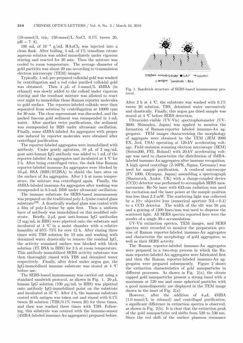
The SERS-based immunoassay was carried out using astandard sandwich protocol, as shown in Fig. 1. 20-µLhuman IgG solution (100 µg/mL in BBS) was pipettedonto antibody IgG-immobilized point on the substrateand incubated at 37 C. After 2 h, the immune substratecoated with antigen was taken out and rinsed with 0.1%tween 20 solution (TBS/0.1% tween 20) for three times,and then was washed three times with TBS. Follow-ing, this substrate was covered with the immuno-sensor(4MBA-labeled immuno-Au aggregates) prepared before.
Fig. 1. Sandwich structure of SERS-based immunoassay pro-tocol.
After 2 h at 4 C, the substrate was washed with 0.1%tween 20 solution, TBS, deionized water successivelyand drastically. Finally, this argon gas dried sample wasstored at 4 C before SERS detection.
Ultraviolet-visible (UV-Vis) spectrophotometer (UV-3600, Shimadzu, Japan) was applied to monitor theformation of Raman-reporter labeled immuno-Au ag-gregates. TEM images characterizing the morphologyof aggregate were obtained by the TEM (JEM 2000EX, Jeol, USA) operating at 120-kV accelerating volt-age. Field emission scanning electron microscopy (SEM)(Sirion200, FEI, Holand) with 20-kV accelerating volt-age was used to characterize the distribution of 4MBA-labeled immuno-Au aggregates after immune recognition.A high speed centrifuge (2-16PK, Sigma, Germany) wasused for sample purification. A confocal microscope(FV 1000, Olympus, Japan) assembling a spectrograph(Sharmrock, Andor, UK) with a charge-coupled device(CCD) detector was performed to accomplish SERS mea-surements. He-Ne laser with 633-nm radiation was usedfor excitation and the laser power at the sample positionwas less than 2.3 mW. The scattering light was collectedby a 10× objective lens (numerical aperture NA = 0.4)to a CCD detector. The width of the slit was 50 µmand a grating of 1200 lines/mm was used to disperse thescattered light. All SERS spectra reported here were theresults of a single 30-s accumulation.
UV-Vis extinction spectra, TEM images, and SERSspectra were recorded to monitor the preparation pro-cess of Raman reporter-labeled immuno-Au aggregatesand characterize the morphology of gold aggregates, aswell as their SERS activity.
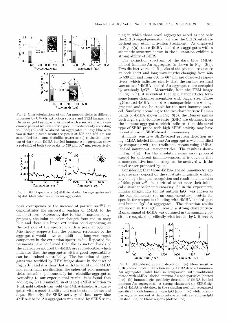
The Raman reporter-labeled immuno-Au aggregateswere prepared in a two-step process in which the Ra-man reporter-labeled Au aggregates were fabricated firstand then the Raman reporter-labeled immuno-Au ag-gregates were prepared subsequently. Figure 2 showsthe extinction characteristics of gold nanoparticles indifferent processes. As shown in Fig. 2(a), the citratecapped gold nanoparticles present a strong band with amaximum at 520 nm and some spherical particles witha good monodispersity are displayed in the TEM imageshown in the inset of Fig. 2(a).
However, after the addition of 4-µL 4MBA(1.0 mmol/L in ethanol) and centrifugal purification,a significant difference in extinction spectra is observed,as shown in Fig. 2(b). It is clear that the extinction peakof the gold nanoparticles red shifts from 520 to 536 nm.Since the red shift of the surface plasmon resonance
March 10, 2010 / Vol. 8, No. 3 / CHINESE OPTICS LETTERS 311
Fig. 2. Characterizations of the Au nanoparticles in differentprosesses by UV-Vis extinction spectra and TEM images. (a)Dispersed gold nanoparticles in red with a surface plasma res-onance peak at 520 nm show a good monodispersity accordingto TEM; (b) 4MBA-labeled Au aggregates in navy blue withtwo surface plasma resonance peaks at 536 and 636 nm areassembled into some chainlike patterns; (c) extinction spec-tra of dark blue 4MBA-labeled immuno-Au aggregates showa red-shift of both two peaks to 539 and 667 nm, respectively.
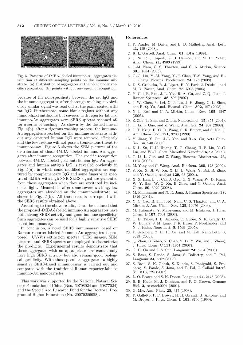
Fig. 3. SERS spectra of (a) 4MBA-labeled Au aggregates and(b) 4MBA-labeled immuno-Au aggregates.
peak corresponds to the increase of particle size[30], itdemonstrates the successful binding of 4MBA to thenanoparticles. Moreover, due to the formation of ag-gregates, the solution color changes from red to navyblue and there is a broad extinction band appearing atthe red side of the spectrum with a peak at 636 nm.Mie theory suggests that the plasmon resonance of theaggregates would have an additional long-wavelengthcomponent in the extinction spectrum[31]. Repeated ex-periments have confirmed that the extinction bands ofthe aggregates induced by 4MBA are reproducible, whichindicates that the aggregates with a good repeatabilitycan be obtained controllably. The formation of aggre-gates was testified by TEM image shown in the inset ofFig. 2(b), and it is clear that with the addition of 4MBAand centrifugal purification, the spherical gold nanopar-ticles assemble spontaneously into chainlike aggregates.According to our experimental results, it is found thatadding 4-µL (1.0 mmol/L in ethanol) 4MBA solution to1-mL gold colloids can yield the 4MBA-labeled Au aggre-gates with a good stability and can be stable for severaldays. Similarly, the SERS activity of those navy blue4MBA-labeled Au aggregates was tested by SERS scan-
ning in which those novel aggregates acted as not onlythe SERS signal-generator but also the SERS substratewithout any other activation treatment. As displayedin Fig. 3(a), those 4MBA-labeled Au aggregates with aschematic structure shown in the illustration exhibits astrong ability of SERS.
The extinction spectrum of the dark blue 4MBA-labeled immuno-Au aggregates is shown in Fig. 2(c).Two distinctive red-shift peaks of the plasmon resonanceat both short and long wavelengths changing from 536to 539 nm and from 636 to 667 nm are observed respec-tively, which indicates clearly that the surface residualvacancies of 4MBA-labeled Au aggregates are occupiedby antibody IgG[6]. Meanwhile, from the TEM imagein Fig. 2(c), it is evident that gold nanoparticles formsome longer chainlike assemblies with bigger size. ThoseIgG-coated 4MBA-labeled Au nanoparticles are well ag-gregated and can be stable for the next immune proto-col. Similarly, according to the two characteristic Ramanbands of 4MBA shown in Fig. 3(b), the Raman signalswith high signal-to-noise ratio (SNR) are obtained fromthe immune aggregates, which indicates that this noveltype of SERS probe with high SERS activity may havepotential use in SERS-based immunoassay.
A highly sensitive SERS-based protein detection us-ing 4MBA-labeled immuno-Au aggregates was identifiedby comparing with the traditional means using 4MBA-labeled immuno-Au nanoparticles. The result is shownin Fig. 4(a). For the absolutely same assay protocolexcept for different immuno-sensors, it is obvious thata more sensitive immunoassay can be achieved with thenovel sensor proposed by us.
Considering that those 4MBA-labeled immuno-Au ag-gregates may deposit on the substrate physically withoutany biologic immune recognition and result in a detectionof false positive[8], it is critical to estimate their inimi-cal disturbance for immunoassay. So in the experiment,human antigen IgG (or rat antigen IgG) was chosen asthe complementary (or un-complementary) protein forspecific (or unspecific) binding with 4MBA-labeled goatanti-human IgG-Au aggregates. The detection resultsare shown in Fig. 4(b). Clearly, a strong characteristicRaman signal of 4MBA was obtained in the sampling po-sition recognized specifically with human IgG. However,
Fig. 4. SERS-based protein detection. (a) More sensitiveSERS-based protein detection using 4MBA-labeled immuno-Au aggregates (solid line) in comparison with traditionalmeans with 4MBA-labeled immuno-Au nanoparticles (dottedline). (b) Immunologic specificity detection of 4MBA-labeledimmuno-Au aggregates. A strong characteristic SERS sig-nal of 4MBA is obtained in the sampling position recognizedspecifically with human antigen IgG (solid line) while no sim-ilar signal is read out at the point coated with rat antigen IgG(dashed line) or blank regions (dotted line).
312 CHINESE OPTICS LETTERS / Vol. 8, No. 3 / March 10, 2010
Fig. 5. Patterns of 4MBA-labeled immuno-Au aggregates dis-tribution at different sampling points on the immune sub-strate. (a) Distribution of aggregates at the point under spe-cific recognition; (b) points without any specific recognition.
because of the non-specificity between the rat IgG andthe immune aggregates, after thorough washing, no obvi-ously similar signal was read out at the point coated withrat IgG. Furthermore, some blank regions without anyimmobilized antibodies but covered with reporter-labeledimmuno-Au aggregates were SERS spectra scanned af-ter a series of washing. As shown by the dashed line inFig. 4(b), after a rigorous washing process, the immuno-Au aggregates absorbed on the immune substrate with-out any captured human IgG were removed efficientlyand the few residue will not pose a tremendous threat toimmunoassay. Figure 5 shows the SEM pictures of thedistribution of those 4MBA-labeled immuno-Au aggre-gates after immune recognition. The specific recognitionbetween 4MBA-labeled goat anti-human IgG-Au aggre-gates and human antigen IgG is revealed obviously inFig. 5(a), in which some modified aggregates are cap-tured by complementary IgG and some fingerprint spec-tra of 4MBA with high SNR SERS signal are generatedfrom those aggregates when they are excitated by inci-dence light. Meanwhile, after some severe washing, fewaggregates are absorbed on the immuno-substrate, asshown in Fig. 5(b). All those results correspond withthe SERS results obtained above.
According to the above results, it can be deduced thatthe proposed 4MBA-labeled immuno-Au aggregates haveboth strong SERS activity and good immune specificity.Such aggregates can be used for a highly sensitive SERSbased immunoassay.
In conclusion, a novel SERS immunoassay based onRaman reporter-labeled immuno-Au aggregates is pro-posed. UV-Vis extinction spectra, TEM images, SEMpictures, and SERS spectra are employed to characterizethe products. Experimental results demonstrate thatthose aggregates with an appropriate size cannot onlyhave high SERS activity but also remain good biologi-cal specificity. With those peculiar aggregates, a highlysensitive SERS-based immunoassay is carried out andcompared with the traditional Raman reporter-labeledimmuno-Au nanoparticles.
This work was supported by the National Natural Sci-ence Foundation of China (Nos. 60708024 and 60877024)and the Specialized Research Fund for the Doctoral Pro-gram of Higher Education (No. 20070286058).
References
1. P. Pandey, M. Datta, and B. D. Malhotra, Anal. Lett.41, 159 (2008).
2. R. L. Garrell, Anal. Chem. 61, 401A (1989).
3. J. Ni, R. J. Lipert, G. B. Dawson, and M. D. Porter,Anal. Chem. 71, 4903 (1999).
4. J.-M. Nam, C. S. Thaxton, and C. A. Mirkin, Science301, 1884 (2003).
5. C.-C. Lin, Y.-M. Yang, Y.-F. Chen, T.-S. Yang, and H.-C. Chang, Biosens. Bioelectron. 24, 178 (2008).
6. D. S. Grubisha, R. J. Lipert, H.-Y. Park, J. Driskell, andM. D. Porter, Anal. Chem. 75, 5936 (2003).
7. Y. Cui, B. Ren, J.-L. Yao, R.-A. Gu, and Z.-Q. Tian, J.Raman Spectrosc. 38, 896 (2007).
8. J.-W. Chen, Y. Lei, X.-J. Liu, J.-H. Jiang, G.-L. Shen,and R.-Q. Yu, Anal. Bioanal. Chem. 392, 187 (2008).
9. N. L. Rosi and C. A. Mirkin, Chem. Rev. 105, 1547(2005).
10. Z. Zhu, T. Zhu, and Z. Liu, Nanotechnol. 15, 357 (2004).
11. T. Li, L. Guo, and Z. Wang, Anal. Sci. 24, 907 (2008).
12. J. T. Krug, II, G. D. Wang, S. R. Emory, and S. Nie, J.Am. Chem. Soc. 121, 9208 (1999).
13. Y. Jiang, Y. Cui, J.-L. Yao, and R.-A. Gu, Acta Chim.Sin. 64, 240 (2006).
14. K.-L. Su, H.-H. Huang, T. C. Chang, H.-P. Lin, Y.-C.Lin, and W.-T. Chen, Microfluid Nanofluid 6, 93 (2009).
15. T. Li, L. Guo, and Z. Wang, Biosens. Bioelectron. 23,1125 (2008).
16. M. Yang and C. Wang, Anal. Biochem. 385, 128 (2009).
17. S. Xu, X. Ji, W. Xu, X. Li, L. Wang, Y. Bai, B. Zhao,and Y. Ozakic, Analyst 129, 63 (2004).
18. X. X. Han, L. J. Cai, J. Guo, C. X. Wang, W. D. Ruan,W. Y. Han, W. Q. Xu, B. Zhao, and Y. Ozakic, Anal.Chem. 80, 3020 (2008).
19. M. Manimaran and N. R. Jana, J. Raman Spectrosc. 38,1326 (2007).
20. Y. C. Cao, R. Jin, J.-M. Nam, C. S. Thaxton, and C. A.Mirkin, J. Am. Chem. Soc. 125, 14676 (2003).
21. M. Futamata, Y. Maruyama, and M. Ishikawa, J. Phys.Chem. B 107, 7607 (2003).
22. C. E. Talley, J. B. Jackson, C. Oubre, N. K. Grady, C.W. Hollars, S. M. Lane, T. R. Huser, P. Nordlander, andN. J. Halas, Nano Lett. 5, 1569 (2005).
23. F. Svedberg, Z. Li, H. Xu, and M. Kall, Nano Lett. 6,2639 (2006).
24. Q. Zhou, G. Zhao, Y. Chao, Y. Li, Y. Wu, and J. Zheng,J. Phys. Chem. C 111, 1951 (2007).
25. G. H. Gu and J. S. Suh, Langmuir 24, 8934 (2008).
26. S. Basu, S. Pande, S. Jana, S. Bolisetty, and T. Pal,Langmuir 24, 5562 (2008).
27. S. Basu, S. K. Ghosh, S. Kundu, S. Panigrahi, S. Pra-haraj, S. Pande, S. Jana, and T. Pal, J. Colloid Interf.Sci. 313, 724 (2007).
28. L. O. Brown and S. K. Doorn, Langmuir 24, 2178 (2008).
29. B. B. Haab, M. J. Dunham, and P. O. Brown, GenomeBiol. 2, research0004 (2001).
30. G. Mie, Ann. Phys. 25, 377 (1908).
31. P. Galletto, P. F. Brevet, H. H. Girault, R. Antoine, andM. Broyer, J. Phys. Chem. B 103, 8706 (1999).