Embed Size (px)
Citation preview


Table of Contents Student Faculty Mentor Page Adaeze Aninweze Hurlbert, Biochemistry 5 J.W. Barrera Barak, Biology 7
Lucas Boncorddo Grossoehme, Biochemistry Evans-Anderson, Biology 8
Lindsay Bradley Abernathy, Mathematics 9
Michelle Corley Harris, Chemistry 11
James Dean Hartel, Chemistry 12
Ian Deas Smith, Biology 36
Tyra Douglas Harris, Chemistry 13
Danielle Gasparik Sumter, Biochemistry 15
Parisa Geranmayeh Smith, Biology 25
Lauren Green Stern, Biology 16
Katja Hall Calloway, Chemistry 17
Brandon Hicks Hanna, Chemistry 18
Anderson Isaac Grattan, Chemistry 19
Bria Jones Sumter, Biochemistry 20
Grace Jones Glasscock, Biology 21
MaLyn Lawhorn Rusinko, Mathematics 22
Jordan Lewis Stern, Biology Frost, Biology 23
Olivia Manley Grossoehme, Biochemistry 24
Kyle McDaniel Smith, Biology 25
Diamond Melendez Hartel Chemistry 26
Alexander Middleton Abernathy, Mathematics 27
Marquet Minor Smith, Biology 28
Rachel Morris Calloway, Chemistry 29
Jamie Murakami Hanna, Chemistry 30
2
Student Faculty Mentor Page Akilah Murray Hurlbert, Biochemistry 31
Denise Peppers Grossoehme, Biochemistry 32
Alexandria Pinnix Hanna, Chemistry 33
Jasmine Richards Calloway, Chemistry 29
Jake Roberts Hanna, Chemistry Lammi, Chemistry 35
Mitchell Schneider Smith, Biology 36
Leigha Stahl Stern, Biology Frost, Biology 37
Kathryn Steverson Stern, Biology 38
Michala Tesney Hurlbert, Biochemistry 39
Danielle Thibault Gelabert, Chemistry 40
Morgan Turnow Grattan, Chemistry 41
James Vinton Birgbauer, Biology 42
Emily Watson Calloway, Chemistry 44
Sarah Wicks Hanna, Chemistry Lammi, Chemistry 45
Deanna Worley Grattan, Chemistry 47
Elijah Wyatt Birgbauer, Biology 48
Jessica Zinna Gelabert, Chemistry 49
Winthrop Students Performing Research at Other Institutions
Kenisha Barber Zachary Schultz University of Notre Dame 6
Kendra Bufkin W. Matthew Leevy University of Notre Dame 10
Esseabasi Etim Marya Lieberman University of Notre Dame 14
Alec Reed Richard Kriwacki St. Jude Children’s Research Hospital 34
Justin Waller W. Matthew Leevy University of Notre Dame 43
Ashley Williams Jie Zheng St. Jude Children’s Research Hospital 46
3

ACKNOWLEDGEMENTS The undergraduate abstract authors were supervised by a vibrant group of faculty in the Biology, Chemistry, and Mathematics Departments. On behalf of the students, faculty, and department administrators, we express our gratitude for the support from the agencies and organizations listed below. The hands-on teaching experiences that faculty provide for these outstanding Winthrop students are only possible through their support.
4
Expression, Purification, and Crystallization of Novel Glycoside Hydrolase Family 30 Xylanases
Adaeze Aninweze (2016) Mentor: Jason C. Hurlbert Identifying alternate energy sources capable of satisfying the demands of the world’s growing population is a topic of major importance in the modern world. One key avenue of research in this field is the development of microbial biocatalysts capable of fermenting renewable resources into ethanol. Lignocellulosic biomass found in plants contains cellulose and hemicellulose that can be used by such biocatalysts. Current biocatalysts are capable of fermenting starches and acid-treated cellulose to ethanol. Unfortunately, the starches used in the process come from corn crops that would normally be used in food production. This resulted in a worldwide spike in the price of food a few years ago when corn was diverted to ethanol production. Using non-edible sources of lignocellulosic biomass like corn husks, sweetgum logs and sugarcane bagasse can prevent such price increases on goods and services. Unfortunately, one of the barriers to utilization of xylans in biofuel production is the resistance of the polymer to enzymatic degradation due to the presence of chemical substitutions on the residues of the main chain. The substitutions vary with plant type: hardwood xylans typically possess a 4-O-methylglucuronic acid substitution for every seventh xylose residue, softwood xylans usually have a 4-O-methylglucuronic acid or arabinose substitution for every third or fourth xylose residue, and the xylans from grasses contain single or multiple substitutions on a majority of xylose residues of the polymer. Xylanases are enzymes that hydrolyze the xylan chains of hemicellulose. They are found in three families of glycoside hydrolase (GH): 10, 11, and 30. The three families of glycoside hydrolase are unique in their ability to identify the different substitutions on the xylan chain. Glycoside hydrolases from family 10 and 11 do not cleave substituted xylan chains very well due to steric hindrance of the substituting residue. Xylanases from GH30 are capable of binding and hydrolyzing substituted xylans chains, in fact the substitution provides a “handle” allowing the enzyme to recognize and bind the substrate. Our research is focused on determining the structures of novel gylcoside hyrdrolase family 30 xylanases from Bacteroides vulgatus, Catenulispora acidiphila, and Paenibacullus mucilaginosus so as to better understand how the enzymes can recognize the substitutions on the xylan chain. GH30 proteins from Bacteroides vulgatus (BvGH30) and Paenibacullus mucilaginosus (PmGH30) were successfully expressed and purified, but the GH30 protein from Catenulispora acidiphila (CaGH30) was only expressed in yields that were too low for crystallographic purposes. The GH30 proteins from Bacteroides vulgatus and Paenibacullus mucilaginosus were subjected to crystallization trials using commercial grid screens and several conditions that yielded small (<0.05 mm) crystals were found. Future work will focus on performing small molecule additive screens on the conditions that yielded crystals of and finding a better condition to express the GH30 protein from Catenulispora acidiphila and later purifying it for crystallization trials. Support for this research was provided by SC-INBRE
5
Fabrication of Surface Enhanced Raman Scattering (SERS) Substrates using
Gold Nanoparticles and Polymethyl methacrylate (PMMA) Kenisha Barber1 (2015) Mentor: Zachary Schultz2
1Winthrop University. Department of Chemistry, Physics and Geology 2University of Notre Dame. Department of Chemistry & Biochemistry
Raman Spectroscopy is a nondestructive way to provide chemically specific information on individual molecules; however, it has poor sensitivity. To improve or increase these Raman signals, Surface Enhanced Raman Scattering (SERS) can be used. SERS occurs when surface plasmons from metals are excited by incident light. This excitation can cause enhance the Raman scattering by 1010. A SERS substrate was fabricated by assembling a film of gold nanoparticles onto a support. Gold nanoparticles were synthesized from hydrogen tetrachloroaurate (III) trihydrate and sodium citrate. The SERS substrate was fabricated using the gold nanoparticles, a PMMA in toluene solution, and ethanol that was slowly added. With the addition of the ethanol, the gold nanoparticles rose to the toluene/water layer while a thin PMMA film formed on top. Upon evaporation of the toluene, the gold nanoparticles were captured in the PMMA film and then placed onto glass or plastic support. The substrate was allowed to dry under vacuum and low heat. To assess the SERS enhancement, the Raman scattering from the thiol, 4-mercaptobenzonitrile (mbn) was measured. Raman spectra were obtained using the Renishaw microscope and laser (633nm) system. The effectiveness of the substrate for SERS activity was compared on the glass, both front and back, and on the plastic media. The substrate on glass showed better enhancement than on the plastic. This substrate shows to have good potential for SERS applications because of its optical transparency and high SERS activity. It is also simple to fabricate.
6
Structure-Function Relationship of White-Tailed Deer’s Primary Bone Tissue in the Femur: Stiffness is Orientation Dependent
J.W. Barrera (2016) Mentor(s): Dr. Meir Barak Bone biomechanical properties such as stiffness are mainly determined by two factors: structural properties and material properties. Ample research was done on the mechanical properties of transverse-isotropic mature secondary bone tissue but much less is known about juvenile primary bone tissue. The purpose of this study was to measure cortical bone stiffness and its correlation to the tissue structure (structural properties) in young animals. We postulated that the juvenile bone tissue will present an orthotropic structure and therefore that tissue stiffness would be the highest in the axial direction followed by the transverse and radial directions. To test this conjecture, we prepared 30 cortical bone cubes (sized 2x2x2mm) from the proximal medial diaphysis of five young white tailed deer femora. Next we measured the cubes stiffness (Young's Modulus (E)) in three orthogonal directions (axial, radial and transverse) under physiological conditions. In addition, several histological slices were prepared from the same region to determine the tissue structure. The histological slides revealed that the bone was composed almost solely of primary osteons in an orthotropic arrangement known as plexiform bone. Furthermore, the mechanical testing reveled that, as predicted, the tissue stiffness was direction-depended, where axial stiffness > transverse stiffness > radial stiffness. This pattern was observed both for the entire cube population but also for the cubes of each of the 5 femora. As young animals tend to fall and injure their limbs much more, an orthotropic structure and stiffness is superior to a transverse-isotropic structure and stiffness. Orthotropic structure yields a higher stiffness in the transverse direction, a feature which is essential to withstand unpredictable loading from various directions. Funded by: INBRE Winthrop Research Council Grant
7
Progress Toward Identification of FoxO Target Genes in C. Intestinalis
Lucas Boncorddo (2015) Mentor: Nicholas E. Grossoehme
Dr. Heather Evans-Anderson
Recent research has identified the organism Ciona intestinalis as a model for heart
regeneration. Ciona intestinalis contains conserved genes that drive heart development and
allow for the regeneration of cardiac myocytes throughout its life. FoxO is an important
transcription factor in this process. The forehead DNA binding region of FoxO is highly
conserved between different organisms. Previous in vitro experiments in our lab have
characterized the protein-DNA interactions between the forehead region of FoxO from C.
intestinalis and sequences of DNA that Human and Drosophila melanogaster FoxO binds
to. This confirmed that the C. intestinalis FoxO DNA Binding Domain (DBD) functions like
that of FoxO from other organisms. Next, we wanted to determine which regions of the genome
FoxO binds to in C. intestinalis. This will help identify which genes are targeted and regulated
by FoxO in C. intestinalis which will help us understand the pathway through which the heart is
regenerated. To do this we utilized the Chromatin Immuno-Precipitation Assay (ChIP Assay) to
identify the sequences of the Ciona intestinalis genome that FoxO binds to. For the ChIP assay,
we opted to make use of the readily available antibody that recognizes hexahistidine tagged
proteins. While Ciona intestinalis naturally expresses FoxO, it is not hexahistidine
tagged. Using standard cloning techniques, a hexahistidine tagged FoxO DBD was inserted into
a pCes, Ciona experession system, expression plasmid. Once confirmed by sequencing, the
expression plasmids were then electroporated into Ciona intestinalis embryos. Once the
hexahistidine tagged FoxO is successfully expressed by Ciona intestinalis a ChIP Assay will be
performed.
Funded by SC-INBRE and Research Corporation Grant 20160.
8
Chronic Myeloid Leukemia and Chemotherapy
Lindsay Bradley (2017) Mentor: Kristen Abernathy Chronic Myeloid Leukemia (CML) begins with a mutation in the Hematopoietic Stem Cells (HSC) that causes the stem cells to produce cancerous Differentiated Cells (DC). In the long run, these tumorous cells out-compete the normal cells and can be fatal if not treated. One of the more common treatments for CML is chemotherapy. In this work, we modify an existing model for CML comprised of growth equations for four types of cells (HSC Normal, DC Normal, HSC Cancerous, and DC Cancerous) to include the effects of chemotherapy treatment. We perform a local stability analysis, sensitivity analysis, and run numerical simulations. We hope to continue our research to explore different types of treatments and their affects upon CML. This work was supported by a Winthrop University Research Council grant.
9

Multimodal Imaging Trials with Zebrafish Specimens Kendra Bufkin1 (2015) Mentor: W. Matthew Leevy2
1Winthrop University. Department of Chemistry, Physics and Geology 2University of Notre Dame, College of Science, Dept. of Biological Sciences
Biomedical imaging is an important technique that can be used for several applications such as cancer research and cardiology. A range of imaging technology, such as PET, SPECT, micro-CT and optical X-ray, is available for imaging. However, many research institutes use rats and mice for preclinical experiments. The purpose of this study is to determine if Zebrafish are compatible for use in pre-clinical imaging, and which modalities and probes work best. Different Zebrafish specimens were tested using five different modalities, and four probes. In order to assess with two-dimensional modalities, both fluorescence and planar X-Ray were performed on the specimens. The fluorescence imaging was acquire using OsteoSense 750x and ProSense 750x as the probes. Through the results, we discovered that OsteoSense did not work as well as the ProSense. The next two modalities represented three-dimensional imaging. These modalities consist of X-ray Computed Tomography (CT) and Positron Emission Tomography (PET), and Single Photon Emission Computed Tomography (SPECT). Sodium Fluoride (NaF), Fludeoxyglucose (FDG) were the two probes tested with PET imaging, while MDP with technetium-99 was used for SPECT.
10
Electrodeposition and Characterization of WSe2/WO3 Layered Thin Films on
Fluorine-Doped Tin Oxide
Michelle Corley (May 2017) Dr. Cliff Harris
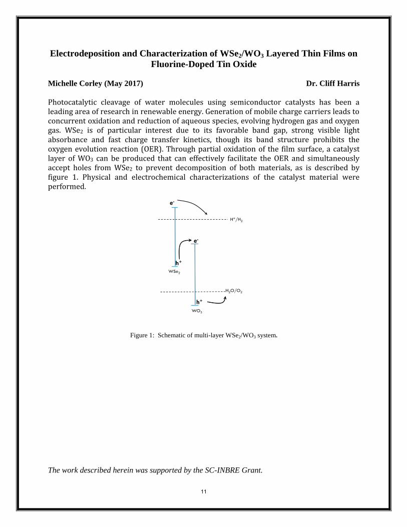
Photocatalytic cleavage of water molecules using semiconductor catalysts has been a leading area of research in renewable energy. Generation of mobile charge carriers leads to concurrent oxidation and reduction of aqueous species, evolving hydrogen gas and oxygen gas. WSe2 is of particular interest due to its favorable band gap, strong visible light absorbance and fast charge transfer kinetics, though its band structure prohibits the oxygen evolution reaction (OER). Through partial oxidation of the film surface, a catalyst layer of WO3 can be produced that can effectively facilitate the OER and simultaneously accept holes from WSe2 to prevent decomposition of both materials, as is described by figure 1. Physical and electrochemical characterizations of the catalyst material were performed.
Figure 1: Schematic of multi-layer WSe2/WO3 system.
The work described herein was supported by the SC-INBRE Grant.
11
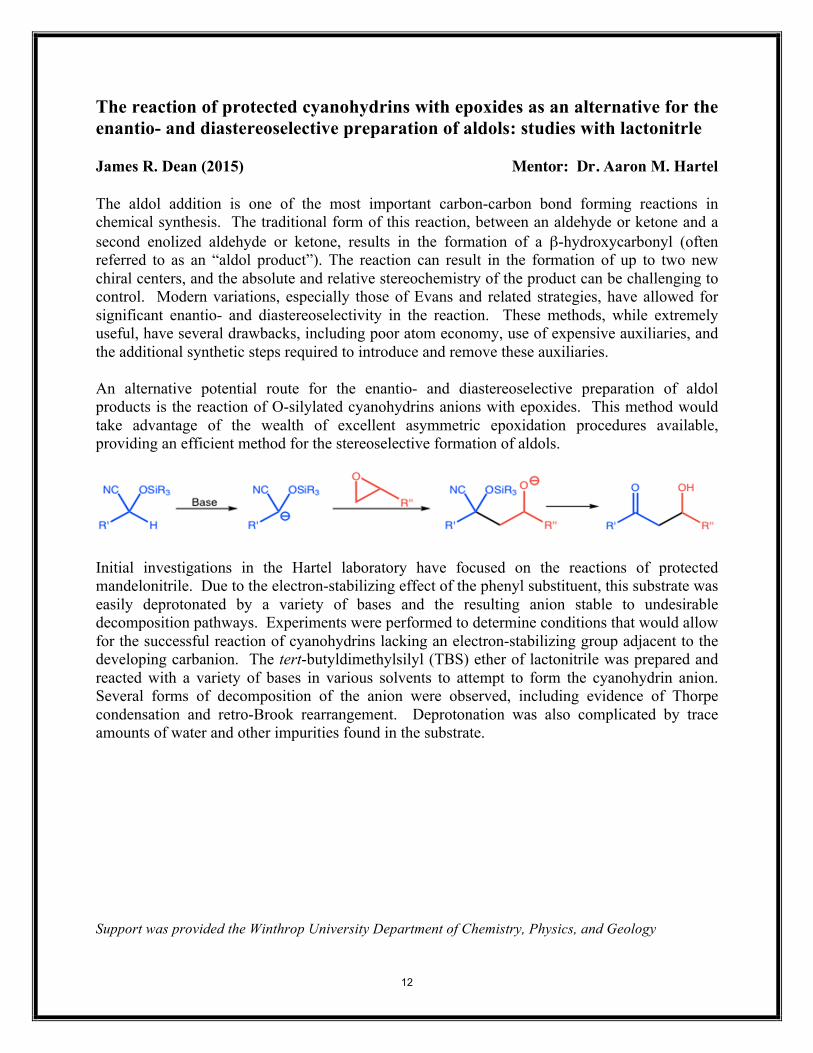
The reaction of protected cyanohydrins with epoxides as an alternative for the enantio- and diastereoselective preparation of aldols: studies with lactonitrle James R. Dean (2015) Mentor: Dr. Aaron M. Hartel The aldol addition is one of the most important carbon-carbon bond forming reactions in chemical synthesis. The traditional form of this reaction, between an aldehyde or ketone and a second enolized aldehyde or ketone, results in the formation of a β-hydroxycarbonyl (often referred to as an “aldol product”). The reaction can result in the formation of up to two new chiral centers, and the absolute and relative stereochemistry of the product can be challenging to control. Modern variations, especially those of Evans and related strategies, have allowed for significant enantio- and diastereoselectivity in the reaction. These methods, while extremely useful, have several drawbacks, including poor atom economy, use of expensive auxiliaries, and the additional synthetic steps required to introduce and remove these auxiliaries. An alternative potential route for the enantio- and diastereoselective preparation of aldol products is the reaction of O-silylated cyanohydrins anions with epoxides. This method would take advantage of the wealth of excellent asymmetric epoxidation procedures available, providing an efficient method for the stereoselective formation of aldols.
Initial investigations in the Hartel laboratory have focused on the reactions of protected mandelonitrile. Due to the electron-stabilizing effect of the phenyl substituent, this substrate was easily deprotonated by a variety of bases and the resulting anion stable to undesirable decomposition pathways. Experiments were performed to determine conditions that would allow for the successful reaction of cyanohydrins lacking an electron-stabilizing group adjacent to the developing carbanion. The tert-butyldimethylsilyl (TBS) ether of lactonitrile was prepared and reacted with a variety of bases in various solvents to attempt to form the cyanohydrin anion. Several forms of decomposition of the anion were observed, including evidence of Thorpe condensation and retro-Brook rearrangement. Deprotonation was also complicated by trace amounts of water and other impurities found in the substrate. Support was provided the Winthrop University Department of Chemistry, Physics, and Geology
12
TiO2/CdS/α-Fe2O3 Multi-Junction Thin Films Tyra Douglas (2015) Mentor: Clifton Harris Although suitable for the reduction of water, CdS is a semiconductor that cannot efficiently oxidize water, leading to a buildup of holes in its valence band. These excess holes drive the decomposition of the film via oxidation of sulfide to sulfur. The objective of this research is to create a TiO2/CdS/α-Fe2O3 thin film for the conversion of solar light into Hydrogen gas while minimizing the decomposition of the CdS. The ability of α-Fe2O3 to scavenge holes from CdS was demonstrated by a two electrode current decay experiment where methyl viologen (MV2+) was used as a redox couple. The TiO2/CdS/α-Fe2O3 and TiO2/CdS thin films were synthesized by first spin coating a layer of TiO2 paste onto fluorine doped tin oxide (FTO) pretreated with TiCl4, followed by separate electrophoretic depositions of CdS and Fe2O3 micelles. Electrochemical data indicates partial suppression of CdS decomposition. Further research is required, including optimization of the Fe2O3 film thickness to improve the kinetics of hole transport from the CdS surface. This work was supported by SC-INBRE.
13
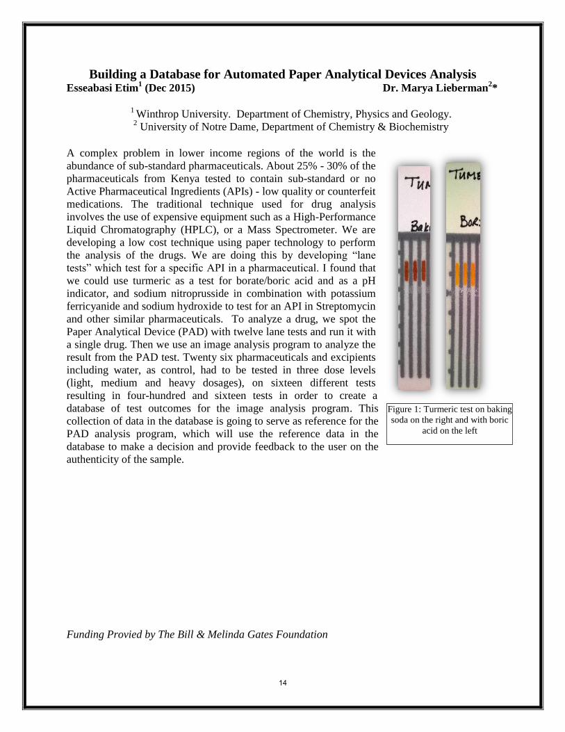
Building a Database for Automated Paper Analytical Devices Analysis Esseabasi Etim
1 (Dec 2015) Dr. Marya Lieberman
2*
1 Winthrop University. Department of Chemistry, Physics and Geology.
2 University of Notre Dame, Department of Chemistry & Biochemistry
A complex problem in lower income regions of the world is the
abundance of sub-standard pharmaceuticals. About 25% - 30% of the
pharmaceuticals from Kenya tested to contain sub-standard or no
Active Pharmaceutical Ingredients (APIs) - low quality or counterfeit
medications. The traditional technique used for drug analysis
involves the use of expensive equipment such as a High-Performance
Liquid Chromatography (HPLC), or a Mass Spectrometer. We are
developing a low cost technique using paper technology to perform
the analysis of the drugs. We are doing this by developing “lane
tests” which test for a specific API in a pharmaceutical. I found that
we could use turmeric as a test for borate/boric acid and as a pH
indicator, and sodium nitroprusside in combination with potassium
ferricyanide and sodium hydroxide to test for an API in Streptomycin
and other similar pharmaceuticals. To analyze a drug, we spot the
Paper Analytical Device (PAD) with twelve lane tests and run it with
a single drug. Then we use an image analysis program to analyze the
result from the PAD test. Twenty six pharmaceuticals and excipients
including water, as control, had to be tested in three dose levels
(light, medium and heavy dosages), on sixteen different tests
resulting in four-hundred and sixteen tests in order to create a
database of test outcomes for the image analysis program. This
collection of data in the database is going to serve as reference for the
PAD analysis program, which will use the reference data in the
database to make a decision and provide feedback to the user on the
authenticity of the sample.
Funding Provied by The Bill & Melinda Gates Foundation
Figure 1: Turmeric test on baking
soda on the right and with boric
acid on the left
14
Progress towards Understanding High Mobility Group A1 (HMGA1) Proteins
Binding DNA Targets
Danielle Gasparik (2016) Mentor: Dr. Takita Sumter
Dr. Robin Lammi
The High Mobility Group A1 gene, encoding HMGA1a and HMGA1b, is considered a
cancer-inducing gene based on its overexpression in human cancers. The transcriptional activity
of HMGA1 proteins are due their DNA binding ability that is mediated by the three highly
conserved DNA binding domains. In general, the proteins are not typical transcription factors but
architectural transcription factors that either activate or repress gene expression depending on the
presence of other proteins with which HMGA interacts. With three DNA binding regions found
in all HMGA1 proteins, it is unclear whether all three of these regions would bind a single DNA
sequence rich in adenine and thymine or if one should expect multimeric interactions between
the DNA and protein. Therefore, we aimed to better elucidate the DNA binding activity and
stoichiometry of HMGA1b. Qualitative assessment of the binding stoichiometry was carried out
by the use of chemiluminescent electrophoretic mobility shift assays (EMSA) with a DNA
sequence bearing three putative HMGA1 binding sites. When DNA concentrations were held
constant and protein concentrations varied to yield one-, two-, and three-fold molar excess, a
significant shift was observed with a marked shift at 1:3 DNA:protein. This data suggests that
each putative HMGA1 binding site within the DNA will bind a single HMGA1 protein.
Interestingly, the addition of a six-fold molar excess of protein yielded a second protein-DNA
complex indicating occupancy of two proteins on the single DNA sequence. In addition,
truncated HMGA1 peptides bearing only the DNA binding domain did not exhibit the same
binding stoichiometry as the full length protein. Collectively, these findings suggest that
HMGA1 proteins bind AT-rich sequences sequentially and that the stoichiometric behavior
changes when various amino acids are deleted. Quantitative fluorescent measurements that allow
specific calculation of stoichiometry and dissociation constants will likely clarify our
understanding of these interactions. Extended application of these findings will improve our
understanding of HMGA1’s function as an architectural transcription factor and provide
significant insight into the discovery of new molecular targets for cancer.
Support was provided by the Winthrop University Department of Chemistry, Physics, and Geology
Funding provided by NIH INBRE (P20 RR-016461) and NIH NCI (1R15CA137520-01)
15
Brain-Penetrating Histone Deacetylase Inhibitor RG2833 Reduces the Growth and Viability of Malignant Melanoma Cells In Vitro
Lauren Green (2016) Mentor(s): Matthew Stern Histone deacetylases (HDACs) play an important role in the epigenetic control of gene expression in both normal and cancer cells. Previous studies have demonstrated that pharmaceutical inhibition of HDACs can kill and/or suppress the growth of cancer cells. RG2833 is a brain-penetrating HDAC inhibitor that targets specific HDACs known to be active in cancer cells. Melanoma cells have previously been shown to respond to HDAC inhibitors that are structurally similar to RG2833. Thus, we hypothesized that the inhibition of HDAC activity by RG2833 would result in the reduced growth and/or death of cells from the malignant melanoma cell lines SK-MEL-5 and SK-MEL-28. To test our hypothesis, we exposed SK-MEL-5 and SK-MEL-28 cells to increasing concentrations of RG2833. We found that concentrations of RG2833 that effectively inhibited HDAC activity also resulted in reduced melanoma cell growth and viability. These results demonstrate the effectiveness of RG2833 in reducing the growth and viability of malignant melanoma cells in vitro and warrant further investigation of the potential therapeutic use of RG2833 and related compounds in the battle against cancer. This project was supported by funding from SC-INBRE and the Winthrop University McNair Scholars Program.
16
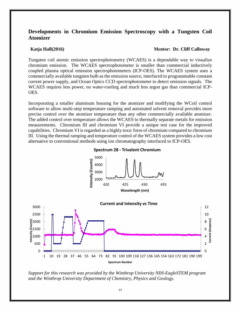
Developments in Chromium Emission Spectroscopy with a Tungsten Coil
Atomizer
Katja Hall(2016) Mentor: Dr. Cliff Calloway
Tungsten coil atomic emission spectrophotometry (WCAES) is a dependable way to visualize
chromium emission. The WCAES spectrophotometer is smaller than commercial inductively
coupled plasma optical emission spectrophotometers (ICP-OES). The WCAES system uses a
commercially available tungsten bulb as the emission source, interfaced to programmable constant
current power supply, and Ocean Optics CCD spectrophotometer to detect emission signals. The
WCAES requires less power, no water-cooling and much less argon gas than commercial ICP-
OES.
Incorporating a smaller aluminum housing for the atomizer and modifying the WCoil control
software to allow multi-step temperature ramping and automated solvent removal provides more
precise control over the atomizer temperature than any other commercially available atomizer.
The added control over temperature allows the WCAES to thermally separate metals for emission
measurements. Chromium III and chromium VI provide a unique test case for the improved
capabilities. Chromium VI is regarded as a highly toxic form of chromium compared to chromium
III. Using the thermal ramping and temperature control of the WCAES system provides a low cost
alternative to conventional methods using ion chromatography interfaced to ICP-OES.
Support for this research was provided by the Winthrop University NIH-EagleSTEM program
and the Winthrop University Department of Chemistry, Physics and Geology.
2000
3000
4000
5000
420 425 430 435
Inte
nsi
ty (
Co
un
ts)
Wavelength (nm)
Spectrum 28 - Trivalent Chromium
0
2
4
6
8
10
12
0
500
1000
1500
2000
2500
3000
1 10 19 28 37 46 55 64 73 82 91 100 109 118 127 136 145 154 163 172 181 190 199
Cu
rre
nt
(Am
pe
res)
Inte
nsi
ty (
Co
un
ts)
Current and Intensity vs Time
Spectrum Number
17
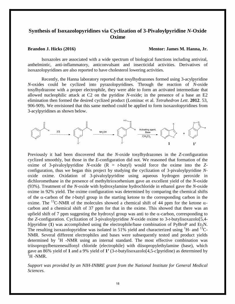
Synthesis of Isoxazolopyridines via Cyclization of 3-Pivaloylpyridine N-Oxide
Oxime
Brandon J. Hicks (2016) Mentor: James M. Hanna, Jr.
Isoxazoles are associated with a wide spectrum of biological functions including antiviral,
anthelmintic, anti-inflammatory, anticonvulsant and insecticidal activities. Derivatives of
isoxazolopyridines are also reported to have cholesterol lowering activities.
Recently, the Hanna laboratory reported that tosylhydrazones formed using 3-acylpyridine
N-oxides could be cyclized into pyrazolopyridines. Through the reaction of N-oxide
tosylhydrazone with a proper electrophile, they were able to form an activated intermediate that
allowed nucleophilic attack at C2 on the pyridine N-oxide; in the presence of a base an E2
elimination then formed the desired cyclized product (Lominac et al. Tetrahedron Lett. 2012. 53,
906-909). We envisioned that this same method could be applied to form isoxazolopyridines from
3-acylpyridines as shown below.
CH2Cl2
Activating agent, Base[O] NH2OH
Previously it had been discovered that the N-oxide tosylhydrazones in the Z-configuration
cyclized smoothly, but those in the E-configuration did not. We reasoned that formation of the
oxime of 3-pivaloylpyridine N-oxide (R = t-butyl) would force the oxime into the Z-
configuration, thus we began this project by studying the cyclization of 3-pivaloylpyridine N-
oxide oxime. Oxidation of 3-pivaloylpyridine using aqueous hydrogen peroxide in
dichloromethane in the presence of methyltrioxorhenium gave an excellent yield of the N-oxide
(93%). Treatment of the N-oxide with hydroxylamine hydrochloride in ethanol gave the N-oxide
oxime in 92% yield. The oxime configuration was determined by comparing the chemical shifts
of the -carbon of the t-butyl group in the starting ketone to the corresponding carbon in the
oxime. The 13
C-NMR of the molecules showed a chemical shift of 44 ppm for the ketone -
carbon and a chemical shift of 37 ppm for that in the oxime. This showed that there was an
upfield shift of 7 ppm suggesting the hydroxyl group was anti to the -carbon, corresponding to
the Z-configuration. Cyclization of 3-pivaloylpyridine N-oxide oxime to 3-t-butylisoxazolo[5,4-
b]pyridine (1) was accomplished using the electrophile/base combination of PyBroP and Et3N.
The resulting isoxazolopyridine was isolated in 51% yield and characterized using 1H- and
13C-
NMR. Several different electrophiles and bases were subsequently tested and product yields
determined by 1H -NMR using an internal standard. The most effective combination was
triisopropylbenzenesulfonyl chloride (electrophile) with diisopropylethylamine (base), which
gave an 86% yield of 1 and a 9% yield of 1′ (3-t-butylisoxazolo[4,5-c]pyridine) as determined by 1H -NMR.
Support was provided by an NIH-INBRE grant from the National Institute for General Medical
Sciences.
1 1′
18
Synthetic Modification of "Zone 2" on a Known Sphingosine Kinase 1
Inhibitor to Improve Oral Bioavailability
Anderson Isaac (2015) Mentor: T. Christian Grattan
Found within the cell membrane, are lipids known as sphingolipids. Besides their distinctive
amphipathic structure, these lipids possess the capability to breakdown under stress to form
sphingolipid metabolites. Metabolites, such as ceramide and sphingosine-1-phosphate (S1P) are
identified as signaling compounds for cell-death (apoptosis) and cell growth (proliferation). The
regulatory enzyme behind the production of these two metabolites is identified as sphingosine-
kinase 1 (SphK1). Due to its proliferative signaling, high levels of S1P have been associated with
cancer patients. As result of this correlation, SK1 has been identified as an oncogene and is
directly associated with cancer making it a viable inhibition target. Since the development of this
observation, numerous inhibitors have been synthesized to cease SphK1 production of SP1.
Smith et. al discovered a group of non-lipid based compounds that inhibit SphK1 proliferative
effect. Despite this scientific milestone, these compounds have only been shown to be effective
in vitro. Since the discovery, scientists have been trying to
increase the compounds potency for in vivo performance
by improving their oral bioavailability.
One of Smith’s compounds has shown significant
activity in vitro allowing our research to use it as a
template structure. Alterations of the template were based
on increasing water solubility due to the fact that the
human body is composed of a high percentage of water.
The template-compound has been identified as having a
log P value of 5.675. A Log P value of 5 or higher is
associated with a compound being less likely to be
absorbed in water.
In order to evaluate the water solubility of the template compound with alterations, logP values
were assessed and then inhibitor compounds were synthesized to determine how these changes
impact the interactions the drug has within the enzyme active site.
Support was provided by an NIH-INBRE grant from the National Center for Research Resources and the National Institute for General Medical Sciences and the Winthrop University Department of Chemistry, Physics, and Geology.
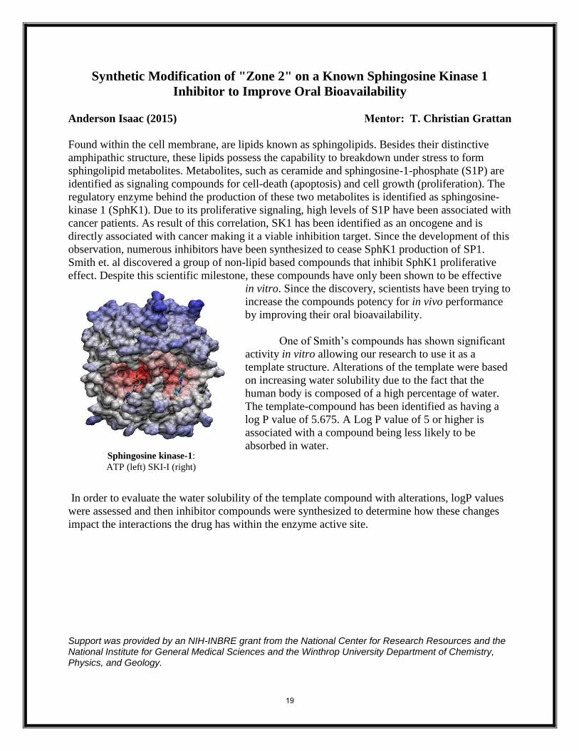
Sphingosine kinase-1:
ATP (left) SKI-I (right)
19
ASSESSING THE BIOLOGICAL ACTIVITY OF
BENZOISOXAZOLO[2,3-a]PYRIDINIUM TETRAFLUOROBORATE
SALTS ON PC3 PROSTATE CANCER CELLS
Bria Jones (2016) Mentor: Dr. Takita F. Sumter
Dr. James Hanna, Jr
Prostate cancer is the second leading cause of cancer death in men and one in every seven men
are likely to be diagnosed with the disease each year. Current therapies for this disease include
surgery chemotherapy and radiation although limited therapeutic indices continue as a challenge. While the initiation and development of cancer results from the deregulation of hundreds of genes most
conventional cancer therapies are nonspecific and lead to adverse side effects. We previously
investigated the activity of a small library of novel benzisoxazolo[2,3-a]pyridinium (Figure 1) compounds
against HCT116 colon cancer cells and recorded activity in the low to mid-micromolar range. The small
planar structure of these novel compounds is a key structural feature of a number of DNA binding drugs.
Based on the successful cytotoxicity observed in HCT 116 colon cancer cells, we aimed to study the
biological activity of the most potent benzisoxazolo[2,3-a]pyridinium derivatives to determine if activity
observed in colon cancer cells was also present in prostate cancer cells. To establish assay conditions, we
first determined the optimal number of cells to use in cell viability assays. We then treated these cells
with the counter ion, tetrafluoroborate, to verify the absence of biological activity. When
compared to sodium chloride, we saw no significant difference between viability of prostate
cancer cells treated with sodium tetrafluoroborate at varying concentrations (p-value=0.5).
Preliminary assessment of the biological activity of methyl substituted benzisoxazolo[2,3-
a]pyridinium tetraflouroborate were inconclusive and additional studies are underway to verify
preliminary findings. Further studies are likely to yield a novel small molecule capable of
specific and effective treatment of prostate cancer.
This work was supported by grants from the NCI (1R15CA137520-01and NIGMS(8 P20 GM103499) of
the National Institutes for Health, the National Science Foundation (MCB0542242).
20
The Role of Urokinase, Plasminogen Activator Inhibitor 1 and Activated
Protein C in Angiogenesis Grace Jones (2015) Mentor: Laura Glasscock The progression of prostate cancer, as with all types of cancer, is dependent upon angiogenesis and invasion. Both processes are dependent upon protease degradation of the extracellular matrix. We investigated the role of urokinase type plasminogen activator (uPA), plasminogen activator inhibitor 1 (PAI-1) and activated protein C (APC) in angiogenesis in vitro. uPA and APC are proteases that can be inhibited by PAI-1. It is widely known that uPA promotes tumor cell invasion and that PAI-1 can inhibit uPA activity. In ovarian cancer, the addition of APC sequesters PAI-1 away from uPA, allowing uPA to continue promoting invasion, as the Glasscock lab also found to be true in prostate cancer. We hypothesized that in angiogenesis, uPA and APC alone will increase endothelial cell microtubule formation, increasing angiogenesis, and that PAI-1 alone will have no effect on microtubule growth but will inhibit the activity of uPA and APC. We also hypothesized that in the presence of APC, uPA, and PAI-, PAI-1 is sequestered from uPA, allowing angiogenesis to resume. To study angiogenesis, we established an in vitro angiogenesis assay using a new endothelial cell line in our lab. We determined that the ideal conditions for this assay are a low passage number, growth factor reduced Matrigel and a cell count of 30,000 cells per well. Using this system, we determined that elevated concentrations of uPA increase angiogenesis and that PAI-1 has no effect on angiogenesis, as was expected. Contrary to our hypothesis, we found that APC had no significant effect on microtubule growth. Currently, this assay is being repeated combining all three of the proteins at varying concentrations to determine if APC sequesters PAI-1 from uPA inhibition, thus promoting angiogenesis. Support for this work was provided by SC-INBRE, the Winthrop University Research Council and the McKay Urology Endowment Fund.
21
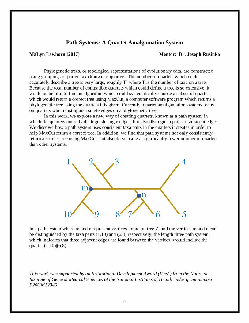
Path Systems: A Quartet Amalgamation System
MaLyn Lawhorn (2017) Mentor: Dr. Joseph Rusinko
Phylogenetic trees, or topological representations of evolutionary data, are constructed
using groupings of paired taxa known as quartets. The number of quartets which could
accurately describe a tree is very large, roughly T4 where T is the number of taxa on a tree.
Because the total number of compatible quartets which could define a tree is so extensive, it
would be helpful to find an algorithm which could systematically choose a subset of quartets
which would return a correct tree using MaxCut, a computer software program which returns a
phylogenetic tree using the quartets it is given. Currently, quartet amalgamation systems focus
on quartets which distinguish single edges on a phylogenetic tree.
In this work, we explore a new way of creating quartets, known as a path system, in
which the quartets not only distinguish single edges, but also distinguish paths of adjacent edges.
We discover how a path system uses consistent taxa pairs in the quartets it creates in order to
help MaxCut return a correct tree. In addition, we find that path systems not only consistently
return a correct tree using MaxCut, but also do so using a significantly fewer number of quartets
than other systems.
In a path system where m and n represent vertices found on tree Z, and the vertices m and n can
be distinguished by the taxa pairs (1,10) and (6,8) respectively, the length three path system,
which indicates that three adjacent edges are found between the vertices, would include the
quartet (1,10)|(6,8).
This work was supported by an Institutional Development Award (IDeA) from the National
Institute of General Medical Sciences of the National Institutes of Health under grant number
P20GM12345
22
Where Escherichia coli is commonly found on the beach? Jordan Lewis (2017) Mentor(s): Matthew Stern
Victoria Frost Sandy beaches are commonly home to pathogens that can severely affect human health. However, we have a limited understanding of how they vary in abundance and distribution within these ecological communities. Understanding how these pathogens are distributed across the beach environment is critical because exposure to or increases in their abundance often can lead to increases in human illnesses and beach closures. In this study, we examined how the distribution and abundance of Escherichia coli (E. coli), an important fecal indicator bacteria species, varied across a set of sandy beaches in Folly Beach, South Carolina. To gather these data, we collected sand samples from three distinct habitats (i.e. dunes, inter-tidal areas, and subtidal areas) at 30 beaches. In addition, we also examined how anthropogenic stressors (i.e. beach re-nourishment) and bacterial levels in source communities (i.e. adjacent marine water sources) influenced our findings. Our results indicate that E. coli is able to persist in all three of the habitat areas of the beach we examined, but in varying abundances. We also determined that renourishment of beaches had a significant influence on the presence and abundance of E. coli and led to an increase in bacterial abundance in inter-tidal communities (which is where they add the re-nourished sand). Finally, we also determined that E. coli was not found in adjacent marine waters, which suggests that terrestrial pollutants and animals may be the likely source of these bacteria. Collectively, our findings suggest that E. coli is likely to be present in all areas of beaches and is an important source of bacterial pollution. Furthermore, our findings imply that bacterial survey methods utilized by organizations like the EPA may be better served by sampling sand for bacteria than adjacent marine water sources. This work was supported by SC-INBRE.
23
The cloning and characterization of nickel uptake regulator (NUR)
mutants from S. coelicolor
Olivia Manley (2016) Mentor: Nicholas E. Grossoehme
Sufficient concentrations of various metal ions within a cell are required for proper
cellular function, but for the same reasons that metal is so vital, too much can become toxic at
high concentrations, notably Zn, Cu, and Fe. Therefore, it is highly important for an organism to
have a mechanism for maintaining metal homeostasis within its cells. Streptomyces coelicolor, a
soil-dwelling bacterium important in the production of antibiotics of medicinal value, utilizes the
nickel uptake regulator (NUR), a dimeric transcriptional repressor protein, to maintain nickel
homeostasis and oxidative response. Previous research describes two key metal-binding sites per
NUR monomer. The crystal structure shows that one site has nickel bound, thus it is called the
Ni-site. The other binding site, called the M-site, was crystallized with zinc bound, though it is
proposed that it can also coordinate nickel. Mutational studies have shown that the specific
amino acids involved in coordinating metal at the Ni-site are His70, His72, and His126, along
with three water molecules in vivo, and that the key amino acids at the M-site are His33, His86,
His88, and His90. Glu101 also forms an electrostatic interaction at the M-site to further secure
the metal ion. Our research seeks to analyze how changes to these binding sites affect the ability
of NUR to coordinate metals and bind to DNA.
NUR mutants containing one or more amino acid substitutions at each binding site have
been cloned. Mutants H86A, E101Q, and H126A were cloned, and the success of the
transformations was confirmed by PCR screening and sequencing results, thus completing the
library of single NUR mutants. We have also created mutants in which all of the metal-binding
potential at each binding site has been abolished, called the Ni and M mutants. The Ni and
M mutants will aid in understanding the specific roles of metal-binding at each binding site.
Complete purification and characterization of these mutants is ongoing.
Previously conducted experiments suggest that NUR must be bound to DNA for metal at
the M-site to be accessible. To confirm this result, an experiment using the strong metal chelator
EDTA to strip WT NUR of metals was conducted. Gel filtration chromatography, which
separates particles by size, was used to examine WT NUR incubated with EDTA and WT NUR
incubated with DNA and EDTA. The zinc content of each of the fractions that eluted from the
column was assessed by atomic absorption spectrophotometry. The experiment showed that
EDTA pulled little metal from NUR when incubated without DNA, but when NUR was
incubated with DNA and EDTA, EDTA removed much more metal from the protein, suggesting
that more metal is exchangeable from NUR once bound to DNA.
Funded by SC-INBRE and Research Corporation Grant 20160.
24
Constructing the Meiofaunal Food Web Via Diagnostic PCR Kyle McDaniel (2016) Parisa Geranmayeh (2015) Rhea Mathew (GSSM 2015)
Mentor: Julian Smith III
Trophic relationships remain a critical cornerstone in determining many other ecological relationships, and this proves to be no exception with the largely unexplored marine interstitial meiofaunal community of the Bogue Banks area in North Carolina. Using diagnostic PCR techniques previously developed in this lab and DNA sequencing, we have made significant progress in identifying the “who eats whom” within meiofaunal communities. Our study has screened over 70 predators for 4 different potential prey taxa using prey-taxon-specific primer sets, and has analyzed macrofaunal-meiofaunal interactions as well as trophic exchange between meiofaunal organisms. Constructing this food web will allow future studies to focus on specific trophic interactions that may determine community structure as wells many other characteristics of the meiofauna, such as behavior and development. This summer work was funded by the Winthrop University Research Council (KM), SC-INBRE (PG), and SC Governor’s School for Science and Mathematics (RM).
25
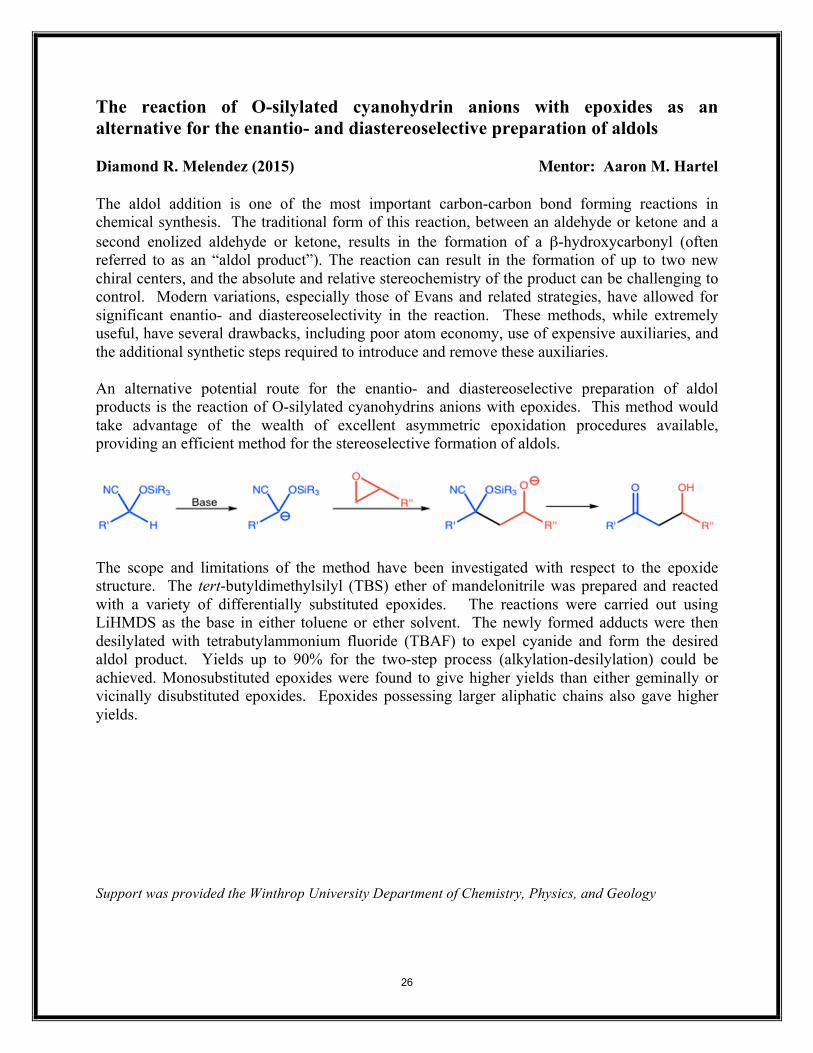
The reaction of O-silylated cyanohydrin anions with epoxides as an alternative for the enantio- and diastereoselective preparation of aldols Diamond R. Melendez (2015) Mentor: Aaron M. Hartel The aldol addition is one of the most important carbon-carbon bond forming reactions in chemical synthesis. The traditional form of this reaction, between an aldehyde or ketone and a second enolized aldehyde or ketone, results in the formation of a β-hydroxycarbonyl (often referred to as an “aldol product”). The reaction can result in the formation of up to two new chiral centers, and the absolute and relative stereochemistry of the product can be challenging to control. Modern variations, especially those of Evans and related strategies, have allowed for significant enantio- and diastereoselectivity in the reaction. These methods, while extremely useful, have several drawbacks, including poor atom economy, use of expensive auxiliaries, and the additional synthetic steps required to introduce and remove these auxiliaries. An alternative potential route for the enantio- and diastereoselective preparation of aldol products is the reaction of O-silylated cyanohydrins anions with epoxides. This method would take advantage of the wealth of excellent asymmetric epoxidation procedures available, providing an efficient method for the stereoselective formation of aldols.
The scope and limitations of the method have been investigated with respect to the epoxide structure. The tert-butyldimethylsilyl (TBS) ether of mandelonitrile was prepared and reacted with a variety of differentially substituted epoxides. The reactions were carried out using LiHMDS as the base in either toluene or ether solvent. The newly formed adducts were then desilylated with tetrabutylammonium fluoride (TBAF) to expel cyanide and form the desired aldol product. Yields up to 90% for the two-step process (alkylation-desilylation) could be achieved. Monosubstituted epoxides were found to give higher yields than either geminally or vicinally disubstituted epoxides. Epoxides possessing larger aliphatic chains also gave higher yields. Support was provided the Winthrop University Department of Chemistry, Physics, and Geology
26
A Nonlinear Model of Cancer Tumor Treatment with Cancer Stem Cells
Alexander D. Middleton (2017) Mentor(s): Dr. Kristen Abernathy
According to the American Cancer Society, cancer is one of the leading causes of
death, second only to heart disease. We present a system of nonlinear, first-order, ordinary
differential equations that describes tumor growth based on healthy cell, tumor cell, and
cancer stem cell populations. We include terms within our model which reflect the differing
effects of chemotherapy and anti-angiogenic therapy to respective cell populations. We
perform stability analysis on the equilibrium solutions to predict the long-term behavior of
the cell populations. With analysis, it is shown that chemotherapy, with the co-
administration of anti-angiogenic treatment, can produce three states: recurrence or
persistence of cancer, and a cure state. Results are supported numerically and bifurcation
diagrams are included to illustrate the different behavior of cell populations depending on
the amount of treatment administered.
This material is based upon work supported by the National Science Foundation under
Grant No. DMS-1358534
27
Characterization of the Hedgehog Signaling Pathway in Isodiametra pulchra (Acoelomorpha) Marquet Minor (2015) Mentor: Julian Smith III The purpose of this study is to distinguish the Hedgehog (Hh) Signaling Pathway in the acoelmorphan, Isodiametra pulchra. The significance of this organism is based on its primitive placement in the bilaterians, along with the complete ciliation of the animal. The role of cilia in Hh signaling can vary amongst animals. Certain organisms have cilia-dependent Hh signaling, while others conduct signaling independently of cilia. Protostomes, for example, require no cilia to conduct signaling, while deuterostomes require some form of cilia to conduct signaling. As previously shown by Emily Bowie in our lab, cyclopamine, which is a Hh pathway inhibitor, depresses cell-cycling in this organism. This down regulation is typical when Hedgehog signaling decreases. Cyclopamine treatment disrupted the integrity of the ciliated epidermis, and this suggests that cilia are required for this organism to properly conduct Hh signaling. This suggested cilia-dependency is consistent with this organism’s phylogenetic placement basally among the deuterostomes. Proteins associated with the Hh pathway were identified with the transcriptome provided from Innsbruck, including orthologues for Hedgehog and multiple possible orthologues for Patched. Primers have been designed and tested via PCR to verify the existence of the transcripts listed. All of the transcripts above have been shown to exist, but only partially verified. Also, the transcriptomic sequences of the wild-type animals (collected on the NC coast) display base variation when compared to the transcriptome the animals cultured in Innsbruck. Next, qPCR primers will be designed and tested to analyze Patched expression. Patched is up-regulated when Hedgehog is activated, so cyclopamine treatment should depress its expression; this technique will allow us to identify the correct orthologue of Patched among the four possible transcripts. Hh plays a critical role in development in many organisms, including humans. The importance of this study is to advance the comprehension of how cilia and Hh are intertwined. This can ultimately lead to control of abnormalities associated with both cilia and the signaling pathway, which can cause several disorders in humans. This work was funded by SC-INBRE.
28
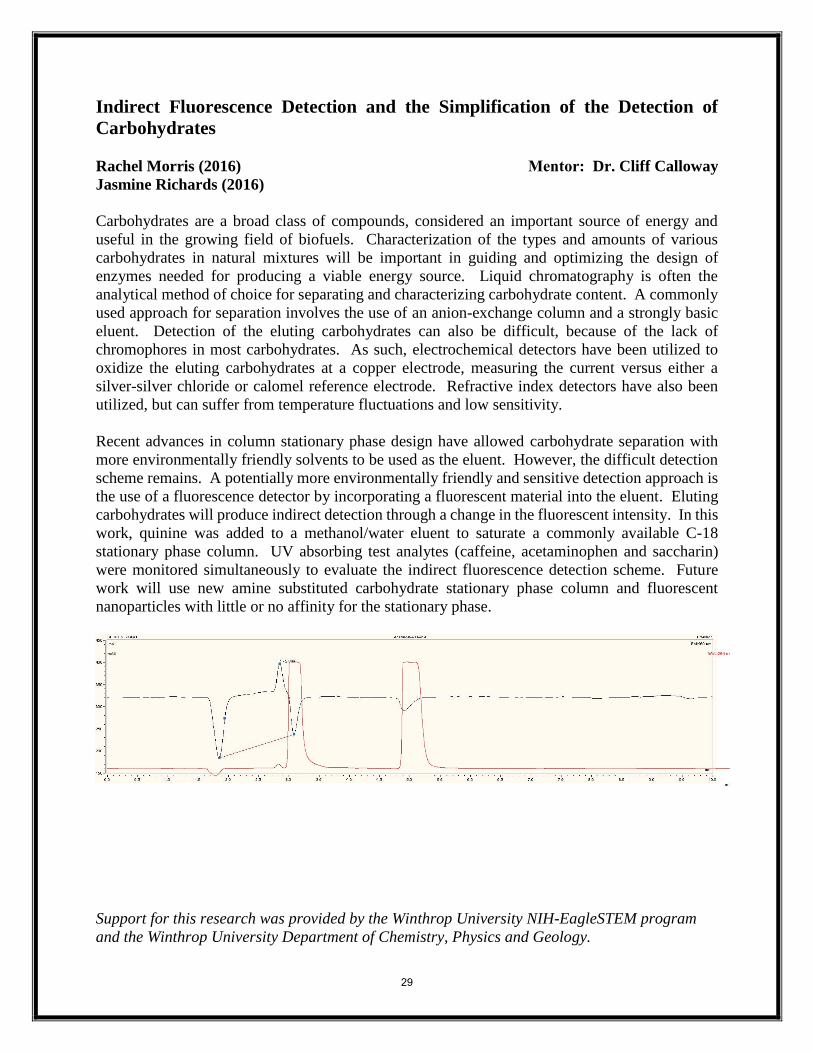
Indirect Fluorescence Detection and the Simplification of the Detection of
Carbohydrates
Rachel Morris (2016)
Jasmine Richards (2016)
Mentor: Dr. Cliff Calloway
Carbohydrates are a broad class of compounds, considered an important source of energy and
useful in the growing field of biofuels. Characterization of the types and amounts of various
carbohydrates in natural mixtures will be important in guiding and optimizing the design of
enzymes needed for producing a viable energy source. Liquid chromatography is often the
analytical method of choice for separating and characterizing carbohydrate content. A commonly
used approach for separation involves the use of an anion-exchange column and a strongly basic
eluent. Detection of the eluting carbohydrates can also be difficult, because of the lack of
chromophores in most carbohydrates. As such, electrochemical detectors have been utilized to
oxidize the eluting carbohydrates at a copper electrode, measuring the current versus either a
silver-silver chloride or calomel reference electrode. Refractive index detectors have also been
utilized, but can suffer from temperature fluctuations and low sensitivity.
Recent advances in column stationary phase design have allowed carbohydrate separation with
more environmentally friendly solvents to be used as the eluent. However, the difficult detection
scheme remains. A potentially more environmentally friendly and sensitive detection approach is
the use of a fluorescence detector by incorporating a fluorescent material into the eluent. Eluting
carbohydrates will produce indirect detection through a change in the fluorescent intensity. In this
work, quinine was added to a methanol/water eluent to saturate a commonly available C-18
stationary phase column. UV absorbing test analytes (caffeine, acetaminophen and saccharin)
were monitored simultaneously to evaluate the indirect fluorescence detection scheme. Future
work will use new amine substituted carbohydrate stationary phase column and fluorescent
nanoparticles with little or no affinity for the stationary phase.
Support for this research was provided by the Winthrop University NIH-EagleSTEM program
and the Winthrop University Department of Chemistry, Physics and Geology.
29
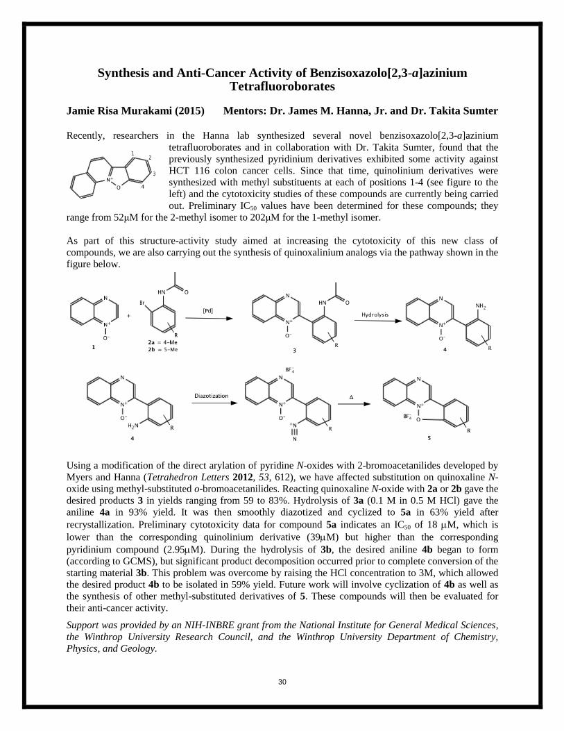
Synthesis and Anti-Cancer Activity of Benzisoxazolo[2,3-a]azinium Tetrafluoroborates
Jamie Risa Murakami (2015) Mentors: Dr. James M. Hanna, Jr. and Dr. Takita Sumter
Recently, researchers in the Hanna lab synthesized several novel benzisoxazolo[2,3-a]azinium
tetrafluoroborates and in collaboration with Dr. Takita Sumter, found that the
previously synthesized pyridinium derivatives exhibited some activity against
HCT 116 colon cancer cells. Since that time, quinolinium derivatives were
synthesized with methyl substituents at each of positions 1-4 (see figure to the
left) and the cytotoxicity studies of these compounds are currently being carried
out. Preliminary IC50 values have been determined for these compounds; they
range from 52μM for the 2-methyl isomer to 202μM for the 1-methyl isomer.
As part of this structure-activity study aimed at increasing the cytotoxicity of this new class of
compounds, we are also carrying out the synthesis of quinoxalinium analogs via the pathway shown in the
figure below.
Using a modification of the direct arylation of pyridine N-oxides with 2-bromoacetanilides developed by
Myers and Hanna (Tetrahedron Letters 2012, 53, 612), we have affected substitution on quinoxaline N-
oxide using methyl-substituted o-bromoacetanilides. Reacting quinoxaline N-oxide with 2a or 2b gave the
desired products 3 in yields ranging from 59 to 83%. Hydrolysis of 3a (0.1 M in 0.5 M HCl) gave the
aniline 4a in 93% yield. It was then smoothly diazotized and cyclized to 5a in 63% yield after
recrystallization. Preliminary cytotoxicity data for compound 5a indicates an IC50 of 18 M, which is
lower than the corresponding quinolinium derivative (39M) but higher than the corresponding
pyridinium compound (2.95M). During the hydrolysis of 3b, the desired aniline 4b began to form
(according to GCMS), but significant product decomposition occurred prior to complete conversion of the
starting material 3b. This problem was overcome by raising the HCl concentration to 3M, which allowed
the desired product 4b to be isolated in 59% yield. Future work will involve cyclization of 4b as well as
the synthesis of other methyl-substituted derivatives of 5. These compounds will then be evaluated for
their anti-cancer activity.
Support was provided by an NIH-INBRE grant from the National Institute for General Medical Sciences,
the Winthrop University Research Council, and the Winthrop University Department of Chemistry,
Physics, and Geology.
30
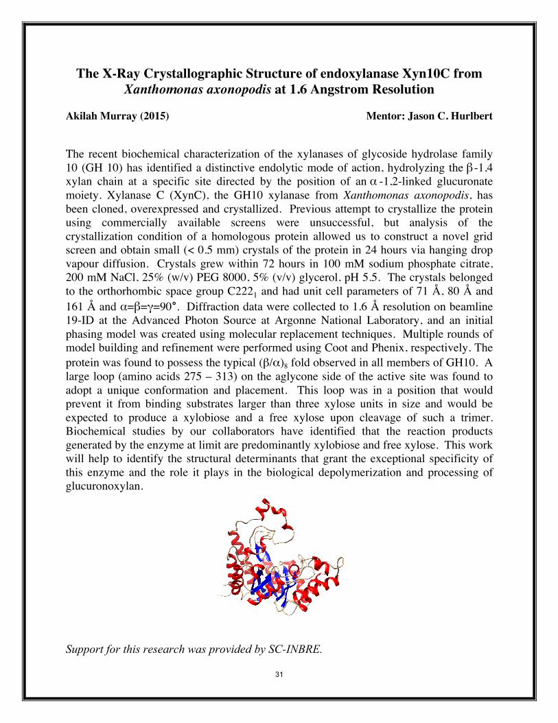
The X-Ray Crystallographic Structure of endoxylanase Xyn10C from Xanthomonas axonopodis at 1.6 Angstrom Resolution
Akilah Murray (2015) Mentor: Jason C. Hurlbert
The recent biochemical characterization of the xylanases of glycoside hydrolase family 10 (GH 10) has identified a distinctive endolytic mode of action, hydrolyzing the β -1,4 xylan chain at a specific site directed by the position of an α -1,2-linked glucuronate moiety. Xylanase C (XynC), the GH10 xylanase from Xanthomonas axonopodis, has been cloned, overexpressed and crystallized. Previous attempt to crystallize the protein using commercially available screens were unsuccessful, but analysis of the crystallization condition of a homologous protein allowed us to construct a novel grid screen and obtain small (< 0.5 mm) crystals of the protein in 24 hours via hanging drop vapour diffusion. Crystals grew within 72 hours in 100 mM sodium phosphate citrate, 200 mM NaCl, 25% (w/v) PEG 8000, 5% (v/v) glycerol, pH 5.5. The crystals belonged to the orthorhombic space group C2221 and had unit cell parameters of 71 Å, 80 Å and 161 Å and α=β=γ=90°. Diffraction data were collected to 1.6 Å resolution on beamline 19-ID at the Advanced Photon Source at Argonne National Laboratory, and an initial phasing model was created using molecular replacement techniques. Multiple rounds of model building and refinement were performed using Coot and Phenix, respectively. The protein was found to possess the typical (β/α)8 fold observed in all members of GH10. A large loop (amino acids 275 – 313) on the aglycone side of the active site was found to adopt a unique conformation and placement. This loop was in a position that would prevent it from binding substrates larger than three xylose units in size and would be expected to produce a xylobiose and a free xylose upon cleavage of such a trimer. Biochemical studies by our collaborators have identified that the reaction products generated by the enzyme at limit are predominantly xylobiose and free xylose. This work will help to identify the structural determinants that grant the exceptional specificity of this enzyme and the role it plays in the biological depolymerization and processing of glucuronoxylan.
Support for this research was provided by SC-INBRE.
31
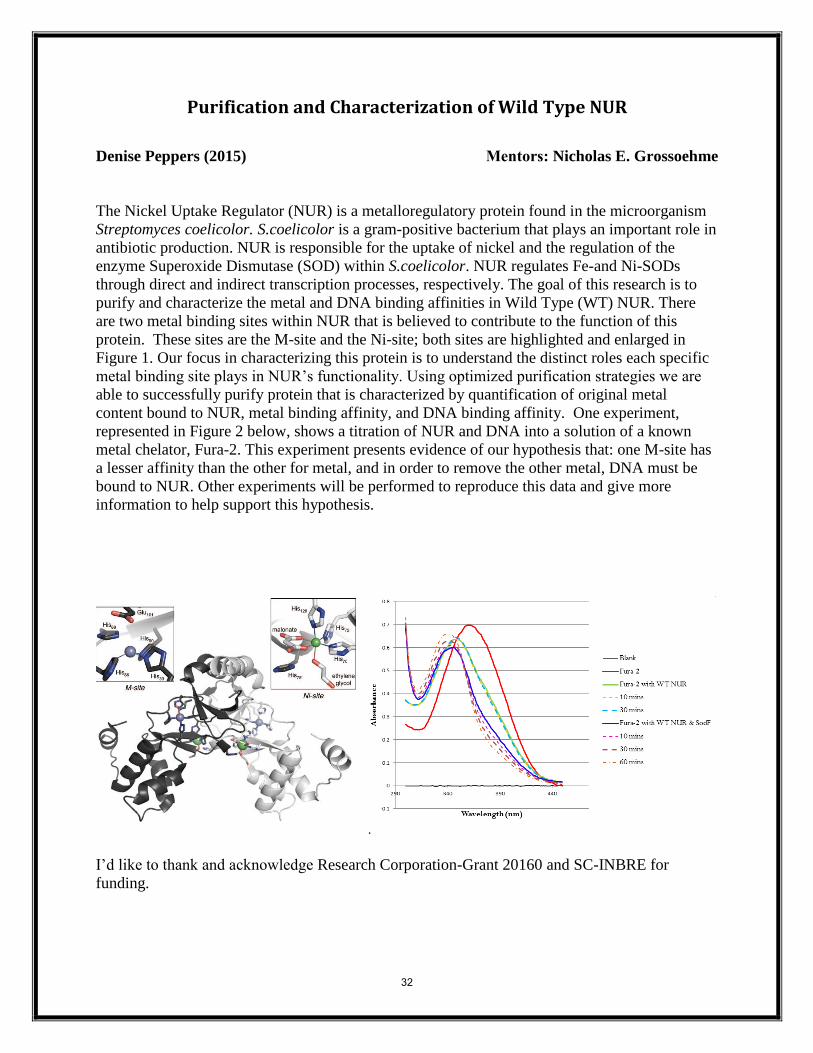
Purification and Characterization of Wild Type NUR
Denise Peppers (2015) Mentors: Nicholas E. Grossoehme
The Nickel Uptake Regulator (NUR) is a metalloregulatory protein found in the microorganism
Streptomyces coelicolor. S.coelicolor is a gram-positive bacterium that plays an important role in
antibiotic production. NUR is responsible for the uptake of nickel and the regulation of the
enzyme Superoxide Dismutase (SOD) within S.coelicolor. NUR regulates Fe-and Ni-SODs
through direct and indirect transcription processes, respectively. The goal of this research is to
purify and characterize the metal and DNA binding affinities in Wild Type (WT) NUR. There
are two metal binding sites within NUR that is believed to contribute to the function of this
protein. These sites are the M-site and the Ni-site; both sites are highlighted and enlarged in
Figure 1. Our focus in characterizing this protein is to understand the distinct roles each specific
metal binding site plays in NUR’s functionality. Using optimized purification strategies we are
able to successfully purify protein that is characterized by quantification of original metal
content bound to NUR, metal binding affinity, and DNA binding affinity. One experiment,
represented in Figure 2 below, shows a titration of NUR and DNA into a solution of a known
metal chelator, Fura-2. This experiment presents evidence of our hypothesis that: one M-site has
a lesser affinity than the other for metal, and in order to remove the other metal, DNA must be
bound to NUR. Other experiments will be performed to reproduce this data and give more
information to help support this hypothesis.
.
I’d like to thank and acknowledge Research Corporation-Grant 20160 and SC-INBRE for
funding.
32
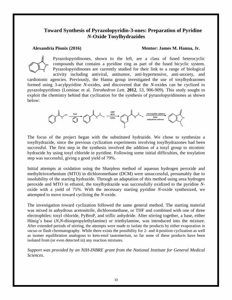
Toward Synthesis of Pyrazolopyridin-3-ones: Preparation of Pyridine
N-Oxide Tosylhydrazides
Alexandria Pinnix (2016) Mentor: James M. Hanna, Jr.
Pyrazolopyridinones, shown to the left, are a class of fused heterocyclic
compounds that contains a pyridine ring as part of the fused bicyclic system.
Pyrazolopyridinones are currently studied for their link to a range of biological
activity including antiviral, antitumor, anti-hypertensive, anti-anxiety, and
cardiotonic agencies. Previously, the Hanna group investigated the use of tosylhydrazones
formed using 3-acylpyridine N-oxides, and discovered that the N-oxides can be cyclized to
pyrazolopyridines (Lominac et al. Tetrahedron Lett. 2012, 53, 906-909). This study sought to
exploit the chemistry behind that cyclization for the synthesis of pyrazolopyridinones as shown
below:
EtOH
TsCl electrophilic additiveUHP
MTO
baseanhydrous solvent
The focus of the project began with the substituted hydrazide. We chose to synthesize a
tosylhydrazide, since the previous cyclization experiments involving tosylhydrazones had been
successful. The first step in the synthesis involved the addition of a tosyl group to nicotinic
hydrazide by using tosyl chloride in pyridine. Following some initial difficulties, the tosylation
step was successful, giving a good yield of 79%.
Initial attempts at oxidation using the Sharpless method of aqueous hydrogen peroxide and
methyltrioxorhenium (MTO) in dichloromethane (DCM) were unsuccessful, presumably due to
insolubility of the starting hydrazide. Through an adaptation of this method using urea hydrogen
peroxide and MTO in ethanol, the tosylhydrazide was successfully oxidized to the pyridine N-
oxide with a yield of 75%. With the necessary starting pyridine N-oxide synthesized, we
attempted to move toward cyclizing the N-oxide.
The investigation toward cyclization followed the same general method. The starting material
was mixed in anhydrous acetonitrile, dichloromethane, or THF and combined with one of three
electrophiles: tosyl chloride, PyBroP, and triflic anhydride. After stirring together, a base, either
Hünig’s base (N,N-diisopropylethylamine) or triethylamine, was introduced into the mixture. After extended periods of stirring, the attempts were made to isolate the products by either evaporation in
vacuo or flash chromatography. While there exists the possibility for 2- and 4-position cyclization as well
as isomer equilibration analogous to keto-enol tautomerism, so far none of these products have been
isolated from (or even detected in) any reaction mixtures.
Support was provided by an NIH-INBRE grant from the National Institute for General Medical
Sciences.
33

NMR Based Drug Design Screening: Targeting Retinoblastoma Cancer
through Activation of the p53 Tumor Suppressor Protein Alec Reed1 (2015) Mentors: David Ban2
Richard Kriwacki2
1Winthrop University. Department of Chemistry, Physics and Geology 2Department of Structural Biology, St. Jude Children’s Research Hospital
Retinoblastoma is a pediatric cancer of the retina caused by inactivation of the tumor suppressor p53. It has been shown that p53 function is abated by overexpression of the negative regulator protein MdmX. Drug design experiments taking advantage of known drug structures failed to produce a candidate with suitable affinity for clinical trials. A novel drug scaffold could be built from drug fragments detected through the use of WaterLOGSY NMR spectroscopy. Twenty possible lead compounds were identified after screening one third of a three thousand fragments.
34
Synthesis and Evaluation of Unsymmetrical Biphenyltetrols as Aggregation Inhibitors for Alzheimer’s Amyloid-β Peptide
Jake Roberts (2017) Mentors: Drs. James M. Hanna, Jr. and Robin K. Lammi
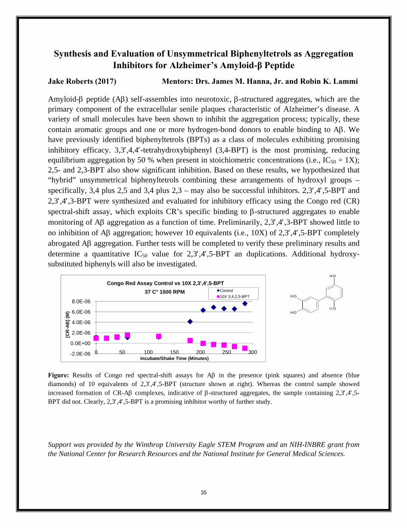
Amyloid-β peptide (Aβ) self-assembles into neurotoxic, β-structured aggregates, which are the primary component of the extracellular senile plaques characteristic of Alzheimer’s disease. A variety of small molecules have been shown to inhibit the aggregation process; typically, these contain aromatic groups and one or more hydrogen-bond donors to enable binding to Aβ. We have previously identified biphenyltetrols (BPTs) as a class of molecules exhibiting promising inhibitory efficacy. 3,3′,4,4′-tetrahydroxybiphenyl (3,4-BPT) is the most promising, reducing equilibrium aggregation by 50 % when present in stoichiometric concentrations (i.e., IC50 = 1X); 2,5- and 2,3-BPT also show significant inhibition. Based on these results, we hypothesized that “hybrid” unsymmetrical biphenyltetrols combining these arrangements of hydroxyl groups – specifically, 3,4 plus 2,5 and 3,4 plus 2,3 – may also be successful inhibitors. 2,3′,4′,5-BPT and 2,3′,4′,3-BPT were synthesized and evaluated for inhibitory efficacy using the Congo red (CR) spectral-shift assay, which exploits CR’s specific binding to β-structured aggregates to enable monitoring of Aβ aggregation as a function of time. Preliminarily, 2,3′,4′,3-BPT showed little to no inhibition of Aβ aggregation; however 10 equivalents (i.e., 10X) of 2,3′,4′,5-BPT completely abrogated Aβ aggregation. Further tests will be completed to verify these preliminary results and determine a quantitative IC50 value for 2,3′,4′,5-BPT an duplications. Additional hydroxy-substituted biphenyls will also be investigated.
Figure: Results of Congo red spectral-shift assays for Aβ in the presence (pink squares) and absence (blue diamonds) of 10 equivalents of 2,3′,4′,5-BPT (structure shown at right). Whereas the control sample showed increased formation of CR-Aβ complexes, indicative of β-structured aggregates, the sample containing 2,3′,4′,5-BPT did not. Clearly, 2,3′,4′,5-BPT is a promising inhibitor worthy of further study.
Support was provided by the Winthrop University Eagle STEM Program and an NIH-INBRE grant from the National Center for Research Resources and the National Institute for General Medical Sciences.
-2.0E-06
0.0E+00
2.0E-06
4.0E-06
6.0E-06
8.0E-06
0 50 100 150 200 250 300
[CR
-AB
] (M
)
Incubate/Shake Time (Minutes)
Congo Red Assay Control vs 10X 2,3',4',5-BPT37 C° 1500 RPM Control
10X 3,4,2,5-BPT
35
Aspects of the Circadian Rhythm in Lower Bilaterians Mitchell Schneider (2016) Kate Carter (Davidson College 2017) Ian Deas (2015)
Mentor: Julian Smith III
Animals exhibit a circadian rhythm in which the periodic synthesis of melatonin and the periodic expression of core clock genes occur, and for which light-sensitive structures that synchronize these responses to the circadian cycle of light and darkness are essential. We have investigated different aspects of these rhythms and their corresponding sensory organs in the primitive flatworm Stenostomum virginianum and in the acoelomorph Isodiametra pulchra. In Stenostomum, previous research in our lab has shown that exogenously applied melatonin suppresses both asexual fission and mitosis. This summer, we developed an assay for melatonin using high performance liquid chromatography (HPLC), and demonstrated that the molecule is present in S. virginianum; we next will use this technique to look for periodic expression of melatonin. Potential orthologues of the two terminal enzymes of melatonin synthesis (AANAT and HIOMT) were identified from the S. virginianum transcriptome, setting the stage for RTPCR studies if melatonin concentration is found to vary. We further demonstrated the presence of possible photoreceptors in S. virginianum using anti-PAX6 immunohistochemistry. As bluelight-sensitive cryptochromes are involved in circadian regulation in other organisms, we examined fission in S. virginianum cultured under blue, red, and white light. Fission was more rapid in blue light than in either red or white light. Although not statistically significant, this suggests that longer-term experiments using the same method might prove fruitful. In culture, Isodiametra. pulchra has been found by others to exhibit a circadian rhythm, laying its eggs during scotophase and failing to lay eggs under continuous illumination. Three proteins in I. pulchra orthologous to the core-clock proteins of fruit flies and mice have been identified and partially sequenced in wild-type specimens by our lab: a single ortholog of Clock and Cycle/Bmal (“Arnt”), an ortholog of Per, and an ortholog of mammalian Timeless (Tim2). Sequencing of these orthologues is ongoing, and will allow the design of real-time PCR primers to study circadian variation in their expression. Work on these organisms should greatly expand our understanding of the evolution of circadian regulation in lower animals. Funded by SC-INBRE and WU McNair Scholars Program.
36
Salinity Tolerance and Survival of Laboratory and Environmental Strains of Escherichia coli
Leigha Stahl (2017) Mentors: Matt Heard
Victoria Frost Escherichia coli are commonly utilized as fecal indicators to assess contamination and water quality in a variety of ecosystems. One group of ecosystems of particular interest is sandy beaches, which are exposed to varying stressors from both terrestrial and marine sources. Historically, beach ecosystems are thought to not be ideal habitats for E. coli because salinity can inhibit growth and survival of these bacteria. However, recent studies have demonstrated that certain strains may be able to persist in these environments. Here we expand upon this recent research and test the effects of salinity on the survival and growth of a laboratory strain of E. coli as well as environmental strains collected from sand samples at Folly Beach, South Carolina. We exposed our strains of E. coli to salt concentrations ranging from 0.5-‐10% and assessed the colony forming units (CFUs) following each of our treatments after a period of 24 hours at 37°C. Our data indicated there was a significant decrease in CFUs and a noticeable reduction in diameter of colony size as salinity increased. In addition, we observed that there is a cut-‐off for salinity tolerance as no colonies were able to grow in salinity concentrations greater than 5%. Collectively, our findings suggest that E. coli can persist on sandy beaches despite the stress of salinity and may be a useful tool in the future for assessing these ecosystems for fecal contamination levels. This work was supported by SC-INBRE.
37
Enhancing the Developmental Potential of Murine Adipose-Derived Mesenchymal Stem Cells
Kathryn Steverson (2016) Mentor(s): Matthew Stern Adipose-derived stem cells (ADSCs) are multipotent somatic stem cells obtained from the microvasculature of adipose tissue. ADSCs cannot match the differentiation potential of pluripotent embryonic stem cells (ES cells). However, previous studies have suggested that the non-traditional method of culturing ADSCs as three-dimensional spheroids can induce the expression of factors associated with pluripotency, including the transcription factor Oct-4. We hypothesize that non-traditional, three-dimensional spheroid culturing of ADSCs can upregulate the expression of several genes associated with pluripotency as well as increase the differentiation potential of ADSCs. Here, we show that murine ES cells cultured in our lab maintain expression of genes associated with the pluripotent state and known to be expressed in ES cells, thereby validating our ES cell culture conditions for future studies. We also show that ADSCs cultured under traditional two-dimensional conditions do not express markers of pluripotency. Interestingly, the expression of several genes know to be expressed in populations of somatic stem cells does vary with the level of confluence of ADSCs and is also affected by medium supplementation with murine leukemia inhibitory factor (mLIF), which is used to maintain pluripotency in cultured murine ES cells. Future work will examine the expression of the same subset of genes in ADSCs cultured as three-dimensional spheroids in the presence/absence of mLIF and murine embryonic fibroblast feeder cells. This project was supported by funding from The Winthrop University Research Council and SC-INBRE.
38
Expression, Purification, and Crystallization of the Xanthmonal Avirulence Protein, AvrBs1.1 and its Target in Capsicum annuum, the Transcription Factor WRKY-1 Michala Tesney (2018) Mentor: Jason C. Hurlbert Brown spots and leaf loss of Capsicum annum, a pepper found in North America and South America, are caused by prolonged activation of the plant’s defense mechanisms to bacterial infection resulting in a phenomenon called the hypersensitive response. In the hypersensitive response, the plant “walls off” the infected tissue with lignin and then a variety of chemical processes occur within the lignified zone which results in the death of both bacterial and plant cells. Certain species-specific bacterial pathogens inject a variety of effector proteins with different activities into the cytosol of the host in an effort to prevent activation of the plant’s defense mechanisms. Our work has focused on the effector protein AvrBs1.1, a dual specificity protein tyrosine produced by Xanthamonas euvesicatoria. Recent work has identified the C. annum transcription factor WRKY-1 as a possible target for AvrBs1.1. We hypothesize that AvrBs1.1 dephosphorylates activated WRKY-1 in the cytosol, thereby preventing it from entering the nucleus and activating the genes of the plant’s defense response. In order to understand the molecular basis of the function of AvrBs1.1, we need to determine the x-ray crystallographic structure of the protein by itself and in complex with WRKY-1. Towards this end, our research has focused on the expression and purification of WRKY-1 and identification of crystallization conditions of AvrBs1.1. We grew large scale cultures of E. coli Rosetta2 (DE3) transformed with a plasmid containing the C. annum WRKY-1 gene and purified the resulting recombinant protein to homogeneity We obtained yields of approximately 0.5 mg of purified WRKY-1 per liter of culture. Using sparse matrix screening and hanging drop vapor diffusion, we found that AvrBs1.1 crystallized in 2.0M lithium sulfate, 2% polyethylene glycol 400, 100mM Tris-acetate pH 8.0, 0.1% v/v β-mercaptoethanol. Using this condition as a base, we employed a 96 condition small molecule additive screen to enhance crystal growth. Several conditions in the additive screen were found to alter the morphology of the crystals grown in the initial conditions, but 0.1M nickel (II) chloride hexahydrate and 2.0M nondetergent sulfobetaine-211 (NDSB-211) were selected for x-ray data collection. The crystals were screened on LS-CAT beamline 21-ID at the Advanced Photon Source at Argonne National Laboratory. The base condition and the NDSB-211 crystals did not diffract and the crystals grown in the presence of the nickel sulfate were salt crystals, not protein. In the future, we will optimize the expression of WRKY-1, continue to follow up on crystallization leads for AvrBs1.1 and attempt to crystallize the AvrBs1.1/WRKY-1 complex. This project was supported by SC-INBRE
39
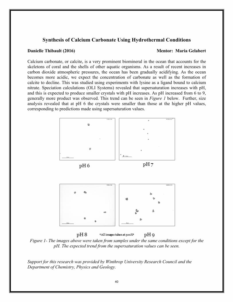
Synthesis of Calcium Carbonate Using Hydrothermal Conditions Danielle Thibault (2016) Mentor: Maria Gelabert Calcium carbonate, or calcite, is a very prominent biomineral in the ocean that accounts for the skeletons of coral and the shells of other aquatic organisms. As a result of recent increases in carbon dioxide atmospheric pressures, the ocean has been gradually acidifying. As the ocean becomes more acidic, we expect the concentration of carbonate as well as the formation of calcite to decline. This was studied using experiments with lysine as a ligand bound to calcium nitrate. Speciation calculations (OLI Systems) revealed that supersaturation increases with pH, and this is expected to produce smaller crystals with pH increases. As pH increased from 6 to 9, generally more product was observed. This trend can be seen in Figure 1 below. Further, size analysis revealed that at pH 6 the crystals were smaller than those at the higher pH values, corresponding to predictions made using supersaturation values.
Figure 1- The images above were taken from samples under the same conditions except for the
pH. The expected trend from the supersaturation values can be seen. Support for this research was provided by Winthrop University Research Council and the Department of Chemistry, Physics and Geology.
40
Synthetic Optimization and Modification of “Zone 4” Reaction on a
Known Sphingosine Kinase 1 Inhibitor to Improve Oral Bioavailability
Morgan Turnow (2016) Mentor: T. Christian Grattan
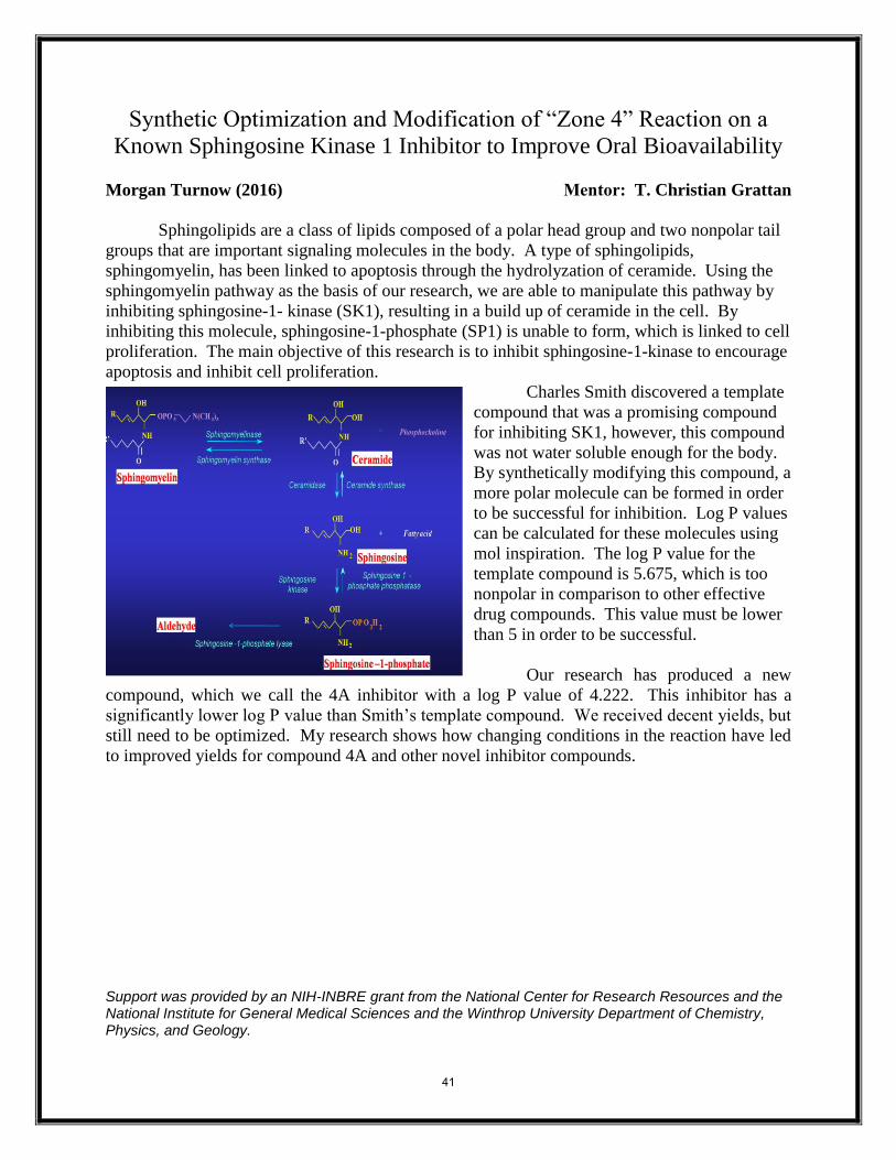
Sphingolipids are a class of lipids composed of a polar head group and two nonpolar tail
groups that are important signaling molecules in the body. A type of sphingolipids,
sphingomyelin, has been linked to apoptosis through the hydrolyzation of ceramide. Using the
sphingomyelin pathway as the basis of our research, we are able to manipulate this pathway by
inhibiting sphingosine-1- kinase (SK1), resulting in a build up of ceramide in the cell. By
inhibiting this molecule, sphingosine-1-phosphate (SP1) is unable to form, which is linked to cell
proliferation. The main objective of this research is to inhibit sphingosine-1-kinase to encourage
apoptosis and inhibit cell proliferation.
Charles Smith discovered a template
compound that was a promising compound
for inhibiting SK1, however, this compound
was not water soluble enough for the body.
By synthetically modifying this compound, a
more polar molecule can be formed in order
to be successful for inhibition. Log P values
can be calculated for these molecules using
mol inspiration. The log P value for the
template compound is 5.675, which is too
nonpolar in comparison to other effective
drug compounds. This value must be lower
than 5 in order to be successful.
Our research has produced a new
compound, which we call the 4A inhibitor with a log P value of 4.222. This inhibitor has a
significantly lower log P value than Smith’s template compound. We received decent yields, but
still need to be optimized. My research shows how changing conditions in the reaction have led
to improved yields for compound 4A and other novel inhibitor compounds.
Support was provided by an NIH-INBRE grant from the National Center for Research Resources and the National Institute for General Medical Sciences and the Winthrop University Department of Chemistry, Physics, and Geology.
41
In vivo confirmation of LPA’s role in axon guidance by the inhibition of autotaxin using siRNA in embryonic chickens
James A Vinton (2014) Mentor: Eric Birgbauer The main principle of axon guidance is to investigate the role of chemical cues in the guidance and termination of growing nerves, both during development and after injury. Lysophosphatidic acid (LPA) is a bioactive lipid that has exhibited characteristics, such as growth cone collapse and neurite retraction, of an axon guidance molecule in vitro. LPA is produced endogenously by the exo-enzyme autotaxin (ATX). Based on previous research, during certain stages of chicken development, autotaxin is expressed posterior to the optic tectum. Retinal ganglion cell (RGC) axons grow to and terminate at the optic tectum during the same stages of development that the expression of ATX was discovered. These findings suggest the possibility that the presence of autotaxin and the subsequent production of LPA could create a chemical barrier preventing the growing RGC axons from growing beyond the tectum. In order to confirm LPA’s role in axon guidance in vivo, we obtained an siRNA for chicken autotaxin from Dr. Ohuchi in Japan. Previously, we produced RCASB-ATXsiRNA-GFP (expressing an siRNA against ATX) and Control retroviruses last summer and had initial success injecting it into the optic tectum of developing chick embryos. However, detection of the virus and growing axons proved to be a limiting factor of the experiment. Therefore, we recently tested several new clearing methods; THF, Ethanol, and Fructose. Clearing is a technique used to make tissues more transparent to allow for better imaging. We also tried new immuno-histochemical axonal staining methods to determine the optimized protocol for our model. Additionally, a new collaboration with MUSC has allowed us the use of their laser scanning multi-photon confocal microscope that, initial results suggest, is able to detect the growing axonal projections in whole mount. In the future, we would like to be able to inject ATX siRNA virus or a control virus into the midbrain of the embryonic chick at E2, incubate them until E12, and using 3D rendering to detect any abnormalities in the termination, growth, or response of the RGC axons as they transverse to the optic tectum. If our experiment can confirm LPA as an axon guidance molecule, it could potentially lead to novel pharmacological or developmental studies in the areas of nerve regeneration and blindness. This research is supported by a grant from the National Eye Institute of the National Institutes of Health under award number R15EY024453. (The content is solely the responsibility of the authors and does not necessarily represent the official views of the National Institutes of Health.)
42
Maximum Constraints of Resolution Detected in Planar (Xtreme) &
Computed Tomographic (CT) X-ray Modalities Justin Waller1 (2015) Mentor: W. Matthew Leevy2
1Winthrop University. Department of Chemistry, Physics and Geology 2University of Notre Dame, College of Science, Dept. of Biological Sciences
X-ray CT is a widely used technique that commonly utilizes anatomical imaging modalities for clinical, professional and research purposes. Computed Tomography is able to integrate three-dimensional data at high resolution with quick attainment to provide a non-intrusive human or specimen imaging platform. Imaging instruments and tools utilizing X-rays were probed for limits of resolution to assess their utility for anatomical imaging. Computerized designs interfaced with 3D-printing enabled the production of resolution phantoms for the acquisition of imaging resolution limitations. Acquisitions of images via In-Vivo Xtreme integrated with the components of the Albira PET/SPEC/CT Imaging system allowed for execution of manipulative actions to efficaciously reconstruct imaging settings to affirm numerical values of limitation. American wire gauge (AWG) copper wires of varying diameter were effectively utilized in the attenuation of X-rays to exploit and confirm resolution constraints. Full width half max (FWHM) values were derived from line intensity profiles, for highly accurate values of the limitations that are existent in imaging equipment and instruments.
43
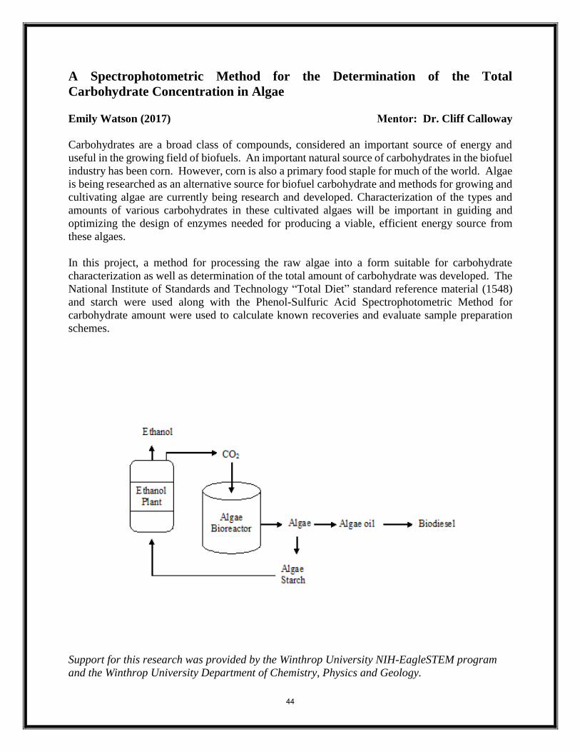
A Spectrophotometric Method for the Determination of the Total
Carbohydrate Concentration in Algae
Emily Watson (2017) Mentor: Dr. Cliff Calloway
Carbohydrates are a broad class of compounds, considered an important source of energy and
useful in the growing field of biofuels. An important natural source of carbohydrates in the biofuel
industry has been corn. However, corn is also a primary food staple for much of the world. Algae
is being researched as an alternative source for biofuel carbohydrate and methods for growing and
cultivating algae are currently being research and developed. Characterization of the types and
amounts of various carbohydrates in these cultivated algaes will be important in guiding and
optimizing the design of enzymes needed for producing a viable, efficient energy source from
these algaes.
In this project, a method for processing the raw algae into a form suitable for carbohydrate
characterization as well as determination of the total amount of carbohydrate was developed. The
National Institute of Standards and Technology “Total Diet” standard reference material (1548)
and starch were used along with the Phenol-Sulfuric Acid Spectrophotometric Method for
carbohydrate amount were used to calculate known recoveries and evaluate sample preparation
schemes.
Support for this research was provided by the Winthrop University NIH-EagleSTEM program
and the Winthrop University Department of Chemistry, Physics and Geology.
44
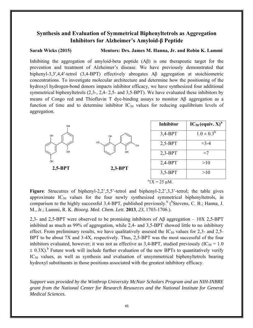
Synthesis and Evaluation of Symmetrical Biphenyltetrols as Aggregation Inhibitors for Alzheimer’s Amyloid-β Peptide
Sarah Wicks (2015) Mentors: Drs. James M. Hanna, Jr. and Robin K. Lammi
Inhibiting the aggregation of amyloid-beta peptide (Aβ) is one therapeutic target for the prevention and treatment of Alzheimer’s disease. We have previously demonstrated that biphenyl-3,3′,4,4′-tetrol (3,4-BPT) effectively abrogates Aβ aggregation at stoichiometric concentrations. To investigate molecular architecture and determine how the positioning of the hydroxyl hydrogen-bond donors impacts inhibitor efficacy, we have synthesized four additional symmetrical biphenyltetrols (2,3-, 2,4- 2,5- and 3,5-BPT). We have evaluated these inhibitors by means of Congo red and Thioflavin T dye-binding assays to monitor Aβ aggregation as a function of time and to determine inhibitor IC50 values for reducing equilibrium levels of aggregation.
Inhibitor IC50 (equiv. X)a
3,4-BPT 1.0 ± 0.3b
2,5-BPT ≈3-4
2,3-BPT ≈7
2,4-BPT >10
3,5-BPT >10
a1X = 25 µM.
Figure: Strucutres of biphenyl-2,2’,5,5’-tetrol and biphenyl-2,2’,3,3’-tetrol; the table gives approximate IC50 values for the four newly synthesized symmetric
b bal biphenyltetrols, in
ly verify 50 values, as well as synthesis and evaluation of unsymmetrical biphenyltetrols bearing
ydroxyl substituents in those positions associated with the greatest inhibitory efficacy.
ed by the Winthrop University McNair Scholars Program and an NIH-INBRE grant from the National Center for Research Resources and the National Institute for General Medical Sciences.
comparison to the highly successful 3,4-BPT, published previously. ( Stevens, C. B.; Hanna, J. M., Jr.; Lammi, R. K. Bioorg. Med. Chem. Lett. 2013, 23, 1703-1706.).
2,3- and 2,5-BPT were observed to be promising inhibitors of Aβ aggregation – 10X 2,5-BPT inhibited as much as 99% of aggregation, while 2,4- and 3,5-BPT showed little to no inhibitory effect. From preliminary results, we have qualitatively assesed the IC50 values for 2,3- and 2,5-BPT to be about 7X and 3-4X, respectively. Thus, 2,5-BPT was the most successful of the four inhibitors evaluated, however; it was not as effective as 3,4-BPT, studied previously (IC50 = 1.0 ± 0.3X).b Future work will include further evaluation of the new BPTs to quantitativeICh
Support was provid
2,5-BPT
2,3-BPT
45
Purification and Structural Studies of Bone Morphogenetic Protein Receptor
Type 1-A (BMPR-1A) Ashley Williams1 (2016) Mentor: Jie Zheng2
1Winthrop University. Department of Chemistry, Physics and Geology 2Department of Structural Biology, St. Jude Children's Research Hospital, Memphis, TN
Bone morphogenetic proteins (BMPs) play an important role in biological functions within the human body, such as embryogenesis, stem cell renewal, and tissue homeostasis1,5. These cell signaling proteins have also been found to be implicated in the pathophysiology of diseases such as arthritis, bone and kidney disorders, and cancers1. Recent reports of adverse side effects as a result of the direct administration of BMP proteins have increased the need for investigation into the development of small molecule activators or deactivators of the BMP signaling pathway. One approach to the structural analysis of BMP signaling is to examine the binding that occurs between the BMP ligand and receptor. We here report our efforts to purify and analyze the structure of bone morphogenetic protein receptor type IA (BMPR-IA) to aid in the investigation of small molecules that can be used at the clinical level to better treat the effects of deregulation in the BMP signaling pathway.
46
SYNTHETIC MODIFICATION OF “ZONE 1” ON A KNOWN SPHINGOSINE KINASE
1 INHIBITOR TO IMPROVE ORAL BIOAVAILABILITY
Deanna Worley (2016) Mentor: T. Christian Grattan
As cancer has become considerably prevalent, much effort and many resources have been
dedicated to the continual search for better treatments. Among these treatments, targeted therapy
has gained an especially high level of interest. Ideally, targeted therapy would decrease cell
proliferation while simultaneously increasing proapoptotic activity in a selective and
noncompetitive way. Two sphingolipid metabolites, ceramide and sphingosine-1-phosphate
(S1P), have been linked to cellular apoptosis and proliferation respectively. Sphingosine kinase 1
(SK1) sustains the molecular equilibrium between ceramide and S1P by the phosphorylation of
sphingosine. However, the targeted inhibition of SK1 can cause concentrations of sphingosine-1
phosphate to decrease, meanwhile forcing the equilibrium to favor ceramide. Several SK1
inhibitors have been successfully identified by Smith et al; however, due to a lack of oral
bioavailability, a number of inhibitor modifications are necessary.
With the aid of enzymatic docking programs, it was possible to make approximations as
to which portions of Smith’s inhibitor interact with SK1’s binding site. Using this information,
we can make synthetic modifications to uninvolved regions of the drug in order to increase
polarity without impacting the enzyme interactions. Calculations of the theoretical Log P and Ki
values were also performed for inhibitor variants. Said calculations directed synthetic changes
and a number of improved SK1 inhibitors have been prepared. In the future, our procedures must
be further optimized and the drug molecules must be purified before effective inhibition analyses
can be performed. Bioassays will then provide information as to how successfully our inhibitors
interact with SK1 and what further modifications need to be made to realize the potential
therapeutic value of these compounds.
Support was provided by an NIH-INBRE grant from the National Center for Research Resources and the National Institute for General Medical Sciences and the Winthrop University Department of Chemistry, Physics, and Geology.
47
Implementation of an in vivo FRET Sensor for RhoA Activation. Elijah Wyatt (2015) Mentor: Dr. Eric Birgbauer Axon guidance is an important factor during development in which nerve cells send out axons to attach to their respective targets. This process is mediated by a plethora of factors and signaling molecules. These factors direct axonal growth through both attraction and repulsion. Furthermore, in greater concentrations, some repulsive factors can cause the growth cone located at the tip of a growing axon to collapse and retract. This phenomenon plays a large role in inhibiting neural injuries from healing. The molecule we are interested in is a bioactive lipid—lysophosphatidic acid (LPA). LPA works through a pathway that results in the activation of RhoA which, in turn, results in cytoskeletal rearrangements that cause growth cone collapse. The detection of molecules like RhoA has been greatly improved through use of techniques with fluorescence resonance energy transfer (FRET). FRET proteins can be used to visualize the presence of various factors because they can be made to change colors, in real time, when a certain molecule is activated. The chief goal of this project was to develop and implement a delivery system for an externally engineered RhoA FRET sensor, then use said sensor to examine RhoA activation in growth cones during LPA assays. An attempt was made to remove the FRET insert from a plasmid, then place that insert into a chicken retrovirus (RCAS-B) for injection, infection, and subsequent expression. While PCR cloning, ClaI digestion, and gel purification were all met with success, the final ligation was unsuccessful after several efforts. The method of delivery for the FRET sensor has been reevaluated and new attempts will be made through use of electroporation. In the future, we will perform a growth cone collapse experiment in which RhoA activation is observed using the FRET sensor in a time-lapse assay. This research is supported by a student co-investigator grant from the Winthrop Research Council and by a grant from the National Eye Institute of the National Institutes of Health under award number R15EY024453. (The content is solely the responsibility of the authors and does not necessarily represent the official views of the National Institutes of Health.)
48
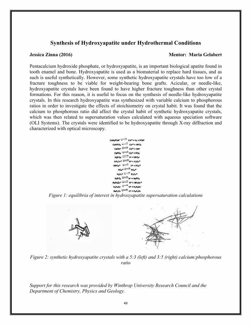
Synthesis of Hydroxyapatite under Hydrothermal Conditions Jessica Zinna (2016) Mentor: Maria Gelabert Pentacalcium hydroxide phosphate, or hydroxyapatite, is an important biological apatite found in tooth enamel and bone. Hydroxyapatite is used as a biomaterial to replace hard tissues, and as such is useful synthetically. However, some synthetic hydroxyapatite crystals have too low of a fracture toughness to be viable for weight-bearing bone grafts. Acicular, or needle-like, hydroxyapatite crystals have been found to have higher fracture toughness than other crystal formations. For this reason, it is useful to focus on the synthesis of needle-like hydroxyapatite crystals. In this research hydroxyapatite was synthesized with variable calcium to phosphorous ratios in order to investigate the effects of stoichiometry on crystal habit. It was found that the calcium to phosphorous ratio did affect the crystal habit of synthetic hydroxyapatite crystals, which was then related to supersaturation values calculated with aqueous speciation software (OLI Systems). The crystals were identified to be hydroxyapatite through X-ray diffraction and characterized with optical microscopy.
Figure 1: equilibria of interest in hydroxyapatite supersaturation calculations
Figure 2: synthetic hydroxyapatite crystals with a 5:3 (left) and 3:5 (right) calcium:phosphorous
ratio Support for this research was provided by Winthrop University Research Council and the Department of Chemistry, Physics and Geology.
49