Embed Size (px)
Citation preview
Buist, et al. (22) followed a group of ‘75 smokers attending a smoking cessation clinic and observed significant improvement in closing volume, closing capacity, and the slope of the alveolar plateau at 6 and 12 months in subjects who stopped smoking. McCarthy, et al. (105) found similar improvement in 131 subjects who stopped smoking; resumption of smoking led to subsequent development of abnormali- ties in the slope of the alveolar plateau and closing capacity. These findings are especially pertinent in view of the suggestion by Cosio, et al. (313 that some of the pathologic changes present when tests of small airway functions are abnormal can be reversed.
As a group, ex-smokers usually perform better on conventional pulmonary function testing than smokers, but they do not perform as well as nonsmokers (67). Several studies have confirmed that there is improvement in performance on standard spirometric function tests following cessation of smoking in small numbers of patients (85, 115, 159), but there is still debate as to whether the normal decline in ventilatory function (i.e., FEV) is accelerated in ex-smokers as compared to nonsmokers. In the Framingham study, Ashley, et al. (8) observed that men and women who continued to smoke had a greater decline in forced vital capacity (FVC) than those who stopped; however, they could not demonstrate consistent changes in the FEVl following smoking cessation. They attributed this to the impreciseness and insensitivity of the FEVi measurement. In women ex-smokers, the decline in FVC was similar to that of female nonsmokers; in male ex- smokers, the decline in FVC was slightly greater than that of male nonsmokers. Fletcher, et al. (57) observed that cessation of smoking halved the rate of loss of FEV and returned the rate of decline in FEV to normal in “susceptible” smokers. However, the lost FEV was not recovered. Smoking cessation had no effect on the normal rate of decline in “unsusceptible” individuals. Similarly, in a two-year followup of 118 continuing ex-smokers, aged 27 to 56, Manfreda, et al. (100) noted that subjects who continued to refrain from smoking had a smaller decline in FEV&FVC ratio than did smokers; in the male ex- smokers, the decline in ventilatory function fell at about the same rate as that for nonsmokers.
In summary, it is clear that smoking cessation leads to improved Performance on standard pulmonary function tests. However there is still debate as to whether the normal decline in ventilatory function is accelerated in ex-smokers as compared to nonsmokers.
Lung Pathology
Auerbach, et al. (10) studied the relationship between age, smoking habits, and emphysematous changes in whole lung sections obtained at autopsy from 1,443 males and 333 females. A total of ‘7,324 sections 1
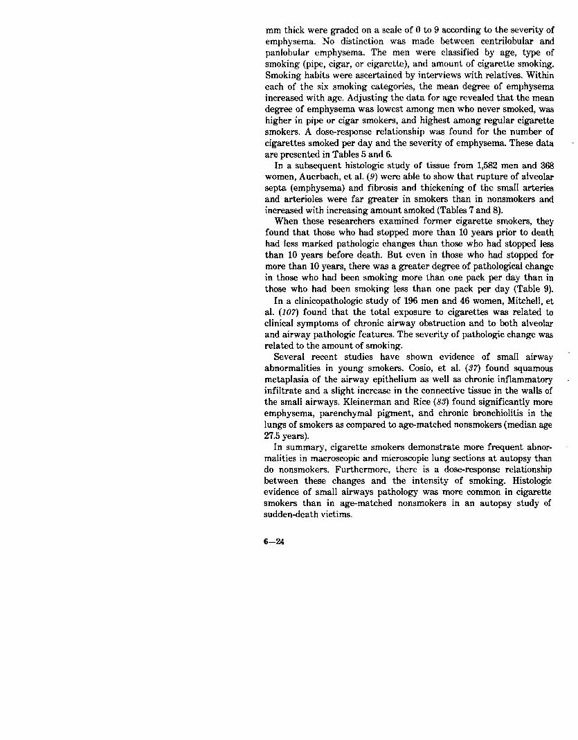
mm thick were graded on a scale of 0 to 9 according to the severity of emphysema. No distinction was made between centrilobular and panlobular emphysema. The men were classified by age, type of smoking (pipe, cigar, or cigarette), and amount of cigarette smoking. Smoking habits were ascertained by interviews with relatives. Within each of the six smoking categories, the mean degree of emphysema increased with age. Adjusting the data for age revealed that the mean degree of emphysema was lowest among men who never smoked, was higher in pipe or cigar smokers, and highest among regular cigarette smokers. A dose-response relationship was found for the number of cigarettes smoked per day and the severity of emphysema. These data are presented in Tables 5 and 6.
In a subsequent histologic study of tissue from 1,582 men and 368 women, Auerbach, et al. (9) were able to show that rupture of alveolar septa (emphysema) and fibrosis and thickening of the small arteries and arterioles were far greater in smokers than in nonsmokers and increased with increasing amount smoked (Tables 7 and 8).
When these researchers examined former cigarette smokers, they found that those who had stopped more than 10 years prior to death had less marked pathologic changes than those who had stopped less than 10 years before death. But even in those who had stopped for more than 10 years, there was a greater degree of pathological change in those who had been smoking more than one pack per day than in those who had been smoking less than one pack per day (Table 9).
In a clinicopathologic study of 196 men and 46 women, Mitchell, et al. (107’) found that the total exposure to cigarettes was related to clinical symptoms of chronic airway obstruction and to both alveolar and airway pathologic features. The severity of pathologic change was related to the amount of smoking.
Several recent studies have shown evidence of small airway abnormalities in young smokers. Casio, et al. (37) found squamous metaplasia of the airway epithelium as well as chronic inflammatory infiltrate and a slight increase in the connective tissue in the walls of the small airways. Kleinerman and Rice (83) found significantly more emphysema, parenchymal pigment, and chronic bronchiolitis in the lungs of smokers as compared to age-matched nonsmokers (median age 27.5 years).
In summary, cigarette smokers demonstrate more frequent abnor- malities in macroscopic and microscopic lung sections at autopsy than do nonsmokers. Furthermore, there is a dose-response relationship between these changes and the intensity of smoking. Histologic evidence of small airways pathology was more common in cigarette smokers than in age-matched nonsmokers in an autopsy study of sudden-death victims.
6-24
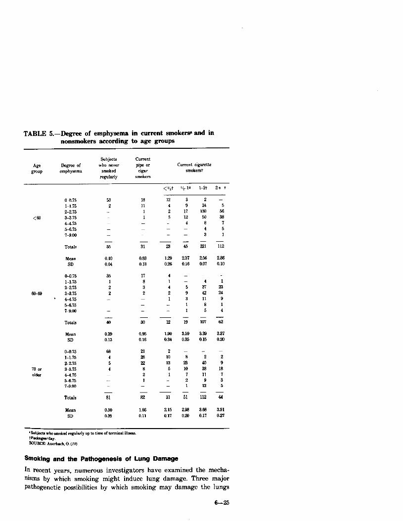
TABLE 5.-Degree of emphysema in current smokew and in nonsmokers according to age groups
Subjects CUlTWIt who never pipe or
smoked cigar regularly smokem
Current cigarette smoke&
< “zt +1t l-2t 2+ t
70 or older
co.75 l-1.75 22.75 u.75 p4.75 5-6.75 7-9.00
Totals
Mean SD
a4l.75 l-l.75 2275
m-69 3-3.75 . 44.75
rx.75 7-9.00
Totals
Mean SD
o-O.75 l-l.75 22.75 3-3.75 4-4.75 5-6.75 7-9.00
Totals
Mean SD
53 2
- - - - -
- 55
0.10 0.04
35 1 2 2
- - -
- 40
0.39 0.13
68 4 5 4
- - -
- 81
0.50 0.39
18 12 11 4
1 2 1 5
- - - - - -
- 31 23
0.83 0.13
129 026
17 4 8 1 3 4 2 2
- 1 - - - -
- 30 12
0.95 0.16
1.90 0.34
21 2 28 10 22 13
8 5 2 1 1 -
- - - a
1.66 0.11
31
215 0.17
- 45
2 24
130 50
8 4 3
iii
237 0.16
256 0.07
- - - 4 5 37 9 42 3 11 1 8 1 5
- - 19 107
3.53 0.35
ii
293 020
3.33 0.15
- 2
40 38 11 9
12
112
3.63 0.17
- 5
56 38
7 5 1
- 112
2.86 0.10
- 1
23 24
9 1 4
-ii
3.37 0.20
- 44
3.91 0.27
*Subjecta who amoked regularly up to time of terminal illnear W=kagw&y. ~LHtCX Auerbsch. 0. (10)
b3king and the Pathogenesis of Lung Damage
In recent years, numerous investigators have examined the mecha- nisms by which smoking might induce lung damage. Three major Pathogenetic possibilities by which smoking may damage the lungs
6-25
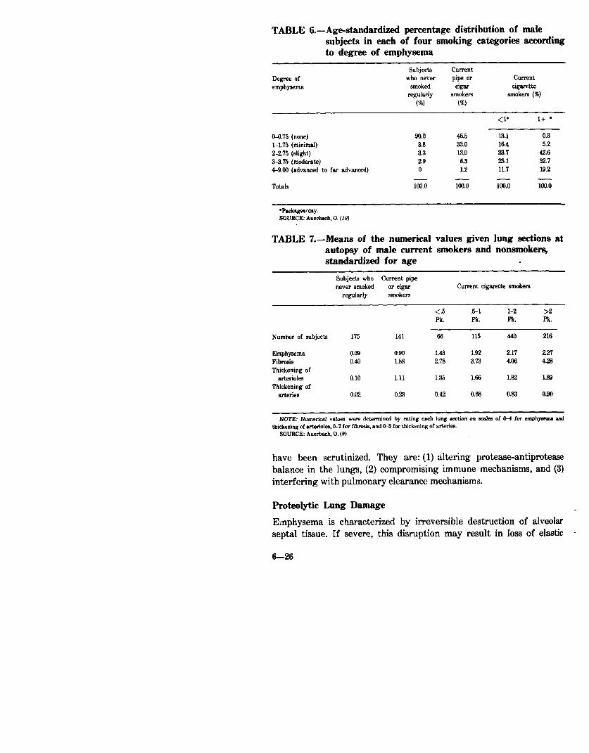
TABLE C.-Age-standardized percentage distribution of male subjects in each of four smoking categories according to degree of emphysema
Degree of emphysema
Subjects current who never pipe or
smoked Gw regularly smokem
(W cv
Current cigarette
smokers (%)
<I’ 1+ l
O-6.75 (none) 90.0 46.5 13.1 0.3 l-l.75 (minimal) 3.8 33.0 16.4 5.2 2-2.75 (slight) 3.3 13.0 33.7 42.6 3-3.75 (moderate) 2.9 6.3 25.1 32.7 44.00 (advanced to far advanced) 0 1.2 11.7 19.2
- - Totals loo.0 Ko 100.0 lao.0
‘Packages/day. SOURCE: Auerbach. 0. (10)
TABLE 7.-Means of the numerical values given lung sections at autopsy of male current smokers and nonsmokers, standardized for age .
Subjects who Current pipe never smoked or cigar Current cigarette smokers
reguldy smoket3
<.5 5-l l-2 >2 Pk. Pk. Pk. Pk.
Number of subjects 175 141 66 115 440 216
Emphysema Fibrosis Thickening of
Wt.&Ok3
Thickening of arteries
0.09 0.90 1.43 1.92 2.17 227 0.40 1.1 278 3.73 4.06 4.26
0.10 1.11 1.35 1.66 1.82 1.89
0.02 0.23 0.42 0.68 0.83 0.90
NOTE: Numerical value8 were determined by rating each lung section on 841% of C-4 for emphysema urd thickening of arterioles. LL? for fibrosis, and CL3 for thickening of art&en.
SOURCE: Auerbach, 0. (9)
have been scrutinized. They are: (1) altering protease-antiprotease balance in the lungs, (2) compromising immune mechanisms, and (3) interfering with pulmonary clearance mechanisms.
Proteolytic Lung Damage
Emphysema is characterized by irreversible destruction of alveolar septal tissue. If severe, this disruption may result in loss of elastic
6-26
TABLE S.-Means of the numerical values given lung sections at autopsy of female current smokers and nonsmokers, standardized for age
Subjects who never smoked
regularly
Current cigarette smokers
<l Pk. 11 Pk
Number oc subjects 262 33 64
Emphywma 0.&5 1.37 1.70 Fibrosis 0.37 239 3.46 Thickening of arterioles 0.06 1.26 1.57 Thickening of arteries 0.01 0.40 0.64
NOTE: Numerical value. were determined by rating each lung section on scales of @4 for emphysma and thickening of the arteriden, O-7 for fibrosis. and Wg for tbiekening of lbe arteries.
60UFccE: Auerbch. 0. (9)
TABLE b.-Means of the numerical values given lung sections at autopsy of male former cigarette smokers, standard&l for age
Formerly Smoked stopped 1 10 yr. supped < 10 yr.
<1 Pk. Pk. <l Pk. Pk.
Number of subjects 35 66 51 131
Emphysema 0.34 0.70 1.0s 1.63 Fibmsis 1.14 1.74 244 3.30 lliclwling of arterides 0.57 0.93 1.25 1.56 Thickening of artwiea 0.04 0.16 0.36 0.61
NOTE: Numeriul values for each finding were determined by rating each lung section on aala, of O-4 for ~pbyaem;l and thiiening of the arimides, W7 for fibrwis, and K3 for thickening of the arteries.
SOURCE: Auerbsb. 0. (9)
recoil, enhanced collapsibility of the airways, and airflow obstruction. The elastic properties of the lung are attributed to the appropriate distribution of elastin in its connective tissue framework. Recent data suggest that the lung damage observed in emphysema may be due to injury of this elastic framework by proteolytic enzymes released (and not inhibited) in the lung. Formulation of this hypothesis was catalyzed by the discovery that emphysema is extremely common in individuals who are severely deficient in alpha-1-antitrypsin (@, a glycoprotein that inhibits several proteases. Subsequently, it was postulated that Conditions interfering with the normal balance between protease and antiprotease activity could give rise to an excess of free protease (i.e., elastase) in the lung and initiate lung destruction (109).
6-27
The proteases are a group of enzymes which probably serve a wide range of functions in the normal host. Proteases with particular elastolytic capability (elastases) are synthesized and released by alveolar macrophages which are found in increased numbers in bronchopulmonary lavage fluid of smokers. They are also present in significant concentrations in polymorphonuclear leukocytes (PMNs).
The antiproteases, of which alpha-1-antitrypsin is the most abun- dant, are found primarily in blood although alveolar macrophages and bronchial secretions are additional sources of antiproteases. An excess of protease within the lung may arise from any circumstances in which there is increased release of protease which is not matched by availability of antiprotease activity at the site of such release. Various types of experimental support for the proteolytically mediated hypothesis of lung damage have been presented in recent years (15, 75, 77,132).
Crude leukocyte extracts can digest lung tissue (76, 92) and homogenates of leukocytes can produce emphysema (101, 103) when instilled into the lungs of animals. The degree of damage depends on the proteolytic activity of the instillate (82). Recently, Senior, et al. (129) instilled purified human leukocyte elastase into the tracheas of hamsters. At two months the lungs of the animals showed mild, patchy
-emphysema. In a related study, Schuyler, et al. (226) administered elastase to hamsters intravenously and demonstrated significant loss of elastic recoil at low lung volumes when their lung histology was normal. The authors suggested that submicroscopic lesions may antedate obvious morphologic evidence of emphysema.
The mechanisms by which cigarette smoking may alter the protease- antiprotease balance have-been the subject of several recent investiga- tions. Janoff and Carp (744 demonstrated .that unfractionated cigarette smoke condensate suppressed antiprotease activity in vitro. Elastin-agarose gels were impregnated with cigarette smoke conden- sate. Elastases were then allowed to diffuse through the gels toward a counterdiffusing sample of antiproteases. The effectiveness of the antiproteases in blocking-the enzyme washet&nined by the elitent of elastin destruction in the plates. -F&Wins, proteases, and antiproteases from different sources, inc!llding purified human leukocyte elastase and human alpha-1-antitrypsin, were tested. In all situations, .t.he
-cigarette smoke condensate suppressed the inhibitory activity of the antiprotease. In a followup study, Carp and Janoff (26) demonstrated that fresh cigarette smoke also Suppressed elastase-inhibitory activity of human serum. In addition, treatment of serum with model oxidants caused a similar suppression of elastase inhibition. These in vitro observations suggested to the researchersthat emphysema in cigarette smokers might be due in part to the suppression of- antipro&% activity by oxidizing agents‘present in cigarette smoke.
6-B
In another study from the same laboratory, Blue and Janoff (16) demonstrated that cigarette smoke condensates elicited the release of elastase from human PMNs. When human PMNs were incubated in vitro with cigarette smoke condensate, three enzymes were released: beta-glucuronidase, acid phosphatase, and elastase. The elastase was active in digesting elastin, even in the continuing presence of cigarette smoke condensate. When mixtures of human PMNs and cigarette smoke condensate were instilled into rat lung in F&O, elastase was released and could be traced to connective tissue targets using immunohistochemical and enzyme-histochemical techniques. This study appears to be particularly relevant in view of previous studies demonstrating that cigarette smoke recruits leukocytes into the lung airways (81, 124), immobilizes them (46), and inhibits their chemotaxis in vitro (17).
The role of the pulmonary macrophage in proteolytic lung damage has been evaluated by several investigators. Alveolar macrophages are normally important in cleansing the lower airways by phagocytising and digesting foreign particulate matter. Bronchopulmonary lavage studies have documented increased total numbers of macrophages in lavage fluid of smokers as compared to nonsmokers (65, 156). Keast and Holt (79) exposed mice to smoke via a special apparatus and found sustained elevations in bronchopulmonary macrophage populations.
Changes in the ultrastructure of macrophages have been reported in smokers. Pratt, et al. (116) observed pigmented cytoplasmic inclusions in macrophages from cigarette smokers, Brody and Craighead (18) observed that the pigmentation appeared to be due, at least in part, to an increased number of lysosomes and phagolysosomes. In addition, distinctive “smoker’s” inclusions were observed within these cyto- plasmic organelles which appeared plate-like and crystallographically consistent with kaolin&e. The authors presented some preliminary evidence that these particles are derived from inhaled tobacco smoke. Kaolinite is a common clay mineral found in the soil in many tobacco growing regions and is sometimes used as a tobacco additive in the production of cigarettes for the purpose of reducing tar content. A few studies have shown that when macrophages engulf kaolinite they release beta-gulcuronidase and lactic acid dehydrogenase, lysosomal enzymes believed to play a role in cell death and fibrogenesis in tiuo (3, 66, 157). In a recent study, Matulionis and Traurig (104) exposed Pulmonary macrophages of mice in situ to cigarette smoke and found: (I) an increase in number, variety, and size of lysosome-like bodies in the macrophage; (2) the appearance of multinucleation; and (3) an increased size of the macrophages. After cessation of smoke exposure, macrophage morphology and population size returned toward normal.
A considerable increase in elastase-like e&erase and protease activity was demonstrated by Harris, et al. (64) in human alveolar macrophages in smokers as compared to nonsmokers. In a subsequent
6-B
study, Rodriguez, et al. (119) demonstrated that human alveolar macrophages from smokers released elastase into serum-free culture medium, unlike those from nonsmokers. Elastase was not detectable in cell homogenates from either smokers or nonsmokers, implying that this enzyme is not stored. The authors suggested that cigarette smokers have the potential for a 20-fold increase in elastase released in the lungs when the increased number of macrophages in lungs of smokers also is considered.
Potentially important effects of cigarette smoke also have been demonstrated on alveolar macrophage pinocytosis (164), cell adhesion (61), cell migration (154), and protein synthesis (94, 95, 163). The data relating the effect of cigarette smoke to alveolar macrophage phagoocytosis and bacteriocidal activity are conflicting (61, 130, 135, 1%‘) but generally have shown cigarette smoke to have a suppressant effect. At least some of the toxic effects of the gas phase of cigarette smoke on macrophage activity may be due to the oxidant, acrolein (74).
In summary, a number of recent investigations have suggested that a destruction of the elastic framework of the lungs seen in COLD may result from a protease-antiprotease imba!ance. Although definitive evidence is lacking, it appears that alveolar macrophages and PMNs are the most important sources for the proteases. Cigarette smoke appears to increase the rate of synthesis and release of elastase in vitro from human alveolar macrophages and increases their numbers. Antiproteases are inhibited from counteracting protease activity in the presence of cigarette smoke in vitro. Possible deleterious effects of cigarette smoke also have been demonstrated on a variety of functions of the human alveolar macrophage.
Interference with Immune Mechanisms The lungs have a highly developed lymphatic system and the capacity to effect local immune responses. Inhalation of tobacco smoke produces significant changes in cellular and humoral immunity in both animal and man. However, the role of such changes in the pathogenesis of lung disease remains speculative. Waldman, et al. (151) reported that cigarette smokers of more than l/2 pack per day had an increased risk of influenza-like illnesses although the length of illness was no different than for nonsmokers.
Finklea, et al. (52) noted that smokers had more frequent subclinical influenza than nonsmokers; subsequently he observed that the serological response (hemaglutination antibody titers) to either vaccination or natural infection with A-Z antigens was similar to that in nonsmokers but not as long lasting (51).
Cigarette smoke appears to adversely affect the nonspecific (phagocytosis) defense mechanisms provided by the alveolar macro- phage. Evidence for an effect on the specific (immune) defense roles
6-30
played by both macrophages and lymphocytes has been offered by several investigators.
The alveolar macrophage system plays an important role in the overall immune response as an antigenic “processor.” Warr and Martin (15.4 studied alveolar macrophages lavaged from four healthy smokers and four healthy nonsmokers. Only two members of each group were reactive to skin tests with Candida albicans. The migration of macrophages from nonsmokers was inhibited by migration inhibitory factor (MIF) whereas macrophages from smokers did not respond to MIF. The cells from smokers were noted to migrate three times faster than those from nonsmokers. When Candida antigen was added to the medium, cells from the nonreactive subjects (both smokers and nonsmokers) were not inhibited. The cells from the reactive nonsmok- ers were inhibited, but not those from reactive smokers. Thus, macrophages from smokers did not respond normally either to MIF or antigenic challenge.
The B and T lymphocytes participate in humoral and cell-mediated immune mechanisms, respectively. Warr, et al. (155) noted that a greater number of T cells and B cells were recovered by human bronchopulmonary lavage from smokers than from nonsmokers. Daniele, et al. (39) examined the T and B cell populations in peripheral blood of smokers versus nonsmokers and found no difference in either the absolute number of cells or the lymphocyte response to phytohema- glutinin (PHA) or concanavalin A. In a lavage study of five smokers the lymphocyte subpopulation did not differ from that in nonsmoking subjects (n=8), but cells from smokers showed a diminished response to PHA and concanavalin A. They concluded that cigarette smoking may impair cellular immune defenses.
In contrast, Silverman, et al. (134 found that young smokers had an increased number of T lymphocytes in peripheral blood and an enhanced response to PHA. No differences were found in the response of older smokers or those with a history of heavier cigarette consumption as compared to controls. A number of other studies have examined the relationship of smoking to T-cell function; these are reviewed in the Chapter on Allergy and Immunity.
Roszman and Rogers (121) noted that both the nicotine and the water-soluble fraction of whole cigarette smoke suppressed the immunoglobulin response of lymphoid cell cultures to antigen chal- lenge. When concentrations of over 200 micrograms per milliliter of nicotine of the water-soluble fraction were added, they were able to SuPpress completely the immunoglobulin response; this suppression also occurred in cells exposed 2 hours prior to the antigenic challenge. In a subsequent experiment, they found suppression of mitogen- induced blastogenesis by cigarette smoke (120). War-r, et al. (156) examined immunoglobulin levels in bronchopulmonary lavage fluid in
6-31
19 smokers and 36 nonsmokers. They could find no difference in IgA levels; however, IgC levels were twice as high in smokers.
In summary, a variety of alterations in the specific immune system have been observed that are presumably due to cigarette smoking. Macrophages from smokers respond abnormally to MIF or antigen challenges. T lymphocytes obtained by bronchopulmonary lavage in smokers showed a diminished response to PHA compared to those of nonsmokers. Cigarette smoke suppresses production of immunoglobu- lin by B lymphocytes in lymphoid cell culture. However, the role of these abnormalities in the pathogenesis of lung damage is unclear.
Effect on Clearance Mechanisms The mucociliary transport system protects the lung against inhaled particulate matter. Its two major components are the respiratory mucus blanket (secreted by submucosal and goblet cells) and the ciliated columnar epithelial cells lining the larger airways. Denudation of epithelium, an increased number of goblet cells, and squamous metaplasia have been demonstrated by Auerbach, et al. (11) in dogs exposed to cigarette smoke via a tracheostoma, and by Leuchtenber- ger, et al. (91) and Rylander (12.4) in mice and guinea pigs exposed to cigarette smoke via their upper airway passages. Similar morphologic abnormalities have been observed in human cigarette smokers (58).
A number of investigators have examined the effects of cigarette smoke on mucociliary function, employing a wide variety of experi- mental techniques. These studies have scrutinized the effects of gas and particulate elements of cigarette smoke in both acute and chronic situations.
Short-term exposure to cigarette smoke causes ciliostasis and decreased mucociliary transport in most animals (152). The ciliotoxic effects of cigarette smoke are not peculiar to tobacco cigarettes; they have been observed in protozoans following exposure to smoke from lettuce and grass cigarettes (60). The data relating these effects to specific particulate or gas phase elements of cigarette smoke are conflicting (38). Moreover, the relevance to human conditions of animal models demonstrating altered mucociliary function in “smoking” (tracheostomized) animals has been questioned, since, in humans, cigarette smoke passes the upper airways which might alter its ciliotoxic capacity for the lower airways (152). Data regarding the effects of acute cigarette exposure on mucociliary clearance in man also are conflicting (2.51).
Long-term exposure to cigarette smoke has been examined in animals and in man. Tracheal mucous velocity has been shown to be decreased in purebred beagle dogs (153) exposed to 100 cigarettes per week for 13.5 months. In donkeys (.2), low level exposure to whole cigarette smoke accelerated tracheobronchial clearance; at intermedi-
ate and high levels whole cigarette smoke had twice the effect of filtered smoke in decreasing clearance.
The long-term effects of cigarette smoking on mucociliary function in man are unclear. Most of the evidence indicates that long-term smoking reduces mucociliary transport (152). Animal and human studies have suggested that cessation of smoking may allow partial recovery of mucociliary function (1,25).
interaction of Smoking with Other Risk Factors for COLD
Alpha-l-antitrypsin Deficiency It would be useful to identify the populations at special risk of developing COLD from smoking so that such populations might be made aware of the risk. Persons with significant deficiencies of alpha- 1-antitrypsin may be such a population.
Eriksson (~8) was the first investigator to observe a relationship between the presence of markedly decreased serum trypsin inhibitory capacity and panlobular emphysema. Since Eriksson’s paper, much research has been published concerning this intriguing observation.
Severe alpha-1-antitrypsin deficiency is due to a rare genetic trait which occurs in approximately 1 in 2,000 people (49). Less severe reductions are found in approximately 2 to 10 percent of the population. Alpha-1-antitrypsin inheritance patterns indicate multiple codominant alleles at one gene locus. Some alleles (notably Z, S, and “null”) are associated with substantially reduced serum levels of alpha- 1-antitrypsin. The autosomal codominant inheritance allows multiple combinations of alleles associated with low or normal serum levels of the antiprotease. For example, extremely low levels are associated with the ZZ homozygous state, intermediate levels with the MZ heterozygous state, and normal levels with the MM state. Thus, a wide range of serum levels may be encountered which depend upon the Particular alleles involved. The particular phenotype of a given patient can be identified by antigen-antibody crossed gel electrophoresis but not by measurement of serum levels alone, because alpha-1-antitrypsin is an acute phase reactant. The pathophysiologic implications of a reduction in antiprotease activity have been discussed in previous sections.
Severe deficiency of alpha-1-antitrypsin has been associated with a Particular type of pulmonary emphysema. While the majority of lungs of emphysematous patients reveal bullous or centrilobular deformities, Particularly of the upper lobes, this hereditary disorder reveals a Panacinar change, most severe in the lower lobes (63, 136, 158). PoPulations with this genetically related form of emphysema have a greater percentage of females than is usually observed in the general emphysema population. Their disease begins earlier, is more severe, is characterized by dyspnea rather than cough, and frequently is
6-33
unassociated with a history of preceding bronchitis (63, 136, 158). Radiographic studies of alpha-1-antitrypsin deficient patients have revealed decreased vascularization of the lower lobes (134).
Several retrospective studies in patients with severe deficiency have demonstrated an association between smoking and the age at which emphysema becomes manifest. However, control nonsmoking subjects with a similar phenotype have not been included. Black and Kueppers (14) evaluated 18 patients with alpha-1-antitrypsin deficiency who had never smoked and had little or no exposure to occupational or urban air pollution and compared them to 36 individuals with similar phenotype (PiZZ) who were (or had been) smokers. A larger percentage of individuals who smoked had impaired lung function early in life. However, there was considerable variability as to clinical course, degree of pulmonary function abnormality, and appearance of the roentgenogram among the nonsmokers. The authors recognized that their study was biased in favor of individuals with symptomatic disease; however, they noted that the rarity of the PiZZ phenotype and the need to identify nonsmokers with no other exposure to respiratory irritants would have required an enormous screening program Prospective studies scrutinizing these relationships are lacking.
The natural history of the states with less severe deficiencies of alpha-1-antitrypsin is unclear (86). Cross-sectional studies have found such a deficiency more frequently in patients with COLD than would be expected by chance alone (87, 93). However, several other reports obtained from population studies have suggested that mild forms of antitrypsin deficiency are not important risk factors for emphysema (30, 34, 111). Mittman (108) recently reviewed the controversy as to whether the MZ phenotype is a significant risk factor for COLD but could not resolve the issue based on current evidence. Longitudinal studies in such individuals have not been reported. Because the natural history of the mild deficiency state is unclear, the effect of smoking on such individuals remains unsettled.
In summary, individuals with severe alpha-1-antitrypsin deficiency have an excessive risk for developing COLD; the onset of symptomatic COLD is probably abbreviated by smoking. The natural history of individuals with mild deficiency states for alpha-1-antitrypsin is unclear, as is the question of whether they represent a group at special risk from cigarette smoking.
Other Genetic Factors Continued interest has been shown in the possible contribution of genetic factors (other than alpha-1-antitrypsin deficiency) to the pathogenesis of COLD. In earlier studies (71, 88, 89), the existence of kindreds with a high incidence of COLD had been noted, but the relative importance of genetic factors and smoking habits was unclear.
6-34

TABLE lO.-Expected and observed prevalence rate (percent) of “cough” among smoking partners to co-twins who either had or had not the symptom “cough” Monozygotic pairs
“Coughing” status in non-smoking partner
No. at risk Prevalence rate for “coughing” among smoking co-twins. percent
EXpXtd OtkWrVd
No “cough” 497 4 12 ‘Tough” 41 24 37
SOURCE: cederlof. R (es,
Cohen, et al. (32, SS), in a family study in Baltimore, Maryland, found an increased prevalence of pulmonary function abnormalities in first- degree relatives of COLD cases as compared to first-degree relatives of nonpulmonary cases, even when Pi variant relatives were excluded. In all groups, smokers demonstrated a higher frequency of function abnormalities. The authors suggested that there is some interaction of familial factors with smoking. In a similar study in rural areas outside Rochester, Minnesota, Miller, et al. (106) found a twofold increased prevalence of functional abnormalities in family members of subjects with COLD as compared to families of controls matched for age, sex, occupation, and smoking exposure.
Cederlof, et al. (27, 28) examined the relationship of smoking to symptom prevalence among monozygotic and dizygotic twins who were both discordant and concordant for smoking habits. They observed that the hypermorbidity for COLD symptoms related to smoking persisted even after controlling for zygosity; they concluded that a causal relationship of smoking and COLD symptoms was supported. However, genetic factors had an appreciable influence.
In a more recent analysis of their twin data, Cederlof, et al. (29) examined the prevalence of cough among monozygotic pairs discordant for smoking. The results are presented in Table 10. They assumed that the nonsmoking symptomatic co-twin had a predisposition to cough. The smoking co-twin had a threefold increase in prevalence of cough compared to his asymptomatic nonsmoking co-twin-a l-112 times increase compared to the symptomatic nonsmoking co-twin. The Prevalence rates were higher in the smoking groups than in non- smoking groups but highest in the “predisposed” smoker. The authors suggested that hereditary factors were equally as important as smoking for the development of cough in the smaller “predisposed” group.
These findings lend support to earlier suspicions that genetic factors may play a role in determining the risk for COLD. Kazazian (78) haq
6-35
suggested that common lung diseases may be due to a combination of risk factors, varying from one individual to another, and that this risk may be modulated by different genes in combination and by different environmental factors (e.g., smoking). Long-term prospective studies are necessary to answer these questions.
Occupational Exposures Exposure to certain occupational environments has been shown to be associated with several forms of non-neoplastic bronchopulmonary disease. An increased prevalence of COLD is found with exposures to coal and granite dust and cotton fiber. This risk is increased further by cigarette smoking. However, in none of these studies is the relationship of COLD to occupation as strong as that to smoking.
A discussion on the proposed modes by which smoking interacts with occupational exposures is presented in the Chapter on the Interaction Between Smoking and Occupational Exposures.
Air Pollution The relationships among air pollution, smoking, and COLD remain controversial. Reasons for this controversy include difficulties in controlling such variables as socioeconomic class, degree of crowding, ethnic differences, and age distribution, as well as in determining the exact type and amount of individual pollution exposure. Measuring individual pollution exposure, even within a small area, is difficult since both amount and type can vary dramatically from street to street (e.g., proximity of a street to a heavily traveled expressway).
In an effort to control as many of these variables as possible, two basic approaches in study design have been utilized. The first approach has been to find areas where different pollution levels have been well- measured and then to select populations that are as similar as possible in these areas. Thus, a population in a low-pollution area can be compared with a similar population in a high-pollution area. The second approach has been to select a population that is as uniform as possible (for example, twins), and then measure individual responses to different pollution exposures.
Using the first approach, the Community Health and Environmental Surveillance System evaluated the excess COLD (i.e., rate of COLD experienced above that of nonsmokers) in subjects in two communities of differing air pollution: Salt Lake City (high), and the Becky Mountain Area (low). Finklea, et al. (5.3) commenting on the data, noted that smoking was the most important risk factor in developing abnormal pulmonary function but that smoking and exposure to air pollution had a synergistic effect.
The relationship among smoking, air pollution, and COLD were analyzed in an autopsy study of tissue samples from St. Louis, Missouri (high pollution) and Winnipeg, Canada (low pollution) (162). Three
6-36
hundred lungs were evaluated as to the extent and degree of emphysema; urban groups were matched for smoking habits, length of residence, age at immigration, and employment history; 25 to 26 percent of each group were nonsmokers. In nonsmokers, emphysema was more frequent and severe in the St. Louis than in the Winnipeg group. In male smokers the incidence of severe emphysema was fourfold higher in the St. Louis than in the Winnipeg group. The author concluded that tobacco smoke may have a cumulative or synergistic action with air-pollution exposure.
Increased prevalence of COLD has been demonstrated in areas of high pollution in the Netherlands (ISO), Yokkaichi, Japan (113), and Cracow, Poland (125). However, these studies were poorly controlled for socioeconomic status.
Several studies have used the second major method of investigating the relationship between smoking, air pollution, and COLD, i.e., to select a uniform population and then to measure individual differences to pollution exposure. Cornstock, et al. (36), in an attempt to control for occupational exposure and socioeconomic class, studied three separate, uniform populations of telephone workers and used as a measure of pollution the location of the place of work and residence. The populations studied were telephone installers and repairmen in Baltimore, New York City, Washington, D.C., and rural Westchester County, New York, in 1962 (survey 1) and in 1967 (survey 2), and telephone installers and repairmen in Tokyo in 1967 (survey 3). The researchers were unable to find any relation between pulmonary symptoms and degree of urbanization of place of work or place of residence (either current or past). They were, however,, able to establish a strong correlation between smoking habits and pulmonary symptoms. Given the crude estimation of pollution exposure used in this study (all workers in each city were treated as though they received the same exposure), a small difference in symptoms due to air pollution could have been missed, whereas the difference due to smoking could be detected both because it was larger and because it Was possible to determine individual exposure more exactly.
Hrubec, et al. (70), in a study of twins from the U.S. Veterans Registry, were unable to show a difference in respiratory symptoms either between individuals with different exposures to air pollution or between members of twin pairs with different air-pollution exposures. However, they too used a crude measure of air-pollution exposure (by each zip code area), and so could have missed a small difference due to air pollution despite being able to relate respiratory symptoms to smoking, socioeconomic status, and alcohol intake.
Volley, et al. (35). in a study of 3,899 persons (2%year-olds born during the last week of March, 1946, in the United Kingdom), were also unable to show a relation between COLD and air pollution. As estimates of air-pollution exposure, they used the domestic coal
6-37
consumption in the towns where the subjects lived. This method of estimating air pollution is subject to the same limitations cited for the previous two studies, i.e., limited sensitivity to small risks due to air pollution.
In summary, if an increased risk of COLD due to air pollution exists, it is small compared to that due to cigarette smoking under conditions of air pollution to which the average person is exposed. The possibility remains that the two kinds of exposure may interact to increase the total effect beyond that contributed by each exposure separately.
Socioeconomic Status In a morbidity survey (117) of the non-institutionalized population of the United States (1964), socioeconomic status appeared to be an important risk factor in determining rates of reporting chronic bronchitis, asthma, and emphysema. Rates were higher among those in lower socioeconomic classes. This relationship had been previously recognized in the United Kingdom (118).
In a recent study, the relationship of smoking to socioeconomic status and chronic respiratory diseases was examined in 9,226 residents of Tecumseh, Michigan, observed from 1962 to 1965 (68). The prevalence of chronic bronchitis was higher in cigarette smokers than in nonsmokers, higher in blue-collar workers than in white-collar workers, and least among men with the most education (Table 11). There was no significant association between the prevalence of asthma and smoking habits, occupation, education, or income. Most of the differences in the prevalence of chronic bronchitis in subjects of differing occupational, educational, or income classes were attributable to differences in smoking habits. Compared with smoking, poor occupations, educational background, and economic circumstances have only a weak deleterious effect.
Childhood Respiratory Illness and Adult Respiratory Disease A connection between pediatric respiratory illness and adult respira- tory disease has long been suspected on clinical grounds. Burrows, et al. (24 recently reported that physician-confirmed chronic bronchitis and/or emphysema and abnormalities in measures of expiratory flow are more common in older subjects with such history. They suggested that childhood respiratory illness leads to an increased susceptibility to the effects of bronchial irritants and respiratory infections.
In a prospective study of lo-year&Is followed since age 2 (n=3699), Colley, et al. (85) found that subjects with a history of respiratory tract illness before age 2 had an increased likelihood of developing respiratory symptoms by age 20. However, cigarette smoking appeared to be an even more important factor in increasing risk for developing these symptoms (Table 12).
6-38
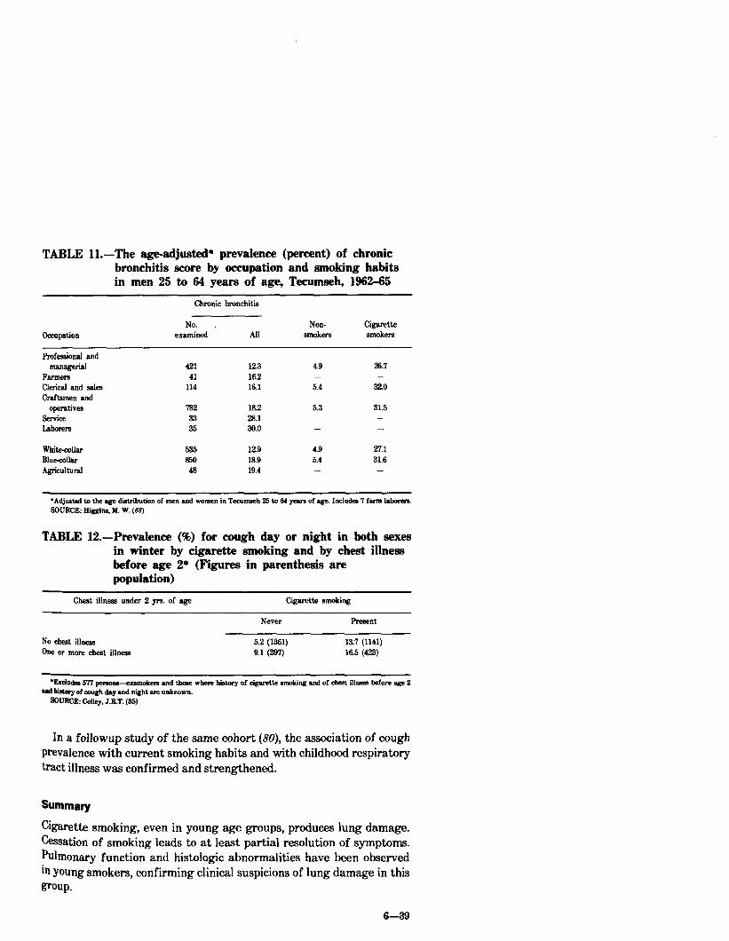
TABLE Il.-The age-adjusted* prevalence (percent) of chronic bronchitis score by occupation and smoking habits in men 25 to 64 years of age, Tecumseh, 1962-65
Chronic bronchitis
Occupation No. ,
examined All Non- Cigarette
smokem sowken
F’rofesaional and managerial
FWlM?TB Clerical and sales Craftsmen and
OperatiVeJ service Labom2
421 123 4.9 26.1 41 162 - -
114 16.1 5.4 32.0
782 18.2 5.3 31.5 33 28.1 - - 35 30.0 - -
whitezollar 53.5 x29 4.9 27.1 BIue-collar 850 18.9 5.4 31.6 Agricultural 48 19.4 -
*Adjusted to the age distribution of men and women in Tecumseh !25 to 64 yeara of agx. Includes 7 farm labwen SOURCE: BiggIna. 116. W. (68)
TABLE 12.-Prevalence (%) for cough day or night in both sexes in winter by cigarette smoking and by cheat illness before age 2* (Figures in parenthesis are population)
Cheat iilness under 2 ye. of age Cigarette smoking
Never Present
No cheat iilneas 5.2 (1361) 13.7 (1141) Ow or more cheat illness 9.1 (397) 16.5 (423)
l hIodea 577 petsons-exmnokera and thaw where history of cigarette smoking and of cheat illnem before age 2 ~6 history of an& day and night ate unknown.
3f)URCE: C&y, J&T. (35)
In a followup study of the same cohort (80), the association of cough prevalence with current smoking habits and with childhood respiratory tract illness was confirmed and strengthened.
Summary
cigarette smoking,-even in young age groups, produces lung damage. Cessation of smoking leads to at least partial resolution of symptoms. Pulmonary function and histologic abnormalities have been observed in young smokers, confirming clinical suspicions of lung damage in this group.
6-B
A variety of pulmonary functional abnormalities believed to represent small airway dysfunction occurs in smokers. Many such individuals demonstrate normal expiratory flow as measured by conventional spirometry. In one prospective study, abnormalities in tests of small airway function appeared to correlate well with pathologic abnormalities of the peripheral airways. It has been suggested that such changes may be precursors of more extensive anatomic-functional abnormalities if smoking were continued. How- ever, prospective studies relating small airway physiological and/or pathological abnormalities to the development of COLD are lacking.
Adult cigarette smokers have respiratory symptoms more frequently than do nonsmokers; some symptoms (i.e., cough and sputum production) increase with a greater dosage of cigarettes. While it is clear that COLD is more common in men than in women, it is uncertain whether men and women with equivalent smoking histories have a similar increase in the prevalence of respiratory symptoms and COLD.
In the majority of epidemiological surveys, a higher prevalence of functional abnormalities has been found in smokers as compared to nonsmokers. There are conflicting data as to the effect of smoking on pulmonary function in different racial groups and whether men and women with equivalent smoking habits have similar reductions in pulmonary function. It is clear that cigarette smoking produces a more rapid decline in FEV and a higher prevalence of productive cough. However, it is unclear whether the presence of productive cough by itself predicts the risk for a more rapid decline in FEV independent of that increased risk associated with cigarette smoking. It has been suggested that there may be a “susceptible” group of smokers whose rate of decline in FEV is much greater than that in both “unsuscepti- ble” smokers and nonsmokers and that “unsusceptible” smokers and nonsmokers have similar rates of decline in FEV. Therefore, preva- lence surveys of functional abnormalities in all smokers may underesti- mate the impact of cigarette smoking in the “susceptible” population.
Several studies have confirmed that there is improvement in standard spirometric function tests following cessation of smoking, but there is still debate as to whether the normal decline in ventilatory function is accelerated in ex-smokers aa compared to nonsmokers.
Cigarette smokers demonstrate more frequent abnormalities in macroscopic and microscopic lung sections at autopsy than do nonsmokers. Furthermore, there is a dose-response relationship between these changes and the intensity of smoking. Histologic evidence of small airways pathology is more common in cigarette smokers than in age-matched nonsmokers in one autopsy study of sudden death victims.
A number of recent investigations have suggested that destructive lung changes seen in the emphysematous form of COLD may result from excess liberation of, or failure to inhibit, proteases in the lung.
6-40
Although definitive evidence is lacking, it appears that PMNs and alveolar macrophages are the most important sources for the proteases. Cigarette smoke appears to increase the rate of synthesis and release of elastase in vitro by human alveolar macrophages. Antiproteases are inhibited in the presence of cigarette smoke in vitro. Cigarette smoke also has been demonstrated to impair a variety of functions of the human alveolar macrophage.
Inhalation of tobacco smoke produces detectable changes in compo- nents of the cellular and humoral immune systems in both animal and man. Macrophages obtained by lung lavage from smokers respond abnormally to MIF or antigen challenge. T lymphocytes obtained from bronchopulmonary lavage show a diminished response to PHA in smokers. Cigarette smoke suppresses production of immunoglobulin by B lymphocytes in lymphoid cell culture. However, the role of these abnormalities in the pathogenesis of lung damage is unclear.
Individuals with severe alpha-1-antitrypsin deficiency have an excessive risk for developing COLD; the onset of symptomatic COLD is probably accelerated by smoking. The natural history of individuals with mild or moderate alpha-1-antitrypsin deficiencies is unclear, as is the effect of smoking on such individuals.
Genetic factors other than alpha-1-antitrypsin deficiency appear to play a role in determining the risk for COLD. Common lung diseases may be due to a combination of risk factors varying from one individual to another. The risk may be modulated by different genes in combination and by different environmental factors (e.g., smoking).
A recent study examined the relationship of smoking to socioeco- nomic status and chronic respiratory disease. The prevalence of chronic bronchitis was higher in cigarette smokers than in nonsmokers, higher in blue-collar workers than white-collar workers, and least among men with the most education. However, most of the differences in the prevalence of chronic bronchitis in subjects of differing occupational, educational, or income classes was attributable to differences in smoking habits. Compared with smoking, poor occupations, educational background, and economic circumstances have only a weak deleterious effect.
Childhood respiratory disease appears to be a risk factor for respiratory symptoms as an adult. However, cigarette smoking appears te be a more important factor in increasing risk for developing these symptoms.
bearch Recommendations The extensive studies already performed have identified several areas that merit particular investigational attention because of their promise in elucidating the effects of smoking and other risk factors upon the development of COLD:
6-41
(1) Current data suggest that early detection of pulmonary functional and histologic changes in asymptomatic smokers may identify populations which are particularly susceptible to COLD. Investigations documenting the relationships between tests for small airways dysfunction, pulmonary histology, and symptoms should be extended. In addition, longitudinal studies are needed to (a) document the impact of smoking cessation upon these early abnormalities, and, most important, to (b) define the relationship of these early abnormali- ties to the development of COLD.
(2) Similar longitudinal studies in patients with welldefined COLD should be carried out to define the effects of smoking cessation on clinical, physiologic, and anatomic parameters.
(3) The protease antiprotease imbalance hypothesis for the patho- genesis of pulmonary elastic tissue injury has received substantial support from investigations reported to date. Observations are available which suggest mechanisms by which cigarette smoke might promote an injury-inducing imbalance in man. Appropriate extensions of both in. vitro and in wivo investigations which bear upon this relationship should be performed. It would appear particularly important to assure that in wivo research be carried out to determine the biologic importance of the expanding body of promising in witro research.
(4) Subjects with genetically-determined severe and mild-moderate deficiencies of alpha-1-antitrypsin appear to be a particularly promis- ing population in which to study the natural history of COLD, the role of cigarette smoking and other risk factors, and the mechanisms responsible for COLD. Carefully designed studies, cross-sectional and longitudinal, of subjects with severe and mild-moderate deficiencies should be undertaken. Multi-center studies with pooling of data should be encouraged.
(5) There are in vitro effects of smoking and cigarette smoke on both the humoral and cellular components of the immune system. Extension of relevant in vitro and in tivo investigations dealing with smoking- immune system interactions should be encouraged.
(6) Further investigations of the relationship between cigarette smoking and the mucociliary (“clearance”) apparatus are warranted.
In all of the above areas, research planning should include attention to the primary goal, i.e., elucidation of the mechanisms responsible for the development of COLD in man and the manner in which smoking impacts upon these mechanisms to promote COLD. Thus, research support should seek a balanced program providing for in vitro and in wivo investigations (in animal models and in man). Such a balanced program also should provide for effective interchange of information among investigators pursuing research in vitro, in animals, and in man.
6-42
Non-Neoplastic Bronchopulmonary Diseases: References (1) ALBERT, R.E., ALESSANDRO, D., BERGER, J., LIPPMANN, M. Long-term
smoking in the donkey: Effect on tracheobronchial particle clearance. Archives of Environmental Health 22: X&19,1971.
(2) ALBERT, R.E., BERGER, J., SANBORN, K., LIPPMANN, M. Effects of cigarette smoke components on bronchial clearance in the donkey. Archives of Environmental Health 29: 96101,1974.
(8) ALLISON, A.C., HARINGTON, J.S., BIRBECK, M. An examination of the cytotoxic effects of silica on macrophagea. Journal of Experimental Medicine 124: 141-153.1966.
(4) AMERICAN COLLEGE OF CHEST PHYSICIANS. AMERICAN THORACIC SOCIETY. Pulmonary terms and symbols. A report of the ACCP-ATS Joint Committee on Pulmonary Nomenclature. Chest 67: 533593,1975.
(5) ANTHONISEN, N.R., BASS, H., ORIOL, A., PLACE, REG., BATES, D.V. Regional lung function in patients with chronic bronchitis. Clinical Science 35(3): 495-511,1963.
(6) ANTHONISEN, N.R., DANSON, J., ROBERTSON, P.C., ROSS, W.R.D. Ah-way closure as a function of age. Respiratory Physiology 3: 53-65,1969-1970.
(r) ARMSTRONG, J.G., WOOLCOCK, A.J. Lung function in asymptomatic cigarette smokers-The single breath nitrogen test. Australia and New Zealand Journal of Medicine 6(2): P&126,1976.
(8) ASHLEY, F., KANNEL, W.B., SORLIE, P.D., MASSON, R Pulmonary function: Relations to aging, cigarette habit, and mortality. The Framingham Study. Annals of Internal Medicine 32(5): 739-745,1975.
(9) AUERB QCH, 0.. GARFINKEL, L., HAMMOND, E.C. Relation of smoking and age to findings in lung parenchyma: A microscopic study. Chest 65(l): 2935, 1974.
(10) AUERBACH, O., HAMMOND, E.C., GARFINKEL, L., BENANTE, C. Relation of smoking and age to emphysema. Wholelung section study. New England Journal of Medicine 236(16): 353357, April 29,1972.
(II) AUERBACH, O., HAMMOND, E.C., KIRMAN, D., GARFINKEL, L., STOUT, A.P. Histologic changes in bronchial tubes of cigarette-smoking dogs. Cancer 20: 2055-2066.1967.
(1.3) BENSON, M.K. The closing volume as a screening test in smokers. Scandinavian Journal of Bespiratory Dii 95(Supplement): &U-90,1974.
(13) BEWLEY, B., BLAND, J.M. Smoking and respiratory symptoms in two groups of schoolchildren. Preventive Medicine 5: 63-69,1976.
(14) BLACK, L.F., KUEPPERS, F. Alpha-l-a titrypsin deficiency in nonsmokers. American Review of Respiratory Disease 117: 421427,1973.
(15) BLACKWOOD, C.F., HOSANNAH, Y., PERMAN, E., KELLER, S., MANDL, I. Experimental emphysema in rata: Elastolytic titer of inducting enzyme as determinent of the response. Proceedings of the Society for Experimental Biology and Medicine 144: 450-454,1973.
(16) BLUE, M.L., JANOFF, A. Possible mechanism of emphysema in a cigarette smokers. Release of elastase from human polymorphonuclear leukocytes by cigarette smoke condensate in vitro. American Review of Respiratory Disease 117(2): 317325, February 1973.
(17) BRIDGES, R.B., KRAAL, J.H., HUANG, L.J.T., CHANCELLOR, M.B. Effects of tobacco smoke on chemotaxis and glucose metabolism of polymorphonuclear leukocytes. Infection and Immunity 15(l): 115-X33,1977.
(18) BRODY, A.R., CRAIGHEAD, J.E. Cytoplasmic inclusions in pulmonary macrophages of cigarette smokers. Laboratory Investigation 32(2): 125132, 1975.
6-43
w
(30)
(34
(33)
(33)
($4)
BROWN, R., WOOLCOCK, A.J., VINCENT, N.J., MACKLEM, P.T. Physiologi- cal effects of experimental airway obstruction with beads. Journal of Applied Physiology 27: 323335,1969.
BUIST, A.S., FLEET, L.V., ROSS, B.B. A comparison of conventional spirometric tests and the test of closing volume in an emphysema screening center. American Review of Respiratory Disease 167: 735743,1973.
BUIST, A.S., ROSS, B.B. Quantitative analysis of the alveolar plateau in the diagnosis of early airway obstruction. American Review of Respiratory Disease 163: 107&1637,1973.
BUIST, A.S., SEXTEN, G.J., NAGY, J.M., ROSS, B.B. The effect of smoking cessation and modification on lung functions. American Review of Respiratory Disease 114: 115-122.1976.
BURROWS, B., KNUDSON, R.J., CLINE, M.G., LEBOWITZ, M.D. Quantative relationships between cigarette smoking and ventilatory function. American Review of Respiratory Dii 115: 195-204,1977.
BURROWS, B., LEBOWITZ, M.D., KNUDSON, R.J. Epidemiologic evidence that childhood problems prodispcee to airways disease in the adult (An association between adult and pediatric respiratory disorders). Pediatric Research ll(3): 21%?20,1977.
CAMNER, P., PHILIPSON, K., ARVIDSSON, T. Withdrawal of cigarette smoking. Arehives of Environmental Health I: 9C%?,l973.
CARP, H., JANOFF, A. Possible mechanisms of emphysema in smokem in vitro suppression of serum elastase-inhibitory capacity by fresh cigarette smoke and prevention by antioxidants. American Review of Respiratory Disesse 118: 61% 621,1973.
CEDERLOF, R, FRIBERG, L., HRUBEC, 2. Cardiovascular and respiratory symptoms in relation to tobacco smoking. Archives of Environmental Health 18(6): 934946, June 1969.
CEDERLOF, R, FRIBERG, L., JONSSON, E., KAIJ, L. Respiratory symptoms and ‘angina pectoris’ in twins with reference to smoking habits. An epidemiological study with mailed questionnaire. Archives of Environmental Health 13(6): 726737, December 1966.
CEDERLGF, R, FRIBERG, L., LUNDMAN, T. The interactions of smoking, environment, and heredity, and their implications for disease etiology. Acta Medica Scandinovica SX?(Supplement): l-128,1977.
CHAN-YEUNG, M., ASHLEY, M.J., COREY, P., MALEDY, H. Pi Phenotypss and the prevalence of chest symptoms and lung function abnormalities in workers employed in dusty industries. American Review of Respiratory Disease 117(2): 239-243,197s.
CHERNIACK, RM. Smoking and chronic airways obstruction. National Heart, Lung and Blood Institute, Division of Lung Diseases. National Technical Information Service PB 272 154, September 1,1977, pp. l-41.
COHEN, B.H., BALL, W.C., JR., BIAS, W.B., BRASHEARS, S., CHASE, G.A., DIAMOND, E.L., HSU, S.H., KREISS, P., LEVY, D.A., MENKES, HA., PERMUTT, S., TOCKMAN, M.S. A genetic epidemiologic study of chronic obstructive pulmonary disease I. Study design and preliminary observations. Johns Hopkins Medical Journal 137: 95164,1975.
COHEN, B.H., BALL, W.C., JR., BRASHEARS, S., DIAMOND, EL., KREISS, P., LEVY, D.A., MENKES, H.A., PERMUTT, S., TOCKMAN, M.S. American Journal of Epidemiology 165(3): 223-231,1977.
COLE, RB., NEVIN, N.C., BLUNDELL, G., MERRE’IT, J.D. MCDONALD, JR., JOHNSTON, W.P. Relation of alpha-1-antitrypsin phenotype to the performance of pulmonary function tests and to the prevalence of respiratory illness in a working population. Thorax 31: 149157,1976.
6-44
(35) CGLLEY, J.R.T., DOUGLAS, J.W.B., REID, D.D. Respiratory disease in young adults: Influence of early childhood lower respiratory tract illness, social class, air pollution, and smoking. British Medical Journal 3(5873): 195-198, July 28, 1973.
(36) COMSTOCK, G.W., STONE, R.W., SAKAI, Y., MATSUYA, T., TONASCIA, J.A. Respiratory findings and urban living. Archives of Environmental Health 27(3): 143-156, September 1973.
(?lr) COSIO, M., GHEZZO, R.H., HOGG, J.C., CORBIN, R., LOVELAND, M., DOSMAN, J., MACKLEM, P.T. The relationships between structural changes in small airways and pulmonary function tests. New England Journal of Medicine 298: X277-1281.1978.
(38) DALHAMN, T. Effect of cigarette smoke on ciliary activity. American Review of Respiratory Disease 93(3)(Supplement): 108114,1966.
($9) DANIELE, R.P., DAUBER, J.H., ALTOSE, M.D., ROWLANDS, D.T., GOREN- BERG, D.J. Lymphocyte studies in asymptomatic cigarette smokers. A comparison between lung and peripheral blood. American Review of Respira- tory Disease 116: 997-1105,1977.
(@) DENSEN, P.M., JONES, E.W., BASS, H.E., BREUER, J., REED, E. A survey of respiratory disease among New York City postal and transit workem. 2 Ventilator-y function tests results.. Environmental Research 2(4): 277-296, July 1969.
(41) DIRKSEN, H., JANZON, L., LINDELL, SE Influence of smoking and cessation of smoking in lung function. A population study of closing volume and nitrogen wash-out. Scandinavian Journal of Respiratory Disease %(Supplement): 266274,1974.
(&?) DOLL, R, PETO, R. Mortality in relation to smoking: 26 years’ observations on male British doctors. British Medical Journal 2: X%-1536,1976.
(43) DOSMAN, J., BODE, F., GHEZZO, R.H., MARTIN, R., MACKLEM, P.T. The relationship between symptoms and functional abnormalities in clinically healthy cigarette smokers. American Review of Respiratory Disease 114(2): 297364, August 1976.
(&) DOSMAN, J.A., GHEZZO, RH., BODE, F., MARTIN, RR., MACKLEM, P.T. Teata of early diagnosis of airway obstruction: A perspective. Revue Francais Maiadiea Respiratoires 5: 377-389,1977.
(45) DOSMAN, J., MACKLEM, P.T. Diseases f o small airways. Advances in Internal Medicine 22: 355376,1977.
(46) EICHEL, B., SHAHRIK, H.A. Tobacco smoke toxicity: Loss of human oral leukocyte function and fluid+ell metabolism. Science 166(3911): 1424-1423, 1969.
(47) ENJEII, S., HAZELWOOD, B., PERMU’PI’, S., MENKES, H., TERRY, P. Pulmonary function in young smokers. Male-female differences. American Review of Respiratory Disease 118: 667675,1978.
(48) ERIKSSON, S. Pulmonary emphysema and alpha-1-antitrypsin deficiency. Acta Medica Scandinavica 175: 197~205,1964.
(U) ERIKSSON, S. Studies in al-antitrypsin deficiency. Acta Medica Scandinavica 177(Supplement 432): l-85,1965.
(SO) FAIRSHTER, RD., WILSON, A.F. Volume of isoflow: Effect of distribution of ventilation. Journal of Applied Physiology: Respiratory, Environmental and Exercise Physiology 43(5): 867-811,1977.
(51) FINKLEA, J.F., HASSELBLAD, V., RIGGAN, W.B., NELSON, W.C., HAM- MER, D.I., NEWILL, V.A. Cigarette smoking hemagglutination inhibition response to infiuenza after natural disease and immunization. American Review of Respiratory Disease 104(3): 368-376, September 1971.
(52) FINKLEA, J.F., SANDIFER, S.H., SMITH, D.D. Cigarette smoking and epidemic influenza. American Journal of Epidemiology 96: 396399,1969.
6-45
(58) FINKLEA, J.F., SHY, C.M., LOVE, C.J., HAYES, C.G., NELSON, R.S., HOUSE, D.E. Health consequences of sulfur oxides: Summary and conclusions baaed upon CHESS studies of 1970-1971. In: Health Consequences of Sulfur Oxides: A Report from CHESS, 1970-1971. U.S. Environmental Protection Agency Publication No. EPA-650/l-74-094,1974.
(54) FLETCHER, C.M. (Editor). Terminology, definitions, classification of chronic pulmonary emphysema and related conditions. A report of the conclusions of a Ciba Guest Symposium. Thorax 14: 2%299,1959.
(55) FLETCHER, C.M., JONES, N.L., BURROWS, B, NIDEN, AH.. American emphysema and British bronchitis. A standardized comparative study. American Review of Respiratory Disease 90: l-13,1934.
(56) FLETCHER, C., PETO, R. The natural history of chronic airflow obstruction. British Medical Journal 1: X45-1648,1977.
(57’) FLETCHER, C., PETO, R., TINKER, C., SPEIZER, F.E. The Natural History of Chronic Bronchitis and Emphysema: An Eight-Year Study of Early Chronic Obstructive Lung Disease in Working Men in London. Oxford, Oxford University Press, 1976,272 pp.
(58) FRASCA, J.M., AUERBACH, O., PARKS, V.R, JAMIESON, J.D. Alveolar ceil hyperplasia in the lungs of smoking dogs. Experimental and Molecular Pathology 21: 300-312,1974.
(56) GELB, A.F., GOLD, W.M., WRIGHT, RR., BRUCH, HR., NADEL, J.A. Physiologic diagnosis of subclinical emphysema. American Review of Beapira- tory Dti 107(l): 50-63,1973.
(60) GRAY, J.P., KENNEDY, JR. Ultrastructure and physiological effecta of nontobacco cigarettes on tetrahymena. Archives of Environmental He&h 28: 2%291,1974.
(61) GREEN, G.M., CAROLIN, D. The depressant effect of cigarette smoke on the in vitro antibacterial activity of alveolar macrophages. New EngIand Journal of Medicine 276(S): 421427,1967.
(62) GREGG, I. A study of the causes of progressive airways obstruction in chronic bronchitis. In: Current Research in Chronic Respiratory Disease, 11th Aspen Emphysema Conference, U.S. Public Health Service, Publication No. 10721, 1968, pp. 235-248.
(65) HAMMARSTEN, J.F., WELCH, M., RICHARDSON, RH., PATTERSON, C.D., GUENTER, C.A. Familial alpha-1-antitrypsin deficiency and pulmonary emphysema. Transactions of the American Clinical and Climatological Association 80(l): 7-14,1969.
(64) HARRIS, J.O., OLSEN, G.N., CASTLE, JR., MALONEY, A.S. Comparison of proteolytic enzyme activity in pulmonary alveolar macrophages and blood leukocytes in smokers and nonsmokers. American Review of Respiratory Disease 111: 579-.586,1975.
(65) HARRIS, J.O., SWENSON, E.W., JOHNSON, J.E., III. Human alveolar macrophages: Comparison of phagocytic ability, glucose utilization, and ultraatructure in smokers and nonsmokers. Journal of Clinical Investigation 49(11): 2086-2096, November 1970.
(66) HEPPLESTON, A.G., STYLES, J.A. Pathology: Activity of a macrophage factor in collagen formation by silica. Nature 214: 521522, April 29, 1937.
(67’) HIGGINS, I.T.T. Epidemiology of chronic respiratory dii: A literature review. U.S. Environmental Protection Agency, Office of Research and Development, 1974, pp. 1-129.
(68) HIGGINS, M.W., KELLER, J.B., METZNER, H.L. Smoking, socioeconomic status, and chronic respiratory disease. American Review of Respiratory Disease 116: 403-410,1977.
6-46
(69) HOGG, J.C., MACKLEM, P.T., THURLBECK, W.M. Site and nature of airway obstruction in chronic obstructive lung disease. New England Journal of Medicine 273(25): 1355-13661963.
(70) HRUBEC, Z., CEDERLOF, R., FRIBERG, L., HORTON, R, OZOLINS, G. Respiratory symptoms in twins. Archives of Environmental Health 2’7(3): 139- 195, September 1973.
(71) HURST, A. Familial emphysema. American Review of Respiratory Disease 36(l): 179136,1959.
(7.8) HUTCHEON, M., GRIFFIN, P., LEVISON, H., ZAMEL, N. Volume of isoflow. A new teat in detection of mild abnormalities of lung mechanics. American Review of Respiratory Disease llo(4): 453465, October 1974.
(78) IMBODEN, C.A., Jr. Rising mortality from chronic respiratory disease. American Journal of Public Health 53: Z?l-222,1963. (Letter)
(74) JAKAB, G.J. Adverse effect of a cigarette smoke component, acrolein, on pulmonary antibacterial defenses and on viral-bacterial interactions in the lung. American Review of Respiratory Disease 115: 33-33,197’7.
(7.&z) JANOFF, A., CARP, H. Possible mechanisms of emphysema in smokem. Cigarette smoke condensate suppresses proteaae inhibition in vitro. American Review of Respiratory Disease 116: 65’72,1977.
(75) JANOFF, A., ROSENBERG, R., GALDSTON, M. Elastaae-like, esteroprotease activity in human and rabbit alveolar macrophage granulea. Pmceedings of the Society for Experimental Biology and Medicine 136(3): 1654-1058, 1971.
(76) JANOFF, A., SANDHAUS, R.A., HOSPELHORN, V.D., ROSENBERG, R. Digestion of lung proteins by human leukocyte granules in vitro. proceedings of the Society for Experimental Biology and Medicine 146: 516-519,1972.
(77) KAPLAN, P.D., KUHN, C., PIERCE, J.A. The induction of emphysema with elastase. I. The evolution of the leaion and the influence of serum. Journal of Laboratory and Clinical Medicine 32(3): 349356,19’73.
(78) KAZAZIAN, H.H. A geneticist’s view of lung disease. American Review of Respiratory Disease 113: 26%X6,1976.
(79) KEAST, D., HOLT, P. Smoking and immune response. New Scientist 61: 396 EO’7, March 23,19’74.
(80) KIERNAN, K.E., COLLEY, J.R.T., DOUGLAS, J.W.B., REID, D. Chronic cough in young adults in relation to smoking habits, childhood environment, and cheat illness. Respiration 33: 236244,19’76.
(81) KILBURN, K.H., MCKENZIE, W. Leukocyte recruitment to airways by cigarette smoke and particle phase in contrast to cytotoxicity of vapor. Science 139: 634637,1975.
(80) KIMBEL, P., WEINBAUM, G. Role of leucoproteases in the genesis of emphysema. In: Junod, A., De Haller, R. (Editors). Lung Metabolism. New York, Academic Press, 19’75, pp. 25-41.
(88) KLEINERMAN, J., RICE, D.B. Evidence for preclinical lesions in lungs of young smokers: A postmortem epidemiologic-pathologic correlative study. In: Steinfeld, J., Griffith, W., Ball, K., Taylor, RM. (Editors). Proceedings of the Third World Conference on Smoking and Health, New York, June 2-5, 1975. Volume II. Health Consequences, Education, Cessation Activities, and Social Action. U.S. Department of Health, Education, and Welfare, Public Health Service, National Institutes of Health, National Cancer Institute, DHEW Publication No. (NIH) 7%1413,19’77, pp. 161-169.
(84) KNUDSON, R.J., LEBOWITZ, M.D., BURTON, A.P., KNUDSON, D.E. The closing volume teat: Evaluation of nitrogen and bolus methods in a random population. American Review of Respiratory Disease 115(3): 423434,197’7.
(8s) KRUMHOLZ, RA., CHEVALIER, R.B., ROSS, J.C. Changes in cardiopulm+ nary functions related to abstinence from smoking. Annals of Internal Medicine 62(2): 197~297,1965.
6-47
![Acute or chronic pulmonary emphysema? Or both?—A ......emphysema or acute alveolar dilation, respectively [3 , 5]. In some cases, an interstitial emphysema is described [, 636]](https://img.pdfslide.us/doc/110x75/6138f505a4cdb41a985b64ce/acute-or-chronic-pulmonary-emphysema-or-bothaa-emphysema-or-acute-alveolar.jpg)