Embed Size (px)
Citation preview

T Cells 1 & 2 IMMU 7630 Fall 2019
1
T CELLS—PARTS 1 & 2
T CELLS. B cell (‘humoral’) immunity is relatively easy to study because B cells secrete antibody, a defined product, which does their work for them; the B cell is not there when antibody does its job. Therapeutic use was made of that fact over a hundred years ago: antisera to treat infectious diseases. T cells, on the other hand, have to be present at the site of interaction with antigen; they do not secrete their receptors. It was difficult to pin down a role for T cells; in fact, the thymus was long thought to be a non-functional organ since you could remove it from an adult human or other animal and nothing much happened. But in 1961, researchers found that ►neonatally thymectomized mice grew up with a wasting syndrome (probably chronic infection) and also had impaired ability to reject foreign skin grafts. A genetically-identical thymic transplant, if not delayed too long, restored them to normal. These experiments marked the beginning of the modern cellular immunology era.
Keep in mind that, unlike B cells which can see free antigen, T cells only see antigenic determinants shown to them by special antigen-presenting molecules on the surfaces of other cells. We’ll explain how that works later in these units; but first, meet the cast of characters:
DIFFERENT TYPES OF T CELLS. There are at least 6 different kinds of T cells, all interesting and important for immune function. They all develop in the thymus and share some properties, but can be distinguished functionally and, to some extent, by surface markers. Right now learning them may be daunting, but from here on we’ll be referring to them every day.1
There are 5 main kinds of helper T cells, and one killer T cell. Let’s start with the helpers (so called because they ‘help’ other cells do things). ►Helpers all express the surface marker CD4. ►Most helpers begin as an undecided precursor: Th0 (zero). These cells are found in the paracortex of lymph nodes, and corresponding positions in other secondary lymphoid tissues. When the antigen their receptors recognize is brought to them by dendritic cells (DC), they begin to divide and differentiate, becoming either Th1, Th17, Th2, Tfh, or Treg cells. ►The previous experience of the DC—the conditions in the periphery during the innate immunity phase when it was activated, what TLR were engaged, what cytokines and chemokines predominated—is the main determinant of the Th0’s ultimate progeny. You end up with some of each kind of helper; it’s the relative proportions that determine the nature of the immune response that you see.
Th1 CELLS. They were once called ‘delayed hypersensitivity T cells’. After activation and proliferation in the lymph node, some of the daughters leave and circulate around the body. When they encounter antigen, presented by macrophages and DC at the infection site, they get reactivated and secrete lymphokines (see terminology table on next page). ►The most important lymphokine secreted by Th1 is interferon gamma (IFN) which is pro-inflammatory, because it’s chemotactic for blood monocytes and tissue macrophages. These cells move in large numbers into the area where the Th1 is recognizing antigen. ►They are also activated by IFN, becoming classically-activated M1 (or ‘angry’) macrophages which avidly ingest and kill bacteria or other foreign invaders. The macrophages release their own cytokines that intensify inflammation, including tumor-necrosis factor alpha (TNFα) and IL-12. This division of labor is efficientthe T cell recognizes, the macrophage attacksbut runs the risk of damage to local tissues by the enraged but imbecilic macrophages. Get a good stimulator
1 A summary of the properties, transcription factors, cytokines etc. of Th cells is in the separate T Cell
Supplementary & Review Material file on http://immuno4ever.org/week05.htm 2 When you have inflammation, you often have fever. IL-1 is the main cause: in the preoptic anterior hypothalamus,
it stimulates the formation of PGE2, which slows the firing rate of certain temperature-control neurons to what they
would normally do at, say 35°C. This activates the heat generation response (e.g., shivering,) producing fever.

T Cells 1 & 2 IMMU 7630 Fall 2019
2
of Th1 cells on your skin—poison ivy is excellent—and you’ll see what I mean. We’ll hear more about this ‘contact hypersensitivity’ later in the course.
►Th1 also secrete IL-2, which helps CTL (killer T cells) get fully activated after they recognize antigen.
Th17 CELLS. There is a more recently described Th subset, called Th17 because it makes the inflammatory lymphokine IL-17 among others. It resembles the Th1 in that its main job seems to be causing inflammation; not surprisingly, then, it has been implicated in several autoimmune diseases (e.g., psoriasis), as has the Th1. It must do something useful for a living, of course; and that is resistance to particularly difficult bacterial and yeast pathogens (such as Candida).3 It is useful to think of Th1 and Th17 as pro-inflammatory, leading to the accumulation of classically activated M1 macrophages at the site of infection; the tactic is a vigorous response to get dangerous pathogens under control quickly. This is highly desirable, but also can get out of control; and if it becomes chronic can result in significant tissue damage.
A QUICK SUMMARY OF TERMINOLOGY
Name Definition Examples Cytokines Short-range peptide mediators made by
any cell, that affect the behavior of the same or another cell.
IL-1, TNFα, IL-12
Lymphokines Short-range mediators made by lymphocytes, which affect the behavior of the same or another cell. A subset of cytokines.
IL-2, IFNγ, IL-4, IL-5, IL-6, IL-10
Chemokines Small (6-14 kD) short-range mediators made by any cell, which primarily cause inflammation.
MIP-1 to -4, RANTES, CCL28, CXCL16, Eotaxin, IL-8
Th2 CELLS. Activated Th2 cells leave the lymph node as do Th1, and circulate through blood and lymph until they encounter their antigen again in the tissues. Here the IL-4 (plus IL-5 and IL-13) they make attracts and activates macrophages, but in a different way than IFNγ does; ►such macrophages are called alternatively activated or M2, and are more involved in healing: debris removal, scar formation, and walling off pathogens that M1 macrophages fail to kill. ►IL-4 is also chemotactic for eosinophils, cells specialized for killing parasites like protozoans and worms. IL-5 stimulates production of more eosinophils in the bone marrow.
►So Th1 are the cells of active, urgent destruction of invaders, via the M1 cells they stimulate; Th2 cells, which tend to appear a bit later in sites of inflammation, are involved via M2 cells in repair and healing. As the yin and yang of T cell immunity they are an awesome pair.
We know that the balance between Th1 and Th2 responses to antigen is very important. Inbred C57BL/6 mice infected with the protozoan Leishmania make a strong initial Th1 response and survive. M1 macrophages destroy the parasites and control the infection; later a wave of Th2 cells helps with the healing process. For genetic reasons BALB/c mice do not respond well with Th1, but preferentially activate Th2 cells; they die after Leishmania exposure. However, if they are treated at the time of infection with IL-12, a cytokine that pushes Th0 to differentiate into Th1, they survive.
3 A bit more information about Th17 cells is in the separate T Cell Supplementary Material file.
T Cells 1 & 2 IMMU 7630 Fall 2019
3
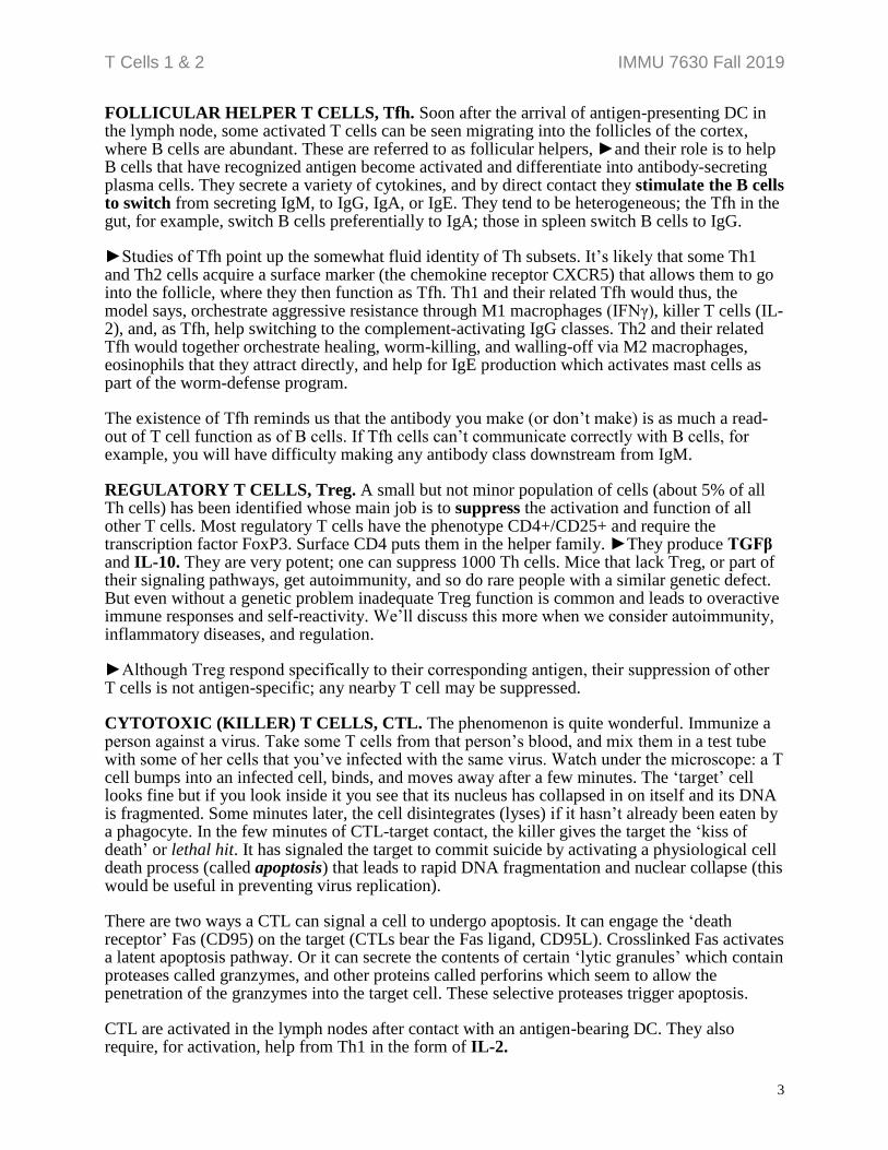
FOLLICULAR HELPER T CELLS, Tfh. Soon after the arrival of antigen-presenting DC in the lymph node, some activated T cells can be seen migrating into the follicles of the cortex, where B cells are abundant. These are referred to as follicular helpers, ►and their role is to help B cells that have recognized antigen become activated and differentiate into antibody-secreting plasma cells. They secrete a variety of cytokines, and by direct contact they stimulate the B cells to switch from secreting IgM, to IgG, IgA, or IgE. They tend to be heterogeneous; the Tfh in the gut, for example, switch B cells preferentially to IgA; those in spleen switch B cells to IgG.
►Studies of Tfh point up the somewhat fluid identity of Th subsets. It’s likely that some Th1 and Th2 cells acquire a surface marker (the chemokine receptor CXCR5) that allows them to go into the follicle, where they then function as Tfh. Th1 and their related Tfh would thus, the model says, orchestrate aggressive resistance through M1 macrophages (IFNγ), killer T cells (IL-2), and, as Tfh, help switching to the complement-activating IgG classes. Th2 and their related Tfh would together orchestrate healing, worm-killing, and walling-off via M2 macrophages, eosinophils that they attract directly, and help for IgE production which activates mast cells as part of the worm-defense program.
The existence of Tfh reminds us that the antibody you make (or don’t make) is as much a read-out of T cell function as of B cells. If Tfh cells can’t communicate correctly with B cells, for example, you will have difficulty making any antibody class downstream from IgM.
REGULATORY T CELLS, Treg. A small but not minor population of cells (about 5% of all Th cells) has been identified whose main job is to suppress the activation and function of all other T cells. Most regulatory T cells have the phenotype CD4+/CD25+ and require the transcription factor FoxP3. Surface CD4 puts them in the helper family. ►They produce TGFβ and IL-10. They are very potent; one can suppress 1000 Th cells. Mice that lack Treg, or part of their signaling pathways, get autoimmunity, and so do rare people with a similar genetic defect. But even without a genetic problem inadequate Treg function is common and leads to overactive immune responses and self-reactivity. We’ll discuss this more when we consider autoimmunity, inflammatory diseases, and regulation.
►Although Treg respond specifically to their corresponding antigen, their suppression of other T cells is not antigen-specific; any nearby T cell may be suppressed.
CYTOTOXIC (KILLER) T CELLS, CTL. The phenomenon is quite wonderful. Immunize a person against a virus. Take some T cells from that person’s blood, and mix them in a test tube with some of her cells that you’ve infected with the same virus. Watch under the microscope: a T cell bumps into an infected cell, binds, and moves away after a few minutes. The ‘target’ cell looks fine but if you look inside it you see that its nucleus has collapsed in on itself and its DNA is fragmented. Some minutes later, the cell disintegrates (lyses) if it hasn’t already been eaten by a phagocyte. In the few minutes of CTL-target contact, the killer gives the target the ‘kiss of death’ or lethal hit. It has signaled the target to commit suicide by activating a physiological cell death process (called apoptosis) that leads to rapid DNA fragmentation and nuclear collapse (this would be useful in preventing virus replication).
There are two ways a CTL can signal a cell to undergo apoptosis. It can engage the ‘death receptor’ Fas (CD95) on the target (CTLs bear the Fas ligand, CD95L). Crosslinked Fas activates a latent apoptosis pathway. Or it can secrete the contents of certain ‘lytic granules’ which contain proteases called granzymes, and other proteins called perforins which seem to allow the penetration of the granzymes into the target cell. These selective proteases trigger apoptosis.
CTL are activated in the lymph nodes after contact with an antigen-bearing DC. They also require, for activation, help from Th1 in the form of IL-2.

T Cells 1 & 2 IMMU 7630 Fall 2019
4
MEMORY T CELLS. After a response to antigen with rapid expansion of the relevant T cell clones, the number of T cells quickly declines, until perhaps 5% of the maximum amount (that’s nevertheless a huge increase over background) are left a few weeks after infection or immunization. These are memory cells, which can replace themselves, and be ready to rapidly differentiate into effector (helper, killer) cells when re-exposed to low antigen concentrations. There are surface markers for distinguishing naïve, memory, and active effector cells. Some memory cells circulate; others stay in the relevant tissue (‘tissue resident memory cells’).
SUBPOPULATION MARKERS. To distinguish the T cell subpopulations physically from each other and from B cells, we take advantage of unique surface molecules. We have monoclonal antibodies against them. These antibodies are tagged with a fluorescent molecule (fluorescein, rhodamine, phycoerythrin, the Alexa Fluor® series), to make it easy to distinguish which cells they bind to. ►B cells are identified using antibodies to immunoglobulins or their chains, or to the surface markers CD19 or CD20. The most useful molecules on T cells are CD3, CD4, and CD8. CD stands for ‘cluster of differentiation,’ a term based on old methods. ►CD3 is on the surface of all T cells; CD4 is on T helpers, CD8 is on CTL. These molecules play a role in T cell activation; we’ll discuss that soon. There are no reliable surface antigens to distinguish Th1 from Th2; you have to look at the lymphokines they make.
MHC RESTRICTION, PART 1. ►This is important. When investigators first studied killer T cells in people immunized with viruses, they found that CTL from someone infected with virus ‘V’ killed his ‘target cells’ (skin fibroblasts are commonly used) if they were infected with ‘V’ but not if infected with a different virus ‘E’. No surprise, that, just immunological specificity. ►But then they found that his anti-V CTL killed only his cells infected with V; not cells from an unrelated person, even if they were infected with V.
ASK YOURSELF: Can you fill in the last two columns? A and B are unrelated people; V and E are unrelated viruses.
CTL
source…
…who is
infected with
Target fibroblasts Killing? What does this result tell us?
Person A Virus V A, not infected
Person A Virus V A, infected with V
Person A Virus E A, infected with V
Person A Virus V B, infected with V
This experiment illustrates a new general principle of T cells: they are restricted in their recognition of antigen, to antigen on the surface of cells (here, the target cells) genetically identical to themselves. That is, they do not ‘see’ antigen alone, but only antigen presented to them on the surface of a genetically-identical cell. How identical? The T cell and the antigen-presenting cell must come from individuals who share alleles at a group of loci collectively called MHC (for Major Histocompatibility Complex), which code for surface glycoprotein molecules. ►Another way of saying this is that the T cell is antigen-specific and MHC-restricted4. MHC molecules are highly variable, that is, there are thousands of alleles in any population. The chances of yours being exactly the same as mine (or A’s the same as B’s) are extremely small.
ASK YOURSELF: You know we can give human antibody (e.g., tetanus antitoxin) to anyone who needs it; does MHC restriction mean we can’t give T cells to someone who needs them?
4 This work earned Peter Doherty and Rolf Zinkernagel the 1996 Nobel Prize in Physiology or Medicine.

T Cells 1 & 2 IMMU 7630 Fall 2019
5
ANTIGEN PRESENTATION TO T CELLS. When an antigen enters the bodylet’s use a virus as an exampleit will infect locally, cause an innate response, and quickly it or its
breakdown products will get ingested by a dendritic cell. Within the endosome viral proteins are broken down to peptides. The endosome fuses with other vesicles which have MHC molecules embedded in their membrane, facing in. Some of the peptides associate with the MHC molecules. The endosome recycles to the cell’s surface and fuses to the plasma membrane,
thus exposing MHC molecules bearing antigenic peptides to the outside world. ►We call cells that do this antigen-presenting cells, APC, and the mechanism just described the extrinsic pathway because it involves antigen from outside the APC. Dendritic cells are the best at this5. It’s this MHC-antigen complex that is presented to the receptor of an appropriate helper T cell. When MHC (Class II, see below) was first crystallized it showed two chains (α, β) folded so that a sort of groove appeared in the end that would be facing towards a T cell. The groove’s base is a beta sheet and its sides are two alpha helices. The MHC molecule had been crystallized in the act of presenting a peptide fragment of an antigen. The picture shows a T cell’s-eye view of this “pMHC” complex. MHC molecules only get to the surface when loaded with a peptide; empty MHC is a rarity.
►Because T cells see antigen only when it is complexed with cell-surface MHC molecules, T cells focus their attention on cell surfaces, and do not interact with free antigen; that is a job for the B cell and its antibodies.
T CELL RECEPTOR. The T cell receptor for antigen (TCR) is structurally related to antibody, and sequence data indicate a common ancestral gene. The two chains are called alpha and beta (don’t confuse these with the α and β of Class II MHC), and each has a constant and a variable portion. The T cell makes its receptor out of V, (D) and J regions—T cell genes, not the ones B cells use—recombined as in B cells, and like antibody, ►each chain has 3 CDRs6; the process takes place in the thymus. Both alpha and beta chains have transmembrane domains, unlike surface Ig, in which only the heavy chains are transmembrane:
5 The other APC are macrophages and B cells. 6 Complementarity-determining regions. Alpha chains are made from V and J segments; beta from V, D, and J (and
so are more diverse than alphas.) Note these are completely different sets of V(D)J from those used by B cells.
APC
T Cell
MHC Class II
antigen

T Cells 1 & 2 IMMU 7630 Fall 2019
6
Intimately associated with the TCR is the complex of molecules called CD3; it has at least 5 chains. It serves to transduce TCR signals for the T cell. This means that when a T cell binds the correct antigen + MHC with its TCR, the actual signal to the T cell is transmitted by CD3 (primarily CD3ζ, zeta). When a Th cell binds to a good APC like a dendritic cell, it receives three sorts of signals. The first is via its TCR—called the TCRpMHC interaction. The second are between many molecules (see the diagram) which are usually only on true APC, and that modify, enhance, or diminish activation. The third involves a variety of cytokines that the APC may secrete to activate, or modulate activation of the T cell, and, reciprocally, the T cell to activate the APC. Biochemical events follow: calcium flux, breakdown of membrane phospholipids, and activation of protein kinases which in turn activate transcription factors. Cytokine receptors are upregulated. The T cell goes into cycle (proliferation) and secretes its lymphokines (differentiation)7. MHC RESTRICTION, PART 2. The major histocompatibility complex is a large group of genes whose products have related functions. We’ll make it more comprehensible later. At this point, though, we’d like to introduce two kinds of MHC genes: Class I and Class II. ►Class I products are on all nucleated cells. ►Class II products are expressed only on the surfaces of dendritic and macrophage-type cells, B cells, and occasionally a few other cell types, all of which are APC: involved in presenting antigenic peptides to Th cells. ►When antigen is endocytosed and presented by a dendritic cell (DC) it associates primarily with Class II MHC molecules in the endocytic vesicle, and these complexes are what the DC presents to a Th cell. Th1, Th17, Tfh, Treg, and Th2 are programmed to recognize peptides on Class II molecules. Class I MHC molecules associate best with peptides that are sampled from proteins ►synthesized within the cell itself, not taken up by endocytosis. Most peptides would be from normal ‘self’ proteins, but antigens derive from abnormal (mutated) molecules and, especially, intracellular parasites. CTL are programmed to see antigen in association with MHC Class I molecules. ►It’s important to know that helpers see antigen + Class II, while CTL see antigen + Class I. And it’s easier to remember if you consider this: CTL are necessary for getting rid of virus by killing virus-infected cells; because they must be able to examine any infected cell, they are naturally ‘restricted’ by the ubiquitous Class I molecules. Helpers have to work together to get the immune response going, and to attract and activate macrophages to eat a foreign invader; it’s natural that they would see antigen presented to them on Class II, which is on APC, the sorts of cells (like DC) that would trap, process, and deliver something foreign to the lymph nodes. Here’s a wonderful thing: The dendritic cell, which gets everything going, is special in that it allows some peptides from antigens it’s eaten to leak over into its ‘intrinsic’ pathway, so that it can present them on Class I as well as Class II MHC at the same time. ►This is called cross-presentation. Thus the DC can bring samples in from the periphery and arrange not just for helpers to respond, but also CTL. Antigen presentation is summarized in diagrams on the last page of this double unit.
7 A note on the opposing properties of CD28 and CTLA-4 (see the Figure) is in the separate T Cell Supplementary &
Review Material file.
CD 4 or 8

T Cells 1 & 2 IMMU 7630 Fall 2019
7
CD4 and CD8 IN T CELL ACTIVATION. CD4 is on the surface of helper T cells. It binds to MHC Class II on the APC; not to the peptide-binding groove, but to the unvarying ‘base’ which is the same in everybody (see the diagram on the previous page). Thus when a Th is seeing antigen + Class II (as it should), the CD4 will help by increasing the strength of the bond. Similarly, the CD8 on CTL binds to the base of Class I, increasing the binding avidity of CTL to antigen + Class I. Without prior TCR binding, the CD molecules binding to MHC don’t activate the cellsthat would lead to chaos. They just increase the avidity of binding that got started by specific (TCRpMHC) recognition. There is evidence that CD4 and CD8 also help transduce activating signals.
T CELLS HELPING B CELLS. For protein antigens a vigorous class-switched antibody response requires help from T cells, as we have mentioned previously. How does a Tfh cell help a B cell get activated and make antibody? Four experimental observations gave us the clues: First, the T cell and the B cell must come from donors with the same MHC Class II. Second: The T cell and the B cell are not specific for the same epitope, but the epitopes they are specific for must both be on the same antigen molecule. Third: If you block the B cell’s ability to endocytose it cannot be helped by a T cell or make antibody. Fourth: The Tfh must contact the B cell.
ASK YOURSELF: It might be interesting for you to stop here, consider what you already know and what you’ve just read, and see whether you can put this together into a model before reading on.
Welcome back. Here’s what happens. The B cell binds the epitope (here, the circle) that its receptor (antibody) is specific for, on an antigen. It then endocytoses the bound molecule, which is broken down in the endocytic vesicle. Peptide fragments bind to MHC Class II molecules brought in by other vesicles that fuse with the endosome, and the peptide-MHC complex moves to the surface; the B cell now displays antigen in Class II. (I hope this sounds familiar; remember, the B cell is also an APC.) Eventually, along comes the correct Tfh and sees its epitope (the square) in Class II on the B cell’s surface. It binds and focuses surface interactions and helper lymphokines on the B cell. ►Note that the epitope that the T cell sees does not have to be the same as the one the B cell saw, and it hardly ever is. Video. You’ll soon see, this is important.
We don’t know for sure if seeing antigen for the first time on the surface of a B cell is enough to activate a resting (naïve) Tfh, or whether it would have to first see it on a dendritic cell. The DC is a better APC because it makes various cytokines to wake up resting T cells, and the B cell makes little or nothing. DC also optimize the secondary interactions shown earlier. In real life, the DC almost surely determines whether there will be a primary response.
Y
Th2B cell
MHC class II
antigen
Y
Th2B cell
MHC class IIY
Th2B cell
MHC class II
antigen
Tfh
T Cells 1 & 2 IMMU 7630 Fall 2019
8
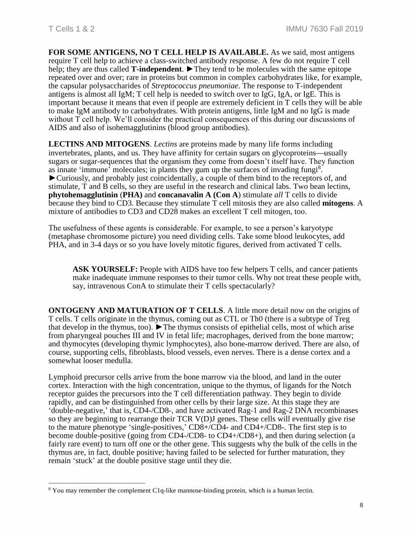
FOR SOME ANTIGENS, NO T CELL HELP IS AVAILABLE. As we said, most antigens require T cell help to achieve a class-switched antibody response. A few do not require T cell help; they are thus called T-independent. ►They tend to be molecules with the same epitope repeated over and over; rare in proteins but common in complex carbohydrates like, for example, the capsular polysaccharides of Streptococcus pneumoniae. The response to T-independent antigens is almost all IgM; T cell help is needed to switch over to IgG, IgA, or IgE. This is important because it means that even if people are extremely deficient in T cells they will be able to make IgM antibody to carbohydrates. With protein antigens, little IgM and no IgG is made without T cell help. We’ll consider the practical consequences of this during our discussions of AIDS and also of isohemagglutinins (blood group antibodies).
LECTINS AND MITOGENS. Lectins are proteins made by many life forms including invertebrates, plants, and us. They have affinity for certain sugars on glycoproteinsusually sugars or sugar-sequences that the organism they come from doesn’t itself have. They function as innate ‘immune’ molecules; in plants they gum up the surfaces of invading fungi8. ►Curiously, and probably just coincidentally, a couple of them bind to the receptors of, and stimulate, T and B cells, so they are useful in the research and clinical labs. Two bean lectins, phytohemagglutinin (PHA) and concanavalin A (Con A) stimulate all T cells to divide because they bind to CD3. Because they stimulate T cell mitosis they are also called mitogens. A mixture of antibodies to CD3 and CD28 makes an excellent T cell mitogen, too.
The usefulness of these agents is considerable. For example, to see a person’s karyotype (metaphase chromosome picture) you need dividing cells. Take some blood leukocytes, add PHA, and in 3-4 days or so you have lovely mitotic figures, derived from activated T cells.
ASK YOURSELF: People with AIDS have too few helpers T cells, and cancer patients make inadequate immune responses to their tumor cells. Why not treat these people with, say, intravenous ConA to stimulate their T cells spectacularly?
ONTOGENY AND MATURATION OF T CELLS. A little more detail now on the origins of T cells. T cells originate in the thymus, coming out as CTL or Th0 (there is a subtype of Treg that develop in the thymus, too). ►The thymus consists of epithelial cells, most of which arise from pharyngeal pouches III and IV in fetal life; macrophages, derived from the bone marrow; and thymocytes (developing thymic lymphocytes), also bone-marrow derived. There are also, of course, supporting cells, fibroblasts, blood vessels, even nerves. There is a dense cortex and a somewhat looser medulla.
Lymphoid precursor cells arrive from the bone marrow via the blood, and land in the outer cortex. Interaction with the high concentration, unique to the thymus, of ligands for the Notch receptor guides the precursors into the T cell differentiation pathway. They begin to divide rapidly, and can be distinguished from other cells by their large size. At this stage they are ‘double-negative,’ that is, CD4-/CD8-, and have activated Rag-1 and Rag-2 DNA recombinases so they are beginning to rearrange their TCR V(D)J genes. These cells will eventually give rise to the mature phenotype ‘single-positives,’ CD8+/CD4- and CD4+/CD8-. The first step is to become double-positive (going from CD4-/CD8- to CD4+/CD8+), and then during selection (a fairly rare event) to turn off one or the other gene. This suggests why the bulk of the cells in the thymus are, in fact, double positive; having failed to be selected for further maturation, they remain ‘stuck’ at the double positive stage until they die.
8 You may remember the complement C1q-like mannose-binding protein, which is a human lectin.

T Cells 1 & 2 IMMU 7630 Fall 2019
9
Single-positive T cells acquire other phenotypic refinements as they mature in the thymus, such as recirculation specification molecules and the various molecules with which they interact with APC. Then they are exported from the medulla. ►Fewer than 2% of thymocytes are exported; the rest will die in the thymus. Why? Because the demands on the T cell repertoire are very strict and not many randomly-generated TCR fill the bill.
REPERTOIRE SELECTION. What are the specifications for a successful T cell?
►A T cell must:
1. Not recognize ‘self,’ that is, not bind with activating affinity to a self-structure (MHC alone, or MHC loaded with a ‘self’ peptide); this would be autoimmunity. 2. Not recognize free antigen (which is antibody’s job). 3. Have the potential to recognize antigenic peptide only when it’s sitting in self MHC. The repertoire is selected within the thymus. Imagine a thymocyte that has just rearranged the genes for the alpha and beta chains of its TCR. It puts the receptors on its surface, and begins percolating through the thymus cortex, during which it will brush against the surfaces of many epithelial, macrophage, and dendritic cells, seeing MHC Class I and II molecules.
Let us say that the variable regions of TCR alpha (V, J) and beta (V, D, J) genes have been selected during evolution to produce receptors that are roughly complementary to the average configuration of an MHC molecule. MHC is very highly polymorphic; there are thousands of alleles in the human species. Since the TCR rearrangements are random, a brand-new thymocyte’s receptors ►will bind to the particular MHC alleles + peptides it finds on cells on its trip through the thymus with either high, low, or no affinity.
POSITIVE SELECTION. The blood-thymus barrier keeps foreign antigens out. ►The thymocyte’s TCR encounters MHC, both Class I and Class II, loaded with endogenous self peptides. If there is low but real affinity of binding between the TCR and the MHC of an epithelial cell (with a ‘self’ peptide in the groove), the cell binds just enough to be told to mature (positive selection). ►Typically, ‘enough’ means that the CDR1s and CDR2s bind to the alpha helices of the MHC groove, but the CDR3s do not bind the endogenous peptide there. The idea here is that this low affinity for self MHC but not self-peptide might turn out in the periphery to be high affinity for self MHC + some foreign peptide. ►This model explains MHC restriction: the T cells that emerge from the thymus of an ‘A’ animal or person see antigen plus ‘A’ MHC, because they were positively selected on ‘A’. There is plenty of evidence to support this: for example, rare patients who genetically have low MHC Class I expression develop normal Th cells but few CTL, because there was not enough Class I in the thymus for their developing CD8+ cells to bind to.
NEGATIVE SELECTION. The next possibility is that the immature T cell’s receptor binds to MHC (which, as we’ve been saying, will have a ‘self’ peptide in it, derived from a normal protein) with high affinity. By high we mean high enough to result in the activation of the T cell; all six of the TCR’s CDRs, including the 2 CDR 3s, are engaged. This is clearly an undesirable cell as its activation would result in autoimmunity. The fate of this immature cell is fascinating: ► either it dies by the process of apoptosis, or it differentiates into a Treg. This latter outcome is very cool—it suggests that if a rogue Th or CTL were trying to become activated by your liver, for example, it might find a Treg next to it saying: Don’t.

T Cells 1 & 2 IMMU 7630 Fall 2019
10
This mechanism could delete T cells reactive against the typical peptides you’d imagine would be expressed in the thymus; but what about liver or thyroid or adrenal-specific gene products?
►Amazingly, the AIRE (autoimmune regulator) gene codes for a transcription factor that causes thymic medullary epithelial cells to express a wide variety of otherwise-inexplicable extrathymic peptides, so that reactive T cells may be removed from the repertoire. In fact, AIRE-deficient people develop multiple autoimmunities. But so do children with Down syndrome, trisomy 21, and they have 3 copies of Aire which is on chromosome 21. Although they develop many Tregs in the thymus, unfortunately those seem to be functionally impaired, probably due to dysregulated signaling.
NON-SELECTION. The third option. Because the repertoire of T cells is generated by random association of V, (D), and J gene segments, it is reasonable to assume that most of the resultant TCR will have no, or not enough, affinity for the particular MHC molecules they find expressed in the thymus. The immature cell thus receives no stimulation through its TCR. Under these circumstances it will die in two or three days, again by apoptosis.
Note that apoptosis is a way in which cells die; its pathways are under genetic control. Almost always when a cell dies for physiological reasons, it is by apoptosis. (But amazingly, the CTL can force its targets to activate this death program, too.)
ASK YOURSELF: This is very hard. Suppose you took a newborn inbred ‘MHC A’ mouse, removed her thymus and replaced it with an irradiated ‘MHC B’ thymus (the radiation would kill the ‘MHC B’ thymocytes in it but not the macrophages, DC, or epithelial cells of the stroma). She will grow a new thymus, with donor MHC B stroma but with her own thymocytes derived from her bone marrow. Let her grow up, in a sterile environment as a precaution. What kind of an immune response do you think she would have? Ask yourself, what MHC molecules will be on the surface of her T cells, B cells, and antigen-presenting cells? What will the MHC restriction of her T cells be? How will they work in her body against infections? Making diagrams will help; don’t get mad at yourself if you can’t figure this out.

T Cells 1 & 2 IMMU 7630 Fall 2019
11
Macrophage
Macrophages phagocytose antigens, digest them in phagolysosomes, load peptides onto MHC Class II, and recycle them to the surface. This is the extrinsic pathway. Of course, like all cells, macrophages also express Class I.
Liver cell
When any cells, like this hepatocyte, make proteins, they shuttle peptides derived from the nascent protein from the cytosol to the endoplasmic reticulum, and thence to the surface, presented in MHC Class I. This is called the intrinsic pathway. In this image the peptide is coded by a virus.
B cell
B cells use surface Ig to bind an epitope of an antigen, then internalize the antigen, and digest it to peptides which are loaded onto MHC Class II and recycled to the surface for interaction with Tfh cells.
Dendritic cell
Dendritic cells, the best APC, take up antigen and process it for MHC Class II as do macrophages or B cells; but there is also ‘cross-presentation’ by the intrinsic pathway, so some peptides are presented on MHC Class I as well. The result is that DC stimulate both Th and CTL.
II
I
A SUMMARY OF ANTIGEN PRESENTATION
I
II
II

T Cells 1 & 2 IMMU 7630 Fall 2019
12
Learning Objectives for T Cells
1. List the main types of T cells, and define their functions. Describe the interplay between
Th1, Th2, and Treg cells.
2. Describe the surface markers that can be used to distinguish between T and B cells in
humans, and between helper and cytotoxic T cells.
3. Define lymphokine, chemokine, and cytokine.
4. Describe an activity of interferon-gamma (IFNγ).
5. Define mitogen, and describe a use for them in the clinical laboratory.
6. Describe what you’d expect to observe in the way of cell proliferation if you added (a) an
antigen, or (b) a mitogen like PHA or ConA, to a mixture of the white cells from normal
human blood.
7. Compare and contrast the antigen receptors of T and B cells.
8. Discuss the structures recognized by T cell receptors (see also Immunogenetics).
9. Distinguish between what is recognized by helper and cytotoxic T cells. Explain the special
role of dendritic cells in this process.
10. Discuss what is meant by ‘MHC-restriction’. Name the classes of MHC molecules by
which CTL and helper T cells are restricted.
11. Describe repertoire selection in the thymus, in terms of positive, negative and non-
selection.
12. Describe the characteristics of T-independent antigens.
13. Outline an experiment that shows that an antibody response can be ‘T-dependent.’
14. Describe how Tfh cells help B cells get activated by antigen and switch immunoglobulin
class.