Embed Size (px)
Citation preview
-4±91 564 DELIVERY SYSTENS FOR BONE MORPflOOENETIC PROTEIN (@IP) IVA- FOR REPAIR OF BATTLE INCURRED BONE INJURIES(U)
CIFORMIR UNIV LOS ANGELES M R URIST 01 NOV 87
NISSFE flhhhhh..h..hh.O 65 M
Imommms
g Lu~! 5.
*I 1.2 1 . 1.
-- fl~'~ TEST CHART
%ATIO4AL S'.P(M OF 5"NOOMd"W "
TZZ %
/ Marshall R. Urist, M.D.
" SECURITY CLASSIFICATION OF THIS PAGE DAND17-84-C-4045 (-I orm Approved11C EILE.C REPORT DOCUMENTATION PAGE OMBNo. 070Ap 1o8
lb RESTRICTIVE MARKINGS
3. DISTRIBUTION /AVAILABILITY OF REPORTApproved for public release;LE distribution unlimited
4. PERFORMING ORGANIZATION REPORT NUMBER(S) S. MONITORING ORGANIZATION REPORT NUMBER(S)
6a. NAME OF PERFORMING ORGANIZATION 6b. OFFICE SYMBOL 7a. NAME OF MONITORING ORG
University of California (ifaplicabe)_________CT_
6c. ADDRESS (City, State, and ZIP Code) 7b. ADDRESS (City, State, and ZIP43
Los Angeles, California 90024 <'11a. NAME OF FUNDING /SPONSORING 8 b. OFFICE SYMBOL 9. PROCUREMENT INSTRUMENT IDENTIFICATION NUMBER.
ORGANIZATION US Army Medical I (if applicable) DAMD17-84-C-4045Research & Development Command
Sc. ADDRESS (City, State and ZIP Code) 10 SOURCE OF FUNDING NUMBERSFort Detrick PROGRAM PROJECT TASK IWORK UNITFrederick, Maryland 21701-5012 ELEMENT NO. NO. 3M1 NO. ACCESSION NO.
61102A 61102BS10 I 371
11. TITLE (Include Security Classification)Deli-ery Systems for Bone Morphogenetic Protein (BMP) for Repair of Battle IncurredBone Injuries
PERSONAL AUTHOR(S)Marshall R. Urist
13a. TYPE OF REPORT 113b. TIME COVERED 114. DATE OF REPORT (Year, Month, Day) 15. PAGE COUNTFinal FROM 7/16/TO_/3n 871 1987 November 1
16. SUPPLEMENTARY NOTATION
17. COSATI CODES 18. SUBJECT TERMS (Continue on reverse if necessary and identify by block number)FIELD GROUP J SUB-GROUP Battle-incurred bone injuries; bone defects; fractures; bone0 regeneration; bone morphogenetic protein; non union; bone
0healing; osteogenesis; fracture compound comminuted.19. ABSTRACT (Continue on reverse if necessary and identify by block number)
___ _--- The attached document is a summary of ten projects on boneregeneration under the influence of a bone morphogenetic protein (BMP),performed in collaboration with Lieutenant Colonel Jeffrey Hollinger andassociates at D1C, /W.1,A:1.'RO in the three-year period from 1984 through1987 inclusive.
The ten projects have in common the objective of determiningefficacy of BMP/NCP in augmentation of bone regeneration process indogs. The tests were performed in standard bone defects of criticalsize for regeneration in adult dogs in skull trephine operations,segmental long bone loss, and spinal fusion. Implants of BMP wereplaced in heterotopic sites in a mouse muscle pouch for preliminarytests for verification of induced bone formation. Four delivery systemswere tested: () a selection of insoluble non-collagenous bone matrix
DISTRIBUTION/AVAILABILITY OF ABSTRACT 21 ABSTRACT SECURITY CLASSIFICATIONOUNCLASSIFIED /UNLIMITED aSAME AS RPT r DTIC USERS Unclassified
22a. NAME OF RESPONSIBLE INDIVIDUAL 22b TELEPHONE (Include Area Code) 22c OFICE SYMBOLMary Frances Bostian 301-663-7325 SGRD-R]1-S
DO Form 1473, JUN 86 Previous editions are obsolete SECUR T CLASSIFICATION OF THIS PAGE
18 82 23
Marshall R. Urist, M.D.DAMD17-84-C-4045
N poteins, which form aggregates with BMP, X-() polylactic acid polymers,,()biodegradable beta tricalcium phosphate, and 4) a
polymethylmethacrylate. The report includes preliminary observations onimplants of human BMP in uman subjects under the guidelines of theHuman Subject Protection mittee of the University of California, LosAngeles, School of Medicine Center for the Health Sciences. Applied to-this-fnnual/Final Report is aof the Materials, Methods, andResulgs of the individual ten projects. The overall results demonstrate.tha% th *eal delivery system for transmission of BM for augmentation
-~ 6f b6nE--rdieneration in long-lived animals such as dogs and humans, mustbe i-ought*.by investigations with the aid of new and improved methods.~These Wt ended investigations should be facilitated by the developmentof a haso$ dneous recombinant BMP, which could become available in.1988-1989.
Accesiol, For
NVTIS CRA &IOTIC TAB o3Unannoiced 0
ByDistr-ibilon,
Availability Codies
Dist Avi ~4.
DTIC ~
copyINSPEC- 0
V.-
Marshall R. Urist, M.D.
DAMD17-84-C-4045
TABLE OF CONTENTS
PAGEReport Documentation Page 1
Table of Contents 3
Title page 4
Introduction 5
1. Craniotomy Defects in Dogs 6
2. Bone Repair Induced by Bone Morphogenetic Protein 7in Ulnar Defects in Dogs /
3. Calcium Phosphate Delivery System for BMP Was 8Tested in Mice before Expensive Experiments WerePerformed in Dogs
4. BMP-Beta Tricalcium Phosphate Composite Delivery 8System in Dogs
5. BMP Polylactic Polyglycollic Acid Polymer Composite 9
in Spinal Fusions in Dogs
6. Craniotomy Defects in Sheep t0
7. Craniotomy Defects in Monkeys 10
8. BMP Delivery System of Bone Matrix Non Collagenous 11Proteins (BMP/NCP) in a Bone Tumor Defect
9. BMP/NCP in a Metaphyseal Defect 12
10. Objections and Specific Aims of Clinical Trials 12of BMP
10A. Repair of Segmental Defects of the Tibia with 13Cancellous Bone and Bone Marrow Augmentation withHuman Bone Morphogenetic Protein (hBMP): A Preliminary
Report of Six Patients
1OB. Bone Morphogenetic Protein Augmentation Grafting 13
of Resistant Femoral Non Unions
List of Publications 15
In Press Publications 18
Submitted Publications 19
Informed Consent Form 20
3521122k'Iot
Marshall R. Urist, M.D.DAMDI7-84-C-4045
AD
DELIVERY SYSTEMS FOR BONE MORPHOGENETIC PROTEIN (BMP)FOR REPAIR OF BATTLE INCURRED BONE INJURIES
FINAL REPORT
NOVEMBER 1, 1987
MARSHALL R. URIST
Supported by
U.S. ARMY MEDICAL RESEARCH AND DEVELOPMENT COD4ANDFort Detrick, Frederick, Maryland 21701-5012
Contract No. DAMD17-84-C-4045
University of California, Los AngelesLos Angeles, California 90024
Approved for public release; distribution unlimited
The findings in this report are not to be construed as an officialDepartment of the Army position unless so designated by otherauthorized documents.
I4
Marshall R. Urist, M.D.
DAMD17-84-C-4045
FINAL REPORT OF DAMD17-84-C-4045 PROJECT NO. 3M1 6102BSI0
INTRODUCTION
Summarized in this report, the 10 projects performed under this
Contract demonstrate the extraordinary potential of BMP research for
improvement of treatment of battle-incurred extensive bone injuries.
Bone differs from other tissue not only in physiochemical structure
but also in its extraordinary capacity for growth, continuous internal
remodeling, and regeneration throughout postfetal life, even in
long-lived higher vertebrates. How much of this capacity can be
accounted for by proliferation of predifferentiated osteoprogenitor
cells and how much can be attributed to induced differentiation of
mesenchymal-type cells have been challenging questions for a long time.
A basic assumption is that regeneration occurs by a combination of the
two processes. The process of induced cell differentiation has been
observed from measurements of the quantities of bone formed in response
to implants of either bone matrix or purified bone morphogenetic protein
(BMP) in extraskeletal and intraskeletal sites. The osteoprogenitor
cell proliferation process has been well known for more than a century
and is measured in reactions of periosteum and endosteum to injury,
diet, vitamins, and hormones. Bone-derived growth factors stimulate
osteoprogenitor cells to proliferate in serum-free tissue culture media.
The mechanisms of action of BMF and growth factors are primarily local,
but secondary systemic imwunologic reactionF could 1hve either
permissive or depressive effects.
The most important and indispensptle substitutes for experiments in
human beings are adult mongrel dogs, monkeys, and sheep. Experimental
.S.
Marshall R. Urist, M.D.
DAMD17-84-C-4045
observations on local conditions and biochemical factors regulating bone
regeneration are recorded in literature dating back over a century. The
largest part of present knowledge was obtained by experiements on
rabbits, guinea pigs, rats, and mice. Relatively little information has
been obtained from research on dogs, a species comparable to man with
respect to regulatory mechanisms, time relations, and surgical problems.
Such information that is available on bone repair in dogs is chiefly
descriptive. Present research was performed on dogs because, as
observed by Coulson, the metabolic activity index (MAI) of the dog is
1.58, remarkably close to that of human beings (I.0); in comparison, the -.
MAI is 5.15 for the rat and 15.60 for the mouse.
Young growing individuals repair cranial trephine defects more
rapidly than adults. Normally, infants can regenerate the entire vault
of the cranium, but in mature individuals, a defect with a diarmeter of
more than 1 cm will heal only with the aid of a transplant of autogeneic
bone. In the adult, the incorporation of the transplant is incomplete
often enough to warrant a search of substitutes for autogeneic bone.
The newest substitute is bone morphogenetic protein (BMP), recently
isolated from bovine and human bone. BMP is the hypothetical paracrine
agent augmenting the procpss cf bone regeneration by induction of the
morphogenetic phase of bone development.
1. CRANIOTOMY DEFECTS IN DOGS.
In the adult dog, a 14-mm skull trephine defect regenerates or, v
incompletely In the lifetime of the individual. Only about half of thc
defect is repaired; the regenerated part develops by extension of growth
from the bony rim. Correlated roentgenographic and histomorphometric
-**<
Marshall R. Urist, M.D.
DAMD17-84-C-4045
methods demonstrate that new bone develops by proliferation of
preexistinp osteoprogenitor cells lining the diploe and perivascular
cells of the bone marrow stroma. An autogeneic bone graft, inc]udinr
bone n.errow, provides a supplementary framework and generally completes
the repair process. Transplants of bone marrow alone fail to repair the
defect. Implants of bovine bone morphogenetic protein (bBMP) and a
carrier consisting of matrix y-carboxyglutamic acid rich protein
(without any additional bone or bone marrow) induce repair almost as
completely as an autograft. BMP-induced bone regeneration is incomplete
in the thin lateral temporal marrow-deficient part of the cranium.
Implants of BMP plus bone marrow induce complete repair, suggesting that
calvarial bone regeneration is bone marrow stroma-dependent for a supply
of target cells. The target for BMP in cranial bone regeneration is the
perivascular connective tissue cells (pericyte) of the host bed marrow
stroma and endosteum. The molecular mechanism of differentiation of
pericytes into osteoprogenitor cells is not known, but the process is
irreversible, heritable, and presents a solvable problem.
2. BONE REPAIR INDUCED BY BONE MORPHOGENETIC PROTEIN IN ULNAR DEFECTS INDOGS.
In dogs, resection of a length of the ulna equal to twice the
diameter of the mid-shaft leaves a defect which consistently fails to
unite. In response to an implant of 100 mg of bovine bone morphogenetic
protein (bBMP), the defect becomes filled by callus consisting of
fibrocartilage, cartilage and woven bone within four weeks. The
cartilage is resorbed and replaced by new bone in four to eight weeks.
Woven bone is then resorbed, colonized by bone marrow cells and
remodeled into lamellar bone. Union of the defect is produced by 12
7
Marshall R. Urist, M.D.
DAMDI7-84-C-4045
weeks. Control defects filled with autogeneic cortical bone chips unite
after the same period.
In regeneration induced by bone morphogenetic protein (BYP) and in
repair enhanced by bone graft, union depends upon the proliferation of
cells within and around the bone ends. Our working hypothesis is that
BMP induces the differentiation of perivascular connective tissue cells
into chondroblasts and osteoprogenitor cells and thereby augments the
process of bone regeneration from the cells already present in the
endosteum and periosteum.
3. CALCIUM PHOSPHATE DELIVERY SYSTEM FOR BMP WAS TESTED IN MICE BEFOREEXPENSIVE EXPERIMENTS WERE PERFORMED IN DOGS.
An aggregate of biodegradable B-tricalcium phosphate and bone
morphogenetic protein (BMP/TCP) induces the differentiation of cartilage
within eight days, cartilage and woven bone within 12 days, and lamellar
bone including bore marrow, within 21 days. The yield of new bone from 4a 1-mg dose was more than 12 times greater from the TCP/BMP than from I
the BMP alone. Whether TCP acts as a slow-release delivery system,
potentiates the activity of BMP, or serves to distribute BMP in a
favorable three-dimensional pattern requires further investigation.
I
4. BMP-BETA TRICALCIUM PHOSPHATE COMPOSITE DELIVERY SYSTEM IN DOGS.
Beta tricalcium phosphate (TCP) was employed as a nonimmunogenic
biodegradable delivery system for bovine bone morphogenetic protein
(bBMP). A bBMP/TCP composite was implanted in adult dogs with skull
trephine defects of a critical size of 1.4 cm that would otherwise .e
remain unhealed in the lifetime of the individual. bBMP/TCP implants
induced 91%-100% incorporation by deposits of new bone. In comparison,
Marshall R. Urist, M.D.DAMD17-84-C-4045
control implants of TCP impregnated with bovine serum albumin (BSA/TCP)
induced 0%-8% incorporation, or only marginal host bed reactive bone
formation. The retention of unabsorbed TCP in the host bone four months
after implantation suggests that further research should be encouraged
to obtain a formulation of sintered calcium phosphate that could be
resorbed more rapidly and in the process more completely replaced by
bone.
5. BMP POLYLACTIC POLYGLYCOLLIC ACID POLYMER COMPOSITE IN SPINAL FUSIONSIN DOGS.
To investigate the action of BMP on the growth of host bed derived
bone in experimental spinal fusions, posterior intervertebral spinal
fusions of the lower thoracic spine were performed in 13 mature mongrel
dogs. The procedures were limited to single intervertebral levels.
Three levels in each dog were employed for controls for BMP levels. The
spinal columns were examined by radiohistomorphometic methods at three
weeks, six weeks, 12 weeks, and six months, and showed the BMP levels to
have two to three times more new bone than control levels. At the BMP
level, an increase in the amount of bone was observed in the interval %
from three to 12 weeks, in contrast to a decrease seen at control -'
levels. Fusion was present in 5/7 of the BMP levels compared with 0/7,
1/7, and 2/7 of control levels. The BMP level exhibited an increased
number and volume of areas de novo cartilage and woven bone formation at
all time intervals of dogs compared to all control levels. The PLA/PGA
polymer was incompletely resorbed and partially retained in the fusion1?
site. These preliminary observations suggest that BMP may serve as a
useful adjunct in spinal fusions, but research is required to find a
more rapidly degradable delivery system. The objective of BMP research
9 _.
Marshall R. Urist, M.D.
DAMD17-84-C-4045
is to augment the host bed capacity for bone generation and regeneration
for patients with missing bone substance from battle-incurred wounds.
6. CRANIOTOMY DEFECTS IN SHEEP.
An aggregate of partially purified bovine bone morphogenetic
protein (bBMP) and bone matrix insoluble noncollagenous proteins (iNCP)
weighing a total of 100 mg of lyophilized BMP/iNCP, was implanted using
ultra thin gelatin capsuled in skull trephine defects in adult sheep.
One hundred milligram samples of freeze-dried bovine serum albumin (BSA)
were similarly implanted in 18-20 mm trephine skull defects and also in
posterior cervical muscle pouches for heterotopic controls. In two out
of five sheep, the trephines were repaired with bone as early as four
weeks after the operation. Eight to 12 weeks after surgery, repair was
complete in the other three sheep. In the control contralateral
trephines, one-third to one-half of the defect was incompletely
repaired. Neither the BMP nor the BSA control implants induced bone
formation in the muscle. While the BMP/INCP prepared from bovine bone
consistently induced regeneration in skull trephine defects, only
fibrous tissue and no extraskeletal bone was induced to form in cervical
muscle pouches in sheep.
7. CRANIOTOMY DEFECTS IN MONKEYS.
Large cranial defects do not always heal spontaneously, especially
in humans; often they have to be obturated with metallic or acrylic
fillers. Bilateral cranial trephine defects, measuring 14-20 mm in
rdiameter, were created in three Rhesus monkeys, providing a typical
primate model system for investigations in comparative physiology of
bone regeneration. Each skull had one control and one experimental
,o0
Marshall R. Urist, M.D.DAMD17-84-C-4045
trephine defect. Control defects were implanted with bovine serum
albumin (BSA). Experimental defects were implanted with 100-200 mg of a
partially purified fraction of bovine bone morphogenetic protein (bBMP)
in two monkeys. In one monkey, the BMP was incorporated in a 1:1
poly(lactic) poly(glycollic) acid copolymer, high-viscosity formula.
The animals were killed at eight weeks, ten weeks, and 16 weeks after
implantation. The tissue responses were analyzed by computed
hist-a-rphometry and routine histologic examination. At each time
interval, the BMP implanted defects produced more complete regeneration
than the control implants. The morphogenetic response occurred in the
following sequences: mesenchymal cell proliferation, chondrogenesis, and
increased bone formation. BMP-induced osteogenesis may initiate the
regenerative process. The copolymer releases BMP but may constitute a .
barrier to the end stages of replacement by new bone and would be more
useful in a low-viscosity rapidly biodegradable form. 'S,
8. BMP DELIVERY SYSTEM OF BONE MATRIX NON COLLAGENOUS PROTEINS (BMP/NCP)IN A BONE TUMOR DEFECT.
A 29-year-old woman with an enchondroma that was expanding and
eroding the palmar cortex of the middle phalanx was successfully treated
by curettage and implantation of bone morphogenetic protein. The
metaphysis and cortex were repaired by lamellar bone within two months.
The medulla was completely filled with trabecular bone by nine months.
The full range of motion and normal functions of finger and hand joints
were restored, and there was no recurrence or abnormalities at follow-up
visits 2.5 years after the o,.eration
Marshall R. Urist, M.D.
DAMD17-84-C-4045
9. BMP/NCP IN A METAPHYSEAL DEFECT.
The response of protodifferentiated and differentiated bone cells
to bovine bone morphc3enetic protein (bBMP) was observed in implants in
the adult rabbit distal femoral metaphysis. Bovine serum albumin and
denatured bBMP were implanted in the contralateral femur of controls.
The changes of the bone marrow reflected the reaction of
protodifferentiated cells. The changes in preexisting trabecular bone
tissue reflected the reaction of differentiated cells to bBMP. 45Ca
radioisotope quantitative methods demonstrated that the bone
morphogenetic response was superimposed upon the reaction to the injury
of surgical implantation. By the end of the fourth week, roentgenograms
and histologic sections showed larger deposits of intrametaphyseal
cartilage and bone in bBMP than in control implanted femurs. By the end
of the eighth week, bone formation was associated with remodeling of the
entire distal femur and expansion of the external diameter of the
metaphysis. These observations indicate the need for investigation of
perisinusoid and perivascular cells of periosteum, endosteum, and marrow
stroma.
10. OBJECTIVES AND SPECIFIC AIMS OF CLINICAL TRIALS OF BMP
Clinical experience is being documented on two groups of patients
selected for preliminary clinical trials of implants of human bone
morphogenetic protein (BMP) in an aggregate of associated bone matrix
non-collagenous proteins BMP/NCP. One group of twelve patients
illustrates the problem of intractable non-unlon of the femur. A second
group consists of six cases of segmental tibia bone defects ranging from
3 to 17 cm in length. Many of the patients had been candidates for
amputation.
12~
Marshall R. Urist, M.D.
DAMD17-84-C-4045
This total series of eighteen patients was investigated with the
objective of determining efficacy of BMP. The specific aim is to
investigate the effects of BMP upon the process of regeneration of bone
defects far beyond the critical dimensions for spontaneous repair in the
lifetime of the individual. The bone defects were incurred in violent
accidents of the character encountered in combat related fractures with
missing substance.
Appended to this report is a sample copy of the informed consent
procedures for patients treated under the guidelines of the Human
Subject Protection Committee, University of California, Los Angeles,
School of Medicine, Center for Health Sciences.
10A. REPAIR OF SEGMENTAL DEFECTS OF THE TIBIA WITH CANCELLOUS BONE ANDBONE MARROW AUGMENTATION WITH HUMAN BONE MORPHOGENETIC PROTEIN(hBMP): A PRELIMINARY REPORT OF SIX PATIENTS.
Human bone morphogenetic protein (hBMP) is a bone cell
differentiation inducing factor. Six patients with traumatic segmental
tibial defects 3 to 17 cm developed solid union by implantation of hBMP,
autogeneic cancellous grafts, and stabilization. There were no
allergic, infectious, or surgical complications. If hBMP augmentation
in biodegradable delivery systems can be established by a prospective,
randomized, double blind investigation, the incidence of successful bone
graft operations for treatment of large segmental defects would be
measurably improved.
1OB. BONE MORPHOGENETIC PROTEIN AUGMENTATION GRAFTING OF RESISTANT
FEMORAL NON UNIONS.
Twelve patients with intractable nonunions of the femoral
diaphyseal or metaphyseal-diaphyseal segments were successfully treated
13
Marshall R. Urist, M.D.DAMDI7-84-C-4045
by a combination of Internal fixation and implants of human bone
morphogenetic protein (hBHP). There vas an average of 3.0 surgical
procedures per patient attempting union prior to hBMP implantation.
Union was obtained in 100% of patients, 11/12 with initial protocol and
in one patient with a repeat stabilization and implantation of hBMP.
Four patients received autogeneic cancellous bone graft and four
patients received allogeneic bone grafts. The BMP implant was prepared
in the form of an aggregate of hBMP and bone matrix water insoluble
noncollagenous proteins (hBMP/INCP). Fifty to 100 mg of hBMP/iNCP was
either implanted In the fracture gap in ultra thin gelatin capsules, or -
incorporated in a strip of polylactic/polyglycollic acid copolymer
(PLA/PGA) and placed as an onlay across the fracture gap. The average
time to union was 4.7 months. Further clinical investigation should be
performed on matched cases with and without B4P augmentation in order to
distinguish hBMP effects from new or improved methods of fracture
fixation and autogeneic cancellous bone grafts.
14
Marshall R. Urist, M.D.DAMD17-84-C-4045
(LIST OF PUBLICATIONS
1. Urist, M.R., Huo, Y.K., Brownell, A.G., Hohl, W.M., Buyske, J.,Lietze, A., Tempst, P., Hunkapillar, M., and DeLange, R.J.:Purification of bovine bone morphogenetic protein by hydroxyapatitechromatography. Proc. Natl. Acad. Sci. 81:371-375, 1984.
2. Sato, K., and Urist, M.R.: Bone morphogenetic protein-inducedcartilage development in tissue culture. Clin. Orthop. 183:180-187,1984.
3. Urist, M.R., Lietze, A., and Dawson, E.: B-tricalcium phosphatedelivery system for bone morphogenetic protein. Clin. Orthop.187:277-280, 1984.
4. Amstutz, H.C., Johnson, E.E., Finerman, G.A.M., Meals, R.A.,Moreland, J.R., Kim, W.C., and Urist, M.R.: Bone morphogenetic proteinand bone-derived growth factors. West. J. Med. 141:71-87, 1984.
5. Urist, M.R., and Hudak, R.T.: Radioimmunoassay of bone morphogeneticprotein in serum: A tissue-specific parameter of bone metabolism.Proc. Soc. Exp. Biol. Med. 176:472-475, 1984.
6. Nilsson, 0., and Urist, M.R.: Response of the rabbit metaphysis toimplants of bovine bone morphogenetic protein (bBMP). Clin. Orthop.195:275-281, 1985.
7. Urist, M.R., and Huo, Y.K.: A hydridoma secreting monoclonal antibonemorphogenetic protein (AntiBMP). Trans. 10th Cong. Intern. Soc. Dev.Biol. August 4-9, 1985.
8. Urist, M.R., Lietze, A., Dawson, E., and Finerman, G.A.M.: Bonemorphogenetic protein (BMP) delivery system. Transactions from the 11thAnnual Meeting of the Society for Biomaterials. 17:24, 1985.
9. Urist, M.R., Brownell, A.B., Huo, Y.K., Lietze, A., Nilsson, 0., \Rasmussen, J., Takahashi, S., Hirota, W., and DeLange, R.: Biologicallyactive N-terminal region of bone morphogenetic protein isolated bypepsin limited proteolysis. Proc. Fed. Am. Soc. Exp. Biol. 69:1956 only,1985.
10. Urist, M.R. Book Review. Surgical Exposures in Orthopedics: TheAnatomic Approach by Stanly Hoppenfeld and Piet deBoer. p. 128.Philadelphia, J.B. Lippincott, 1984. New Eng. J. Med. Jan. 10, 1985.
11. Urist, M.R., Hudak, R.T., Huo, Y.K., and Rasmussen, J.K.:Osteoporosis: a bone morphogenetic protein autoimmune disease. InSecond Intern. Conf. on Bone Growth. p. 77-96. Jan. 3-5, 1985. Edited byA. Dixon and B.G. Sarnat. New York: A.R. Liss, Inc., 1985.
12. Urist, M.R., Hudak, R., Huo, Y.K., Rasmussen, J., Hirota, W. andLietze, A.: Bone morphogenetic protein (BMP) and anti-BMP immunoassay inpatients with osteoporosis. In: The Chemistry and Biology of MineralizedTissues. p. 62-69. Edited by W.T. Butler. Birmingham, Ebsco Media Inc.,
1985.
15
Marshall R. Urist, M.D.DAMD17-84-C-4045
13. Sato, K., and Urist,-M.R.: Induced Regeneration of calvaria by bonemorphogenetic protein (BMP) in dogs. Clin. Orthop. 197:301-311, 1985.
14. Urist, M.R., and Huo, Y.K.: A hybridoma secreting monoclonalanti-bone morphogenetic protein. Cell Differentiation. 16(Suppl):1355,1985.
15. Canalis, E., Centrella, M. and Urist, M. R.: Effect of partiallypurified bone morphogenetic protein on DNA synthesis and cellreplication in calvarial and fibroblast cultures. Clin. Orthop.198:289-296, 1985.
16. Syftestad, G.T., Triffitt, J.T., Urist, M.R., and Caplan, A.I.: Anosteo-inductive bone matrix extract stimulates the in vitro conversionof mesenchyme into chondrocytes. Calcif. Tissue Int. 36:625-627, 1984.
17. Urist, M.R., Nilsson, O.S., Hudak, R., Huo, Y.K., Rasmussen, J.,Hirota, W., and Lietze, A.: Immunologic evidence of a bone morphogeneticprotein in the milieu interieur. Ann. Biol. Clin., 43:755-766, 1985.
18. Urist, M.R., Kovacs, S., and Yates, K.A.: Regeneration of anenchondroma defect under the influence of an implant of human bonemorphogenetic protein. J. Hand Surgery 11A:417-419, 1986.
19. Johnson, E., Finerman, G.A.M., and Urist, M.R.: Treatment ofdifficult non-union with bone morphogenetic protein augmentation.Abstract for the American Academy of Orthopaedic Surgeons, Fifty-FourthAnnual Meeting, San Francisco, CA, January 22-27, 1987.
20. Takahashi, S., and Urist, M.R.: Differentiation of cartilage onthree substrata under the influence of an aggregate of morphogeneticprotein and other bone tissue noncollagenous proteins (BMP/iNCP). Clin.Orthop. 207:227-238, 1986.
21. Urist, M.R.: Book Review. Bone and Mineral Research Annual 3.Edited by W.A. Peck, Elsevier, Amsterdam and New York, Under auspices ofthe Int. Conf. Calcium Regulating Hormones, 1985. Quart. Rev. Biol.61:303 only, 1986.
22. Nilsson, O.S., Urist, M.R., Dawson, E.G., Schmalzried, T.P., andFinerman, G.A.M.: Bone repair induced by bone morphogenetic protein inulnar defects in dogs. J. of Bone and Joint Surg. 68B(vol. 4):635-642,1986.
23. Urist, M.R., Nilsson, 0., Rasmussen, J., Hirota, W., Lovell, T.,Schmalzreid, T., and Finerman, G.A.M.: Bone regeneration under theinfluence of a bone morphogenetic protein (BMP) beta tricalciumphosphate (TCP) composite in skull trephine defects in dogs. Clin.Orthop. 214:295-304, 1987.
24. Urist, M.R., Huo, Y.K., Chang, J.J., Hudak, R.T., Rasmussen, J.K.,Hirota, W., Lietze, A., Brownell, A.G., Finerman, G.A.M., and DeLange,R.J.: hydroxyapatite affinity, electroelution, and radioimmunoassay forIdentification of human and bovine bone morphogenetic proteins andpolypeptides. pp. 149-176. In: Development and Diseases of Cartilage and
16 V
Marshall R. Urist, M.D.DAMD17-84-C-4045
Revised February 16, 1988
In press:
Lovell, T.P., Dawson, E.G., Nilsson, O.S., Urist, M.R.: Bonemorphogenetic protein augmented experimental spinal fusion in dogs.Clin. Orthop. 227:265-268, 1988 in press.
Lindholm, T.S., Lindholm, T.C., Alitalo, I. and Urist, M.R.: Bovinebone morphogenetic protein (bBMP) induced repair of skull trephinedefects in sheep. Clin. Orthop. 1988 in press.
Urist, M.R.: Bone morphogenetic protein, bone regeneration, heterotopicossification and the bone-bone marrow consortium. pp. 00-00. In: Boneand Mineral Research, Vol. 6. Edited by William A. Peck, ElsevierScience Publishers, New York, N.Y., 1988 in press.
Johnson, E.E., Urist, M.R., and Finerman, G.A.M.: Bone morphogeneticprotein augmentation grafting of resistant femoral nonunions: Apreliminary report. Clin. Orthop. 1988 in press.
Johnson, E.E., Urist, M.R., and Finerman, G.A.M.: Repair of segmentaldefects of the tibia with cancellous bone grafts augmented with humanbone morphogenetic protein (hBMP): A preliminary report. Clin. Orthop.1988 in press.
Urist, M.R.: Symposium on Bone Transplantation: Update on Osteochondraland Allograft Surgery. Foreward. Berlin, Springer Verlag, 1988 inpress.
Urist, M.R.: Symposium on Bone Transplantation: Update onOsteochrondral and Allograft Surgery. Bone alternatives. Berlin,Springer Verlag, 1988 in press.
Urist, M.R.: Symposium on Bone Transplantion: Update on Osteochondraland Allograft Surgery. Bone morphogenetic protein (BMP) induced boneformation and the bone bone-marrow consortium. Berlin, Springer Verlag,1988 in press.
Kawai, K. and Urist, M.R.: Quantitative computation of the inducedheterotopic bone formation by an image analyzing system. Clin. Orthop.1988 in press.
Urist, M.R.: Bone morphogenetic protein induced bone formation inexperimental animals and patients with large bone defects. pp. 00-00.In: Cell and Molecular Biology of Vertebrate Hard Tissues. Edited byCiba, Symposium no.00, Ciba Foundation, London, 1988 in press.
Oklund, S.A., Prolo, D.J., Urist, M.R., and King, S.E.: Repair ofcanine skull defects implanted with polylactic acid copolymer (PLA)wafers containing BMP. Am. Assn. Tissue Banks. 11th Annual Meeting, bSept. 28-30, 1987 Washington. D.C.
18
Marshall R. Urist, M.D.DAND1I7-84-C-4045
Bone Matrix, Edited by A. Sen and T. Thornhill, New York City, N.Y.,Alan L. Liss, 1987.
25. Ferguson, D., Davis, W.L., Urist, M.R., Hurt, W.C.: Bovine bonemarphogenetic protein (bBMP) fraction-induced repair of craniotomydefects in the rhesus monkey (Macaca speciosa). Clin. Orthop. 219:251-258, 1987.
17h
Marshall R. Urist, M.D.
DAMD17-84-C-4045
Submitted for Publication:
Urist, M.R., Huo, Y.K., Kawai, T., Kawamura, M. and DeLange, R.:Further purification of bone morphogenetic protein by heparin andcalmodulin sepharose affinity chromatography.
Mahy, P.R. and Urist, M.R.: IL-i-like cytokine activity initiated byBone Morphogenetic Protein.
Kawamura, M. and Urist, M.R.: The effect of fibrin on osteoinductivityof bone morphogenetic protein. Clin. Orthop. 1988.
Urist, M.R.: Book Review. The Osteoporotic Syndrome: Detection,Prevention and Treatment. Edited by Louis V. Avioli, M.D. University ofCalifornia Printing Services Radiology Research and EducationFoundation, San Francisco, CA, J. Am. Med. Assn. 1988, in press.
Urist, M.R.: Book Review. Osteoporosis Update, 1987. Edited by HarryK. Genant, M.D. University of California Printing Services RadiologyResearch and Education Foundation, San Francisco, CA, J. Am. Med. Assn.1988, in press.
191 I N
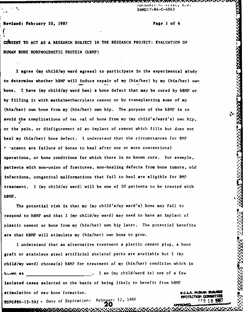
DAMD17-84-C-4045
Revised: February 10. 1987 Page I of 6
COASENT TO ACT AS A RESEARCH SUBJECT IN THE RESEARCH PROJECT: EVALUATION OF
HUMAN BONE MORPHOGENETIC PROTEIN (hBMP)
I agree (my child/my ward agrees) to participate in the experimental study
to determine whether hBMP will induce repair of my (his/her) by my (his/her) own
bone. I have (my child/my ward has) a bone defect that may be cured by hBMP or
by filling it with methalmethacrylate cement or by transplanting some of my
(his/her) own bone from my (his/her) own hip. The purpose of the hBMP is to
avoid the complications of reL val of bone from my (my child's/ward's) own hip,
or the pain, or disfigurement of an implant of cement which fills but does not
heal my (his/her) bone defect. I understand that the circumstances for BMP
-atment are failure of bones to heal after one or more conventional
operations, or bone conditions for which there is no known cure. For example,
patients with non-union of fractures, non-healing defects from bone tumors, old
infections, congenital malformations that fail to heal are eligible for BMP
treatment. I (my child/my ward) will be one of 50 patients to be treated with
hBMP.
The potential risk is that my (my child's/my ward's) bone may fail to I
respond to hBMP and that I (my child/my ward) may need to have an implant of
plastic cement or bone from my (his/her) own hip later. The potential benefits 'Uare that hBMP will stimulate my (his/her) own bone to grow.
I understand that as alternative treatment a plastic cement plug, a bone
graft or stainless steel artificial skeletal parts are available but I (my
child/my ward) choose(s) hBMP for treatment of my (his/her) condition which is
k,.,ovn as . I am (my child/ward is) one of a few
Isolated cases selected on the basis of being likely to benefit from hBMP
stimulation of new bone formation. A L. MUM SJA
C 6 2 Date of Exp iration February 12 , 1988 rr& Go w s-2SPC#86-120592 ,n . 5 I
Marshall R. Urst, I D.DAMD17-84-C-40
45 Page 2 of 6
I have (my child/my ward has) the choice of having a plastic cement plug,
or bone from my (his/her) own hip or leg, or bone donated by another individual,
or the Implant of hBMP. The hBMP Is a purified protein that is the active
ingredient of a bone graft. If.the hBMP is used it will be placed in the bone
defect in my (his/her) . The implantation of hBMP will
be made at the same operation that treats my (his/her)
and does not require a separate operation
unless. at the time of surgery, my (his/her) condition is found to require
additional bone from another individual, or some bone removed from my (his/her)
hip.
, The part of the study that is experimental is the substitution of hBMP, a
chemically purified crystalline protein for plastic cement bone graft, or
stainless steel parts. The present experimental procedure consists of
implantation of hBMP in an otherwise non-healing bone defect. The hBMP is
obtained from human bone and is purified by a series of chemical fractionation
processes which are known to remove all the non-BMP substances from the BMP. '"
The transplantation antigens which are known to cause rejection of a bone graft
are removed in the process of manufacturing hBMP.
HAZARDS. I (my child/my ward) understand(s) that the hazards of \.•%!
experimental hBMP concerns the possibility of toxicity and antigenicity. 'U
Toxicity means the quality of being poisonous. Toxic chemicals cause skin rash,
nausea, allergic reactions, inflammation, tumors and other adverse reactions.
None of these have been detected in animals of many different species treated
with BMP. No toxicity has been found In experiments of 100 chickens, 200 mice,
2000 rats, 50 guinea pigs, 1000 rabbits, 6 dogs and 2 monkeys treated with BMP
in the period from 1975 to 1982. These toxicity studies in animals have shown
no observable organ damage and no allergic response. An allergic response means
the rejection of the implanted BMP by local inflammation or a systemic reaction
such as a skin rash, flushing of the skin, nausea, vomiting or fainting. None
HSPC #86-12-592 21U .~ 4 ~ e5.
Marshall Fage s t '
DAMD17-84 - 45
of these reactions have been noted in animals thus far treated with BMP, nor in
.e more than 10 human patients treated to date. Another hazard is the
possibility of a wound infection from either the implant or the surgical
operation on my (my child's/ward's) bone. Based on experiments on the animals
listed above and more than 10 human patients successfully treated with hBMP,
there Is an 85Z chance that hBMP will stimulate bone formation when implanted in
a bone defect.
In 6 patients with 4 to 6 failed operations for non-healing leg bone
fractures, all six have healed following implantation of hBMP. In one patient
with a failure of treatment of a 2 1/2 year-old fracture of the femur, treated
by electrical stimulation for 10 months, healing occurred within 6 months after
implantation of hBMP. In another patient with a bone defect in the finger
caused by excision of a bone tumor, the bone healed within three months after
3lntation of hBMP. In two patients with bone decay of the head of the femur
(hip), decompression with implantation of hBUP has saved the patients from
replacement of the joint with an artificial hip. No evidence of toxicity has
been seen in these 10 patients in doses of 2.0 mg/kilo, possibly because hBMP is
a normal constituent of the bone and blood. The effective dose is 0.1 mg/kilo: 1.03
in a phalangeal bone defect 1.0 cm in volume. These doses are 100 to 1000
times less than the safe doses in laboratory animals. The long term effect of
hBMP in humans is not known.
Alternative procedures. Instead of using an implant of hBMP my (my
child's/ward's) bone defect can be filled with plastic cement, or my (his/her)
hip or leg may have a second operation for transfer of a bone graft into the
bone defect in the . The plastic cement has
been judged to be safe but it is not well tolerated because it does not bind to
Ltae bone but floats in the space between the bone and the soft parts and is a
cause of pain and irritation. The bone that is transplanted from the patient's
own hip is the best tolerated but requires a separate incision and a second
RSPC 086-12-592 ' 22 %
C 17Marsha ,%_rJ VDAMD 17-8-C- 4 5 -- M.D.
operation, which in turn can cause a painful scar, lump. infection, and/or blood
clots. This is the reason it is desirable to test hBMP as a substitute for hip
bone or leg bone transplants. I understand that I have (my child/my ward has)
the option to have the transplant or plastic cement if the hBMP does not
successfully induce repair of my (his/her) bone defect.
I have (my child/ward has) read the description of the experimental
procedures to be used in this study as they are explained above, and I (my
child/ward) give(s) my (his/her) consent to Marshall R. Urist, M.D. and his
associate orthopedic and other surgical specialists to perform the operation
named
I understand that as a consequence of my (my child/ward) signing, hBMP will be
used.
Although I (my child/ward) now consent(s) to participate in this study by
clinical follow-up visits, routine laboratory analyses, complete blood count,
tests of kidney function and liver function, disability evaluations, and x-ray
examinations for a period of 5 years, I (my child/ward) may elect to discontinue
participation in this study at any time without prejudice to continued care or itreatment. I (my child/ward) consent(s) to the blood tests that require 2
tablespoons of blood, and 24 hour samples of urine (not more than 3 times) for
the above laboratory specimens when I am (he/she is) in the hospital. I (my
child/ward) understand(s) that the potential hazards of venipuncture for blood a
collection in the above noted amounts are almost nil but could include pain,
bleeding, light headedness, fainting, and rarely infection or blood clots.
I (my child/ward) agree(s) to have follow-up x-ray examinations at 2 month
intervals for 6 months, and at one year intervals thereafter for 5 years. These
x-rays are requested only at intervals that are not known to cause harmful side
effects of radiation. Two to 5 years is the time necessary to determine all the
HSPC #86-12-592&
23
, , r . a., a , , , ,' , , V , V- V=- ,: 'V -
Marshall R. tat .6DAMD17-84-C-4045
benefits of hbMP. I agree that I or my (my child's) insurance company will be
( responsible for all costs of the follow-up examinations. I understand that hBMP will be
supplied at no cost to a third party carrier or to me (my child/my ward).
Information about hBMP will be available to me (my child/ward) at any time.
Any questions that may occur to me (him/her) will be answered at any time by Dr.
Marshall R. Urist (Principal Investigator), 1000 Veteran Avenue, Room A3-34, Los
Angeles, California 90024, (213) 825-6521, and/or his associates. Although the
results of this study may be published, my (my child's/ward's) identity will not
be disclosed unless (my child/my ward) I give(s) separate consent or unless
required by law. I understand that If the investigational design or use of the
information is to be changed, I will be so informed and my (his/her) consent
reqbtained. I do (my child/ward does) not expect a specific benefit from the use
of hBMP other than to possibly help heal my (my child's/ward's) bone defect. It
is my (his/her) understanding that the results of the investigation are personal
and will be treated confidentially.
I understand that I (my child/ward has) have the right to refuse to
participate in, or to withdraw from, this research at any time without prejudice
to my (my child's/ward's) future medical care at UCLA.
I understand that circumstances may arise which might cause the
investigator to terminate my (his/her) participation before completion of the
study.
In signing this consent form, I acknowledge receipt of a copy of the form,
as well as a copy of the "Subject's Bill of Rights."
I understand that if I am injured as a direct results of research
procedures not done primarily for my own benefit, I will receive medical
treatment at no cost. The University of California does not provide any other
form of compensation for injury.
IN
HSPC 086-12-592
24
* .,. . • Marsh1lRk Vri&t, M.D.DA4 I #f t
I understand that if I have further questions, comments, or concerns about
the study or the informed consent process, I may write or call the office of the I
Vice Chancellor-Research Programs, 3134 Murphy Hall, UCLA, Los Angeles, CA,
90024, (213) 825-8714.
Witnessed Signed
Date# Dated
Signature of Father or Guardian, Signature of Mother or Guardian, if if
patient is 18 years old or patient is 18 years old or under.
under.
Signature of minor, if between Dated
12 and 18 years old.
pRQTtCTIO' COMMIlENSPC-l86-12-592 "FB1 l S
Expiration Date: February 12, 1988
25I

- -~- -,or. .a
/7Mf