Embed Size (px)
Citation preview
Systems/Circuits
Distinctive Modulation of Dopamine Release in the NucleusAccumbens Shell Mediated by Dopamine and AcetylcholineReceptors
Jung Hoon Shin,* Martin F. Adrover,* and X Veronica A. AlvarezLaboratory on Neurobiology of Compulsive Behaviors, National Institute on Alcohol Abuse and Alcoholism, National Institutes of Health, Bethesda,Maryland 20892
Nucleus accumbens (NAc) shell shows unique dopamine (DA) signals in vivo and plays a unique role in DA-dependent behaviors such asreward-motivated learning and the response to drugs of abuse. A disynaptic mechanism for DA release was reported and shown to requiresynchronized firing of cholinergic interneurons (CINs) and activation of nicotinic acetylcholine (ACh) receptors (nAChRs) in DA neuron(DAN) axons. The properties of this disynaptic mechanism of DA transmission are not well understood in the NAc shell. In this study, invitro fast-scan cyclic voltammetry was used to examine the modulation of DA transmission evoked by CINs firing in the shell of mice andcompared with other striatal regions. We found that DA signals in the shell displayed significant degree of summation in response to trainstimulation of CINs, contrary to core and dorsal striatum. The summation was amplified by a D2-like receptor antagonist and experi-ments with mice with targeted deletion of D2 receptors to DANs or CINs revealed that D2 receptors in CINs mediate a fast inhibitionobserved within 100 ms of the first pulse, whereas D2 autoreceptors in DAN terminals are engaged in a slower inhibition that peaks at�500 ms. ACh also contributes to the use-dependent inhibition of DA release through muscarinic receptors only in the shell, where higheractivity of acetylcholinesterase minimizes nAChR desensitization and promotes summation. These findings show that DA signals aremodulated differentially by endogenous DA and ACh in the shell, which may underlie the unique features of shell DA signals in vivo.
Key words: D2 receptors; G-protein coupled receptors; muscarinic receptors; nicotinic receptors; optogenetics; voltammetry
IntroductionThe shell is the most ventral part of nucleus accumbens (NAc)and has unique anatomical features that define a separate cir-
cuitry within the basal ganglia (Humphries and Prescott, 2010).The shell receives inputs from limbic structures (Lu et al., 1998;Zhou et al., 2003; Britt et al., 2012) and also projects widely out-side of the basal ganglia, including to lateral hypothalamus,pedunculopontine nucleus, and thalamus, but not to substantianigra or subthalamic nucleus (Mogenson et al., 1985; Yang andMogenson, 1987; Groenewegen et al., 1993; Lavín and Grace,1994; Pennartz et al., 1994). The NAc shell encodes specific as-pects of reward processing such as context and cue information(Bossert et al., 2007; West and Carelli, 2016) and it is critical inmediating reinforcing and rewarding properties of stimulantdrugs (McBride et al., 1999; Kelley, 2004; Wise, 2004; Everitt andRobbins, 2005; Crombag et al., 2008).
Collective evidence from in vivo studies suggest that dopa-mine (DA) transmission in the NAc shell is regulated differen-
Received March 2, 2017; revised Sept. 29, 2017; accepted Oct. 6, 2017.Author contributions: J.H.S., M.F.A., and V.A.A. designed research; J.H.S. and M.F.A. performed research; J.H.S.
and M.F.A. analyzed data; J.H.S., M.F.A., and V.A.A. wrote the paper.This work was funded by the Intramural Programs of National Institute on Alcohol Abuse and Alcoholism and
National Institute of Neurological Disorders and Stroke (Grant ZIA-AA000421). We thank Roland Bock (NationalInstitute on Alcohol Abuse and Alcoholism–National Institutes of Health) for developing the analysis software andDavid Lovinger and the members of the Alvarez laboratory for valuable comments on this manuscript.
The authors declare no competing financial interests.*J.H.S. and M.F.A. contributed equally to this work.Correspondence should be addressed to Dr. Veronica A. Alvarez, National Institute on Alcohol Abuse and Alco-
holism, National Institutes of Health, 5625 Fishers Lane, Bethesda, MD 20892. E-mail: [email protected]:10.1523/JNEUROSCI.0596-17.2017
Copyright © 2017 the authors 0270-6474/17/3711166-15$15.00/0
Significance Statement
The present study reports that dopamine (DA) release evoked by activation of cholinergic interneurons displays a high degree ofsummation in the shell and shows unique modulation by endogenous DA and acetylcholine. Desensitization of nicotinic receptors,which is a prevailing mechanism for use-dependent inhibition in the nucleus accumbens core and dorsal striatum, is also minimalin the shell in part due to elevated acetylcholinesterase activity. This distinctive modulation of DA transmission in the shell mayhave functional implications in the acquisition of reward-motivated behaviors and reward seeking.
11166 • The Journal of Neuroscience, November 15, 2017 • 37(46):11166 –11180
tially by D2/3 receptors to give rise to larger increase of DA levelsupon cocaine exposure than in other regions (Jones et al., 1996;Di Chiara and Bassareo, 2007; Aragona et al., 2008; Aragona et al.,2009; Dreyer et al., 2016). Furthermore, antipsychotic drugs,mostly D2/3 receptor antagonists, produce larger increases of DAconcentration in the NAc shell compared with core and prefron-tal cortex (Tanda et al., 2015). The properties of DA release invitro have been studied in the shell using electrical stimulationand modulation by acetylcholine (ACh) through the nicotinicACh receptors (nAChRs) and muscarinic ACh receptors (mAChRs)has been proposed (Threlfell et al., 2010; Patel et al., 2012; Shin etal., 2015). However, DA release can be evoked in the striatum bydifferent mechanisms: a monosynaptic mechanism that involvesdirect firing of action potentials in DA neuron (DAN) axons andby a disynaptic mechanism that requires synchronized firing ofcholinergic interneurons (CINs) and activation of nAChRs onDAN axons (Cachope et al., 2012; Threlfell et al., 2012; Wang etal., 2014; Shin et al., 2015). These two mechanisms differ in thedependence of the DA response to the frequency of stimulation.For example, in the dorsal striatum, DA signals show summa-tion in the amplitude of the DA signal in response to trains ofstimulation when evoked by the monosynaptic but not by thedisynaptic mechanism (Threlfell et al., 2012). This lack ofsummation is likely due to the inhibition of subsequent DArelease during the train stimulation.
Desensitization of nAChRs has been suggested as a mecha-nism for the lack of summation of DA release evoked by trainstimulation of CINs (Threlfell et al., 2012). nAChRs expressed inDAN axons contain �2-subunits and are highly prone to desen-sitization, which depends on the extracellular concentration ofACh (Giniatullin et al., 2005). The degree of nAChR desensitiza-tion is then in part regulated by the balance between release andclearance mechanisms for ACh. Acetylcholinestease, an extracel-lular enzyme for ACh degradation, has been shown to have ahigher activity the NAc shell than other striatal subregions inseveral animal species (Zaborszky et al., 1985; Voorn et al., 1994;Franklin and Paxinos, 2007). In this study, we explored whetheracetylcholinestease activity regulates the degree of desensitizationand if desensitization of nAChRs is less dominant in the NAcshell, rendering a different frequency summation in this region.Gi/o-protein coupled D2Rs and mAChRs in CINs (Yan andSurmeier, 1996; Yan et al., 1997; Alcantara et al., 2003) andDAN axons (Sesack et al., 1994) modulate the release of AChand DA (Threlfell et al., 2010; Cachope et al., 2012; Adrover etal., 2014; Shin et al., 2015; Sulzer et al., 2016). We also ex-plored whether these receptors are activated by endogenouslyreleased DA and ACh from previous activity and play a role inthe inhibition of subsequent DA and ACh release in a use-dependent manner.
Materials and MethodsAnimals. Heterozygote B6.SJL-Slc6a3 tm1.1(cre)Bkmn/J mice (006660; TheJackson Laboratory), referred to as DAT-cre mice; heterozygote B6N.129S6(B6)-Chat tm2(cre)Lowl/J mice (018957; The Jackson Laboratory),referred as ChAT-cre mice; and the crosses of DAT-cre or ChAT-cremice with B6.129S4(FVB)-Drd2 tm1.1Mrub/J mice (020631; The JacksonLaboratory), referred as autoDrd2KO mice (Bello et al., 2011) andCINDrd2KO mice (Kharkwal et al., 2016), respectively, were used forexperiments. All mice were male in C57BL/6 background and housed ona 12 h light/dark cycle (06:30 –18:30 light) with food and water ad libitum.All experiments were performed in accordance with guidelines from theNational Institute on Alcohol Abuse and Alcoholism (NIAAA) AnimalCare and Use Committee.
Stereotaxic injection of AAV-ChR2 vectors. Injections were performedas described previously Adrover et al. (2014). Briefly, mice (5– 6 weeksold) anesthetized by a mixture of isoflurane-oxygen were placed in astereotaxic frame (David Kopf). Adeno-associated virus (AAV) vectorscarrying the sequence for Cre-dependent expression of Channelrhodop-sin2 (ChR2) protein, AAV5-EF1a-DIO-hChR2(H134R)-EYFP (4 � 10 12
IU/ml), were bilaterally injected into VTA/SNc (AP: �3.30, ML: � 0.50,DV: �4.50) of DAT-cre or AutoDrd2KO mice or in the NAc (AP: �1.35,ML: � 1.05, DV: �5.00) or dorsal striatum (AP: �1.05, ML: � 1.20, DV:�3.20) of ChAT-cre or CINDrd2KO mice. All stereotaxic coordinatesare in millimeters from bregma according to the atlas of Franklin andPaxinos (2007). The injection volumes were 300 nl at a flow rate of 100nl/min. Recordings were made after a minimum of 3 weeks of incuba-tion. Viral vectors were purchase from the Gene Therapy Center VectorCore at University of North Carolina.
Slice preparation. Brains were sliced in a vibratome (VT-1200S; Leica)in an ice-cold cutting solution containing the following (in mM): 225sucrose, 13.9 NaCl, 26.2 NaHCO3, 1 NaH2PO4, 1.25 glucose, 2.5 KCl, 0.1CaCl2, 4.9 MgCl2, and 3 kynurenic acid. Sagittal slices (240 �m) wererecovered for 20 min at 33°C in artificial CSF (ACSF) containing thefollowing (in mM): 124 NaCl, 1 NaH2PO4, 2.5 KCl, 1.3 MgCl2, 2.5 CaCl2,20 glucose, 26.2 NaHCO3, and 0.4 ascorbic acid and were maintained inACSF in the dark at room temperature prior recordings. Slices wererecorded with continuous superperfusion (2 ml/min) of ACSF at 32°Cusing an inline heater (Harvard Apparatus).
Fast scan cyclic voltammetry (FSCV). Carbon fiber microelectrodeswere prepared using a carbon fiber (7 �m diameter) inserted into a glasspipette (�150 �m of exposed fiber). The carbon-fiber microelectrodewas held at �0.4 V versus Ag/AgCl and a triangular voltage ramp from�0.4 V to �1.2 V and back to �0.4 V (400 V/s) was delivered every100 ms. DA transients were evoked by illuminating a brief (0.6 ms) lightpulse using an optical fiber (200 �m/0.22 NA) connected to a 470 nmLED (2 mW; ThorLabs). The light pulses were delivered every 2 min.Data were collected with a retrofitted head stage (CB-7B/EC with 5 M�resistor) using a Multiclamp 700B amplifier (Molecular Devices), withdigitization at 100 kHz using pClamp10 software (Molecular Devices)or using custom-written software in Igor Pro (Wavemetrics) runningmafPC (courtesy of M.A. Xu-Friedman). All data collected were analyzedwith the custom-written software. The current peak amplitudes of theevoked DA transients were converted to DA concentrations according tothe postexperimental calibration using 1–3 �M DA. FSCV was performedin the NAc shell, NAc core, and dorsal striatum regions.
Cell-attached recording. Cell-attached recording was performed usingglass pipettes (�4 M�) filled with Cs-based internal saline (Adrover etal., 2014). CINs from ChAT-cre mice infected with AAV5-EF1a-DIO-hChR2(H134R)-EYFP vector were identified with fluorescence and con-firmed by their characteristic spontaneous firing pattern. The recordingswere performed at holding potential of 0 mV with digitization at 5 kHzusing pClamp10 software. Action potentials were evoked using the samelight stimulation used to evoke DA transients (see above).
Drugs. Sulpiride, dihydro-�-erythroidine hydrobromide (DH�E),scopolamine, and ambenonium were purchased from Tocris Bioscience.Kynurenic acid (sodium salt) was obtained from Ascent Scientific. Allother chemicals were from Sigma-Aldrich.
Experimental design and statistical analysis. For each dataset, record-ings were made from at least five slices or cells derived from a total of atleast three animals. Statistical analysis was performed with Prism soft-ware (GraphPad) using linear regression, unpaired two-tailed Student’s ttest, and one- and two-way ANOVA. Tukey’s or Bonferroni’s test formultiple comparisons were used for post hoc analysis.
ResultsFrequency-dependent summation of DA signals in theshell regionTo study the use-dependent modulation of DA release evoked byactivation of CINs (Cachope et al., 2012; Threlfell et al., 2012),ChR2-EYFP was expressed in CINs in the NAc (Fig. 1A) and thedorsal striatum regions of ChAT-cre mice by bilateral local ad-
Shin, Adrover et al. • Distinctive Dopamine Release Modulation in Shell J. Neurosci., November 15, 2017 • 37(46):11166 –11180 • 11167

ministration of AAV5 vector with Cre-dependent ChR2-EYFP DAtransients were evoked by brief (0.6 ms) 470 nm light pulsesaround the area showing the expression of EYFP and measuredusing FSCV. DA transients evoked by optogenetic stimulation ofCINs (CIN-oDA) showed the characteristic oxidation and reduc-tion profile of DA (Fig. 1B). In the core region, the amplitude ofCIN-oDA in response to train stimulation were practically indis-
tinguishable from the DA transient in response to single stimu-lation (Fig. 1C) as previously reported in the dorsal striatumregion (Threlfell et al., 2012). Both the peak amplitude (Fig. 1D)and the area underneath the curve (Fig. 1E) of CIN-oDA in thecore and dorsal striatum revealed the insensitivity to stimula-tion frequency between 2 and 10 Hz (F � 0.83, p � 0.45 for thepeak and area, n � 6 slices/5 mice), except for a slight decrease
A
E
D
B
C
ires/cre+DIO:ChR2-EYFP in ChAT+/IRES-cre (Chat-cre)
acaac
VPShell
Core
2 Hz 5 Hz 10 Hz1pShell
3.0
2.0
1.0
kaeP
ADo-
NIC
) FS
CA ni p 1 ot .
mro n(
1p 2 Hz 5 Hz 10 Hz
Dorsal striatum 3.0
2.0
1.0
1p 2 Hz 5 Hz 10 Hz
Core
ACSF + sulp
3.0
2.0
1.0
1p 2 Hz 5 Hz 10 Hz
Shell
*
***
5.0
3.0
1.0
aerA
ADo-
NIC
)FS
CA ni p 1 ot .
mro n(
1p 2 Hz 5 Hz 10 HzFrequency
*****
5.0
3.0
1.0
1p 2 Hz 5 Hz 10 HzFrequency
***
ACSF + sulp
5.0
3.0
1.0
1p 2 Hz 5 Hz 10 HzFrequency
****
+ sulp
2 Hz 5 Hz 10 Hz1pCore
ACSF
15
10
5
0
-5
)An( tnerru
C
1.20.80.4-0.4Voltage (V)
Figure 1. DA release evoked by optogenetic stimulation of CIN shows a frequency-dependent summation only in the NAc shell region of the striatum in the presence of D2 receptor antagonist.A, Left, Fluorescence image of sagittal brain section of ChAT �/IRES-cre (ChAT-cre) mouse showing labeled CINs expressing ChR2-EYFP overlaid onto corresponding sections from the mouse brain atlas(Franklin and Paxinos, 2007). Scale bar, 400 �m. Right, Fluorescence image of labeled cell bodies and processes of CINs in the NAc shell area. Scale bar, 50 �m. B, Representative current–voltageplot of CIN-oDA. C, Representative CIN-oDA traces with single stimulation (1p), or trains of 4 at 2, 5, or 10 Hz in ACSF (top) and in the presence of the D2 receptor antagonist sulpiride (1 �M, bottom)either in (left) core or in (right) shell area of NAc. Dotted lines represent the baseline and the peak amplitude of CIN-oDA with single stimulation. Blue ticks represent the optogenetic stimulations.Scale bars, 200 nM and 2 s. D, E, Peak amplitudes (D) or areas (E) normalized by CIN-oDA transients by single pulse (1p) in ACSF plotted as a function of stimulation patterns in dorsal striatum (left),core (middle), and shell (right). *p 0.05, **p 0.01, ***p 0.001.
11168 • J. Neurosci., November 15, 2017 • 37(46):11166 –11180 Shin, Adrover et al. • Distinctive Dopamine Release Modulation in Shell
in the peak at 5 Hz in dorsal striatum (0.96 � 0.01 of 1p,p � 0.001).
However, in the shell region, the peak amplitude of CIN-oDAevoked by train stimulation at 10 Hz was significantly larger com-pared with single pulse (33 � 10% increase, n � 9 slices/5 mice,p � 0.036 from Tukey’s multiple-comparisons test; Fig. 1C,D).The frequency-dependent summation became more evidentwhen the areas under the curve of CIN-oDA were compared(52 � 14% increase, p � 0.02 with 2 Hz; 36 � 12%, p � 0.067with 5 Hz; 88 � 22%, p � 0.016 with 10 Hz; n � 9 slices/5 mice;Fig. 1E). These results suggest that DA transients in the shell showsummation compared with other striatal subregions when stim-ulated with train of pulses at different frequency.
To determine whether the lack of summation of CIN-oDA inthe core and dorsal striatum is due to the activation of D2/3receptors by initial DA release, we performed similar train stim-ulation experiments in the presence of the DA D2-like receptorantagonist sulpiride (1 �M) and frequency-dependent summa-tion of CIN-oDA was observed in all three regions (Fig. 1C,E).Sulpiride had no effect on the peak or area of CIN-oDA evoked bysingle pulses (p 0.999 for peaks and areas from all three re-gions; Fig. 1D,E), in agreement with previous studies using elec-trical stimulation (Trout and Kruk, 1992; Wieczorek and Kruk,1995). Sulpiride increased the peak amplitude of CIN-oDAevoked by train only in the shell (F1.02, 7.13 � 9.47, p � 0.017; Fig.1C,D), with no increase in the core and dorsal striatum (F1.72, 8.59 �2.89, p � 0.114 for the core region and F1.98, 9.88 � 2.04, p � 0.182for the dorsal striatum region; Fig. 1C,D).
For the area of the CIN-oDA transients, sulpiride had effectsat most frequencies in all striatal regions (Fig. 1E). In the core anddorsal striatum, the area of CIN-oDA with train increased afterblocking the D2/3 receptors (core: 105 � 2% in ACSF vs 168 �22% in sulpiride, p � 0.003 for 2 Hz and 100 � 3% in ACSF vs153 � 21% in sulpiride, p � 0.02 for 5 Hz; dorsal striatum: 116 �9% in ACSF vs 205 � 15% in sulpiride, p 0.001 for 2 Hz and100 � 6% in ACSF vs 147 � 17% in sulpiride, p � 0.004 for 5 Hz),but decreased with 10 Hz frequency. In the shell, the increase wasmore pronounced where the responses to trains were larger afterblocking D2/3 receptors (136 � 12% in ACSF vs 359 � 69%% insulpiride, p � 0.002 for 5 Hz and 188 � 22% in ACSF vs 291 �84% in sulpiride, p � 0.006 for 10 Hz, Bonferroni’s multiple-comparisons test). These results suggest that, during the trainstimulation, D2/3 receptors are activated by the endogenouslyreleased DA and suppress subsequent release of DA, exertinguse-dependent inhibition. Furthermore, the data indicate thatthis D2/3-receptor-mediated inhibition is more evident in theshell region.
D2/3 receptors in CIN and DAN terminals mediate differentcomponents of use-dependent inhibition of DA releaseTo further investigate how DA release is modulated by D2/3receptors during train stimulation in the shell, we conductedexperiments measuring CIN-oDA evoked by paired-pulse stim-ulations using different intervals (Fig. 2). For shorter intervals,the responses to the second pulse (P2) were assessed offline bysubtracting the responses to single pulse from paired-pulse re-sponses (Fig. 2A, dotted line). The paired-pulse ratios (PPRs;P2/P1) were plotted as a function of interstimulus interval (ISI)(Fig. 2B, black) and showed a marked depression in response tothe P2 stimulus at all ISIs. The long-lasting component of P2depression was reported previously in the nucleus accumbensand dorsal striatum (Cachope et al., 2012; Wang et al., 2014). Thisphenomenon may be caused by the intrinsic release properties of
DAN terminals because DA release evoked by direct optogeneticstimulation of DAN terminals also shows a slow recovery of theP2 depression that takes up to hundreds of seconds (Adrover etal., 2014), which might reflect vesicle depletion (Wang et al.,2014).
The P2 depression of CIN-oDA was even more pronouncedwith short ISIs (100 –500 ms) than longer ISIs. We thus testedwhether this was due to a lack of fidelity of ChR2-evoked firing ofCINs during the second pulse because ChR2 undergoes inactiva-tion (Lin, 2011) and fails to evoke action potentials reliably atfrequencies 25 Hz (Lin et al., 2009). When optogenetic evokedaction potentials were recorded in CINs from the NAc shell incell-attached mode (Fig. 2C), the fidelity was near 100% for ISIsof 100 ms or longer (100 � 0% for 100 ms; 96 � 4% for 200 ms;96 � 4% for 500 ms; p 0.05 for all the ISIs; n � 9 cells/4 mice;Fig. 2C). With 50 ms, the second pulse evoked an action potentialon 88 � 8% and 11 � 11% for ISI of 10 ms. Therefore, this resultrules out that the marked depression in P2 is due to the lack offidelity of action potential generation during the second pulse for100 ms or longer ISIs.
The D2/3 receptor antagonist sulpiride (1 �M) partially re-lieved the depression of responses to P2, especially with ISIsshorter than 1 s (PPRsulp � PPRACSF: 0.19 � 0.06, p � 0.01 for 200ms; 0.27 � 0.06, p 0.001 for 500 ms; Fig. 2B). The time courseof D2/3-receptor-mediated depression of P2 was assessed by sub-tracting the PPR curve in ACSF from the curve in sulpiride (Fig.2E), which shows that D2/3 receptor activation by endogenouslyreleased DA takes place as early as 100 ms and reaches a maxi-mum by �500 ms. This onset of D2/3-receptor-mediated inhi-bition of CIN-oDA is within the range previously reported withelectrically evoked DA transients (Phillips et al., 2002).
D2/3 receptor-mediated inhibition of CIN-oDA could be me-diated by one or multiple sources of D2 receptors and by D3receptors known to be more abundant in the shell (Maina andMathews, 2010). D2 receptors in CINs that inhibit ACh releasecan subsequently diminish DA release evoked by CINs and D2/3autoreceptors can also inhibit DA release directly at DAN termi-nals. In this study, we focus on the contribution of the two poolsof D2 receptors to the use-dependent inhibition and generatedcell-specific Drd2 knock-out mice with targeted deletion of D2receptors from CINs (ChAT-cre � Drd2 loxP/loxP, referred asCINDrd2KO) to isolate its contribution (Fig. 2D). Mice lackingD2 receptors in CINs showed less P2 depression of CIN-oDA atshorter ISIs compared with control ChAT-cre animals in ACSF(PPRCINDrd2KO � PPRChAT-cre: 0.58 � 0.06 for 100 ms; 0.30 �0.06 for 200 ms; 2-way RM ANOVA genotype � PPI interactionF(6,126) � 13.03, p 0.001; post hoc t test, p 0.001), but notdifferent at ISIs � 500 ms (Fig. 2B,D). In these knock-out ani-mals, addition of sulpiride had no effect on P2 depression at the100 ms interval, but D2/3 receptor modulation at �500 ms waslarger than in ChAT-cre controls (Fig. 2D,E). The onset of D2/3-receptor-mediated inhibition was delayed, suggesting that D2receptors in CINs are responsible for the early component of theuse-dependent inhibition at �100 ms. This finding also impliesthat the remaining component of the use-dependent inhibition isnot due to D2 receptors in CINs, but rather is likely due to D2/3autoreceptors in DAN terminals.
To isolate the contribution of D2 autoreceptors, we measuredDA transients evoked by optogenetic stimulation of DAN axons(DAN-oDA) using paired-pulse stimulation in the shell region ofDAT-cre mice. The transients evoked by DAN axons stimulationhad similar amplitudes (253 � 16 nM for DAN-oDA, n � 31
Shin, Adrover et al. • Distinctive Dopamine Release Modulation in Shell J. Neurosci., November 15, 2017 • 37(46):11166 –11180 • 11169

slices/9 mice; 261 � 15 nM for CIN-oDA, n � 45/17 mice; un-paired t test p � 0.71), but showed slower decay (0.35 � 0.02 s forDAN-oDA; 0.25 � 0.01 for CIN-oDA; p 0.001; Fig. 3A), pos-sibly reflecting differences in reuptake, as described previously(O’Neill et al., 2017). The PPR curve of DAN-oDA showed the
characteristic U-shape, as described previously (Adrover et al.,2014; Fig. 3B). The increase in PPRs at shorter ISIs suggests that asupralinear relationship exists between Ca 2� concentration andthe release probability in DAN terminals in addition to the afore-mentioned vesicle depletion hypothesis.
A
B
ED
C
+ sulp
0.5 s 2 s0.2 s s 1s 1.0
ACSF
Shell
1.0
0.8
0.6
0.4
0.2
0.0
ADo-
NIC fo
RP
P
40.1
2 41
2 410
2
ChAT-cre
*****
ACSF + sulp
60
40
20
0
2drD yb nois se rpe
D %
40.1
2 41
2 410
2
ISI (sec)
ChAT-cre CINDrd2KO
100
80
60
40
20
0.
mits dn2 rof ytilediF %
0.01 0.1 1ISI (sec)
100 ms
1.0
0.8
0.6
0.4
0.2
0.0
ADo-
NIC fo
RP
P
40.1
2 41
2 410
2
ISI (sec)
CINDrd2KO
*
****
ACSF + sulp
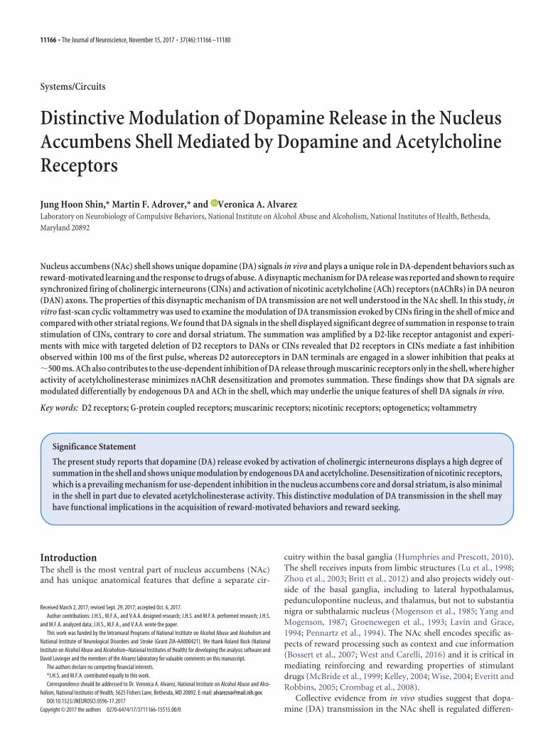
Figure 2. Early phase of the D2-receptor-mediated depression of CIN-oDA in the shell is through D2 receptors in CINs. A, Representative traces of CIN-oDA evoked by paired-pulse stimulationswith different ISIs (0.1, 0.2, 0.5, 1, or 2 s) in ACSF (top) and in the presence of sulpiride (1 �M, bottom) in the shell area of ChAT-cre mice. CIN-oDA transients evoked by the second pulse (dotted) werecalculated by subtracting the single-pulse CIN-oDA transients for ISIs between 0.1 and 2 s. Blue ticks represent the optogenetic stimulations. B, PPRs (P2/P1) of CIN-oDA measured in the shell areaof ChAT-cre mice were plotted as a function of ISI in ACSF (black) or in the presence of sulpiride (red). C, Left, Representative traces of cell-attached patch recordings from a CIN showingaction potentials in response to paired optogenetic stimulations (blue ticks) with different ISIs: 0.01, 0.05, 0.1, 0.2, and 0.5 s. Right, Fidelity for second stimulation was calculated as thepercentage of successfully evoked action potentials by the second stimulation and plotted as a function of ISI. D, PPR of CIN-oDA measured in the shell area of ChAT-cre � Drd2 loxP/loxP
(CIND2rdKO) mice plotted as a function of ISI in ACSF (black) or in the presence of sulpiride (red). E, Degree of depression in P2 due to DA D2/3 receptor activation was assessed bysubtracting the PPR curves in ACSF from the PPR curve in the presence of sulpiride either from ChAT-cre mice (thick) or from CINDrd2KO mice (thin). *p 0.05, **p 0.01, ***p 0.001.
11170 • J. Neurosci., November 15, 2017 • 37(46):11166 –11180 Shin, Adrover et al. • Distinctive Dopamine Release Modulation in Shell

Application of sulpiride significantly attenuated the depres-sion of P2 for ISIs of 200 ms, 500 ms, and 1 s [PPRsulp � PPRACSF:0.10 � 0.03 for 200 ms; 0.17 � 0.03 for 500 ms; 0.14 � 0.03 for 1 s;2-way repeated-measures (RM) ANOVA pharmacology � ISIinteraction F(6,168) � 6.174, p 0.001; post hoc t test p � 0.003 for200 ms and p 0.001 for 500 ms and 1 s; Fig. 3B,D]. Sulpiridehad no effect on ISIs longer than 2 s, indicating that P2 depressionat longer intervals is not mediated by D2/3 receptors.
Because DAN-oDA is independent of CIN activity andnAChR activation, this sulpiride-mediated relief in P2 depressionis most likely due to D2 and D3 autoreceptors in DAN terminals.In fact, when D2 autoreceptors were deleted selectively in DANs(DAT-cre � Drd2 loxP/loxP, referred as autoDrd2KO), most ofsulpiride relief of the DAN-oDA was lost (2-way ANOVA nointeraction F(6,55) � 0.85, p � 0.54; no main effect with pharma-cology F(1,55) � 2.59, p � 0.11; Fig. 3C,D). Furthermore, inautoDrd2KO mice, the PPR curve in ACSF was almost overlap-ping with the curve in sulpiride. A small fraction of remaining
inhibition was observed (Fig. 3C,D) and is likely mediated by D3autoreceptors in DAN terminals. Therefore, in the shell, D2 au-toreceptors are responsible for most of the use-dependent inhi-bition of DA release of DAN-oDA.
This D2 autoreceptor-mediated inhibition of P2 is not pres-ent at 100 ms and only becomes apparent after 200 ms, with amaximum at �500 ms (no effect seen for ISIs 2 s). Therefore,the onset of the D2-autoreceptor-mediated inhibition of DAN-oDA (Fig. 3D) is delayed compared with the onset of the D2-receptor-mediated inhibition of P2 in CIN-oDA from controlChAT-cre mice (Fig. 2E). In addition, the time course of theautoreceptor-mediated inhibition of DAN-oDA resembles thetime course of CIN-oDA from mice lacking the D2 receptor in CINs(CINDrd2KO; Fig. 2E). These findings suggest that the early onset(�100 ms) of the use-dependent modulation by D2 receptors ismediated by D2 receptors in CINs and the late component (�500ms) is mediated by D2 autoreceptors in DAN terminals.
A
DC
B
0.4
0.3
0.2
0.1
0.0
)ces( uat yaceD
CIN-oDA DAN-oDA
***
60
40
20
0
2drD yb noisserpe
D%
60.1
2 4 61
2 4 610
2
ISI (sec)
DAT-cre autoDrd2KO
1.2
1.0
0.8
0.6
0.4
0.2
0.0
ADo-
NA
D fo R
PP
60.1
2 4 61
2 4 610
2
ISI (sec)
DAT-cre
***
*****
ACSF + sulp
1.2
1.0
0.8
0.6
0.4
0.2
0.0
A
Do-N
AD fo
RP
P
60.1
2 4 61
2 4 610
2
ISI (sec)
autoDrd2KO
ACSF + sulp
1 s
DAN-oDA CIN-oDA
Figure 3. D2 autoreceptors on the DAN terminals are responsible for the late phase of D2-receptor-mediated inhibition of DA release. A, Top, Representative traces of DAN-oDA (black) andCIN-oDA (gray). Bottom, Decay tau’s plotted as bar graph (average � SEM). B, C, PPRs of DAN-oDA measured in the shell area of either DAT-cre (B) or DAT-cre � Drd2 loxP/loxP (autoDrd2KO; C) miceplotted as a function of ISI in ACSF (black) or in the presence of sulpiride (1 �M, red) in the shell area. D, Degree of depression in P2 due to DA D2 autoreceptors activation was assessed as describedpreviously and plotted as a function of ISI either from DAT-cre mice (thick line) or from autoDrd2KO (thin line). **p 0.01, ***p 0.001.
Shin, Adrover et al. • Distinctive Dopamine Release Modulation in Shell J. Neurosci., November 15, 2017 • 37(46):11166 –11180 • 11171
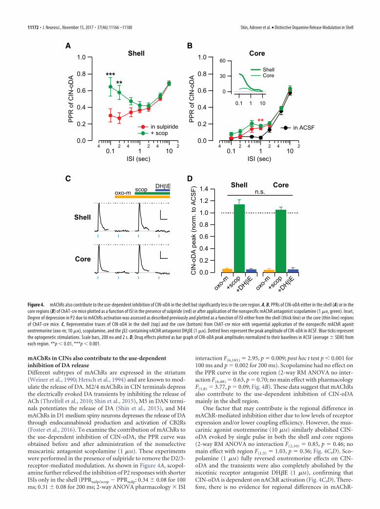
mAChRs in CINs also contribute to the use-dependentinhibition of DA releaseDifferent subtypes of mAChRs are expressed in the striatum(Weiner et al., 1990; Hersch et al., 1994) and are known to mod-ulate the release of DA. M2/4 mAChRs in CIN terminals depressthe electrically evoked DA transients by inhibiting the release ofACh (Threlfell et al., 2010; Shin et al., 2015), M5 in DAN termi-nals potentiates the release of DA (Shin et al., 2015), and M4mAChRs in D1 medium spiny neurons depresses the release of DAthrough endocannabinoid production and activation of CB2Rs(Foster et al., 2016). To examine the contribution of mAChRs tothe use-dependent inhibition of CIN-oDA, the PPR curve wasobtained before and after administration of the nonselectivemuscarinic antagonist scopolamine (1 �M). These experimentswere performed in the presence of sulpiride to remove the D2/3-receptor-mediated modulation. As shown in Figure 4A, scopol-amine further relieved the inhibition of P2 responses with shorterISIs only in the shell (PPRsulp/scop � PPRsulp: 0.34 � 0.08 for 100ms; 0.31 � 0.08 for 200 ms; 2-way ANOVA pharmacology � ISI
interaction F(6,181) � 2.95, p � 0.009; post hoc t test p 0.001 for100 ms and p � 0.002 for 200 ms). Scopolamine had no effect onthe PPR curve in the core region (2-way RM ANOVA no inter-action F(6,48) � 0.63, p � 0.70; no main effect with pharmacologyF(1,8) � 3.77, p � 0.09; Fig. 4B). These data suggest that mAChRsalso contribute to the use-dependent inhibition of CIN-oDAmainly in the shell region.
One factor that may contribute is the regional difference inmAChR-mediated inhibition either due to low levels of receptorexpression and/or lower coupling efficiency. However, the mus-carinic agonist oxotremorine (10 �M) similarly abolished CIN-oDA evoked by single pulse in both the shell and core regions(2-way RM ANOVA no interaction F(2,10) � 0.85, p � 0.46; nomain effect with region F(1,5) � 1.03, p � 0.36; Fig. 4C,D). Sco-polamine (1 �M) fully reversed oxotremorine effects on CIN-oDA and the transients were also completely abolished by thenicotinic receptor antagonist DH�E (1 �M), confirming thatCIN-oDA is dependent on nAChR activation (Fig. 4C,D). There-fore, there is no evidence for regional differences in mAChR-
A
C D
B1.0
0.8
0.6
0.4
0.2
0.0
ADo-
NIC fo
RP
P
40.1
2 41
2 410
2
ISI (sec)
Core
** in ACSF
1.0
0.8
0.6
0.4
0.2
0.0
ADo-
NIC fo
RP
P
40.1
2 41
2 410
2
ISI (sec)
Shell
*****
in sulpiride + scop
60
30
0
0.1 1 10
Shell Core
Core
Shell
scop DHβEoxo-m 1.4
1.2
1.0
0.8
0.6
0.4
0.2
0.0
)FS
CA ot .
mron( kaep A
Do-NI
C
oxo-m
+sco
p
+DHβ
Eox
o-m+s
cop
+DHβ
E
CoreShelln.s.
Figure 4. mAChRs also contribute to the use-dependent inhibition of CIN-oDA in the shell but significantly less in the core region. A, B, PPRs of CIN-oDA either in the shell (A) or in thecore regions (B) of ChAT-cre mice plotted as a function of ISI in the presence of sulpiride (red) or after application of the nonspecific mAChR antagonist scopolamine (1 �M, green). Inset,Degree of depression in P2 due to mAChRs activation was assessed as described previously and plotted as a function of ISI either from the shell (thick line) or the core (thin line) regionsof ChAT-cre mice. C, Representative traces of CIN-oDA in the shell (top) and the core (bottom) from ChAT-cre mice with sequential application of the nonspecific mAChR agonitoxotremorine (oxo-m; 10 �M), scopolamine, and the �2-containing nAChR antagonist DH�E (1 �M). Dotted lines represent the peak amplitude of CIN-oDA in ACSF. Blue ticks representthe optogenetic stimulations. Scale bars, 200 nM and 2 s. D, Drug effects plotted as bar graph of CIN-oDA peak amplitudes normalized to their baselines in ACSF (average � SEM) fromeach region. **p 0.01, ***p 0.001.
11172 • J. Neurosci., November 15, 2017 • 37(46):11166 –11180 Shin, Adrover et al. • Distinctive Dopamine Release Modulation in Shell

mediated inhibition and the lack of relief of the paired-pulsedepression by scopolamine in the core region is unlikely due tolower expression or coupling efficiency of mAChRs.
Another possible explanation is that this regional difference isdue to a difference in DA release properties of DAN terminals. Toaddress this, we compared the PPR curves and the response totrains of stimulation pulses for DAN-oDA in the different striatalregions. The PPR curves for DAN-oDA showed higher valuesthan CIN-oDA at short intervals for all striatal regions (p 0.001for ISIs � 500 ms in shell; p 0.05 ISIs � 2 s in core; p 0.001for ISIs � 200 ms in dorsal striatum; Bonferroni’s multiple-comparisons test; Fig. 5A,D), suggesting that DAN terminals arecapable of more release at short intervals than the release seenduring CINs evoked DA release. Sulpiride further increased thePPR values for DAN-oDA also in the core for 200 –1000 ms ISIs,but not at shorter intervals (Fig. 5E), similar to data from shell(Fig. 3B). There was no relief of DAN-oDA depression bysulpiride in the dorsal striatum at all ISIs tested. Therefore, smallregional differences were seen at the shortest interval where theshell showed the highest values of PPR compared with the coreand the dorsal striatum both in ACSF (PPRShell � PPRCore: 0.23 �0.03 for 100 ms, post hoc) and in sulpiride (PPRShell � PPRCore:0.19 � 0.05 for 100 ms, post hoc t test p 0.001; 0.12 � 0.05 for200 ms, p � 0.08; 2-way RM ANOVA region � PPI interactionF(6,108) � 2.38, p � 0.034; Fig. 5). Because DAN terminals can
undergo depletion after the initial release (Wang et al., 2014),PPR values may depend on the P1 amplitude, which may explainthe differences between regions and between CIN-oDA andDAN-oDA. However, the P1 amplitudes were not significantlydifferent between regions and between stimulations (CIN-oDA:292.4 � 52.0 nM for dorsal striatum, 338.7 � 59.8 nM for core,257.2 � 18.7 nM for shell; DAN-oDA: 337.9 � 52.5 nM for dorsalstriatum, 345.2 � 61.8 nM for core, 248.6 � 14.4 nM for shell;F(5,46) � 1.846, p � 0.1225; one-way ANOVA). Furthermore,there was no correlation between P1 and PPR values from all therecordings with ISI � 0.1 s when the degree of depletion may behighest (CIN-oDA: F(1,25) � 2.028, p � 0.17, r 2 � 0.075; DAN-oDA: F(1,23) � 3.311, p � 0.08, r 2 � 0.126; linear regression; Fig.5F). Differences in PPR between regions and between stimula-tions cannot be explained by depletion.
The response to trains of pulses also showed significant sum-mation in the peak of DAN-oDA in all striatum regions (dorsalstriatum: 30.3 � 6.4%, p � 0.02 for 10 Hz, n � 6 slices/2 mice;core: 31.0 � 8.1%, p � 0.04 for 5 Hz and 95.2 � 6.7%, p 0.001for 10 Hz, n � 6 slices/3 mice; shell: 48.4 � 10.9%, p � 0.009 for5 Hz and 129.9 � 9.6% increase for 10 Hz, p 0.001, n � 9slices/3 mice; all from Tukey’s multiple-comparisons test; Fig.6A,B). This summation in the response to trains of pulse wasmore apparent when scored with the area of DAN-oDA (dorsalstriatum: 16.4 � 3.7%, p � 0.03 for 2 Hz, 35.7 � 7.6%, p � 0.02
CA B
D E F1.0
0.8
0.6
0.4
0.2
0.0
ADo-
NA
D fo R
PP
40.1
2 41
2 410
2
ISI (sec)
1.0
0.8
0.6
0.4
0.2
0.0
A
Do-NI
C fo R
PP
40.1
2 41
2 410
2
ISI (sec)
ACSF
1.0
0.8
0.6
0.4
0.2
0.04
0.12 4
12 4
102
ISI (sec)
1.0
0.8
0.6
0.4
0.2
0.04
0.12 4
12 4
102
ISI (sec)
+ sulpiride1.0
0.8
0.6
0.4
0.2
0.04
0.12 4
12 4
102
ISI (sec)
+ sulpirideand scopolamine
Shell Core dorsal striatum
1.0
0.8
0.6
0.4
0.2
0.0
RP
P)FS
CA ,ces 1.0 = ISI(500300100
P1 amplitude (nM)500300100
DAN-oDACIN-oDA
p=0.16
p=0.08
Figure 5. PPR curves of CIN-oDA and DAN-oDA from three different striatal regions for each pharmacological condition. A–C, PPR curves of CIN-oDA from three different striatal regionsplotted for comparison in ACSF (A), with sulpiride (1 �M; B), and with sulpiride and scopolamine (1 �M; C). D, E, PPR curves of DAN-oDA in ACSF (D) and with sulpiride (E). F, PPR valuesof CIN-oDA (left) and DAN-oDA (right) with 0.1 s interval in ACSF plotted as a function of the P1 amplitudes for individual recordings from three different striatal regions. The blue linesrepresent the linear regression between the PPR values and the P1 amplitudes from all the striatal regions in each group. The p-values represent significant difference from zero slope.
Shin, Adrover et al. • Distinctive Dopamine Release Modulation in Shell J. Neurosci., November 15, 2017 • 37(46):11166 –11180 • 11173
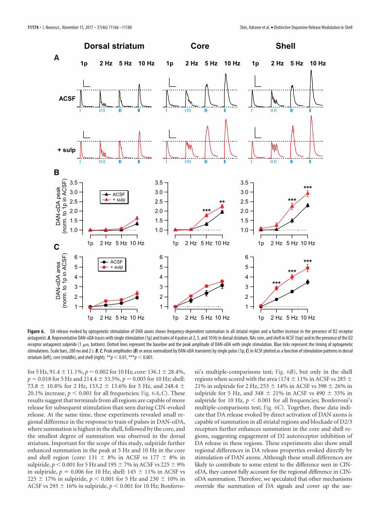
for 5 Hz, 91.4 � 11.1%, p � 0.002 for 10 Hz; core: 136.1 � 28.4%,p � 0.018 for 5 Hz and 214.4 � 33.5%, p � 0.005 for 10 Hz; shell:73.8 � 10.8% for 2 Hz, 153.2 � 13.6% for 5 Hz, and 248.4 �20.1% increase, p 0.001 for all frequencies; Fig. 6A,C). Theseresults suggest that terminals from all regions are capable of morerelease for subsequent stimulation than seen during CIN-evokedrelease. At the same time, these experiments revealed small re-gional difference in the response to train of pulses in DAN-oDA,where summation is highest in the shell, followed by the core, andthe smallest degree of summation was observed in the dorsalstriatum. Important for the scope of this study, sulpiride furtherenhanced summation in the peak at 5 Hz and 10 Hz in the coreand shell region (core: 131 � 8% in ACSF vs 177 � 8% insulpiride, p 0.001 for 5 Hz and 195 � 7% in ACSF vs 225 � 9%in sulpiride, p � 0.006 for 10 Hz; shell: 145 � 11% in ACSF vs225 � 17% in sulpiride, p 0.001 for 5 Hz and 230 � 10% inACSF vs 293 � 16% in sulpiride, p 0.001 for 10 Hz; Bonferro-
ni’s multiple-comparisons test; Fig. 6B), but only in the shellregions when scored with the area (174 � 11% in ACSF vs 285 �21% in sulpiride for 2 Hz, 253 � 14% in ACSF vs 398 � 26% insulpiride for 5 Hz, and 348 � 21% in ACSF vs 490 � 35% insulpiride for 10 Hz, p 0.001 for all frequencies; Bonferroni’smultiple-comparisons test; Fig. 6C). Together, these data indi-cate that DA release evoked by direct activation of DAN axons iscapable of summation in all striatal regions and blockade of D2/3receptors further enhances summation in the core and shell re-gions, suggesting engagement of D2 autoreceptor inhibition ofDA release in these regions. These experiments also show smallregional differences in DA release properties evoked directly bystimulation of DAN axons. Although these small differences arelikely to contribute to some extent to the difference seen in CIN-oDA, they cannot fully account for the regional difference in CIN-oDA summation. Therefore, we speculated that other mechanismsoverride the summation of DA signals and cover up the use-
B
A
C
3.5
3.0
2.5
2.0
1.5
1.0
1p 2 Hz 5 Hz 10 Hz
******
Dorsal striatum
ACSF
1p 2 Hz 5 Hz 10 Hz
Shell
1p 2 Hz 5 Hz 10 Hz
Core
1p 2 Hz 5 Hz 10 Hz
+ sulp
3.5
3.0
2.5
2.0
1.5
1.0
kaep A
Do-N
AD
)FS
CA ni p1 ot .
mron(
1p 2 Hz 5 Hz 10 Hz
ACSF + sulp
3.5
3.0
2.5
2.0
1.5
1.0
1p 2 Hz 5 Hz 10 Hz
*****
6
5
4
3
2
1
aera A
Do-N
AD
)FS
CA ni p1 ot .
mron(
1p 2 Hz 5 Hz 10 Hz
ACSF + sulp
6
5
4
3
2
1
1p 2 Hz 5 Hz 10 Hz
***
******
6
5
4
3
2
1
1p 2 Hz 5 Hz 10 Hz
Figure 6. DA release evoked by optogenetic stimulation of DAN axons shows frequency-dependent summation in all striatal region and a further increase in the presence of D2 receptorantagonist. A, Representative DAN-oDA traces with single stimulation (1p) and trains of 4 pulses at 2, 5, and 10 Hz in dorsal striatum, NAc core, and shell in ACSF (top) and in the presence of the D2receptor antagonist sulpiride (1 �M, bottom). Dotted lines represent the baseline and the peak amplitude of DAN-oDA with single stimulation. Blue ticks represent the timing of optogeneticstimulations. Scale bars, 200 nM and 2 s. B, C, Peak amplitudes (B) or areas normalized by DAN-oDA transients by single pulse (1p; C) in ACSF plotted as a function of stimulation patterns in dorsalstriatum (left), core (middle), and shell (right). **p 0.01, ***p 0.001.
11174 • J. Neurosci., November 15, 2017 • 37(46):11166 –11180 Shin, Adrover et al. • Distinctive Dopamine Release Modulation in Shell

dependent mAChR- and D2-receptor-mediated inhibition of DArelease in the core region.
Clearance of ACh affects release of DAEndogenously released ACh is cleared mostly by enzymatic deg-radation by acetylcholinesterase (Quinn, 1987). Histochemicalassays report regional differences in the level of acetylcholinest-erase activity in the striatum (Zaborszky et al., 1985; Voorn et al.,1994; Franklin and Paxinos, 2007). The acetylcholinesterase ac-tivity in the core region is lower compared with the shell, suggest-ing that the clearance of ACh is slower and ACh lasts longer in thecore region. It is also known that, once nAChRs are activated,they undergo desensitization when ACh is present continuously(Giniatullin et al., 2005). Therefore, we hypothesized that AChreleased from the first pulse desensitizes nAChRs, causing themassive depression of subsequent pulses delivered at short inter-vals in the core region. nAChR desensitization might mask theuse-dependent mAChR- and D2-receptor-mediated modulationin this region. Furthermore, due to the higher level of acetylcho-linesterase, released ACh would be cleared more quickly from theshell than the core region, which may account for faster recoveryfrom nAChR desensitization in the shell region and for lesspaired-pulse depression of CIN-oDA. If this hypothesis is cor-rect, then we can predict that prolonging the increase of ACh inthe shell region will enhance nAChR desensitization, resulting inmore pronounced paired-pulse depression as in the core region.
To test this hypothesis, we applied the acetylcholinesteraseinhibitor ambenonium and obtained the PPR curve in the pres-ence of sulpiride and scopolamine from the shell and core regions(Fig. 7). In the shell region, ambenonium (50 nM) increased theamplitude of CIN-oDA in response to single pulse stimulation(21.4 � 7.14% increase from the baseline, n � 8 slices/5 mice),indicating that ACh clearance can modulate the amplitude ofCIN-oDA. This finding is in agreement with data described pre-viously (Zhang et al., 2004) and it is likely caused by larger in-crease in the extracellular concentration of ACh upon release andmore nAChR activation. However, ambenonium decreased thePPR values in the shell region significantly (in sulpiride and sco-polamine vs after ambenonium: 0.65 � 0.12 vs 0.02 � 0.00 for100 ms, p 0.001; 0.59 � 0.09 vs 0.03 � 0.01 for 200 ms, p 0.001; 0.48 � 0.05 vs 0.05 � 0.02 for 500 ms, p 0.001; 0.42 �0.04 vs 0.08 � 0.05 for 1 s, p � 0.001; from Bonferroni’s multiple-comparisons test; Fig. 7A) and to a lesser extent in the core region(in sulpiride and scopolamine vs after ambenonium: 0.25 � 0.04vs 0.08 � 0.04 for 500 ms, p � 0.03; 0.21 � 0.03 vs 0.01 � 0.01 for1 s, p � 0.008; 0.25 � 0.03 vs 0.04 � 0.01 for 2 s, p � 0.006; 0.41 �0.02 vs 0.22 � 0.07 for 5 s, p � 0.01; 0.64 � 0.03 vs 0.46 � 0.11 for10 s, p � 0.02; from Bonferroni’s multiple-comparisons test; Fig.7B). In the presence of ambenonium, the PPR curves from thetwo regions look very similar. Furthermore, the large summationof CIN-oDA seen only in the shell in response to train stimulationwas completely abolished by this concentration of ambenonium(in sulpiride and scopolamine vs after ambenonium: 163 � 12%of CIN-oDA peak to single stimulation vs 105 � 4% for 5 Hz, p 0.001; 250 � 20% vs 99 � 4% for 10 Hz, p 0.001; from Bon-ferroni’s multiple-comparisons test; Fig. 7C–F). In summary, thedata provide evidence that the activity level of acetylcholinest-erase can determine the degrees of summation of the DA signalsevoked by CIN and the frequency-dependent modulation of DArelease, which are different in the NAc shell compared with thecore. Therefore, in agreement with the previous hypothesis byThrelfell et al. (2012), these findings suggest that nAChR desen-sitization plays an important role in shaping the use-dependent
depression of DA signals in the core and dorsal striatum regions,which is less prominent in the shell region, where frequency-dependent summation of DA signals evoked by CIN is larger andsignals are more susceptible to use-dependent modulation bymAChR and D2 receptors and autoreceptors.
DiscussionThis study characterized the use-dependent inhibition of DAtransients evoked by optogenetic stimulation of CIN. The NAcshell shows unique properties that contribute to distinctive mod-ulation of DA transmission not observed in the NAc core anddorsal striatum: frequency-dependent summation in response totrain stimulation; depression of subsequent release by D2 recep-tors and mAChRs; and higher sensitivity to acetylcholinesteraseblockade, suggestive of faster degradation of ACh in the shell.
CIN-oDA transients result from a disynaptic mechanism thatinvolves ACh release from CINs, activation of nAChRs in DANaxons, and DA release from DAN terminals (Fig. 8A). Here, weshow that CIN-oDA in the core region has a frequency insensi-tivity similar to the dorsal striatum (Threlfell et al., 2012),whereas CIN-oDA in the shell shows modest summation by trainstimulation at 10 Hz (Fig. 1). Indeed, the PPR of CIN-oDA at 100ms interval was significantly higher in the shell region than coreand dorsal striatum, where the response to the second pulse wascompletely depressed (Fig. 5). In the presence of a D2/3 receptorantagonist, CIN-oDA showed greater frequency-dependentsummation in the shell region (Fig. 1), but only minor changes inthe core and dorsal striatum regions. Similarly, D2/3 receptorantagonism increases the PPRs at shorter intervals, which werefurther increased by addition of a mAChR antagonist in the shell.Therefore, these results indicate that D2 receptors and mAChRsin the shell contribute to the use-dependent inhibition of CIN-oDA, whereas in the core and dorsal striatum, these receptorscontribute far less.
This regional difference in the summation by train stimula-tion may be explained by the release properties of DA and/orACh. DA transients evoked by direct stimulation of DAN axonsshowed small regional difference in the PPR curves (Fig. 5) andboth core and shell showed some degree of summation of DAN-oDA signals with train stimulation at 5 and 10 Hz (Fig. 6). Thediversity of DANs across the VTA-SNc area with regard tothe genetic and electrophysiological properties could underlie theregional differences in release properties seen at the terminals.Therefore, whereas these regional differences in DAN-oDA aresmall and cannot fully account for the differences, they can con-tribute to the regional differences seen in CIN-oDA.
A recent study evaluated ACh release in the dorsal striatumusing G-protein-activated inwardly rectifying potassium chan-nels overexpressed in medium spiny neurons (Mamaligas and Ford,2016), where the P2 response was depressed by half at 100 ms ISI.Therefore, it is unlikely that the depression of ACh release entirelyaccounts for the use-dependent depression of CIN-oDA seen in thisregion.
As for the regional difference in modulation of CIN-oDA byD2 or mACh receptors, one possibility is that the expressionand/or efficacy of these receptors is low in the core and dorsalstriatum. However, the D2 receptor agonist quinpirole sup-presses CIN-oDA in dorsal striatum (Threlfell et al., 2012) andso does the mAChR agonist oxotremorine in dorsal striatum(Threlfell et al., 2012) and in the core (Fig. 4), indicating thatCIN-oDA in core and dorsal striatum can be modulated by thesereceptors. Indeed, D2/3 receptor antagonism relieves the depres-sion of P2 at 1 s ISI, but not at shorter ISIs (Figs. 4, 5), and also
Shin, Adrover et al. • Distinctive Dopamine Release Modulation in Shell J. Neurosci., November 15, 2017 • 37(46):11166 –11180 • 11175

relieves CIN-oDA evoked by train stimulation at 2 and 5 Hz, butnot at 10 Hz (Fig. 1C,E), in the core and dorsal striatum. There-fore, it is apparent that there is a mechanism suppressing DArelease in response to subsequent stimulations after initial stim-ulation, which lasts up to 500 ms and masks D2/3 receptor- ormAChR-mediated depression in the core and dorsal striatum.
�2-containing nAChRs in DAN axons are responsible for trig-gering DA release evoked by synchronous activation of CINs (Ca-chope et al., 2012; Threlfell et al., 2012), which are prone todesensitization during sustained increases in extracellular ACh orin response to prolonged application of agonists (Giniatullin etal., 2005). nAChR desensitization was proposed as one of the
BA
C
FE
D
1.0
0.8
0.6
0.4
0.2
0.0
ADo-
NIC fo
RP
P
40.1
2 41
2 410
2
ISI (sec)
Core
***
*
*
**
1.0
0.8
0.6
0.4
0.2
0.0
ADo-
NIC fo
RP
P
40.1
2 41
2 410
2
ISI (sec)
Shell
***
*** **
***
in sulp and scop +amb
80
40
0
0.1 1 10
Shell Core
+ amb
1p 2 Hz 5 Hz 10 Hz
in sulp and scop
1p 2 Hz 5 Hz 10 Hz
in sulp and scop
1p 2 Hz 5 Hz 10 Hz
3.0
2.0
1.0
kaeP
ADo-
NIC
)p1 ot .mron(
1p 2 Hz 5 Hz 10 Hz
*** ***
Shell in sulp and scop Core in sulp and scop
Shell + amb
Figure 7. Clearance of ACh by acetylcholinesterase shapes the time course of the use-dependent inhibition of CIN-oDA. A, B, PPRs of CIN-oDA either in the shell (A) or in the core regions(B) of ChAT-cre mice plotted as a function of ISI in the presence of sulpiride and scopolamine (green) or after application of the acetylcholinesterase blocker ambenonium (50 nM; blue).Inset, Degree of depression in P2 due to blocking acetylcholinesterase was assessed as described previously and plotted as a function of ISI either from the shell (thick line) or the core (thinline) regions of ChAT-cre mice. C–E, Representative CIN-oDA traces with single stimulation (1p) or trains of 4 at 2, 5, or 10 Hz in the presence of sulpiride and scopolamine (left) in the shell(C), core (D), and in the shell after further application of ambenonium (blue; E). Dotted lines represent the baseline and the peak amplitude of CIN-oDA with single stimulation. Blue ticksrepresent the optogenetic stimulations. Scale bars, 200 nM and 2 s. F, Peak amplitudes normalized by CIN-oDA transients by single pulse (1p) plotted as a function of stimulation patterns.*p 0.05, **p 0.01, ***p 0.001.
11176 • J. Neurosci., November 15, 2017 • 37(46):11166 –11180 Shin, Adrover et al. • Distinctive Dopamine Release Modulation in Shell

ACh
DA
A + Ch
DAN
CIN
A + Ch ACh
DA
AChA + Ch
DA
100 ms after P1
Shell Core/Dorsal Striatum
D2 receptorsDA transporter
mAChR
nAChR (available)
Acetylcholinesterase
Acetylcholine (ACh)
Dopamine (DA)
nAChR (desensitized)
1
2
3
0.1 1 10Time (sec)
Modulation by GPCR
DANCIN
E
mAChR D2 receptors
0
50
100%
0.1 1 10Time (sec)
nAChR availability
Core/Dorsal Striatum
Shell
D
P2 (t = 100 ms)P1 (t = 0 ms)P2 (t = 100 ms)P1 (t = 0 ms)
B C
A
Figure 8. Model of use-dependent inhibition of CIN-evoked DA release by D2 receptors and ACh receptors. A, Diagram showing a disynaptic mechanism of CIN-evoked DA release through: (1) AChrelease from CIN terminal, (2) nAChR activation, and (3) DA release from DAN terminal. B, C, Top, Diagrams showing different states 100 ms after the initial stimulation of CIN (P1) in either the shell(C) or the core/dorsal striatum (D). Different levels of acetylcholinesterase activity produce different rate of ACh clearance in two regions. The higher level of ACh in core/dorsal striatum causesdesensitization of nAChRs, resulting in no subsequent DA release by P2 (at 100 ms) independent of ACh release. Bottom, DA transients reflecting DA release at P1 and P2 in the corresponding striatalsubregions. D, E, Proposed time courses of nAChR availability (blue; D) after the initial stimulation (P1) in the shell or core/dorsal striatum regions and modulation of DA release by DA Drd2 (red;E) and mAChR (green; E) in CINs and DAN terminals (drawn in arbitrary scale).
Shin, Adrover et al. • Distinctive Dopamine Release Modulation in Shell J. Neurosci., November 15, 2017 • 37(46):11166 –11180 • 11177

mechanisms responsible for the lack of summation in CIN-oDAin dorsal striatum (Threlfell et al., 2012). ACh released from CINsis cleared mainly by acetylcholinesterase and the time course ofACh clearance depends on acetylcholinesterase activity. There-fore, we hypothesized that the activity of acetylcholinesterasecould affect the degree of nAChR desensitization upon ACh re-lease, with higher activity leading to lower desensitization andvice versa (Fig. 8B,C). Indeed, higher acetylcholinesterase activ-ity in the shell region has been reported in different species (Za-borszky et al., 1985; Voorn et al., 1994; Franklin and Paxinos,2007). In this study, we found that impairing ACh clearance witha low dose of the acetylcholinesterase inhibitor ambenoniumdramatically depressed subsequent CIN-oDA more in the shellthan core region without depressing the P1 response. After theapplication of the inhibitor, both the PPR and the response totrains from the shell region resembled those from the core anddorsal striatum (Fig. 7). These results suggest that full recoveryfrom nAChR desensitization takes up to 500 ms in the core anddorsal striatum regions, but much less time in the shell region(Fig. 8D). Further, these findings provide evidence that acetyl-cholinesterase expression and activity levels are critical factorsdetermining the extent of nAChR desensitization and shaping theuse-dependent inhibition of CIN-oDA across striatal regions. Re-cently, Salinas et al. (2016) reported that striosome and matrix inthe striatum show different dopamine dynamics and cocainesensitivity using single electrical stimulations. Because these sub-compartments are known to have differential expression of sev-eral proteins including acetylcholinesterase, it will be interestingto see whether these subcompartments also show different use-dependent modulation similar to our findings.
Our study provides evidence that D2 receptors expressed inCINs and DAN terminals both contribute to the depression ofCIN-oDA as early as 100 ms and peak at 500 ms before disappear-ing at 2 s (Fig. 2B,E). The early onset of the depression (100 ms)was absent in mice lacking D2 receptors in CIN (CINDrd2KO),suggesting that this early component of the depression is due toD2 receptors on CIN terminals (Fig. 2D,E). The late componentof the depression of CIN-oDA (500 ms) persisted in CINDrd2KOmice and we hypothesized that it is mediated by D2 receptors inDAN terminals (D2 autoreceptors). The time course of the D2-receptor-mediated depression of DAN-oDA developed later (notseen at 100 ms) and reached maximum at 500 ms (Fig. 3B,D),similarly to CIN-Drd2KO. Mice lacking D2 autoreceptors onDANs confirmed the hypothesis and showed dramatically re-duced depression of DAN-oDA with similar late onset.
It is worth noting that the D2-receptor-mediated depressionof CIN-oDA observed at the earliest time point measured (100 ms)is in agreement with previous reports of D2-receptor-mediated re-sponses in DANs and CINs (Beckstead et al., 2004; Gantz et al.,2013; Chuhma et al., 2014). This D2-receptor-mediated depres-sion of CIN-oDA develops faster than the depression mediatedby D2 autoreceptors on DAN terminals (Fig. 8E), suggesting thatD2 receptors in CINs are in close proximity to DA release sitesand act as postsynaptic receptors to synaptically released DA.Conversely, the slow onset of D2 autoreceptors could reflectphysical distance between the release site and the location of D2autoreceptors. It is also possible that the activity of the DA trans-porter, which is widely expressed on DAN axons (Zhang et al.,2015), limits the DA concentration around D2 autoreceptors(Fig. 8A). Interestingly, mAChRs, which we speculate act as au-toreceptors in CINs, showed a fast onset of CIN-oDA depressionin the shell that resemble the depression by D2 receptors in CINs(Fig. 8E).
In this study, all forms of Gi-coupled receptor modulation ofDA release evaluated produced effects on P2 only when deliveredwithin 2 s from P1, indicating that mechanisms other than G-protein-coupled receptor modulation are responsible for thelong-lasting P2 depression. Vesicle depletion, replenishment, andvesicle loading are other processes likely to contribute to the slowrecovery of P2 and dominate over G-protein modulation of re-lease at longer intervals (Wang et al., 2014).
Mouse lines with cell-specific deletion of D2 receptors haveproven useful for studying the contribution of D2 receptors indifferent cell types. However, findings show that these geneticmodifications can also cause long-term changes to the intrinsicproperties of DA release (Bello et al., 2011). In the presence ofsulpiride, the PPR curves from ChAT-cre and CINDrd2KO aredifferent at ISIs from 100 to 500 ms and overlapping for longerISIs (Fig. 2B,D). There was less P2 depression in CINDrd2KO atshort ISIs, suggesting that the probability of release of AChand/or DA is changed after genetic deletion of D2 receptors fromCINs. These findings are not entirely surprising considering thewell established role of Gi-coupled receptors in long-term plas-ticity at striatal synapses (Atwood et al., 2014).
In conclusion, this study shows that, although nAChR desen-sitization is the dominant mechanism in the core and dorsal stria-tum regions, the frequency insensitivity in the shell region ismainly achieved through the use-dependent activation of D2 re-ceptors (both auto and hetero) and mAChRs. This result alsoimplies that alterations in D2 receptor availability in the shellcould have an impact on use-dependent modulation of DA trans-mission in the shell. Our findings showing distinctive modula-tion by endogenous DA and ACh in vitro could account for theunique features of in vivo DA signals in the shell and have far-reaching implications for the functional role of NAc shell regionin reward-motivated learning.
ReferencesAdrover MF, Shin JH, Alvarez VA (2014) Glutamate and dopamine trans-
mission from midbrain dopamine neurons share similar release proper-ties but are differentially affected by cocaine. J Neurosci 34:3183–3192.CrossRef Medline
Alcantara AA, Chen V, Herring BE, Mendenhall JM, Berlanga ML (2003)Localization of dopamine D2 receptors on cholinergic interneurons of thedorsal striatum and nucleus accumbens of the rat. Brain Res 986:22–29.CrossRef Medline
Aragona BJ, Cleaveland NA, Stuber GD, Day JJ, Carelli RM, Wightman RM(2008) Preferential enhancement of dopamine transmission within thenucleus accumbens shell by cocaine is attributable to a direct increasein phasic dopamine release events. J Neurosci 28:8821– 8831. CrossRefMedline
Aragona BJ, Day JJ, Roitman MF, Cleaveland NA, Wightman RM, Carelli RM(2009) Regional specificity in the real-time development of phasic dopa-mine transmission patterns during acquisition of a cue-cocaine associa-tion in rats. Eur J Neurosci 30:1889 –1899. CrossRef Medline
Atwood BK, Lovinger DM, Mathur BN (2014) Presynaptic long-term de-pression mediated by Gi/o-coupled receptors. Trends Neurosci 37:663–673. CrossRef Medline
Beckstead MJ, Grandy DK, Wickman K, Williams JT (2004) Vesicular do-pamine release elicits an inhibitory postsynaptic current in midbrain do-pamine neurons. Neuron 42:939 –946. CrossRef Medline
Bello EP, Mateo Y, Gelman DM, Noaín D, Shin JH, Low MJ, Alvarez VA,Lovinger DM, Rubinstein M (2011) Cocaine supersensitivity and en-hanced motivation for reward in mice lacking dopamine D2 autorecep-tors. Nat Neurosci 14:1033–1038. CrossRef Medline
Bossert JM, Poles GC, Wihbey KA, Koya E, Shaham Y (2007) Differentialeffects of blockade of dopamine D1-family receptors in nucleus accum-bens core or shell on reinstatement of heroin seeking induced by contex-tual and discrete cues. J Neurosci 27:12655–12663. CrossRef Medline
Britt JP, Benaliouad F, McDevitt RA, Stuber GD, Wise RA, Bonci A (2012)
11178 • J. Neurosci., November 15, 2017 • 37(46):11166 –11180 Shin, Adrover et al. • Distinctive Dopamine Release Modulation in Shell

Synaptic and behavioral profile of multiple glutamatergic inputs to thenucleus accumbens. Neuron 76:790 – 803. CrossRef Medline
Cachope R, Mateo Y, Mathur BN, Irving J, Wang HL, Morales M, LovingerDM, Cheer JF (2012) Selective activation of cholinergic interneuronsenhances accumbal phasic dopamine release: setting the tone for rewardprocessing. Cell Rep 2:33– 41. CrossRef Medline
Chuhma N, Mingote S, Moore H, Rayport S (2014) Dopamine neuronscontrol striatal cholinergic neurons via regionally heterogeneous dopa-mine and glutamate signaling. Neuron 81:901–912. CrossRef Medline
Crombag HS, Bossert JM, Koya E, Shaham Y (2008) Review. Context-inducedrelapse to drug seeking: a review. Philos Trans R Soc Lond B Biol Sci 363:3233–3243. CrossRef Medline
Di Chiara G, Bassareo V (2007) Reward system and addiction: what dopa-mine does and doesn’t do. Curr Opin Pharmacol 7:69 –76. CrossRefMedline
Dreyer JK, Vander Weele CM, Lovic V, Aragona BJ (2016) Functionallydistinct dopamine signals in nucleus accumbens core and shell in thefreely moving rat. J Neurosci 36:98 –112. CrossRef Medline
Everitt BJ, Robbins TW (2005) Neural systems of reinforcement for drugaddiction: from actions to habits to compulsion. Nat Neurosci 8:1481–1489. CrossRef Medline
Foster DJ, Wilson JM, Remke DH, Mahmood MS, Uddin MJ, Wess J, Patel S,Marnett LJ, Niswender CM, Jones CK, Xiang Z, Lindsley CW, Rook JM,Conn PJ (2016) Antipsychotic-like effects of M4 positive allosteric mod-ulators are mediated by CB2 receptor-dependent inhibition of dopaminerelease. Neuron 91:1244 –1252. CrossRef Medline
Franklin KBJ, Paxinos G (2007) The mouse brain in stereotaxic coordinates,Ed 3. Amsterdam: Elsevier.
Gantz SC, Bunzow JR, Williams JT (2013) Spontaneous inhibitory synapticcurrents mediated by a G protein-coupled receptor. Neuron 78:807– 812.CrossRef Medline
Giniatullin R, Nistri A, Yakel JL (2005) Desensitization of nicotinic AChreceptors: shaping cholinergic signaling. Trends Neurosci 28:371–378.CrossRef Medline
Groenewegen HJ, Berendse HW, Haber SN (1993) Organization of the out-put of the ventral striatopallidal system in the rat: ventral pallidal effer-ents. Neuroscience 57:113–142. CrossRef Medline
Hersch SM, Gutekunst CA, Rees HD, Heilman CJ, Levey AI (1994) Distri-bution of m1–m4 muscarinic receptor proteins in the rat striatum: lightand electron microscopic immunocytochemistry using subtype-specificantibodies. J Neurosci 14:3351–3363. Medline
Humphries MD, Prescott TJ (2010) The ventral basal ganglia, a selectionmechanism at the crossroads of space, strategy, and reward. Prog Neuro-biol 90:385– 417. CrossRef Medline
Jones SR, O’Dell SJ, Marshall JF, Wightman RM (1996) Functional and an-atomical evidence for different dopamine dynamics in the core and shellof the nucleus accumbens in slices of rat brain. Synapse 23:224 –231.CrossRef Medline
Kelley AE (2004) Memory and addiction: shared neural circuitry and mo-lecular mechanisms. Neuron 44:161–179. CrossRef Medline
Kharkwal G, Brami-Cherrier K, Lizardi-Ortiz JE, Nelson AB, Ramos M, DelBarrio D, Sulzer D, Kreitzer AC, Borrelli E (2016) Parkinsonism drivenby antipsychotics originates from dopaminergic control of striatal cholin-ergic interneurons. Neuron 91:67–78. CrossRef Medline
Lavín A, Grace AA (1994) Modulation of dorsal thalamic cell activity by theventral pallidum: its role in the regulation of thalamocortical activity bythe basal ganglia. Synapse 18:104 –127. CrossRef Medline
Lin JY (2011) A user’s guide to channelrhodopsin variants: features, limita-tions and future developments. Exp Physiol 96:19 –25. CrossRef Medline
Lin JY, Lin MZ, Steinbach P, Tsien RY (2009) Characterization of engi-neered channelrhodopsin variants with improved properties and kinetics.Biophys J 96:1803–1814. CrossRef Medline
Lu XY, Ghasemzadeh MB, Kalivas PW (1998) Expression of D1 receptor,D2 receptor, substance P and enkephalin messenger RNAs in the neu-rons projecting from the nucleus accumbens. Neuroscience 82:767–780. Medline
Maina FK, Mathews TA (2010) A functional fast scan cyclic voltammetryassay to characterize dopamine D2 and D3 autoreceptors in the mousestriatum. ACS Chem Neurosci 1:450 – 462. CrossRef Medline
Mamaligas AA, Ford CP (2016) Spontaneous synaptic activation of musca-rinic receptors by striatal cholinergic neuron firing. Neuron 91:574 –586.CrossRef Medline
McBride WJ, Murphy JM, Ikemoto S (1999) Localization of brain rein-forcement mechanisms: intracranial self-administration and intracra-nial place-conditioning studies. Behav Brain Res 101:129 –152. CrossRefMedline
Mogenson GJ, Swanson LW, Wu M (1985) Evidence that projections fromsubstantia innominata to zona incerta and mesencephalic locomotorregion contribute to locomotor activity. Brain Res 334:65–76. CrossRefMedline
O’Neill B, Patel JC, Rice ME (2017) Characterization of optically and elec-trically evoked dopamine release in striatal slices from digenic knock-inmice with DAT-driven expression of channelrhodopsin. ACS Chem Neu-rosci 8:310 –319. CrossRef Medline
Patel JC, Rossignol E, Rice ME, Machold RP (2012) Opposing regulation ofdopaminergic activity and exploratory motor behavior by forebrain andbrainstem cholinergic circuits. Nat Commun 3:1172. CrossRef Medline
Pennartz CM, Groenewegen HJ, Lopes da Silva FH (1994) The nucleus ac-cumbens as a complex of functionally distinct neuronal ensembles: anintegration of behavioural, electrophysiological and anatomical data.Prog Neurobiol 42:719 –761. CrossRef Medline
Phillips PE, Hancock PJ, Stamford JA (2002) Time window of autoreceptor-mediated inhibition of limbic and striatal dopamine release. Synapse 44:15–22. CrossRef Medline
Quinn DM (1987) Acetylcholinesterase: enzyme structure, reaction dynam-ics, and virtual transition-states. Chem Rev 87:955–979. CrossRef
Salinas AG, Davis MI, Lovinger DM, Mateo Y (2016) Dopamine dynamicsand cocaine sensitivity differ between striosome and matrix compartments ofthe striatum. Neuropharmacology 108:275–283. CrossRef Medline
Sesack SR, Aoki C, Pickel VM (1994) Ultrastructural localization of D2receptor-like immunoreactivity in midbrain dopamine neurons and theirstriatal targets. J Neurosci 14:88 –106. Medline
Shin JH, Adrover MF, Wess J, Alvarez VA (2015) Muscarinic regulation ofdopamine and glutamate transmission in the nucleus accumbens. ProcNatl Acad Sci U S A 112:8124 – 8129. CrossRef Medline
Sulzer D, Cragg SJ, Rice ME (2016) Striatal dopamine neurotransmission:regulation of release and uptake. Basal Ganglia 6:123–148. CrossRef Medline
Tanda G, Valentini V, De Luca MA, Perra V, Serra GP, Di Chiara G (2015) Asystematic microdialysis study of dopamine transmission in the accum-bens shell/core and prefrontal cortex after acute antipsychotics. Psycho-pharmacology 232:1427–1440. CrossRef Medline
Threlfell S, Clements MA, Khodai T, Pienaar IS, Exley R, Wess J, Cragg SJ(2010) Striatal muscarinic receptors promote activity dependence of do-pamine transmission via distinct receptor subtypes on cholinergic in-terneurons in ventral versus dorsal striatum. J Neurosci 30:3398 –3408.CrossRef Medline
Threlfell S, Lalic T, Platt NJ, Jennings KA, Deisseroth K, Cragg SJ (2012)Striatal dopamine release is triggered by synchronized activity in cholin-ergic interneurons. Neuron 75:58 – 64. CrossRef Medline
Trout SJ, Kruk ZL (1992) Differences in evoked dopamine efflux in rat cau-date putamen, nucleus accumbens and tuberculum olfactorium in theabsence of uptake inhibition: influence of autoreceptors. Br J Pharmacol106:452– 458. CrossRef Medline
Voorn P, Brady LS, Schotte A, Berendse HW, Richfield EK (1994) Evidencefor two neurochemical divisions in the human nucleus accumbens. EurJ Neurosci 6:1913–1916. CrossRef Medline
Wang L, Zhang X, Xu H, Zhou L, Jiao R, Liu W, Zhu F, Kang X, Liu B, TengS, Wu Q, Li M, Dou H, Zuo P, Wang C, Wang S, Zhou Z (2014) Tem-poral components of cholinergic terminal to dopaminergic terminaltransmission in dorsal striatum slices of mice. J Physiol 592:3559 –3576.CrossRef Medline
Weiner DM, Levey AI, Brann MR (1990) Expression of muscarinic acetyl-choline and dopamine receptor mRNAs in rat basal ganglia. Proc NatlAcad Sci U S A 87:7050 –7054. CrossRef Medline
West EA, Carelli RM (2016) Nucleus accumbens core and shell differentiallyencode reward-associated cues after reinforcer devaluation. J Neurosci36:1128 –1139. CrossRef Medline
Wieczorek W, Kruk ZL (1995) Influences of neuronal uptake and D2 auto-receptors on regulation of extracellular dopamine in the core, shell androstral pole of the rat nucleus accumbens. Brain Res 699:171–182. CrossRefMedline
Wise RA (2004) Dopamine, learning and motivation. Nat Rev Neurosci5:483– 494. Medline
Shin, Adrover et al. • Distinctive Dopamine Release Modulation in Shell J. Neurosci., November 15, 2017 • 37(46):11166 –11180 • 11179

Yan Z, Surmeier DJ (1996) Muscarinic (m2/m4) receptors reduce N- andP-type Ca2� currents in rat neostriatal cholinergic interneurons througha fast, membrane-delimited, G-protein pathway. J Neurosci 16:2592–2604. Medline
Yan Z, Song WJ, Surmeier J (1997) D2 dopamine receptors reduce N-typeCa2� currents in rat neostriatal cholinergic interneurons through amembrane-delimited, protein-kinase-C-insensitive pathway. J Neuro-physiol 77:1003–1015. Medline
Yang CR, Mogenson GJ (1987) Hippocampal signal transmission to the pe-dunculopontine nucleus and its regulation by dopamine D2 receptors inthe nucleus accumbens: an electrophysiological and behavioural study.Neuroscience 23:1041–1055. CrossRef Medline
Zaborszky L, Alheid GF, Beinfeld MC, Eiden LE, Heimer L, Palkovits M
(1985) Cholecystokinin innervation of the ventral striatum: a morphologi-cal and radioimmunological study. Neuroscience 14:427–453. CrossRefMedline
Zhang L, Zhou FM, Dani JA (2004) Cholinergic drugs for Alzheimer’s dis-ease enhance in vitro dopamine release. Mol Pharmacol 66:538 –544.CrossRef Medline
Zhang S, Qi J, Li X, Wang HL, Britt JP, Hoffman AF, Bonci A, Lupica CR,Morales M (2015) Dopaminergic and glutamatergic microdomains ina subset of rodent mesoaccumbens axons. Nat Neurosci 18:386 –392.CrossRef Medline
Zhou L, Furuta T, Kaneko T (2003) Chemical organization of projectionneurons in the rat accumbens nucleus and olfactory tubercle. Neurosci-ence 120:783–798. CrossRef Medline
11180 • J. Neurosci., November 15, 2017 • 37(46):11166 –11180 Shin, Adrover et al. • Distinctive Dopamine Release Modulation in Shell


![An Introduction to Genomic Ranges Classes · | b chr2 [2, 8] + | 0.888888888888889 c chr2 [3, 9] + | 0.777777777777778-----seqinfo:](https://img.pdfslide.us/doc/110x75/60d780e78509dc5ecd7adc88/an-introduction-to-genomic-ranges-classes-b-chr2-2-8-0888888888888889.jpg)


![Regression analysis of properties of [Au(IPr)(CHR2)] complexes](https://img.pdfslide.us/doc/110x75/61cf39bf763fd5586f043155/regression-analysis-of-properties-of-auiprchr2-complexes.jpg)