Embed Size (px)
Citation preview
LARGE-SCALE BIOLOGY ARTICLE
Systems Analysis of Auxin Transport in the ArabidopsisRoot ApexW OPEN
Leah R. Band,a,1,2 Darren M. Wells,a,1 John A. Fozard,a Teodor Ghetiu,a Andrew P. French,a Michael P. Pound,a
Michael H.Wilson,a Lei Yu,aWenda Li,a Hussein I. Hijazi,a JaesungOh,a SimonP. Pearce,aMiguel A. Perez-Amador,b
Jeonga Yun,c Eric Kramer,d Jose M. Alonso,c Christophe Godin,e Teva Vernoux,f T. Charlie Hodgman,a
Tony P. Pridmore,a Ranjan Swarup,a John R. King,a and Malcolm J. Bennetta
a Centre for Plant Integrative Biology, University of Nottingham, Nottingham LE12 5RD, United Kingdomb Instituto de Biología Molecular y Celular de Plantas, Universidad Politécnica de Valencia–Consejo Superior de InvestigacionesCientíficas, Ciudad Politécnica de la Innovación, 46022 Valencia, Spainc Department of Genetics, North Carolina State University, Raleigh, North Carolina 27695d Physics Department, Bard College at Simon’s Rock, Great Barrington, Massachusetts 01230e Virtual Plants Project Team, Unité Mixte de Recherche, Amélioration Génétique des Plantes Méditerranéennes et Tropicales, InstitutNational de Recherche en Informatique et en Automatique/Centre de Coopération Internationale en Recherche Agronomique pour leDéveloppement, 34095 Montpellier, Francef Laboratoire de Reproduction et Developpement des Plantes, CNRS, INRA, Ecole Normale Supérieure Lyon, Université Claude BernardLyon 1, Université de Lyon, 69364 Lyon, France
Auxin is a key regulator of plant growth and development. Within the root tip, auxin distribution plays a crucial role specifyingdevelopmental zones and coordinating tropic responses. Determining how the organ-scale auxin pattern is regulated at thecellular scale is essential to understanding how these processes are controlled. In this study, we developed an auxin transportmodel based on actual root cell geometries and carrier subcellular localizations. We tested model predictions using the DII-VENUS auxin sensor in conjunction with state-of-the-art segmentation tools. Our study revealed that auxin efflux carriers alonecannot create the pattern of auxin distribution at the root tip and that AUX1/LAX influx carriers are also required. We observedthat AUX1 in lateral root cap (LRC) and elongating epidermal cells greatly enhance auxin’s shootward flux, with this flux beingpredominantly through the LRC, entering the epidermal cells only as they enter the elongation zone. We conclude that thenonpolar AUX1/LAX influx carriers control which tissues have high auxin levels, whereas the polar PIN carriers control thedirection of auxin transport within these tissues.
INTRODUCTION
The plant hormone auxin is an important regulator of plant growthand development (Benjamins and Scheres, 2008) and plays a keyrole in organ initiation and in tropic responses (Marchant et al.,1999; Benková et al., 2003). Auxin is transported through plant tis-sues via specialized carriers on the cell membranes; the cellular andsubcellular location of these membrane proteins can create auxinmaxima and directed fluxes on the organ scale (Swarup et al., 2005;Grieneisen et al., 2007).
Within the plant root, auxin moves in a rootward direction withinthe stele, redistributes at the root tip, and moves in a shootwarddirection through the root’s outer layers, a flux pattern often referred
to as a reversed fountain (Grieneisen et al., 2007). These fluxes arethought to be essential both for patterning the root tip, specifying theposition of the quiescent center (QC) and developmental zones(Sabatini et al., 1999; Blilou et al., 2005; Jiang and Feldman, 2005;Grieneisen et al., 2007), and for controlling the gravitropic response,communicating an auxin asymmetry formed at the tip to the elon-gating tissues (Marchant et al., 1999; Rashotte et al., 2000;Ottenschläger et al., 2003; Swarup et al., 2005). How these organ-scale auxin fluxes are created by regulation at the cellular scale is anactive research area. Studies have primarily focused on the role ofthe PIN membrane proteins that facilitate auxin efflux from the cy-toplasm to the apoplast (Blilou et al., 2005). These proteins havetissue-specific subcellular locations; for example, PIN1 is observedon the rootward faces of the stele (Gälweiler et al., 1998) and PIN2on the shootward faces of epidermal cells (Blilou et al., 2005) (seeFigure 1A for the location of the different root tissues). Severalmodeling studies have shown that this PIN distribution can createthe reversed fountain auxin flux pattern (Grieneisen et al., 2007,2012; Stoma et al., 2008; Santuari et al., 2011; Mironova et al., 2012).Genetic studies suggest that the auxin distribution in the root
is governed not only by the PINs but also by the AUX1/LAX
1 These authors contributed equally to this work.2 Address correspondence to [email protected] author responsible for distribution of materials integral to the findingspresented in this article in accordance with the policy described in theInstructions for Authors (www.plantcell.org) is: Malcolm J. Bennett([email protected]).W Online version contains Web-only data.OPENArticles can be viewed online without a subscription.www.plantcell.org/cgi/doi/10.1105/tpc.113.119495
The Plant Cell, Vol. 26: 862–875, March 2014, www.plantcell.org ã 2014 American Society of Plant Biologists. All rights reserved.
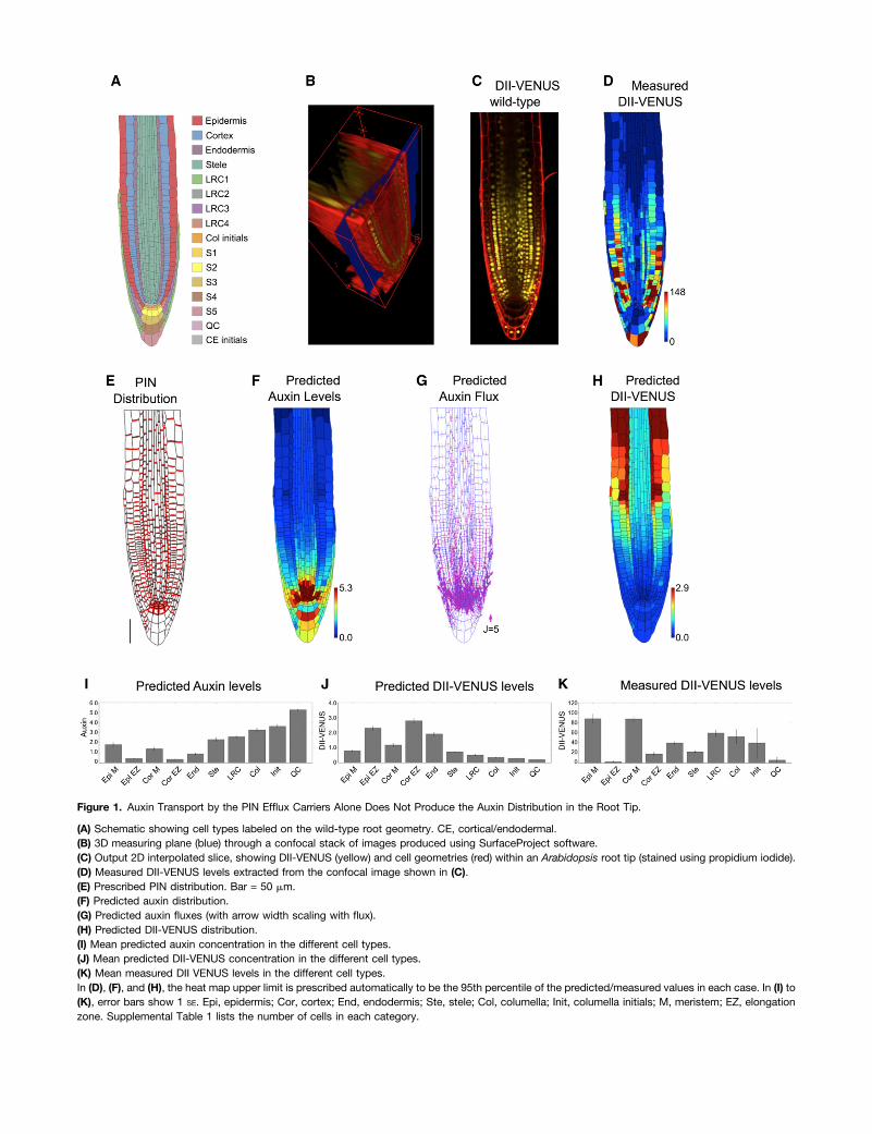
Figure 1. Auxin Transport by the PIN Efflux Carriers Alone Does Not Produce the Auxin Distribution in the Root Tip.
(A) Schematic showing cell types labeled on the wild-type root geometry. CE, cortical/endodermal.(B) 3D measuring plane (blue) through a confocal stack of images produced using SurfaceProject software.(C) Output 2D interpolated slice, showing DII-VENUS (yellow) and cell geometries (red) within an Arabidopsis root tip (stained using propidium iodide).(D) Measured DII-VENUS levels extracted from the confocal image shown in (C).(E) Prescribed PIN distribution. Bar = 50 mm.(F) Predicted auxin distribution.(G) Predicted auxin fluxes (with arrow width scaling with flux).(H) Predicted DII-VENUS distribution.(I) Mean predicted auxin concentration in the different cell types.(J) Mean predicted DII-VENUS concentration in the different cell types.(K) Mean measured DII VENUS levels in the different cell types.In (D), (F), and (H), the heat map upper limit is prescribed automatically to be the 95th percentile of the predicted/measured values in each case. In (I) to(K), error bars show 1 SE. Epi, epidermis; Cor, cortex; End, endodermis; Ste, stele; Col, columella; Init, columella initials; M, meristem; EZ, elongationzone. Supplemental Table 1 lists the number of cells in each category.
membrane proteins, which facilitate anionic auxin influx from theapoplast to the cytoplasm (Kramer, 2004; Swarup and Péret,2012). The presence of AUX1 is essential for the root’s gravitropicresponse (Marchant et al., 1999). This finding was corroborated bya computational model of the three outer root layers of the elon-gation zone, which showed that AUX1 expression within the epi-dermis, but not the cortex and endodermis, causes the epidermalauxin concentration to be high, resulting in a shootward flux(Swarup et al., 2005). Models have also demonstrated the impor-tance of AUX1 in controlling the auxin distribution during root hairinitiation (Jones et al., 2009) and root nodule formation (Perrine-Walker et al., 2010). Despite these findings, the spatial distributionof AUX1/LAX proteins has not been incorporated into recent roottip models (Grieneisen et al., 2007, 2012; Stoma et al., 2008;Mironova et al., 2012). Other classes of membrane proteins havealso been shown affect auxin transport; for example, members ofthe ABCB transporter family contribute to auxin efflux (Spalding,2013), and nitrate and potassium transporters have been shownto facilitate auxin movement (Vicente-Agullo et al., 2004; Krouket al., 2010).
In this study, we seek to develop realistic models of auxintransport within the root tip of the model plant Arabidopsis thaliana.Previous experimental work has suggested that the lateral rootcap (LRC) is important in creating the shootward auxin fluxes(Swarup et al., 2005). However, this tissue has also not beenincluded in most previous auxin transport models (Swarup et al.,2005; Grieneisen et al., 2007; Grieneisen et al., 2012; Mironovaet al., 2012): These represented root cells (or subcellular com-partments) by rectangular geometries and hence could not captureaccurately cell-to-cell connectivities. To address these issues, webase our model on actual cell geometries segmented from confocalmicroscopy image data.
To test our model predictions, we use the recently developedauxin sensor DII-VENUS (Santuari et al., 2011; Vernoux et al.,2011; Brunoud et al., 2012). Until recently, modelers have had torely on the auxin response reporter DR5 (Ulmasov et al., 1997) fora qualitative verification of their predictions. However, as an outputof the auxin signaling pathway, the DR5 response depends on thecell’s level of intermediate proteins (namely, members of the Aux/IAA and ARF families). These intermediate protein levels are likelyto vary significantly between different tissues, making quantitativecomparisons subject to error (Wells et al., 2013). In contrast, DII-VENUS is closely related to auxin concentrations: Within each cell,auxin degrades Aux/IAA by first binding with the TIR1/AFB re-ceptors, forming complexes that can bind with domain II of theAux/IAA, promoting their degradation via the ubiquitination path-way (Supplemental Figure 1; Dharmasiri et al., 2005); with domainII of the Aux/IAA fused to the VENUS fluorescent protein, the DII-VENUS sensor does not depend on the levels of Aux/IAA and ARFproteins (Brunoud et al., 2012; Wells et al., 2013), enabling it to beused as a quantitative tool (Band et al., 2012). Again, in contrastwith previous modeling studies, we explicitly incorporate part ofthe auxin signaling pathway in order predict the DII-VENUS dis-tribution (using the parameterized model of Band et al., 2012). Bycomparing these predictions with DII-VENUS measurements fromthe same root geometry, we can test our predictions on a cell-by-cell basis, providing a quantitative assessment of the model re-sults. This systems approach reveals that both influx and efflux
carriers are essential to create the auxin distribution at the root tip,with the influx carriers determining the sites of auxin accumulation.
RESULTS
Creating a Virtual Root Model by Integrating CellGeometries and Auxin Efflux Carrier Distributions
Recent models that use regular cell geometries have suggestedthat the pattern of PIN efflux carriers can create the auxin distri-bution at the root tip (Grieneisen et al., 2007, 2012; Stoma et al.,2008; Mironova et al., 2012) with an auxin maxima in the QC re-gion (Petersson et al., 2009). We therefore first considered the roleof the PINs and predicted what auxin distribution they createwhen modeling transport within a root tip comprised of actual rootcell geometries. We used a static root tip geometry, which isreasonable because auxin-transport time scales are considerablyshorter than the time scale of growth (see discussion in Band andKing, 2012).We used stacks of confocal microscopy images of DII-VENUS–
expressing roots stained with propidium iodide to capture rootcell geometries and reveal cellular organization (Figure 1B; seeMethods). As expected, different cells were in focus in differentplanes within the image stack; therefore, to produce a 2D ge-ometry suitable for our analysis, we developed a software tool,called SurfaceProject, to extract a plane from the stack (Figure1B; Supplemental Figure 2; see Methods). Using this plane (Figure1C), we segmented the multicellular root tip geometry and nuclearfluorescence channel using the CellSeT tool (Pound et al., 2012);these geometries (Supplemental Table 1) and fluorescence in-tensities (Figure 1D) were read into a vertex-based model, basedon the OpenAlea modeling framework (Pradal et al., 2008).We prescribed a PIN distribution on our virtual root tissue (Figure
1E) based on reports in the literature and our own observationsusing anti-PIN antibodies raised against the central loop of PIN1, 2,3, 4, and 7 (Supplemental Figure 3, Supplemental Tables 2 and 3,and Supplemental Methods, specifying the carrier distributions).We specified PIN proteins to be on the shootward-facing mem-branes in the LRC, elongation-zone cortex, and elongation-zoneand distal-meristem epidermis, on the rootward-facing membranesin the stele, endodermis and meristem cortex, and proximal-meristem epidermis, and on all faces of cells in the QC, columellainitial, and S1 and S2 tiers of the columella (Friml et al., 2002a,2002b; Peer et al., 2004; Blilou et al., 2005; Abas et al., 2006; Mülleret al., 1998). Given that the relative rates of auxin transport by thedifferent PIN proteins are unknown, we did not distinguish thedifferent members of the PIN family and supposed that membranefaces that are observed to contain PIN proteins have the same PIN-mediated efflux permeability.The model also incorporated a weak background efflux to ac-
count for any low levels of nonpolar PIN and for the presence ofABCB transporters. ABCBs appear to have nonpolar subcellulardistributions with different members of the ABCB family collectivelypresent within most root tissues (Spalding, 2013); we couldtherefore capture the ABCB-facilitated efflux by specifying a back-ground efflux on the cell membranes.We also included auxin production and degradation: Auxin
has been shown to be synthesized in the root tip (Ljung et al.,
864 The Plant Cell
2005), and high levels of auxin biosynthesis enzymes have beenobserved in the QC and columella initials (Stepanova et al.,2008). We therefore specified small production and degradationrates within every cell and higher auxin production in the QC andcolumella initials. The model also captured auxin diffusionthrough the apoplast, as measured by Kramer et al. (2007).Auxin diffusion within the cytoplasm is thought to be muchquicker than that in the apoplast (Swarup et al., 2005; Krameret al., 2007), and we treated auxin concentrations as uniformwithin each cell. Spatial auxin variations in the root tip can beconsidered negligible due to the small size of cells in this re-gion (Band and King, 2012). This is in contrast with the largercells further from the root tip, where subcellular variations inauxin have previously been considered (Laskowski et al., 2008;Payne and Grierson, 2009).
The resulting model can be described by a system of ordinarydifferential equations (ODEs) for the auxin concentrations withineach cell and each segment of apoplast (Supplemental Meth-ods, modeling auxin dynamics in the root tip); these equationsdepend on a number of model parameters, as detailed inSupplemental Tables 4 to 6. We represented the supply of auxinfrom the shoot by prescribing a non-zero auxin concentrationwithin the stele cells at the boundary of the modeled tissue andallowed auxin to leave that tissue through the outer layers bysetting the auxin concentrations in the epidermal, cortical, andendodermal cells at the boundary to zero. Starting from an initialcondition in which all remaining concentrations equal zero, wesimulated the ODEs until the concentrations and fluxes reacheda steady state. In addition, we developed a graphical user interface(GUI), named SimuPlant: The Virtual Root to enable thoseinterested to interact with the model (see Methods and Discussionbelow).
Auxin Distribution in the Root Tip Depends on Mechanismsin Addition to the Efflux Carriers
In agreement with previous studies that used rectangular cellgeometries (Grieneisen et al., 2007, 2012), the model predicteda hormone maxima at the root tip, with auxin levels reducing ina shootward direction (Figures 1F and 1I). We predicted thereversed fountain flux pattern, with rootward fluxes throughthe stele and shootward fluxes through the roots outer layers(Figure 1G).
To test quantitatively our predicted auxin distributions, wealso modeled the DII-VENUS auxin sensor. The dynamic re-lationship between auxin and DII-VENUS has recently beencharacterized using a parameterized mathematical model (Bandet al., 2012), and we used this model to predict the DII-VENUSlevel within each cell (Figures 1H and 1J). These predictionswere compared with observed DII-VENUS levels extracted usingthe CellSeT tool (Figures 1D and 1K). Interestingly, there weresignificant differences between the predicted and measured DII-VENUS distributions. We predicted DII-VENUS levels to be lowclose to the QC and to increase in a shootward direction (Fig-ures 1H and 1J) (the reverse of the auxin pattern), whereas ourmeasurements revealed that DII-VENUS is high in the meriste-matic epidermal and cortical cells, reducing as these cells enterthe elongation zone (Figures 1D and 1K) (Brunoud et al., 2012).
To explore the robustness of the model results, we consid-ered several modifications to our model. We predicted how theauxin distribution is affected by (1) different cases of wild-typecell geometries (Supplemental Figure 4), (2) the presence ofPINs on the inner periclinal membranes (i.e., the membranesfacing the root center) of the epidermal, cortical, and LRC cells(Supplemental Figure 5), (3) removing auxin production anddegradation (Supplemental Figure 6), and (4) removing thenonpolar auxin efflux (Supplemental Figure 7). We found thatthese changes modified the gradient of auxin away from theQC but did not qualitatively affect the predicted auxin and DII-VENUS distributions. We concluded that carrier-mediated effluxalone cannot reproduce the auxin distribution at the root tip.
The Interplay between PIN/ABCB-Mediated Efflux andAUX1/LAX-Mediated Influx Can Capture the AuxinDistribution at the Root Tip
Since the efflux carriers alone could not recreate the auxin dis-tribution, we extended our model to incorporate active influx viathe nonpolar AUX1/LAX membrane proteins. As described in theliterature and shown in Supplemental Figure 8, we took AUX1 tobe present in the LRC, elongation zone epidermis, and S1, S2,and S3 tiers of the columella (Swarup et al., 2001, 2005), LAX2 tobe present in the QC, columella initials, and rootward half of themeristematic stele (Péret et al., 2012), and LAX3 to be presentonly in the S2 tier of the columella (Swarup et al., 2008; Péretet al., 2012).Prescribing the observed AUX1/LAX distributions on our
virtual root tissue (Figure 2A; Supplemental Table 7 andSupplemental Methods, specifying the carrier distributions)and simulating the model dynamics, we predicted the steadystate distributions and fluxes (Figures 2B to 2F), obtaininghigh auxin levels within the QC, LRC, and elongating epi-dermal cells (Figures 2B and 2E). Auxin flows toward the QCthrough the stele and away from the QC though the LRC,flowing into the epidermis at the end of the LRC, as cellsenter the elongation zone (Figure 2C).The predicted DII-VENUS distribution is in reasonably good
agreement with our measurements (compare Figures 2D and 2Fto Figures 1D and 1K): DII-VENUS is predicted to be low in theQC region and LRC, whereas in the epidermis, DII-VENUS ispredicted to be high in the meristem but to reduce significantlyas cells emerge from under the LRC to enter the elongation zone(Figure 2D). Tissue-specific auxin measurements using cell-sorting techniques have also revealed the epidermal auxinconcentration to be low in the meristem (Petersson et al.,2009), consistent with our model predictions (Figures 2B and2E) and DII-VENUS measurements (Figures 1D and 1K) (seeSupplemental Figure 9 for replicates). We conclude that influxcarriers play crucial roles in controlling the auxin distributionat the root tip.
AUX1 Is Essential to Create the Auxin Distribution at theRoot Tip
Although extending our model to incorporate the AUX1/LAXinflux carriers enabled us to predict the observed wild-type
Systems Analysis of Auxin Distribution in Root Tissues 865

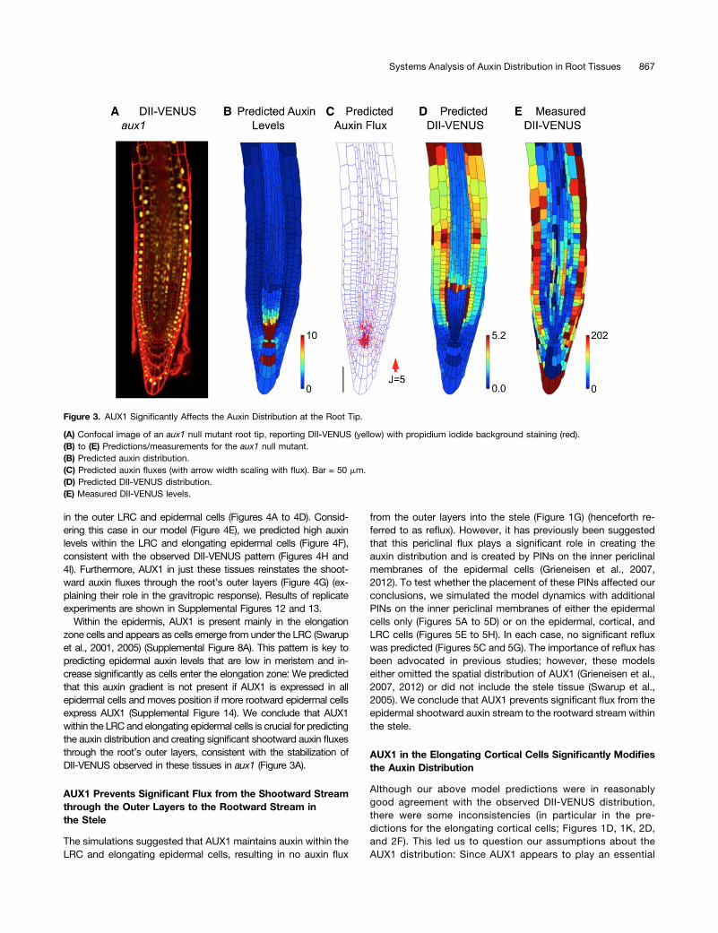
DII-VENUS distribution, this does not rule out other explan-ations. We tested our model predictions further by consideringan aux1 null mutant by crossing the aux1-22 allele (Swarup et al.,2004) with the DII-VENUS reporter. Using root cell geometriesextracted from an image of this line (Figure 3A), we predictedthat removing AUX1 would result in a buildup of auxin within theQC and surrounding cells, with little difference between theauxin levels in the different tissue layers and only small shoot-ward fluxes through the root’s outer layers (Figures 3B and 3C).The predicted and measured DII-VENUS levels were in rea-sonably good agreement, with low DII-VENUS (corresponding tothe pool of high auxin) surrounding the QC (Figures 3D and 3E)(see Supplemental Figure 10 for replicates). We conclude thatAUX1 is necessary to produce the observed auxin distributions.
To investigate further the role of the PIN efflux carriers, wesimulated the auxin dynamics with no PINs present (SupplementalFigure 11). The predicted auxin distributions are similar both with
and without polar PINs (Figure 2B; Supplemental Figure 11A);however, omitting the polar PINs led to predicted auxin fluxes thatare considerably smaller (Supplemental Figure 11B). These resultssuggested that the polar PINs create the directed auxin fluxes buthave little effect on the auxin distribution. We conclude that theAUX1/LAX influx carriers control which tissues have high auxinlevels, whereas the polar PIN carriers control the direction of auxintransport within these tissues.
AUX1 in LRC and Elongation Zone Epidermal Cells IsRequired for the Auxin Dynamics
AUX1 within LRC and elongating epidermal cells has beenshown to be essential for the gravitropic response (Swarupet al., 2005). To investigate the role of these tissues further,we crossed the aux1, DII-VENUS reporter line with aux1,J0951>>AUX1 (Swarup et al., 2005), which expresses AUX1 solely
Figure 2. The Auxin Distribution in the Root Tip Is Due to the Interplay between PIN and AUX1/LAX-Mediated Active Transport.
(A) Prescribed distributions of AUX1 (green and purple), LAX2 (blue), and LAX3 (purple). Bar = 50 mm.(B) Predicted auxin distribution.(C) Predicted auxin fluxes (with arrow width scaling with flux).(D) Predicted DII-VENUS distribution.(E) Mean predicted auxin concentration in the different cell types.(F) Mean predicted DII-VENUS concentration in the different cell types.
866 The Plant Cell
in the outer LRC and epidermal cells (Figures 4A to 4D). Consid-ering this case in our model (Figure 4E), we predicted high auxinlevels within the LRC and elongating epidermal cells (Figure 4F),consistent with the observed DII-VENUS pattern (Figures 4H and4I). Furthermore, AUX1 in just these tissues reinstates the shoot-ward auxin fluxes through the root’s outer layers (Figure 4G) (ex-plaining their role in the gravitropic response). Results of replicateexperiments are shown in Supplemental Figures 12 and 13.
Within the epidermis, AUX1 is present mainly in the elongationzone cells and appears as cells emerge from under the LRC (Swarupet al., 2001, 2005) (Supplemental Figure 8A). This pattern is key topredicting epidermal auxin levels that are low in meristem and in-crease significantly as cells enter the elongation zone: We predictedthat this auxin gradient is not present if AUX1 is expressed in allepidermal cells and moves position if more rootward epidermal cellsexpress AUX1 (Supplemental Figure 14). We conclude that AUX1within the LRC and elongating epidermal cells is crucial for predictingthe auxin distribution and creating significant shootward auxin fluxesthrough the root’s outer layers, consistent with the stabilization ofDII-VENUS observed in these tissues in aux1 (Figure 3A).
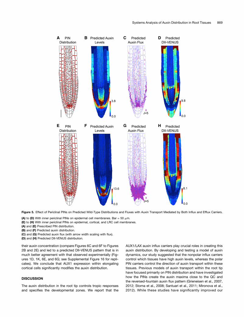
AUX1 Prevents Significant Flux from the Shootward Streamthrough the Outer Layers to the Rootward Stream inthe Stele
The simulations suggested that AUX1 maintains auxin within theLRC and elongating epidermal cells, resulting in no auxin flux
from the outer layers into the stele (Figure 1G) (henceforth re-ferred to as reflux). However, it has previously been suggestedthat this periclinal flux plays a significant role in creating theauxin distribution and is created by PINs on the inner periclinalmembranes of the epidermal cells (Grieneisen et al., 2007,2012). To test whether the placement of these PINs affected ourconclusions, we simulated the model dynamics with additionalPINs on the inner periclinal membranes of either the epidermalcells only (Figures 5A to 5D) or on the epidermal, cortical, andLRC cells (Figures 5E to 5H). In each case, no significant refluxwas predicted (Figures 5C and 5G). The importance of reflux hasbeen advocated in previous studies; however, these modelseither omitted the spatial distribution of AUX1 (Grieneisen et al.,2007, 2012) or did not include the stele tissue (Swarup et al.,2005). We conclude that AUX1 prevents significant flux from theepidermal shootward auxin stream to the rootward stream withinthe stele.
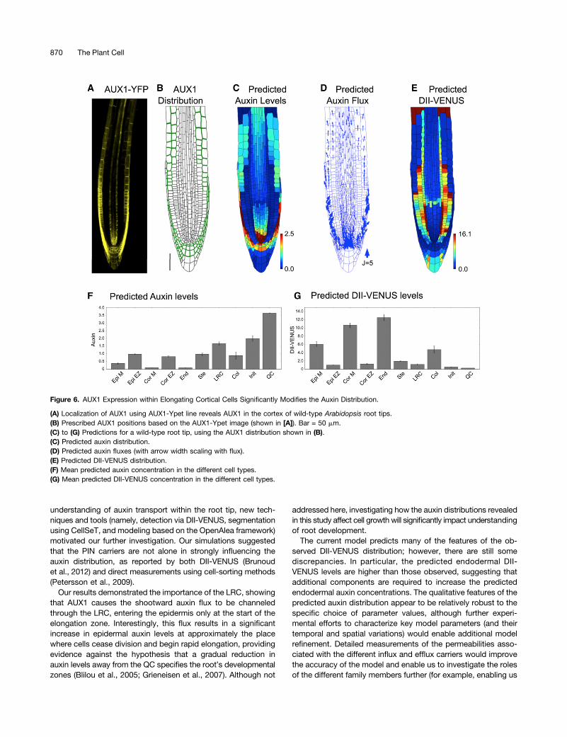
AUX1 in the Elongating Cortical Cells Significantly Modifiesthe Auxin Distribution
Although our above model predictions were in reasonablygood agreement with the observed DII-VENUS distribution,there were some inconsistencies (in particular in the pre-dictions for the elongating cortical cells; Figures 1D, 1K, 2D,and 2F). This led us to question our assumptions about theAUX1 distribution: Since AUX1 appears to play an essential
Figure 3. AUX1 Significantly Affects the Auxin Distribution at the Root Tip.
(A) Confocal image of an aux1 null mutant root tip, reporting DII-VENUS (yellow) with propidium iodide background staining (red).(B) to (E) Predictions/measurements for the aux1 null mutant.(B) Predicted auxin distribution.(C) Predicted auxin fluxes (with arrow width scaling with flux). Bar = 50 mm.(D) Predicted DII-VENUS distribution.(E) Measured DII-VENUS levels.
Systems Analysis of Auxin Distribution in Root Tissues 867
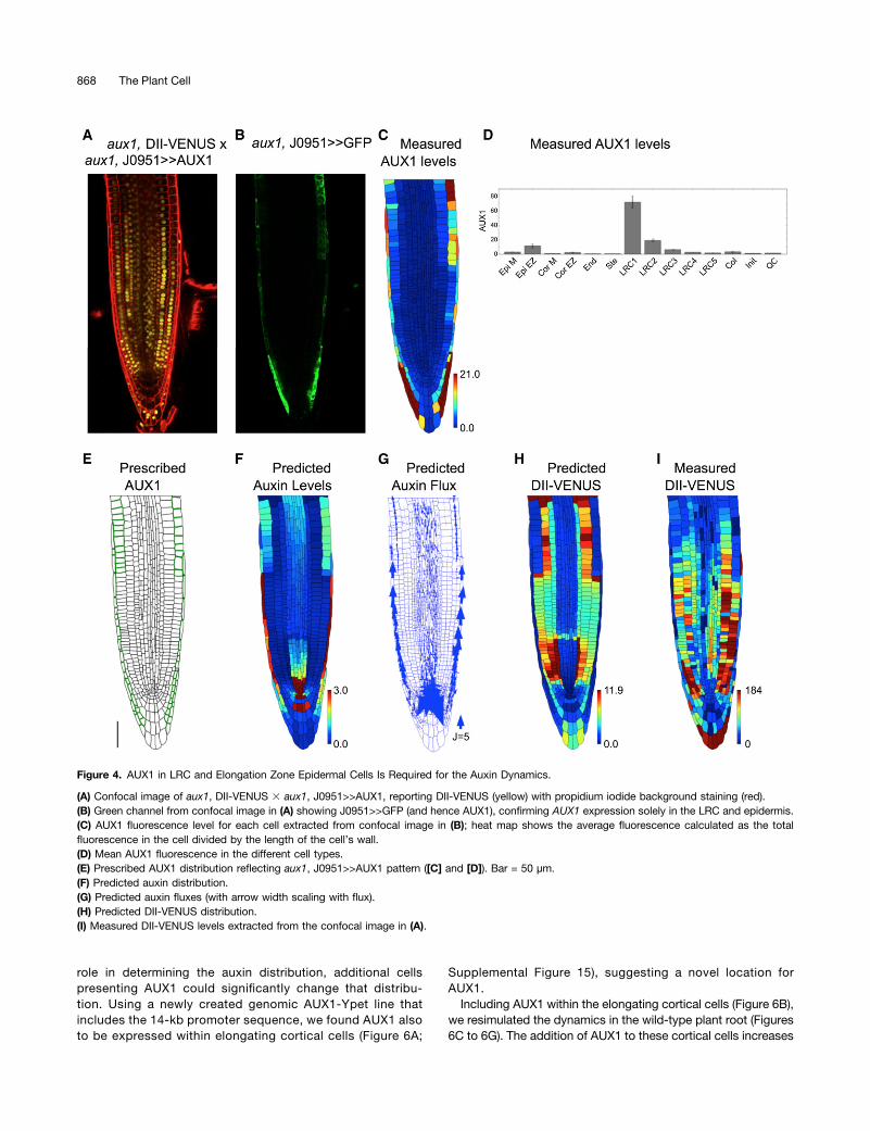
role in determining the auxin distribution, additional cellspresenting AUX1 could significantly change that distribu-tion. Using a newly created genomic AUX1-Ypet line thatincludes the 14-kb promoter sequence, we found AUX1 alsoto be expressed within elongating cortical cells (Figure 6A;
Supplemental Figure 15), suggesting a novel location forAUX1.Including AUX1 within the elongating cortical cells (Figure 6B),
we resimulated the dynamics in the wild-type plant root (Figures6C to 6G). The addition of AUX1 to these cortical cells increases
Figure 4. AUX1 in LRC and Elongation Zone Epidermal Cells Is Required for the Auxin Dynamics.
(A) Confocal image of aux1, DII-VENUS 3 aux1, J0951>>AUX1, reporting DII-VENUS (yellow) with propidium iodide background staining (red).(B) Green channel from confocal image in (A) showing J0951>>GFP (and hence AUX1), confirming AUX1 expression solely in the LRC and epidermis.(C) AUX1 fluorescence level for each cell extracted from confocal image in (B); heat map shows the average fluorescence calculated as the totalfluorescence in the cell divided by the length of the cell’s wall.(D) Mean AUX1 fluorescence in the different cell types.(E) Prescribed AUX1 distribution reflecting aux1, J0951>>AUX1 pattern ([C] and [D]). Bar = 50 µm.(F) Predicted auxin distribution.(G) Predicted auxin fluxes (with arrow width scaling with flux).(H) Predicted DII-VENUS distribution.(I) Measured DII-VENUS levels extracted from the confocal image in (A).
868 The Plant Cell
their auxin concentration (compare Figures 6C and 6F to Figures2B and 2E) and led to a predicted DII-VENUS pattern that is inmuch better agreement with that observed experimentally (Fig-ures 1D, 1K, 6E, and 6G; see Supplemental Figure 16 for repli-cates). We conclude that AUX1 expression within elongatingcortical cells significantly modifies the auxin distribution.
DISCUSSION
The auxin distribution in the root tip controls tropic responsesand specifies the developmental zones. We report that the
AUX1/LAX auxin influx carriers play crucial roles in creating thisauxin distribution. By developing and testing a model of auxindynamics, our study suggested that the nonpolar influx carrierscontrol which tissues have high auxin levels, whereas the polarPIN carriers control the direction of auxin transport within thesetissues. Previous models of auxin transport within the root tiphave focused primarily on PIN distribution and have investigatedhow the PINs create the auxin maxima close to the QC andthe reversed-fountain auxin flux pattern (Grieneisen et al., 2007,2012; Stoma et al., 2008; Santuari et al., 2011; Mironova et al.,2012). While these studies have significantly improved our
Figure 5. Effect of Periclinal PINs on Predicted Wild-Type Distributions and Fluxes with Auxin Transport Mediated by Both Influx and Efflux Carriers.
(A) to (D) With inner periclinal PINs on epidermal cell membranes. Bar = 50 mm.(E) to (H) With inner periclinal PINs on epidermal, cortical, and LRC cell membranes.(A) and (E) Prescribed PIN distribution.(B) and (F) Predicted auxin distribution.(C) and (G) Predicted auxin flux (with arrow width scaling with flux).(D) and (H) Predicted DII-VENUS distribution.
Systems Analysis of Auxin Distribution in Root Tissues 869
understanding of auxin transport within the root tip, new tech-niques and tools (namely, detection via DII-VENUS, segmentationusing CellSeT, and modeling based on the OpenAlea framework)motivated our further investigation. Our simulations suggestedthat the PIN carriers are not alone in strongly influencing theauxin distribution, as reported by both DII-VENUS (Brunoudet al., 2012) and direct measurements using cell-sorting methods(Petersson et al., 2009).
Our results demonstrated the importance of the LRC, showingthat AUX1 causes the shootward auxin flux to be channeledthrough the LRC, entering the epidermis only at the start of theelongation zone. Interestingly, this flux results in a significantincrease in epidermal auxin levels at approximately the placewhere cells cease division and begin rapid elongation, providingevidence against the hypothesis that a gradual reduction inauxin levels away from the QC specifies the root’s developmentalzones (Blilou et al., 2005; Grieneisen et al., 2007). Although not
addressed here, investigating how the auxin distributions revealedin this study affect cell growth will significantly impact understandingof root development.The current model predicts many of the features of the ob-
served DII-VENUS distribution; however, there are still somediscrepancies. In particular, the predicted endodermal DII-VENUS levels are higher than those observed, suggesting thatadditional components are required to increase the predictedendodermal auxin concentrations. The qualitative features of thepredicted auxin distribution appear to be relatively robust to thespecific choice of parameter values, although further experi-mental efforts to characterize key model parameters (and theirtemporal and spatial variations) would enable additional modelrefinement. Detailed measurements of the permeabilities asso-ciated with the different influx and efflux carriers would improvethe accuracy of the model and enable us to investigate the rolesof the different family members further (for example, enabling us
Figure 6. AUX1 Expression within Elongating Cortical Cells Significantly Modifies the Auxin Distribution.
(A) Localization of AUX1 using AUX1-Ypet line reveals AUX1 in the cortex of wild-type Arabidopsis root tips.(B) Prescribed AUX1 positions based on the AUX1-Ypet image (shown in [A]). Bar = 50 mm.(C) to (G) Predictions for a wild-type root tip, using the AUX1 distribution shown in (B).(C) Predicted auxin distribution.(D) Predicted auxin fluxes (with arrow width scaling with flux).(E) Predicted DII-VENUS distribution.(F) Mean predicted auxin concentration in the different cell types.(G) Mean predicted DII-VENUS concentration in the different cell types.
870 The Plant Cell
to assess whether the different members of the PIN family fa-cilitate different rates of auxin transport). Similarly, the relativeroles of the carrier-mediated and passive transport componentsdepend on pH measurements: For example, a larger apoplasticpH would lead to a higher proportion of auxin being anionic andentering the cells via the influx carriers. Thus, increases/decreasesin apoplastic pH leads to the influx carriers having larger/smallereffects on the predicted distributions (Supplemental Figures 17and 18).
Auxin distributions are also affected by spatial variations in auxinproduction and degradation. We prescribed high auxin productionin the QC and in the columella initials (reflecting observed bio-synthesis enzyme distributions; Stepanova et al., 2008); increasing/decreasing the production rate in these tissues increases/decreases their auxin levels (Supplemental Figures 19 and 20).Furthermore, simulations suggested that apoplastic diffusion andnonpolar efflux (as mediated by nonpolar PINs and/or ABCBs, forexample) promote periclinal auxin transport, enabling auxin tomove between epidermal, cortical, and endodermal tissues; re-moving these fluxes reduces the agreement between our predictedand observed DII-VENUS levels (Supplemental Figures 21 and 22).Measurements of apoplast thicknesses and characterization of the
mechanisms underlying nonpolar transport would allow furtherfine-tuning of our predictions and understanding.Our study also highlights the benefits of developing and
modeling fluorescent reporters (Wells et al., 2013). Until recently,the auxin response has been visualized using the DR5 reporter(Ulmasov et al., 1997). Although observed DR5 patterns quali-tatively agree with those for DII-VENUS, the latter provides amore detailed readout of the auxin distribution, enabling us totest model predictions quantitatively on a cell-by-cell basis.Since using DII-VENUS in conjunction with state-of-the-artsegmentation tools enabled us to gain a unique perspective onthis key plant hormone, we envisage the procedure being ap-plied to auxin studies in other plant organs and species, withsimilar integrative approaches benefiting the use of fluorescentlylabeled hormone variants developed for gibberellin (Shani et al.,2013) and brassinosteroids (Irani et al., 2012).Finally, to enable researchers to interact with our models,
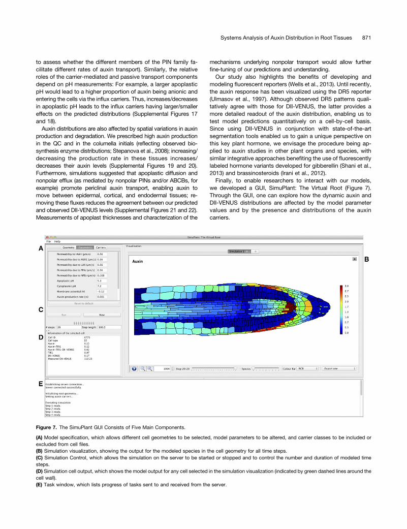
we developed a GUI, SimuPlant: The Virtual Root (Figure 7).Through the GUI, one can explore how the dynamic auxin andDII-VENUS distributions are affected by the model parametervalues and by the presence and distributions of the auxincarriers.
Figure 7. The SimuPlant GUI Consists of Five Main Components.
(A) Model specification, which allows different cell geometries to be selected, model parameters to be altered, and carrier classes to be included orexcluded from cell files.(B) Simulation visualization, showing the output for the modeled species in the cell geometry for all time steps.(C) Simulation Control, which allows the simulation on the server to be started or stopped and to control the number and duration of modeled timesteps.(D) Simulation cell output, which shows the model output for any cell selected in the simulation visualization (indicated by green dashed lines around thecell wall).(E) Task window, which lists progress of tasks sent to and received from the server.
Systems Analysis of Auxin Distribution in Root Tissues 871
METHODS
Plant Material and Growth Conditions
All Arabidopsis thaliana lines were in the Columbia-0 background. Six-day-old DII-VENUS seedlings were grown from surface-sterilized seedsas described previously (Holman et al., 2010). To generate reporter plantswith tissue-specific AUX1 expression (Figures 4A and 4B), the aux1, DII-VENUS line (Figure 3A) was crossed with the aux1, J0951>>AUX1 line(Swarup et al., 2005), in which AUX1 expression is targeted to the LRC andexpanding epidermal tissues in the aux1mutant background using a GAL4-based transactivation expression approach. The Ypet fusion to AUX1(Figure 6A) was generated by employing a recombineering approach (Zhouet al., 2011). In brief, a PCR fragment containing the Ypet coding sequencewas recombined in frame into the AUX1 genomic sequence (encoded ona TAC plasmid in Escherichia coli) to create an AUX1-VENUS protein fusionwhose expression was driven by the native 14-kb promoter sequence.
Immunolocalization and Imaging
Four-day-old seedlings were fixed, and immunolocalization experimentswere performed as described previously (Péret et al., 2012) and visualizedusing confocal laser scanningmicroscopy. Primary antibodieswere normallyused at 1:100 to 1:200; Alexa Fluor–coupled secondary antibodies (In-vitrogen) were used at 1:200 dilution. Live imaging was performed using anSP5 spectral detection confocal laser scanning microscope (Leica). Rootswere stained with propidium iodide (Sigma-Aldrich) to visualize apoplastorganization. Microscope settings were as detailed by Pound et al. (2012).
Segmentation
Image stacks were taken about the midline of roots to include all nucleirequired for quantification (typically 30 steps at 1-µm intervals). 2D planeswere constructed from the 3D stacks using the SurfaceProject plug-in forthe Fiji image processing package (Schindelin et al., 2012). Details ofSurfaceProject can be found in the Supplemental Methods, and the soft-ware and user documentation are provided at www.simuplant.org. Cellgeometries and DII-VENUS and GFP fluorescence intensities were ex-tracted from the confocal images using the CellSeT segmentation tool, asdescribed by Pound et al. (2012).
Modeling
The geometrical and fluorescence measurements from CellSeT were readinto an OpenAlea data structure (Pradal et al., 2008). Using the resultingmulticellular tissue structure, auxin transport was modeled using a vertex-based approach (Supplemental Figure 23). The auxin and DII-VENUS dy-namics described in the main text were captured by a system of ODEs,which depend on a number of parameter values (Supplemental Tables 4 to6), which were sourced from the literature (Scott and Allen, 1999; Sze et al.,1999; Fasano et al., 2001; Swarup et al., 2005; Heisler and Jönsson, 2006;Kramer et al., 2007). Simulations were performed in the Python pro-gramming language (www.python.org) using the ODE solver LSODES fromthe ODEPACK suite (Hindmarsh, 1983) to approximate numerically sol-utions of the system of differential equations. This solver exploits thesparsity of the Jacobian of the system of equations; the sparsity pattern ofthe Jacobian was supplied to the numerical solver to improve the speed ofthe simulations. Simulations were run until the distributions and fluxesreached their steady states. These steady state solutions were then pre-sented using the Python library matplotlib (Hunter, 2007). Plotting thesteady state concentration distributions required us to choose a range forthe heat map, which we prescribed automatically to minimize subjectivity.Plotting histograms of the predicted and measured concentrations of thedifferent cells (for example, Supplemental Figure 24), we observed that the
lower limit of the concentration distribution is (unsurprisingly) zero for bothauxin and DII-VENUS and that the distributions are negatively skewed.Although we could have chosen the upper limit of the heat map range to bemaximum predicted/measured concentration in each case, outliers in theconcentration distribution would significantly affect the appearance of thepresented distribution. We therefore prescribed the heat map upper limit tobe the 95th percentile of the concentration values. In the main text andsupplemental figures, we present our predicted distributions of auxin andDII-VENUS. The model also predicted the concentration distribution of thefree and bound TIR1/AFB receptors; in Supplemental Figure 25, we showsample predictions of these that correspond to the simulations in Figures 3and 6 (see Supplemental Methods for further details).
SimuPlant GUI
A GUI called SimuPlant was developed to enable those interested to in-teract directly with the simulations presented in this study. SimuPlant iswritten in Java 7, which is easily installable on most modern operatingsystems (OSX/Windows/Linux). The client connects to a simulation server,where all the computationally intensive operations are performed. Thedefault behavior is for the client to connect to the server provided by theUniversity of Nottingham, available via www.simuplant.org. However, forintensive use, it is also possible to install the server locally. In this case, thesoftware is available from Sourceforge; links to this software can be foundon the GUI website www.simuplant.org. The client permits the user tochoose between the nine different root tip geometries presented herein,each acquired from confocal images using SurfaceProject and CellSeT;three of these are from wild-type plants (Figure 1C; Supplemental Figures4A and 4G), three from aux1 null mutant plants (Figure 3A; SupplementalFigures 10A and 10F), and three from the aux1, DII-VENUS 3 aux1,J0951>>AUX1 line (Swarup et al., 2005) in which AUX1 is expressed solelyin the outer LRC and epidermal cells (Figure 4A; Supplemental Figures 12Aand 13A). The parameters in the auxin transport model may be varied by theuser, and the user can select different options for the placement of the auxininflux and efflux carriers. Default settings correspond to the parametervalues listed in Supplemental Tables 4 and 6 and the carrier distributionsdetailed in Supplemental Tables 3 and 7. Simulation outputs can be dis-played in the GUI. The GUI shows the temporal evolution of the solutiontoward a steady state, allowing one to visualize the flux of auxin throughthe root tissues. Simulation outputs can be saved in a portable networkgraphics format, and one can select individual cells to view the predictedconcentrations within that cell. Further information about the GUI is pro-vided in the user documentation available at www.simuplant.org.
Supplemental Data
The following materials are available in the online version of this article.
Supplemental Figure 1. The Network of Interactions That Leads toAuxin-Mediated Degradation of DII-VENUS.
Supplemental Figure 2. Constructing a 2D Plane from a 3D ConfocalImage Stack Using SurfaceProject.
Supplemental Figure 3. In Situ Immunodetection of PIN Proteins inthe Arabidopsis Root Tip.
Supplemental Figure 4. Replicates of Results for the Wild-Type RootTip Considering Efflux Carrier–Mediated Transport Only (i.e., with NoInflux Carriers Present).
Supplemental Figure 5. Effect of Periclinal PINS on the PredictedDistributions and Fluxes with Efflux Carrier–Mediated Transport only(i.e., with No Influx Carriers Present).
Supplemental Figure 6. Effect of Omitting Auxin Synthesis andDegradation on the Predicted Distributions and Fluxes with Efflux Carrier–Mediated Transport Only (i.e., with No Influx Carriers Present).
872 The Plant Cell
Supplemental Figure 7. Effect of Omitting Nonpolar Efflux (asMediated by Nonpolar PINs and/or ABCBs) on the Predicted Distri-butions and Fluxes with Efflux Carrier–Mediated Transport Only (i.e.,with No Influx Carriers Present).
Supplemental Figure 8. Expression Patterns of AUX1/LAX AuxinTransporters in the Arabidopsis Root Tip.
Supplemental Figure 9. Replicates of Predictions for the Wild-TypePlant Root with Auxin Transport Mediated by Both Efflux and InfluxCarriers.
Supplemental Figure 10. Replicates of Results for the aux1 NullMutant Plant Root.
Supplemental Figure 11. Effect of Omitting the Polar PINs on thePredicted Wild-Type Distributions and Fluxes (with Auxin TransportMediated by Both Influx and Efflux Carriers).
Supplemental Figure 12. Replicate 1 Results for the aux1J0951>>AUX1 Mutant Plant Root in Which AUX1 Is Expressed Solelyin the Outer LRC and Epidermal Cells.
Supplemental Figure 13. Replicate 2 Results for the aux1J0951>>AUX1 Mutant Plant Root in Which AUX1 Is Expressed Solelyin the Outer LRC and Epidermal Cells.
Supplemental Figure 14. Effect of AUX1 Being Expressed inMeristematic Epidermal Cells on the Predicted Wild-Type Distributionsand Fluxes (with Auxin Mediated by Both Influx and Efflux Carriers).
Supplemental Figure 15. Replicates for the AUX1-Ypet Line.
Supplemental Figure 16. Replicates of Simulations for the Wild-TypeRoot Tip with AUX1 Expressed in the Elongating Cortical Cells.
Supplemental Figure 17. Effect of Apoplastic pH on the PredictedDistributions and Fluxes in the Wild-Type Root Tip.
Supplemental Figure 18. Effect of Apoplastic pH on the PredictedDistributions and Fluxes in the aux1 Null Mutant Root Tip.
Supplemental Figure 19. Effect of Auxin Production in the QC andColumella Initials on the Predicted Distributions and Fluxes in the Wild-Type Root Tip.
Supplemental Figure 20. Effect of Auxin Production in the QC andColumella Initials on the Predicted Distributions and Fluxes in the aux1Null Mutant Root Tip.
Supplemental Figure 21. Effect of Omitting Cell Wall Diffusion and/orNonpolar Efflux on the Predicted Distributions and Fluxes in Wild-TypeRoot Tip.
Supplemental Figure 22. Effect of Omitting Apoplastic Diffusion and/or Nonpolar Efflux on the Predicted Distributions and Fluxes in an aux1Null Mutant Root Tip.
Supplemental Figure 23. Schematics Illustrating the Vertex-BasedInterpretation of the Multicellular Plant Tissues.
Supplemental Figure 24. Histograms Showing the Distributions ofPredicted/Measured Auxin and DII-VENUS Levels.
Supplemental Figure 25. Predicted Distributions of the Free andBound TIR1/AFB Receptors.
Supplemental Table 1. Number of Each Cell Type in Each Cell Geometry.
Supplemental Table 2.Observed Distributions of Different Members ofthe PIN Family, Based on the Images Shown in Supplemental Figure 3.
Supplemental Table 3. Rules Used to Prescribe the PIN Distributionon Our Virtual Root Geometries.
Supplemental Table 4. Parameter Values Used in the Auxin-TransportModel.
Supplemental Table 5. Description of Parameters in the DII-VENUSNetwork Model (See Equation 3.1 in Supplemental Methods).
Supplemental Table 6. Parameters Used in the Reduced DII-VENUSNetwork Model (See Equation 3.11 in Supplemental Methods).
Supplemental Table 7. Distributions of the AUX1/LAX Influx Carriers,Based on the Images Shown in Supplemental Figure 8.
Supplemental Methods.
ACKNOWLEDGMENTS
This project was supported by the Biotechnology and Biological SciencesResearch Council (BBSRC) and Engineering and Physical Sciences Re-search Council funding to the Centre for Plant Integrative Biology. In addition,we acknowledge the support of the Leverhulme Trust (L.R.B.), the Al-TajirWorld of Islam Trust (H.I.H.), National Science Foundation Grant DBI0820755(J.M.A.), the Royal Society and Wolfson Foundation (J.R.K.), and BBSRCProfessorial Research Fellowship funding (M.J.B.). We thank Antoine Larrieu,Caroline Howells, and Edward Venison for assistance in generation of theAUX1:VENUS line.
AUTHOR CONTRIBUTIONS
L.R.B. and D.M.W. designed the research, performed the research,contributed new tools, analyzed data, and wrote the article. J.A.F. performedresearch, contributed new tools, and analyzed data. A.P.F. and M.P.P.performed research and contributed new tools. T.G., M.H.W., L.Y., W.L.,H.I.H., J.O., S.P.P., M.A.P.-A., J.Y., J.M.A., C.G., and T.V. contributed newtools. E.K. and T.C.H. designed the research. T.P.P. designed theresearch and contributed new tools. R.S. designed the research,performed research, and contributed new tools. J.R.K. and M.J.B.designed the research, analyzed data, and wrote the article. All authorsdiscussed the results and commented on the article.
Received October 9, 2013; revised January 6, 2014; accepted February14, 2014; published March 14, 2014.
REFERENCES
Abas, L., Benjamins, R., Malenica, N., Paciorek, T., Wi�sniewska, J.,Moulinier-Anzola, J.C., Sieberer, T., Friml, J., and Luschnig, C.(2006). Intracellular trafficking and proteolysis of the Arabidopsisauxin-efflux facilitator PIN2 are involved in root gravitropism. Nat.Cell Biol. 8: 249–256. Erratum. Nat. Cell Biol. 8: 424.
Band, L.R., and King, J.R. (2012). Multiscale modelling of auxin transportin the plant-root elongation zone. J. Math. Biol. 65: 743–785.
Band, L.R., et al. (2012). Root gravitropism is regulated by a transientlateral auxin gradient controlled by a tipping-point mechanism.Proc. Natl. Acad. Sci. USA 109: 4668–4673.
Benjamins, R., and Scheres, B. (2008). Auxin: The looping star inplant development. Annu. Rev. Plant Biol. 59: 443–465.
Benková, E., Michniewicz, M., Sauer, M., Teichmann, T.,Seifertová, D., Jürgens, G., and Friml, J. (2003). Local, efflux-dependent auxin gradients as a common module for plant organformation. Cell 115: 591–602.
Blilou, I., Xu, J., Wildwater, M., Willemsen, V., Paponov, I., Friml, J.,Heidstra, R., Aida, M., Palme, K., and Scheres, B. (2005). The PINauxin efflux facilitator network controls growth and patterning inArabidopsis roots. Nature 433: 39–44.
Systems Analysis of Auxin Distribution in Root Tissues 873
Brunoud, G., Wells, D.M., Oliva, M., Larrieu, A., Mirabet, V.,Burrow, A.H., Beeckman, T., Kepinski, S., Traas, J., Bennett,M.J., and Vernoux, T. (2012). A novel sensor to map auxinresponse and distribution at high spatio-temporal resolution. Nature482: 103–106.
Dharmasiri, N., Dharmasiri, S., and Estelle, M. (2005). The F-boxprotein TIR1 is an auxin receptor. Nature 435: 441–445.
Fasano, J.M., Swanson, S.J., Blancaflor, E.B., Dowd, P.E., Kao,T.H., and Gilroy, S. (2001). Changes in root cap pH are required forthe gravity response of the Arabidopsis root. Plant Cell 13: 907–921.
Friml, J., Benková, E., Blilou, I., Wisniewska, J., Hamann, T., Ljung,K., Woody, S., Sandberg, G., Scheres, B., Jürgens, G., andPalme, K. (2002b). AtPIN4 mediates sink-driven auxin gradientsand root patterning in Arabidopsis. Cell 108: 661–673.
Friml, J., Wi�sniewska, J., Benková, E., Mendgen, K., and Palme, K.(2002a). Lateral relocation of auxin efflux regulator PIN3 mediatestropism in Arabidopsis. Nature 415: 806–809.
Gälweiler, L., Guan, C., Müller, A., Wisman, E., Mendgen, K.,Yephremov, A., and Palme, K. (1998). Regulation of polar auxintransport by AtPIN1 in Arabidopsis vascular tissue. Science 282:2226–2230.
Grieneisen, V.A., Scheres, B., Hogeweg, P., and M Marée, A.F.(2012). Morphogengineering roots: Comparing mechanisms ofmorphogen gradient formation. BMC Syst. Biol. 6: 37.
Grieneisen, V.A., Xu, J., Marée, A.F., Hogeweg, P., and Scheres, B.(2007). Auxin transport is sufficient to generate a maximum andgradient guiding root growth. Nature 449: 1008–1013.
Heisler, M., and Jönsson, H. (2006). Modeling auxin transport andplant development. J. Plant Growth Regul. 25: 302–312.
Hindmarsh, A.C. (1983). ODEPACK: A systematized collection ofODE solvers. In Scientific Computing: IMACS Transactions onScientific Computation, Vol. 1, R.S. Stepleman, ed (North-Holland,Amsterdam: IMACS), pp. 55–64.
Holman, T.J., Wilson, M.H., Kenobi, K., Dryden, I.L., Hodgman,T.C., Wood, A.T., and Holdsworth, M.J. (2010). Statisticalevaluation of transcriptomic data generated using the Affymetrixone-cycle, two-cycle and IVT-Express RNA labelling protocols withthe Arabidopsis ATH1 microarray. Plant Methods 6: 9–10.
Hunter, J.D. (2007). Matplotlib: A 2D graphics environment. Comput.Sci. Eng. 9: 90–95.
Irani, N.G., et al. (2012). Fluorescent castasterone reveals BRI1signaling from the plasma membrane. Nat. Chem. Biol. 8: 583–589.
Jiang, K., and Feldman, L.J. (2005). Regulation of root apicalmeristem development. Annu. Rev. Cell Dev. Biol. 21: 485–509.
Jones, A.R., Kramer, E.M., Knox, K., Swarup, R., Bennett, M.J.,Lazarus, C.M., Leyser, H.M., and Grierson, C.S. (2009). Auxintransport through non-hair cells sustains root-hair development.Nat. Cell Biol. 11: 78–84.
Kramer, E.M. (2004). PIN and AUX/LAX proteins: Their role in auxinaccumulation. Trends Plant Sci. 9: 578–582.
Kramer, E.M., Frazer, N.L., and Baskin, T.I. (2007). Measurement ofdiffusion within the cell wall in living roots of Arabidopsis thaliana. J.Exp. Bot. 58: 3005–3015.
Krouk, G., et al. (2010). Nitrate-regulated auxin transport by NRT1.1defines a mechanism for nutrient sensing in plants. Dev. Cell 18:927–937.
Laskowski, M., Grieneisen, V.A., Hofhuis, H., Hove, C.A.T.,Hogeweg, P., Marée, A.F.M., and Scheres, B. (2008). Root systemarchitecture from coupling cell shape to auxin transport. PLoS Biol.6: e307.
Ljung, K., Hull, A.K., Celenza, J., Yamada, M., Estelle, M.,Normanly, J., and Sandberg, G. (2005). Sites and regulation ofauxin biosynthesis in Arabidopsis roots. Plant Cell 17: 1090–1104.
Marchant, A., Kargul, J., May, S.T., Muller, P., Delbarre, A., Perrot-Rechenmann, C., and Bennett, M.J. (1999). AUX1 regulates rootgravitropism in Arabidopsis by facilitating auxin uptake within rootapical tissues. EMBO J. 18: 2066–2073.
Mironova, V.V., Omelyanchuk, N.A., Novoselova, E.S., Doroshkov,A.V., Kazantsev, F.V., Kochetov, A.V., Kolchanov, N.A.,Mjolsness, E., and Likhoshvai, V.A. (2012). Combined in silico/invivo analysis of mechanisms providing for root apical meristem self-organization and maintenance. Ann. Bot. (Lond.) 110: 349–360.
Müller, A., Guan, C., Gälweiler, L., Tänzler, P., Huijser, P.,Marchant, A., Parry, G., Bennett, M., Wisman, E., and Palme, K.(1998). AtPIN2 defines a locus of Arabidopsis for root gravitropismcontrol. EMBO J. 17: 6903–6911.
Ottenschläger, I., Wolff, P., Wolverton, C., Bhalerao, R.P.,Sandberg, G., Ishikawa, H., Evans, M., and Palme, K. (2003).Gravity-regulated differential auxin transport from columella tolateral root cap cells. Proc. Natl. Acad. Sci. USA 100: 2987–2991.
Payne, R.J.H., and Grierson, C.S. (2009). A theoretical model forROP localisation by auxin in Arabidopsis root hair cells. PLoS ONE4: e8337.
Peer, W.A., Bandyopadhyay, A., Blakeslee, J.J., Makam, S.N.,Chen, R.J., Masson, P.H., and Murphy, A.S. (2004). Variation inexpression and protein localization of the PIN family of auxin effluxfacilitator proteins in flavonoid mutants with altered auxin transportin Arabidopsis thaliana. Plant Cell 16: 1898–1911.
Péret, B., et al. (2012). AUX/LAX genes encode a family of auxin influxtransporters that perform distinct functions during Arabidopsisdevelopment. Plant Cell 24: 2874–2885.
Perrine-Walker, F., et al. (2010). Auxin carriers localization drivesauxin accumulation in plant cells infected by Frankia in Casuarinaglauca actinorhizal nodules. Plant Physiol. 154: 1372–1380.
Petersson, S.V., Johansson, A.I., Kowalczyk, M., Makoveychuk,A., Wang, J.Y., Moritz, T., Grebe, M., Benfey, P.N., Sandberg, G.,and Ljung, K. (2009). An auxin gradient and maximum in theArabidopsis root apex shown by high-resolution cell-specificanalysis of IAA distribution and synthesis. Plant Cell 21: 1659–1668.
Pound, M.P., French, A.P., Wells, D.M., Bennett, M.J., andPridmore, T.P. (2012). CellSeT: Novel software to extract andanalyze structured networks of plant cells from confocal images.Plant Cell 24: 1353–1361.
Pradal, C., Dufour-Kowalski, S., Boudon, F., Fournier, C., andGodin, C. (2008). OpenAlea: A visual programming and component-based software platform for plant modeling. Funct. Plant Biol. 35:751–760.
Rashotte, A.M., Brady, S.R., Reed, R.C., Ante, S.J., and Muday,G.K. (2000). Basipetal auxin transport is required for gravitropism inroots of Arabidopsis. Plant Physiol. 122: 481–490.
Sabatini, S., Beis, D., Wolkenfelt, H., Murfett, J., Guilfoyle, T.,Malamy, J., Benfey, P., Leyser, O., Bechtold, N., Weisbeek, P.,and Scheres, B. (1999). An auxin-dependent distal organizer ofpattern and polarity in the Arabidopsis root. Cell 99: 463–472.
Santuari, L., Scacchi, E., Rodriguez-Villalon, A., Salinas, P.,Dohmann, E.M., Brunoud, G., Vernoux, T., Smith, R.S., andHardtke, C.S. (2011). Positional information by differential endocytosissplits auxin response to drive Arabidopsis root meristem growth. Curr.Biol. 21: 1918–1923.
Scott, A.C., and Allen, N.S. (1999). Changes in cytosolic pH withinArabidopsis root columella cells play a key role in the early signalingpathway for root gravitropism. Plant Physiol. 121: 1291–1298.
Schindelin, J., et al. (2012). Fiji: An open-source platform forbiological-image analysis. Nat. Methods 9: 676–682.
Shani, E., Weinstain, R., Zhang, Y., Castillejo, C., Kaiserli, E.,Chory, J., Tsien, R.Y., and Estelle, M. (2013). Gibberellins
874 The Plant Cell
accumulate in the elongating endodermal cells of Arabidopsis root.Proc. Natl. Acad. Sci. USA 110: 4834–4839.
Spalding, E.P. (2013). Diverting the downhill flow of auxin to steergrowth during tropisms. Am. J. Bot. 100: 203–214.
Stepanova, A.N., Robertson-Hoyt, J., Yun, J., Benavente, L.M., Xie,D.Y., Dolezal, K., Schlereth, A., Jürgens, G., and Alonso, J.M.(2008). TAA1-mediated auxin biosynthesis is essential for hormonecrosstalk and plant development. Cell 133: 177–191.
Stoma, S., Lucas, M., Chopard, J., Schaedel, M., Traas, J., andGodin, C. (2008). Flux-based transport enhancement as a plausibleunifying mechanism for auxin transport in meristem development.PLOS Comput. Biol. 4: e1000207.
Swarup, K., et al. (2008). The auxin influx carrier LAX3 promoteslateral root emergence. Nat. Cell Biol. 10: 946–954.
Swarup, R., and Péret, B. (2012). AUX/LAX family of auxin influxcarriers-an overview. Front Plant Sci 3: 225.
Swarup, R., Friml, J., Marchant, A., Ljung, K., Sandberg, G., Palme,K., and Bennett, M. (2001). Localization of the auxin permeaseAUX1 suggests two functionally distinct hormone transportpathways operate in the Arabidopsis root apex. Genes Dev. 15:2648–2653.
Swarup, R., et al. (2004). Structure-function analysis of the presumptiveArabidopsis auxin permease AUX1. Plant Cell 16: 3069–3083.
Swarup, R., Kramer, E.M., Perry, P., Knox, K., Leyser, H.M., Haseloff,J., Beemster, G.T., Bhalerao, R., and Bennett, M.J. (2005). Rootgravitropism requires lateral root cap and epidermal cells for transportand response to a mobile auxin signal. Nat. Cell Biol. 7: 1057–1065.
Sze, H., Li, X., and Palmgren, M.G. (1999). Energization of plant cellmembranes by H+-pumping ATPases. Regulation and biosynthesis.Plant Cell 11: 677–690.
Ulmasov, T., Murfett, J., Hagen, G., and Guilfoyle, T.J. (1997).Aux/IAA proteins repress expression of reporter genes containingnatural and highly active synthetic auxin response elements.Plant Cell 9: 1963–1971.
Vernoux, T., et al. (2011). The auxin signalling network translatesdynamic input into robust patterning at the shoot apex. Mol. Syst.Biol. 7: 508.
Vicente-Agullo, F., Rigas, S., Desbrosses, G., Dolan, L., Hatzopoulos,P., and Grabov, A. (2004). Potassium carrier TRH1 is required for auxintransport in Arabidopsis roots. Plant J. 40: 523–535.
Wells, D.M., Laplaze, L., Bennett, M.J., and Vernoux, T. (2013).Biosensors for phytohormone quantification: challenges, solutions,and opportunities. Trends Plant Sci. 18: 244–249.
Zhou, R., Benavente, L.M., Stepanova, A.N., and Alonso, J.M. (2011). Arecombineering-based gene tagging system for Arabidopsis. Plant J. 66:712–723.
Systems Analysis of Auxin Distribution in Root Tissues 875
DOI 10.1105/tpc.113.119495; originally published online March 14, 2014; 2014;26;862-875Plant Cell
Hodgman, Tony P. Pridmore, Ranjan Swarup, John R. King and Malcolm J. BennettPerez-Amador, Jeonga Yun, Eric Kramer, Jose M. Alonso, Christophe Godin, Teva Vernoux, T. Charlie
Michael H. Wilson, Lei Yu, Wenda Li, Hussein I. Hijazi, Jaesung Oh, Simon P. Pearce, Miguel A. Leah R. Band, Darren M. Wells, John A. Fozard, Teodor Ghetiu, Andrew P. French, Michael P. Pound,
Root ApexArabidopsisSystems Analysis of Auxin Transport in the
This information is current as of September 3, 2018
Supplemental Data /content/suppl/2014/03/10/tpc.113.119495.DC2.html /content/suppl/2014/03/10/tpc.113.119495.DC1.html
References /content/26/3/862.full.html#ref-list-1
This article cites 56 articles, 20 of which can be accessed free at:
Permissions https://www.copyright.com/ccc/openurl.do?sid=pd_hw1532298X&issn=1532298X&WT.mc_id=pd_hw1532298X
eTOCs http://www.plantcell.org/cgi/alerts/ctmain
Sign up for eTOCs at:
CiteTrack Alerts http://www.plantcell.org/cgi/alerts/ctmain
Sign up for CiteTrack Alerts at:
Subscription Information http://www.aspb.org/publications/subscriptions.cfm
is available at:Plant Physiology and The Plant CellSubscription Information for
ADVANCING THE SCIENCE OF PLANT BIOLOGY © American Society of Plant Biologists