Embed Size (px)
Citation preview

Research ArticleSystemic Use of ArnicaMontana for the Reduction of PostsurgicalSequels following Extraction of Impacted Mandibular 3rd Molars:A Pilot Study
Hani Mawardi ,1 Shaima Ghazalh,2 Ahmad Shehatah,3 Amnah Abdelwahid,4
Amin Aljohani,3 Osama Felemban ,5 Soulafa Almazrooa ,1
Lena Elbadawi,6 and Hazem Shawky7,8
1Department of Oral Diagnostic Sciences, King AbdulAziz University, Faculty of Dentistry, Jeddah, Saudi Arabia2Ministry of Health, Madinah, Saudi Arabia3Ministry of Health, Taif, Saudi Arabia4Private Practice, Jeddah, Saudi Arabia5Department of Pediatric Dentistry, King AbdulAziz University, Faculty of Dentistry, Jeddah, Saudi Arabia6Department of Periodontics, King AbdulAziz University, Faculty of Dentistry, Jeddah, Saudi Arabia7Department Oral and Maxillofacial Surgery, Faculty of Oral and Dental Medicine, Cairo University, Cairo, Egypt8Department Oral and Maxillofacial Surgery, Al-Farabi Private College for Dentistry and Nursing, Jeddah, Saudi Arabia
Correspondence should be addressed to Hani Mawardi; [email protected]
Received 22 June 2020; Revised 4 September 2020; Accepted 24 November 2020; Published 12 December 2020
Academic Editor: Vincenzo De Feo
Copyright © 2020HaniMawardi et al.+is is an open access article distributed under the Creative Commons Attribution License,which permits unrestricted use, distribution, and reproduction in any medium, provided the original work is properly cited.
Background. Postsurgical sequels (PSS) are a group of complications commonly encountered following invasive dental surgicalprocedures such as bone grafting procedures, external sinus grafting, and 3rd molar extractions. +ese include pain, intraoral andextraoral bruising, and edema.+e aim of this study is to evaluate the clinical efficacy of arnica montana (AM) in the managementof PSS following extraction of impacted mandibular 3rd molars. +e investigators null hypothesis includes no significant role ofAM in reducing PSS following dental extraction.Materials andMethods.+e investigators implemented a case-control pilot studyenrolling twenty-three patients with impacted mandibular 3rd molars. +ese patients were allocated to AM or control group.Baseline clinical measurements were collected and included: (1) length of the surgical procedure, (2) pain score, (3) maximummouth opening, and (4) facial measurements to evaluate edema levels. Subjects in active group received systemic AM tabletsfollowing the manufacturer instructions. All study subjects were followed up on Days 2, 4, and 7. Data was analyzed for statisticalsignificance. Results. A total of 30 impacted mandibular 3rd molars were extracted, in which 22 completed with AM.+ere were 16females, and the average age was 26 years. On Day 2, subjects in the AM group reported significantly lower VAS compared tocontrol group (3.09± 2.22 versus 4.75± 1.28). In addition, bleeding, extraoral bruising, edema, and decrease in maximum mouthopening were significantly less reported in the AM group. Conclusions. +is study describes the potential benefit of AM inreducing PSS following dental extractions.
1. Introduction
Postsurgical sequels (PSS) are a group of complicationscommonly encountered following invasive dental surgicalprocedures such as periodontal regenerative surgeries,maxillary sinus augmentation, and extraction of impacted
3rd molars [1]. +ese include pain, extraoral bruising, tris-mus, edema, and few more [1, 2]. +e degree and severity ofPSS are often associated with several factors such as com-plexity of the surgical procedure, patient’s underlyingmedical condition, age, procedure duration, and dentalpractitioner’s experience [3]. +e use of nonsteroidal anti-
HindawiEvidence-Based Complementary and Alternative MedicineVolume 2020, Article ID 6725175, 9 pageshttps://doi.org/10.1155/2020/6725175

inflammatory medications (NSAID) and corticosteroids(CS) to prevent and manage PSS has been a commonpractice for decades with acceptable outcome and patientsatisfaction [4–8]. Even with variability of outcome based onfactors such as the severity of the surgery, patient’s age, andsurgical technique, special group of patients with chronicunderlying medical conditions (e.g., hypertension, kidneydisease, and diabetes mellitus) may not be eligible for suchapproach due to potential adverse events and toxicities af-fecting various body organs. As a result, other alternativepharmacological and homeopathic therapies with bettersafety profile should be considered [9, 10].
Several homeopathic remedies for management of PSSand other inflammatory conditions have been in use sincethe eighteenth century. Arnica Montana supplements (AM)is one, which has been introduced as an effective agent toreduce PSS and specifically edema [10]. It is an extract from aplant species which belong to the Asteraceae family com-prising arnica montana, arnica fulgens, and arnica cha-missonis commonly found in Central Europe and theSiberian mountains [11]. AM has been marketed in differentformulation including topical application for skin and as asystemic preparations for oral consumption as well [9].Emerging evidence from themedical literature has suggestedfor AM to replace common practices of prescribing NSAIDsand other available agents to reduce dental PSS with lesspotential toxicities [9]. In general, AM is considered safe forhuman consumption and has been long utilized withminimal reported adverse events [12]. As of today, the lit-erature on AM application and efficacy in the dental field isstill lacking.
+e objective of this study is to evaluate the clinicalefficacy of systemic AM in the management of PSS followingextraction of impacted mandibular 3rd molars. We believethis case-control study will be a great addition to the lit-erature of PSS management in invasive dental procedures.
2. Methods
A human research ethical approval was obtained through Al-Farabi Private College, School of Dentistry, Jeddah, SaudiArabia. +e study included patients with impacted man-dibular 3rd molars indicated for extraction. Eligibility criteriafor participation included (1) adult patients 18 years old orolder; (2) impacted mandibular 3rd molar with classes II–IIIsurgical difficulty and depth level of B or C indicated forextraction based on Pell-Gregory classification (class II: thespace between the second molar and the ramus of themandible is less than the mesiodistal diameter of the thirdmolar; class III: all or most of the third molar is in the ramusof the mandible; Depth B: the occlusal plane of the impactedtooth is between the occlusal plane and the cervical line of thesecond molar; and depth C: the impacted tooth is below thecervical line of the second molar); (3) no recent history ofusing NSAIDs or CS in the last 2 weeks; (4) no knownunderlying medical conditions which may affect the outcomeof the study; and (5) no smoking history for at least the past 2weeks prior to the surgery [13]. +e study exclusion criteriaincluded (1) patients with contraindication to AM therapy
such as pregnancy and/or breastfeeding women; (2) activesmoking of any type; and (3) known allergy history to AM.
+e study aim and design were explained to all partic-ipants in detail prior to signing the consent form. Data onpatient age, gender, medical history, and medications wascollected for each subject. Baseline clinical measurements forall subjects included (1) classification of the impactedmandibular 3rd molar; (2) length of the surgical proceduredefined as the time between starting the 1st surgical incisionup until the last suture; (3) pain levels recorded on a visualanalogue scale (VAS) of 10 cm with a score ranging from 0(no pain) to 10 (the worst pain possible); (4) maximummouth opening determined by measuring the interincisaldistance; and (5) facial measurements to evaluate edemalevel at baseline was completed using facial anatomiclandmarks as described previously by Neupert et al. [14].Based on this protocol, the mandibular angle was used as amain reference point to measure the linear distances to thefollowing facial landmarks: mandibular angle to tragus (A),to lateral canthus (B), to alar base (C), to lip commissure (D),and to pogonion (E) for each participant (Figure 1). +efacial landmarks were identified using an indelible ink. Allmeasurements were completed using a 3-0 silk suture fol-lowing the face contour and documented in centimeters.
+e extraction of all 3rd molar procedures was performedby a single oral surgeon (HS) in order to limit potentialdifferences in clinical skills and techniques which mayimpact the study outcome (Day 0). Extraction of mandibular3rd molars were completed under local anesthesia using 2%lidocaine with 1 :100,000 epinephrine injected via inferioralveolar nerve (IAN) combined with buccal and lingualnerve block. A flap following Lotter design was raised using a15C blade followed by guttering of the buccal and distal sidesof the impacted tooth using an externally irrigated straighthand piece with surgical carbide bur (30,000 rpm) [15].Teeth sectioning was completed as needed followed by toothremoval and curettage with smoothing of bone edges.Minimal modifications to the extraction steps took place forfew cases as indicated which did not affect the study out-come. +e extraction site was sutured with a 3-0 silk sutureand hemostasis achieved. Ice pack was applied to the surgicalside continuously for 20 minutes. Postsurgical instructionsincluded antibiotics (amoxicillin 500mg/3 times a day orclindamycin 300mg/3 times a day for 5 days), paracetamol500mg, or paracetamol 500mg/codeine, 8mg/caffeine, and30mg (Solpadine®) every 6 hours for 3 days and then asneeded until Day 7. Chlorhexidine gluconate antisepticsolution 0.12% was prescribed twice/day swish and spitstarting Day 1 and for 7 days. No NSAIDs were prescribedafter surgical procedure per study protocol.
Eligible patients were allocated to have the extraction ofimpacted mandibular 3rd molar completed with or withoutAM. For cases with bilateral impaction of mandibular 3rdmolars, one side was extracted without AM first followed byextraction of the opposite side on AM with 2 weeks inbetween in order to avoid any latent effect on the controlside. +e protocol for study group included receiving AMtablets 30X (Hyland’s Inc., Los Angeles, California) fol-lowing the manufacturer instructions via dissolving each
2 Evidence-Based Complementary and Alternative Medicine

tablet under the tongue and then swallowing it per thefollowing sequence: (1) 4 tablets 1 hour before the procedure(Day 0); (2) 4 tablets× 4 times/day starting 1 hour followingextraction of mandibular 3rd molar (Day 0; total of 16tablets); (3) 4 tablets× 4 times/day on Day 1 (total of 16tablets); (4) 4 tablets× 4 times/day on Day 2 (total of 16tablets); and (5) 4 tablets× 4 times/day on Day 3 (total of 16tablets).
On Days 2 and 4, participants were contacted by a studycoinvestigator by phone to assess pain level (0� lowest;10� highest). In addition, questions on the presence ofactive bleeding, skin bruising (ecchymosis), and limitation inmouth opening were asked using none/mild/moderate/se-vere scale. Patients were also asked about extraoral swelling(edema) using the following grading: 0 for no swelling, 1 formild swelling, 2 for moderate swelling, and 3 for severeswelling.
All study subjects were followed up in the dental clinicon Day 7. During this visit, reevaluation of maximummouthopening and facial edema was completed. In addition,healing progress of the surgical site in terms of bleeding,signs for infection or dry socket, and extraoral bruising wasevaluated. Bleeding events were graded usingWHO scale fororal bleeding defined as follows: Grade 1, total duration of allbleeding episodes in previous 24 hours is <30 minutes withpetechiae of oral mucosa (mild); Grade 2, total duration ofall episodes in previous 24 hours is >30 minutes (moderate);and Grade 3, any bleeding requiring RBC transfusion overroutine transfusion needs (severe). In addition, clinicalimages were obtained at baseline and follow-up visits forcomparison purposes.
Collected data were found to be nonnormally distributed.Continuous variables were analyzed using Mann–Whitney U
test, while categorical variables were analyzed using Chi-Square test and Fisher Exact tests to compare the AM andcontrol groups at the significance level of 0.05. SPSS Statisticsfor Windows®, Version 23.0 (Armonk, NY: IBM Corp) wasused to analyze the data.
3. Results
+ere were 16 patients with unilateral and 7 patients withbilateral impacted mandibular 3rd molars indicated forextraction. All 23 patients have completed the study, inwhich 16 patients (69.5%) were females and overall averageage was 26 years (range 18–35). Study participants wereasymptomatic at baseline and referred from the ortho-dontics service for extraction of impacted 3rd molars. Ingeneral, all subjects were healthy without significant medicalhistory and taking no medications. In addition, all subjectswere not active smokers. Complete demographic data aresummarized in Table 1.
Total of 30 mandibular 3rd molars were extracted, inwhich 22 were in the AM group (Figures 2 and 3). Nostatistical difference was detected in terms of mandibular 3rdmolar depth (p � 0.825) or class (p � 1.00) between bothAM and control groups. +e mean extraction procedureduration was 37 minutes (range 30–45 minutes) for AMgroup and 39 minutes (range 32–43 minutes) for controlgroup. On the day of the surgery, average maximum mouthopening for patients who received AM supplements was4.79± 0.44 cm compared to 4.75± 1.28 cm for patients whodid not receive it (p � 0.872). In addition, all patients hadpain VAS of 0 out of 10.
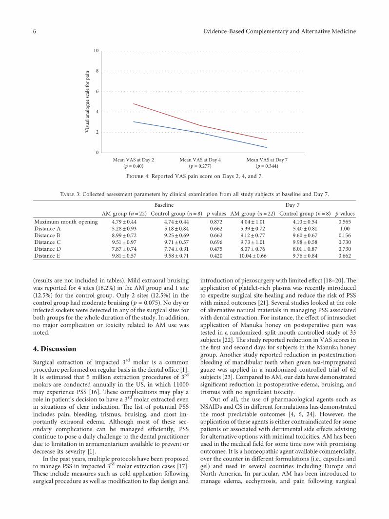
Postsurgery follow-ups of study participants werecompleted via phone calls on Days 2 and 4 (Table 2). On Day2, subjects in the AM group reported significantly lower painVAS of 3.09± 2.22 compared to 4.75± 1.28 for control group(p � 0.040). On Days 4 and 7, the AM group showedconsistently lower mean pain VAS, but the differences werenot statistically significant. On Day 4, the reported pain VASwas 1.86± 1.49 for AM group and 2.63± 1.60 for the controlgroup (p � 0.277). At the same time, pain VAS on Day 7 forAM group was 0.45± 1.26 and 1.25± 1.91 for the controlgroup (p � 0.344) (Figure 4). Bleeding was significantly lessreported in the AM group compared to the control group(p � 0.004) on Day 2. +e majority of sites (72.7%) in theAM group had no bleeding reported, while the majority ofsites in the control group (75.0%) reported Grade 1 bleeding.None of the patients in both groups reported any bleedingon Day 4.
In terms of extraoral swelling (edema), no statisticallysignificant difference was found between the groups re-garding the distribution of swelling severity on Day 2(p � 0.085). However, the swelling was significantly lesssevere in the AM group compared to the control group(p � 0.027). In the AM group, 7 sites (31.8%) had noswelling, 12 sites (54.5%) had mild swelling, 2 sites (9.1%)had moderate swelling, and 1 site (4.5%) had severe swelling.For the control group, 1 site (12.5%) had no swelling, 2 sites(25%) had mild swelling, 1 site (12.5%) had moderateswelling, and 4 sites (50%) had severe swelling. On Day 2,
A
E
B
C
D
Figure 1: Facial landmarks and distances to angle of the mandibleused to evaluate study subjects for facial edema at baseline and Day7 (A� distance to tragus, B� distance to lateral canthus,C� distance to alar base, D� distance to lip commissure, andE� distance to pogonion).
Evidence-Based Complementary and Alternative Medicine 3
extraoral bruising was significantly less severe in the AMgroup compared to the control group (p � 0.029). Mildextraoral bruising was reported for 2 sites (9.1%) in the AMgroup compared to 4 sites (50%) in the control sites. By Day4, no extraoral bruising was reported for 16 sites (72.2%) inthe AM group; however, 3 sites (13.6%) had mild and 3 sites(13.6%) hadmoderate bruising. For the control group, 5 sites(62.5%) had no bruising, 1 site (12.5%) had mild, 1 site(12.5%) had moderate, and 1 site (12.5%) had severebruising. +e differences in extraoral bruising were notsignificant between both groups (p � 0.526).
+e distribution of the reported severity of limitedmouth opening was significantly less severe on Day 2 amongAM group compared to control group (p � 0.016). In theAM group, no limitation in maximum moth opining wasreported in 5 sites (22.7%). However, 12 sites (54.65%) had
mild grade, 4 sites (18.2%) had moderate grade, and 1 site(4.5%) had severe limitation in mouth opening. Comparingthese numbers to control group, 1 site (12.5%) had nolimitation in mouth opening, 2 sites (25%) had mild grade,and 5 sites (62.5%) had moderate grade. Similar findings interms of distribution of limited mouth opening severity werereported between both groups on Day 4. In the AM group,limitation in maximum moth opining was absent in 7 sites(31.8%); however, 11 sites (50%) had mild grade, 3 sites(13.6%) had moderate grade, and 1 site (4.5%) had severelimitation in mouth opening. Comparing these numbers tocontrol group, 1 site (12.5%) had no limitation in mouthopening, 2 sites (25%) had mild grade, 3 sites (37.5%) hadmoderate grade, and 2 sites (25%) had severe grade.
On Day 7, all subjects had a scheduled follow-up visit inthe dental clinic to assess healing following the extraction
Table 1: Demographics of included patients.
AM group (n� 22 sites) Control group (n� 8 sites)Age (mean/years) 26 24Gender 15 females/7 males 8 femalesClassification of impacted mandibular 3rd molarDepth B 12 (54.5%) 4 (50.0%)Depth C 10 (45.5%) 4 (50.0%)Class II 11 (50.0%) 4 (50.0%)Class III 11 (50.0%) 4 (50.0%)
(a) (b)
(c) (d)
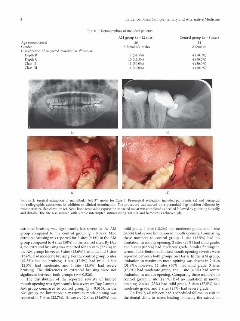
Figure 2: Surgical extraction of mandibular left 3rd molar for Case 1. Presurgical evaluation included panoramic (a) and periapical(b) radiographic assessment in addition to clinical examination. +e procedure was started by a pyramidal flap incision followed bymucoperiosteal flab elevation (c). Next, bone removal to expose the impacted molar was completed as needed followed by guttering buccallyand distally. +e site was sutured with simple interrupted sutures using 3-0 silk and hemostasis achieved (d).
4 Evidence-Based Complementary and Alternative Medicine
procedure. No difference in maximum mouth openingbetween AM and control group was noted (p � 0.565). Inaddition, the average facial measurements for edema
assessment (distances A, B, C, D, and E) between bothgroups were comparable with no statistical significance(Table 3). Extraoral bruising was also assessed on Day 7
(a) (b)
(c) (d)
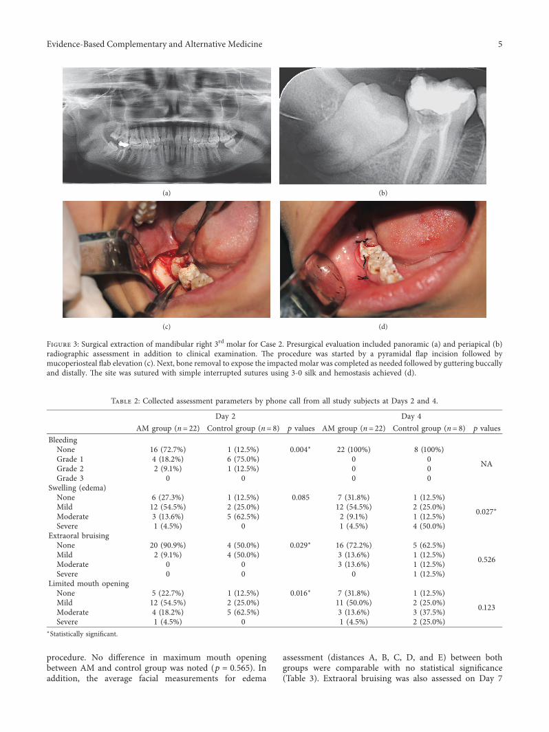
Figure 3: Surgical extraction of mandibular right 3rd molar for Case 2. Presurgical evaluation included panoramic (a) and periapical (b)radiographic assessment in addition to clinical examination. +e procedure was started by a pyramidal flap incision followed bymucoperiosteal flab elevation (c). Next, bone removal to expose the impacted molar was completed as needed followed by guttering buccallyand distally. +e site was sutured with simple interrupted sutures using 3-0 silk and hemostasis achieved (d).
Table 2: Collected assessment parameters by phone call from all study subjects at Days 2 and 4.
Day 2 Day 4AM group (n� 22) Control group (n� 8) p values AM group (n� 22) Control group (n� 8) p values
BleedingNone 16 (72.7%) 1 (12.5%) 0.004∗ 22 (100%) 8 (100%)
NAGrade 1 4 (18.2%) 6 (75.0%) 0 0Grade 2 2 (9.1%) 1 (12.5%) 0 0Grade 3 0 0 0 0
Swelling (edema)None 6 (27.3%) 1 (12.5%) 0.085 7 (31.8%) 1 (12.5%)
0.027∗Mild 12 (54.5%) 2 (25.0%) 12 (54.5%) 2 (25.0%)Moderate 3 (13.6%) 5 (62.5%) 2 (9.1%) 1 (12.5%)Severe 1 (4.5%) 0 1 (4.5%) 4 (50.0%)
Extraoral bruisingNone 20 (90.9%) 4 (50.0%) 0.029∗ 16 (72.2%) 5 (62.5%)
0.526Mild 2 (9.1%) 4 (50.0%) 3 (13.6%) 1 (12.5%)Moderate 0 0 3 (13.6%) 1 (12.5%)Severe 0 0 0 1 (12.5%)
Limited mouth openingNone 5 (22.7%) 1 (12.5%) 0.016∗ 7 (31.8%) 1 (12.5%)
0.123Mild 12 (54.5%) 2 (25.0%) 11 (50.0%) 2 (25.0%)Moderate 4 (18.2%) 5 (62.5%) 3 (13.6%) 3 (37.5%)Severe 1 (4.5%) 0 1 (4.5%) 2 (25.0%)
∗Statistically significant.
Evidence-Based Complementary and Alternative Medicine 5
(results are not included in tables). Mild extraoral bruisingwas reported for 4 sites (18.2%) in the AM group and 1 site(12.5%) for the control group. Only 2 sites (12.5%) in thecontrol group had moderate bruising (p � 0.075). No dry orinfected sockets were detected in any of the surgical sites forboth groups for the whole duration of the study. In addition,no major complication or toxicity related to AM use wasnoted.
4. Discussion
Surgical extraction of impacted 3rd molar is a commonprocedure performed on regular basis in the dental office [1].It is estimated that 5 million extraction procedures of 3rdmolars are conducted annually in the US, in which 11000may experience PSS [16]. +ese complications may play arole in patient’s decision to have a 3rd molar extracted evenin situations of clear indication. +e list of potential PSSincludes pain, bleeding, trismus, bruising, and most im-portantly extraoral edema. Although most of these sec-ondary complications can be managed efficiently, PSScontinue to pose a daily challenge to the dental practitionerdue to limitation in armamentarium available to prevent ordecrease its severity [1].
In the past years, multiple protocols have been proposedto manage PSS in impacted 3rd molar extraction cases [17].+ese include measures such as cold application followingsurgical procedure as well as modification to flap design and
introduction of piezosurgery with limited effect [18–20].+eapplication of platelet-rich plasma was recently introducedto expedite surgical site healing and reduce the risk of PSSwith mixed outcomes [21]. Several studies looked at the roleof alternative natural materials in managing PSS associatedwith dental extraction. For instance, the effect of intrasocketapplication of Manuka honey on postoperative pain wastested in a randomized, split-mouth controlled study of 33subjects [22]. +e study reported reduction in VAS scores inthe first and second days for subjects in the Manuka honeygroup. Another study reported reduction in postextractionbleeding of mandibular teeth when green tea-impregnatedgauze was applied in a randomized controlled trial of 62subjects [23]. Compared to AM, our data have demonstratedsignificant reduction in postoperative edema, bruising, andtrismus with no significant toxicity.
Out of all, the use of pharmacological agents such asNSAIDs and CS in different formulations has demonstratedthe most predictable outcomes [4, 6, 24]. However, theapplication of these agents is either contraindicated for somepatients or associated with detrimental side effects advisingfor alternative options with minimal toxicities. AM has beenused in the medical field for some time now with promisingoutcomes. It is a homeopathic agent available commercially,over the counter in different formulations (i.e., capsules andgel) and used in several countries including Europe andNorth America. In particular, AM has been introduced tomanage edema, ecchymosis, and pain following surgical
Mean VAS at Day 2(p = 0.40)
Mean VAS at Day 4(p = 0.277)
Mean VAS at Day 7(p = 0.344)
0
2
4
6
8
10
Visu
al an
alog
ue sc
ale f
or p
ain
Figure 4: Reported VAS pain score on Days 2, 4, and 7.
Table 3: Collected assessment parameters by clinical examination from all study subjects at baseline and Day 7.
Baseline Day 7AM group (n� 22) Control group (n� 8) p values AM group (n� 22) Control group (n� 8) p values
Maximum mouth opening 4.79± 0.44 4.74± 0.44 0.872 4.04± 1.01 4.10± 0.54 0.565Distance A 5.28± 0.93 5.18± 0.84 0.662 5.39± 0.72 5.40± 0.81 1.00Distance B 8.99± 0.72 9.25± 0.69 0.662 9.12± 0.77 9.60± 0.67 0.156Distance C 9.51± 0.97 9.71± 0.57 0.696 9.73± 1.01 9.98± 0.58 0.730Distance D 7.87± 0.74 7.74± 0.91 0.475 8.07± 0.76 8.01± 0.87 0.730Distance E 9.81± 0.57 9.58± 0.71 0.420 10.04± 0.66 9.76± 0.84 0.662
6 Evidence-Based Complementary and Alternative Medicine

procedures. +e current study report on the potential ap-plication of AM to prevent or manage PSS associated withextraction of impacted mandibular 3rd molars.
As of today, almost all of the data available on AM isoriginating from the medical literature. A case series in-cluding 13 subjects who went through rhinoplasty surgerywith osteotomies and received AM for 3 days was reported[25]. All patients had accelerated postoperative healing anddecrease in bruising and ecchymosis. Out of all, a singlepatient experienced mild itching and rash which resolvedduring the study follow-up duration. A recent study re-ported a faster resolution of postoperative sore throat,dysphagia, aphonia, and hoarseness in 2 patients followinglaryngeal mask insertion [26]. Both patients were treatedwith 3 doses of AM and reported symptoms resolutionwithin 36 hours. On the other hand, a randomized, double-blinded clinical trial was conducted and included 27 subjectsto evaluate the effect of topical AM hydrogel pads on ec-chymosis following upper blepharoplasty [27]. After 30 days,the study failed to demonstrate a statistical significancebetween both groups in terms of ecchymosis or healingperiod.
In the dental literature, a single study from the 1980slooked at the potential use of AM in dental surgeries. In thisrandomized double-blinded clinical trial, the effect of AMand metronidazole on PSS following extraction of man-dibular 3rd molars was evaluated in 118 patients [28]. Studyparticipants were randomly assigned to three groups: Group1 received metronidazole (400mg twice daily); Group 2received AM (200mg twice daily); Group 3 received placebotablets. Pain score, edema, and trismus parameters were usedto compare the outcomes of AM and metronidazole. At theend of the study, metronidazole reduced the incidence ofpain and edema and enhanced the healing process followingsurgical extraction compared to AM and placebo groups buthad no effect on trismus. In addition, AM was less effectivethan the placebo in this clinical study. +ese results ingeneral contradict our data, which could be contributed tofactors such as difference in assessment methods, surgicaltechnique used, and classification of impacted teethextracted during the course of the study. Considering thelimited available literature on AM in the dental field, itsapplication may still benefit patients at risk of PSS based onthe such as in cases of external sinus augmentation andmajor bone grafting procedures on a case-by-case basis.+erefore, the use of AM in the current study was justifiablebased on the comparative, recent medical literature[9, 29, 30].
In order to better evaluate the effect of AM, severalparameters were assessed at different time points in thisstudy. On Day 2, patients in AM group had better paincontrol and milder PSS in terms of intraoral bleeding,extraoral bruising, and trismus compared to control group.In addition, extraoral edema was significantly less reportedin the AM group compared to control. On Day 4, the dif-ference in postoperative edema was more evident betweenboth groups as 31.8% of AM sites had no swelling comparedto 12.5% sites of control group. During the follow-up visit onDay 7, most of PSS have resolved completely for both groups
as anticipated for similar cases in general. However, lowerpain scores for AM sites were reported compared to controlsites (0.45± 1.26 versus 1.25± 1.91).
As of today, limited measures are available to prevent orreduce PSS following surgical extraction of impacted 3rdmolars. Hyaluronic acid (HA) is linear polysaccharides ofthe extracellular matrix which can be found in various bodytissues. Several studies investigated the effect of HA depo-sition in extraction sockets of impacted 3rd molars anddemonstrated decrease in postoperative pain [31]. However,no role in reducing other PSS was reported. A randomized,double-blind, crossover study was conducted to comparebetween the effect of etodolac, naproxen, and diclofenac asPSS prophylaxis for patients receiving surgical extraction of3rd molars [32]. A total of 42 patients were included in thestudy and allocated to either Group A of etodolac (200mg),Group B of naproxen sodium (275mg), or Group C ofdiclofenac potassium (50mg) to be administered 1 hourbefore the procedure and continued for 3–5 days afterward.At the end of the study, diclofenac potassium was signifi-cantly more effective in reducing postsurgical edemacompared to other groups.
+e effect of CS either through injectable or systemicroutes on PSS has been investigated extensively in the past. Asplit-mouth, randomized triple-blind controlled clinical trialwas conducted to compare the effect of dexamethasone(8mg/day) to diclofenac sodium (50mg/day) and codeine(50mg/day) on patients receiving extraction of bilateralmandibular 3rd molars [33]. +e study demonstrateddexamethasone to be the most efficient in controllingpostoperative pain and edema. In a randomized clinical trial,intralesional and intravenous dexamethasone given 1 hourbefore procedure was effective and superior to oral dexa-methasone in decreasing postoperative pain and edemafollowing surgical extraction of 3rd molars [7]. Combinationof CS with other agents such as NSAIDs for synergistic effecthas also been investigated and was superior to dexameth-asone and NSAIDs alone in controlling postoperative edemaand pain [34].
Based on our data, AM showed significant potential todecrease the degree of edema in addition to pain experiencefollowing surgical extraction of impacted mandibular 3rdmolars. Even with all patients received antibiotic therapy,none were prescribed NSAIDs to eliminate the confoundingrisk. One factor to consider when assessing AM effect is thepurity of product and additive contents if any. +e com-mercial supplements are not overseen by food and drugadministration inmost countries; significant variations in AMcontents may exist among manufacturers potentially affectingthe overall clinical outcome [35]. AM dosing is another factorto consider which varied between conducted studies in theliterature and has to be investigated in future studies andassessed for bias [36]. +e literature on AM safety profile isalso lacking. However, several larger studies have investigatedthe effect of AM in managing PSS associated with differentprocedures and failed to report major complications in in-cluded subjects even in the setting of chronic underlyingmedical conditions such as diabetes, hypertension, and renaldisease [9, 12]. Overall, the literature reported side effects were
Evidence-Based Complementary and Alternative Medicine 7

limited to mild itching and rash [25]. Hence, it may bereasonable to consider AM as a fairly safe product for humanconsumption. As of today, AM interaction with othermedication is not clear and should be evaluated on a case bycase basis.
+e current study is aimed at highlighting the potentialrole of AM in dental setting, specifically in extraction ofimpacted mandibular molar. Based on our data, AM couldbe offered to selective group of patients with concerns overpostoperative edema, bruising, and/or pain. It is fair toanticipate the same benefit with using AM in other invasivedental surgeries such as sinus augmentation and guidedtissue regeneration. However, future studies needed betterevidence-based application.
+is study has several limitations. First, the enrollment oflarger number of study and control sites may have allowed forbetter evaluation of the study outcome and more justificationfor clinical application in the daily practice. However, thisstudy helps to shed more light on the potential benefit of AMin reducing PSS associated with impacted 3rd molars ex-traction and help to support future studies to confirm thesefinding. Second, a single dose of AM was used in this casesseries. Comparison between different manufactures’ productsand dosing would have given better understating on the bestway to apply AM in the dental field. +ird, assessment ofpostsurgical pain, bleeding, and edema was self-reported onDays 2 and 4 whichmay have been biased and underreported.In-clinic patient assessment on Days 2 and 4 would beconsidered in the future. Fourth, factors such as unequalnumber of enrolled subjects in relation to gender, race,variability of used postsurgical medications, and comparingamount of bone removed during the extraction may haveaffected our results.
5. Conclusion
Based on the current findings, AM seems to have a potentialbenefit in management of secondary complications fol-lowing surgical extraction of impacted 3rd molars specificallyfor pain, ecchymosis, and edema. Further randomizedclinical trials with a larger group of subjects are warranted toconfirm these findings.
Data Availability
+e data supporting the conclusions of the study can beprovided upon request to the corresponding author byemailing.
Ethical Approval
A human research ethical approval was obtained throughAl-Farabi Private College, School of Dentistry, Jeddah, SaudiArabia.
Conflicts of Interest
+e authors declare that there are no conflicts of interestregarding the publication of this article.
References
[1] P. Glera-Suarez, D. Soto-Penaloza, D. Penarrocha-Oltra, andM. Penarrocha-Diago, “Patient morbidity after impactedthird molar extraction with different flap designs. A sys-tematic review and meta-analysis,” Medicina oral, patologiaoral y cirugia bucal, vol. 25, no. 2, pp. e233–e239, 2020.
[2] S. I. Yamada, T. Hasegawa, S. Soutome et al., “Prevalence ofand risk factors for postoperative hemorrhage after lowerthird molar extraction on Warfarin therapy: a multicenterretrospective study in Japan,” Odontology, vol. 108, no. 3,pp. 462–469, 2019.
[3] W. Zhang, J. Li, Z. B. Li, and Z. Li, “Predicting postoperativefacial swelling following impacted mandibular third molarsextraction by using artificial neural networks evaluation,”Scientific Reports, vol. 8, no. 1, p. 12281, 2018.
[4] E. Shoohanizad and M. Parvin, “Comparison the dexa-methasone administration effects on post-operative sequelaebefore and after “third molar” extraction surgeries,” Endo-crine, Metabolic & Immune Disorders—Drug Targets, vol. 20,no. 3, pp. 356–364, 2019.
[5] R. Brignardello-Petersen, “Similar pain, swelling, and trismusafter third-molar surgical extraction when receiving a phy-totherapeutic drug, ibuprofen, or a placebo,” 7e Journal ofthe American Dental Association, vol. 150, no. 5, p. e50, 2019.
[6] R. Brignardello-Petersen, “+ere are probably no addedbenefits of intravenous ketamine for postoperative pain inpatients undergoing third-molar extraction who receiveperioperative antibiotics, ibuprofen, and dexamethasone,”7e Journal of the American Dental Association, vol. 151, no. 1,p. e1, 2020.
[7] M. Brucoli, M. De Andreis, M. Bonaso, P. Boffano, andA. Benech, “Comparative assessment of dexamethasone ad-ministration routes for the management of postoperativesymptoms following third molar surgery,” Journal of Sto-matology, Oral and Maxillofacial Surgery, vol. 120, no. 6,pp. 529–533, 2019.
[8] F. Sortino and M. Cicciu, “Strategies used to inhibit post-operative swelling following removal of impacted lower thirdmolar,” Dental Research Journal, vol. 8, no. 4, pp. 162–171,2011.
[9] R. Knackstedt and J. Gatherwright, “Perioperative homeo-pathic arnica and bromelain,” Annals of Plastic Surgery,vol. 84, no. 3, pp. e10–e15, 2020.
[10] P. Kriplani, K. Guarve, and U. S. Baghael, “Arnica montanaL.—a plant of healing: review,” Journal of Pharmacy andPharmacology, vol. 69, no. 8, pp. 925–945, 2017.
[11] M. Sutovska, P. Capek, M. Kocmalova et al., “Characterizationand pharmacodynamic properties of Arnica montana com-plex,” International Journal of Biological Macromolecules,vol. 69, pp. 214–221, 2014.
[12] D. Ho, J. Jagdeo, and H. A. Waldorf, “Is there a role for arnicaand bromelain in prevention of post-procedure ecchymosis oredema? A systematic review of the literature,” DermatologicSurgery, vol. 42, no. 4, pp. 445–463, 2016.
[13] G. B. Pell, “Impacted mandibular third molars:classificationand modified techniques for removal,” 7e Dental Digest,vol. 39, pp. 330–338, 1933.
[14] E. A. Neupert III, J. W. Lee, C. B. Philput, and J. R. Gordon,“Evaluation of dexamethasone for reduction of postsurgicalsequelae of third molar removal,” Journal of Oral andMaxillofacial Surgery, vol. 50, no. 11, pp. 1177–1182, 1992.
8 Evidence-Based Complementary and Alternative Medicine
[15] R. Lotter, “Periodontal considerations in the extraction ofretained wisdom teeth. Varying technics,” Le Chirurgien-dentiste de France, vol. 54, no. 252, pp. 35–44, 1984.
[16] J. W. Friedman, “+e prophylactic extraction of third molars:a public health hazard,” American Journal of Public Health,vol. 97, no. 9, pp. 1554–1559, 2007.
[17] L. Laino, G. Troiano, M. Dioguardi et al., “Patient discomfortduring and after surgically assisted rapid maxillary expansionunder local anaesthesia,” Journal of Craniofacial Surgery,vol. 27, no. 3, pp. 772–775, 2016.
[18] A. Ali, S. J. Shah, A. A. Shah, and S. Aslam, “Comparison ofcomma incision with Ward’s incision in third molar ex-traction in terms of postoperative sequel—a clinical study,”National Journal of Maxillofacial Surgery, vol. 10, no. 2,pp. 200–205, 2019.
[19] R. Brignardello-Petersen, “Inconsistent evidence regardingthe benefits of cryotherapy in minimizing postoperativecomplications after third-molar extraction,”7e Journal of theAmerican Dental Association, vol. 150, no. 9, p. e134, 2019.
[20] C. Patil, A. Jadhav, R. M. Borle, and A. Mishra, “Piezosurgeryvs bur in impacted mandibular third molar surgery: evalu-ation of postoperative sequelae,” Journal of Oral Biology andCraniofacial Research, vol. 9, no. 3, pp. 259–262, 2019.
[21] J. V. Dos Santos Canellas, F. G. Ritto, and P. J. D. A. Medeiros,“Efficacy of platelet-rich fibrin after mandibular third molarextraction: a systematic review and meta-analysis,” Journal ofOral and Maxillofacial Surgery, vol. 75, no. 8, pp. 1576-1577,2017.
[22] N. M. Al-Khanati and Y. Al-Moudallal, “Effect of intrasocketapplication of Manuka honey on postsurgical pain of im-pacted mandibular third molars surgery: split-mouth ran-domized controlled trial,” Journal of Maxillofacial and OralSurgery, vol. 18, no. 1, pp. 147–152, 2019.
[23] R. Soltani, A. Haghighat, M. Fanaei, and G. Asghari, “Eval-uation of the effect of green tea extract on the prevention ofgingival bleeding after posterior mandibular teeth extraction:a randomized controlled trial,” Evidence-Based Complemen-tary and Alternative Medicine, vol. 2014, Article ID 857651,4 pages, 2014.
[24] R. Brignardello-Petersen, “Submucosal dexamethasone re-duces pain, swelling, and trismus after impacted third-molarextraction,” 7e Journal of the American Dental Association,vol. 148, no. 5, p. e64, 2017.
[25] S. R. Chaiet and B. C. Marcus, “Perioperative Arnica montanafor reduction of ecchymosis in rhinoplasty surgery,” Annals ofPlastic Surgery, vol. 76, no. 5, pp. 477–482, 2016.
[26] D. Tsintzas and G. Vithoulkas, “Treatment of postoperativesore throat with the aid of the homeopathic remedy Arnicamontana: a report of two cases,” Journal of Evidence-BasedComplementary & Alternative Medicine, vol. 22, no. 4,pp. 926–928, 2017.
[27] B. S. Kotlus, D.M. Heringer, and R.M. Dryden, “Evaluation ofhomeopathic Arnica montana for ecchymosis after upperblepharoplasty: a placebo-controlled, randomized, double-blind study,” Ophthalmic Plastic & Reconstructive Surgery,vol. 26, no. 6, pp. 395–397, 2010.
[28] G. S. N. Kaziro, “Metronidazole (Flagyl) and Arnica montanain the prevention of post-surgical complications, a compar-ative placebo controlled clinical trial,” British Journal of Oraland Maxillofacial Surgery, vol. 22, no. 1, pp. 42–49, 1984.
[29] W. Lee, W. Mack, S. Seiff, J. Kang, and K. Tran, “Assessing theeffectiveness of Arnica montana and Rhododendrontomentosum (Ledum palustre) in the reduction of ecchymosisand edema after oculofacial surgery: preliminary results:
Erratum,” Ophthalmic Plastic and Reconstructive Surgery,vol. 34, no. 2, p. 188, 2018.
[30] L. Sorrentino, S. Piraneo, E. Riggio et al., “Is there a role forhomeopathy in breast cancer surgery? A first randomizedclinical trial on treatment with Arnica montana to reducepostoperative seroma and bleeding in patients undergoingtotal mastectomy,” Journal of Intercultural Ethno-pharmacology, vol. 6, no. 1, pp. 1–8, 2017.
[31] N. Yilmaz, N. Demirtas, H. O. Kazancioglu, S. Bayer,A. H. Acar, and A. Mihmanli, “+e efficacy of hyaluronic acidin postextraction sockets of impacted third molars: a pilotstudy,” Nigerian Journal of Clinical Practice, vol. 20, no. 12,pp. 1626–1631, 2017.
[32] N. Akbulut, E. Ustuner, C. Atakan, and G. Colok, “Com-parison of the effect of naproxen, etodolac and diclofenac onpostoperative sequels following third molar surgery: arandomised, double-blind, crossover study,” Medicina OralPatologıa Oral Y Cirugia Bucal, vol. 19, no. 2, pp. e149–e156,2014.
[33] T. C. Lima, E. Bagordakis, S. G. M. Falci, C. R. R. Dos Santos,and M. L. P. Pinheiro, “Pre-emptive effect of dexamethasoneand diclofenac sodium associated with codeine on pain,swelling, and trismus after third molar surgery: a split-mouth,randomized, triple-blind, controlled clinical trial,” Journal ofOral and Maxillofacial Surgery, vol. 76, no. 1, pp. 60–66, 2018.
[34] B. O. Bamgbose, J. A. Akinwande, W. L. Adeyemo,A. L. Ladeinde, G. T. Arotiba, andM. O. Ogunlewe, “Effects ofco-administered dexamethasone and diclofenac potassium onpain, swelling and trismus following third molar surgery,”Head & Face Medicine, vol. 1, p. 11, 2005.
[35] P. Bellavite, M. Marzotto, and C. Bonafini, “Arnica montanaexperimental studies: confounders and biases?” Journal ofIntegrative Medicine, vol. 16, no. 2, pp. 72–76, 2018.
[36] P. Bellavite, M. Marzotto, and C. Bonafini, “Critical com-ments and methodological variations in Arnica montana’sresearch studies,” Journal of Ayurveda and Integrative Med-icine, vol. 9, no. 3, pp. 238-239, 2018.
Evidence-Based Complementary and Alternative Medicine 9


![Research Article Propagation and Introduction of Arnica ...arnica in many European countries [ , ]. A. montana habitats have been fragmented, especially at the edge of its dense geographical](https://img.pdfslide.us/doc/110x75/60b4c536b7f73a47361060d6/research-article-propagation-and-introduction-of-arnica-arnica-in-many-european.jpg)