Embed Size (px)
Citation preview
SYSTEMIC PATHOLOGY I - VPM 222
Pathology of the
Endocrine System
Lecture 1
General Mechanisms & Pituitary Gland (Web Review)
Paul Hanna Fall 2012

Reference Texts
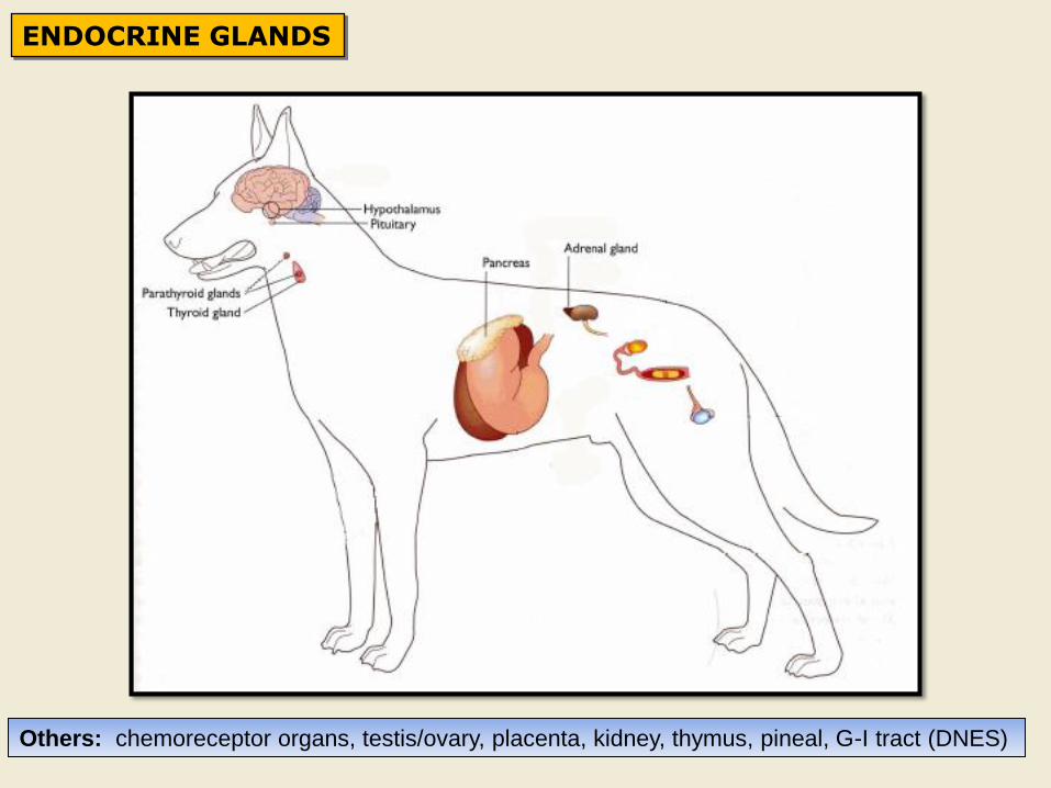
ENDOCRINE GLANDS
Others: chemoreceptor organs, testis/ovary, placenta, kidney, thymus, pineal, G-I tract (DNES)
From McGavin & Zachary: Pathologic Basis of Veterinary Disease, 4th ed
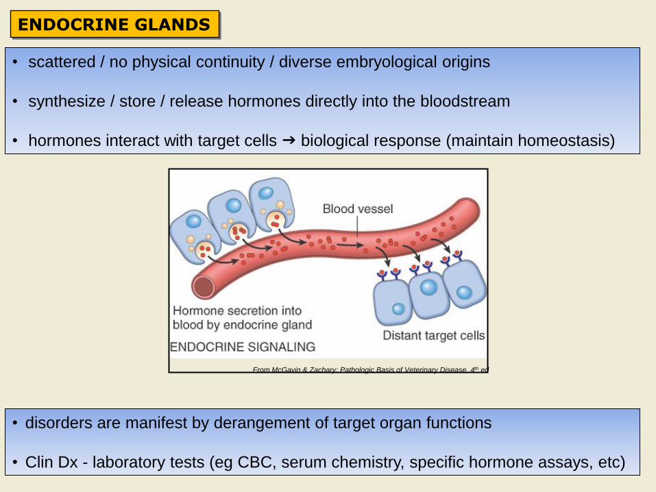
ENDOCRINE GLANDS
• scattered / no physical continuity / diverse embryological origins
• synthesize / store / release hormones directly into the bloodstream
• hormones interact with target cells biological response (maintain homeostasis)
• disorders are manifest by derangement of target organ functions
• Clin Dx - laboratory tests (eg CBC, serum chemistry, specific hormone assays, etc)
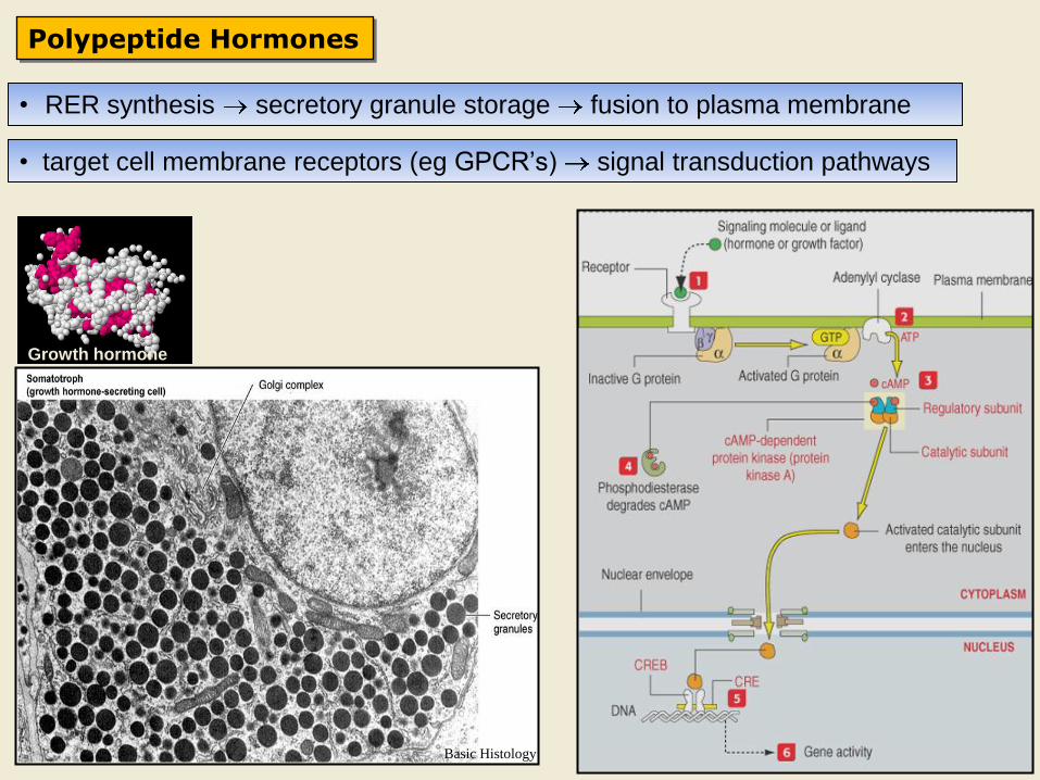
Polypeptide Hormones
Basic Histology
Growth hormone
• RER synthesis secretory granule storage fusion to plasma membrane
• target cell membrane receptors (eg GPCR’s) signal transduction pathways
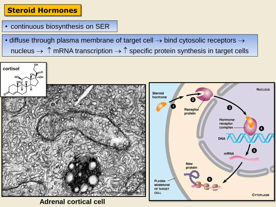
Steroid Hormones
[Bloom & Fawcett: A textbook of Histology]
Adrenal cortical cell
cortisol
• continuous biosynthesis on SER
• diffuse through plasma membrane of target cell bind cytosolic receptors
nucleus mRNA transcription specific protein synthesis in target cells
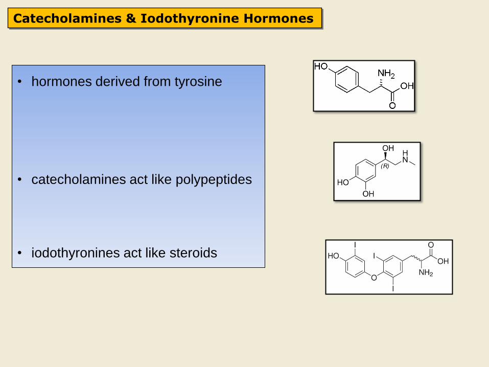
Catecholamines & Iodothyronine Hormones
• hormones derived from tyrosine
• catecholamines act like polypeptides
• iodothyronines act like steroids
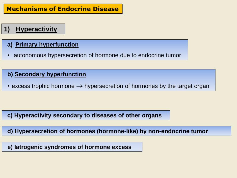
Mechanisms of Endocrine Disease
1) Hyperactivity
a) Primary hyperfunction
• autonomous hypersecretion of hormone due to endocrine tumor
b) Secondary hyperfunction
• excess trophic hormone hypersecretion of hormones by the target organ
c) Hyperactivity secondary to diseases of other organs
d) Hypersecretion of hormones (hormone-like) by non-endocrine tumor
e) Iatrogenic syndromes of hormone excess
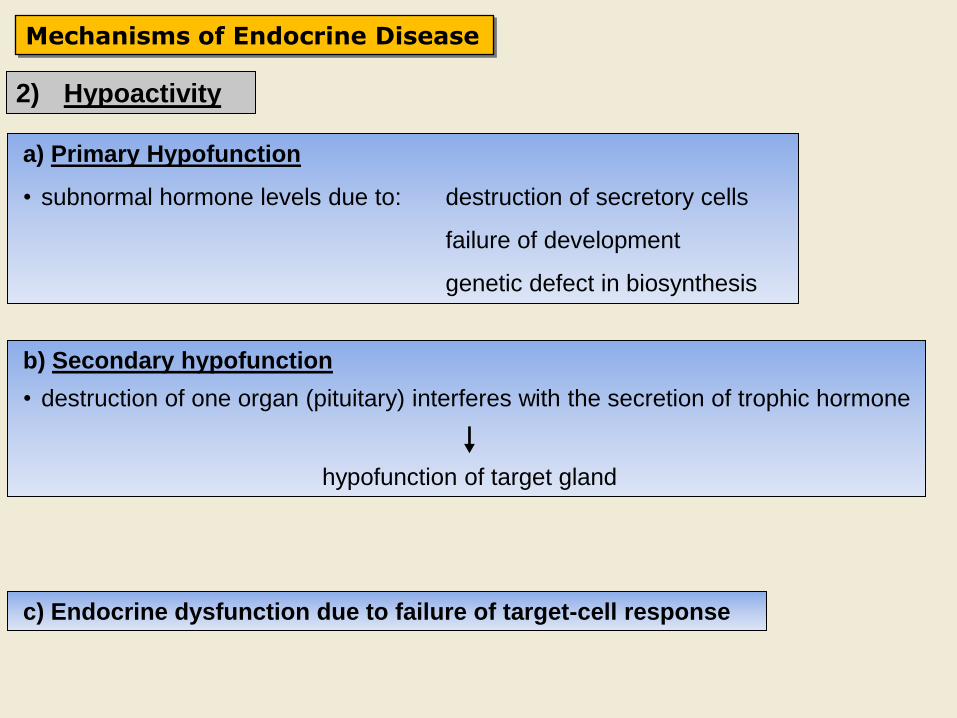
Mechanisms of Endocrine Disease
2) Hypoactivity
a) Primary Hypofunction
• subnormal hormone levels due to: destruction of secretory cells
failure of development
genetic defect in biosynthesis
b) Secondary hypofunction
• destruction of one organ (pituitary) interferes with the secretion of trophic hormone
hypofunction of target gland
c) Endocrine dysfunction due to failure of target-cell response

Pituitary Gland (Hypophysis)
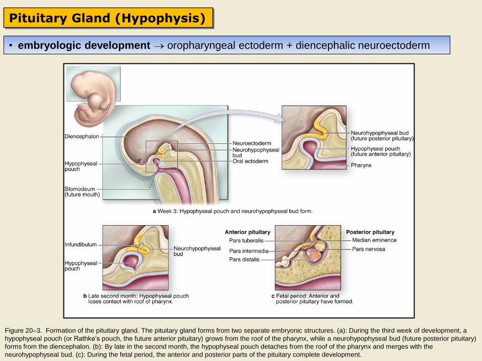
Figure 20–3. Formation of the pituitary gland. The pituitary gland forms from two separate embryonic structures. (a): During the third week of development, a
hypophyseal pouch (or Rathke’s pouch, the future anterior pituitary) grows from the roof of the pharynx, while a neurohypophyseal bud (future posterior pituitary)
forms from the diencephalon. (b): By late in the second month, the hypophyseal pouch detaches from the roof of the pharynx and merges with the
neurohypophyseal bud. (c): During the fetal period, the anterior and posterior parts of the pituitary complete development.
• embryologic development oropharyngeal ectoderm + diencephalic neuroectoderm
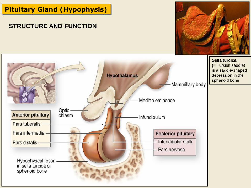
STRUCTURE AND FUNCTION
Pituitary Gland (Hypophysis)
Sella turcica
(= Turkish saddle)
is a saddle-shaped
depression in the
sphenoid bone
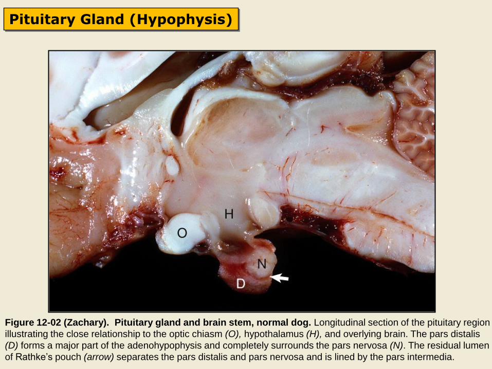
Pituitary Gland (Hypophysis)
Figure 12-02 (Zachary). Pituitary gland and brain stem, normal dog. Longitudinal section of the pituitary region
illustrating the close relationship to the optic chiasm (O), hypothalamus (H), and overlying brain. The pars distalis
(D) forms a major part of the adenohypophysis and completely surrounds the pars nervosa (N). The residual lumen
of Rathke’s pouch (arrow) separates the pars distalis and pars nervosa and is lined by the pars intermedia.
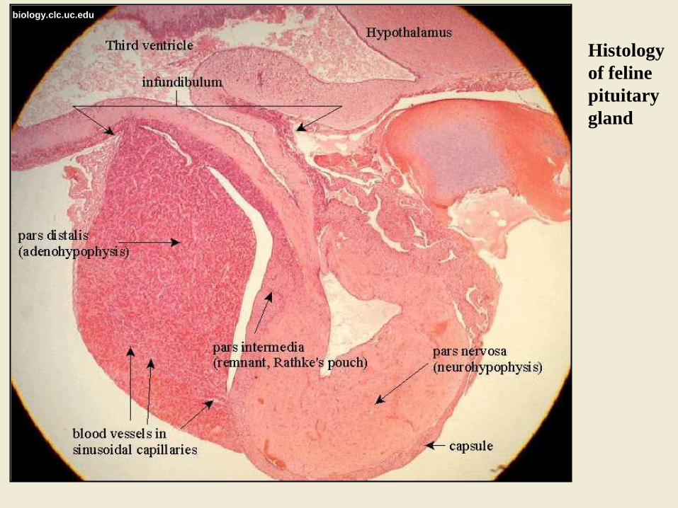
Histology
of feline
pituitary
gland
biology.clc.uc.edu
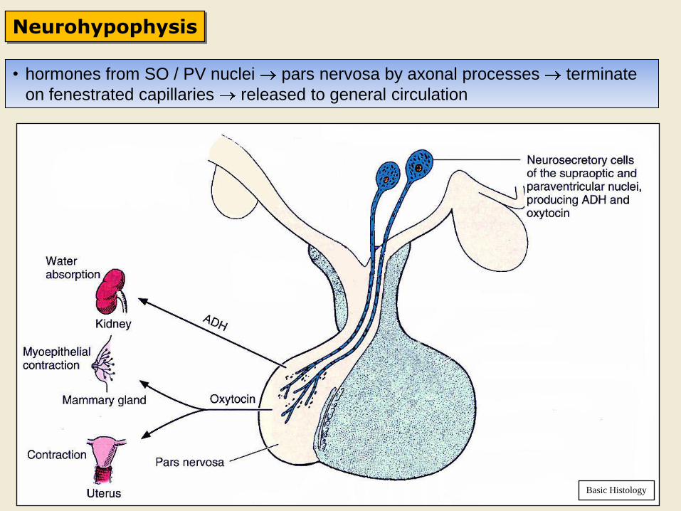
Neurohypophysis
Basic Histology
• hormones from SO / PV nuclei pars nervosa by axonal processes terminate
on fenestrated capillaries released to general circulation
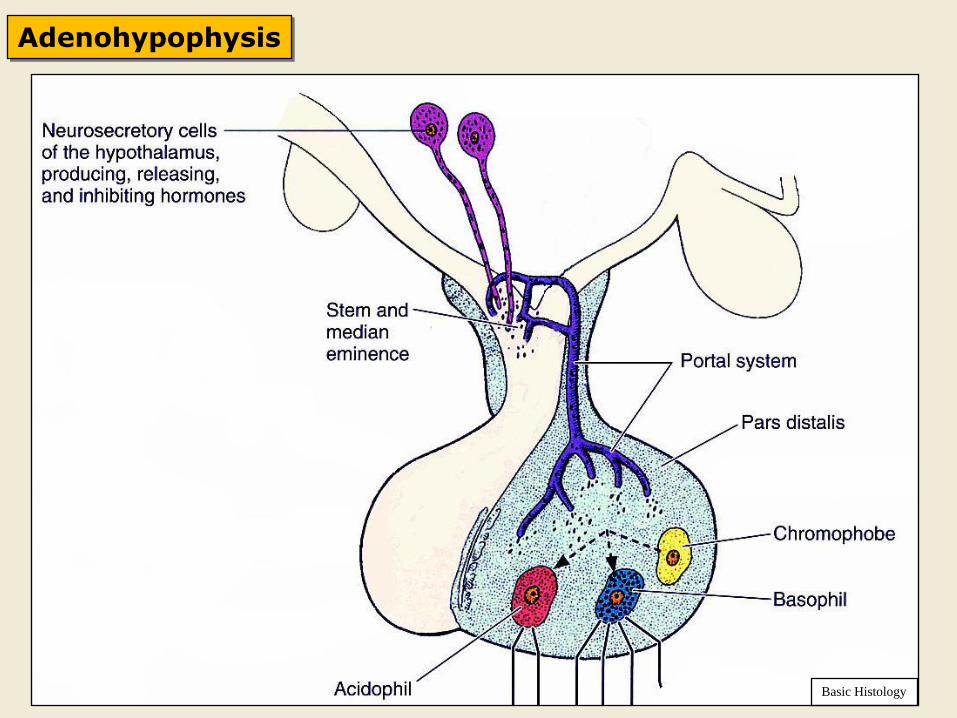
Adenohypophysis
Basic Histology
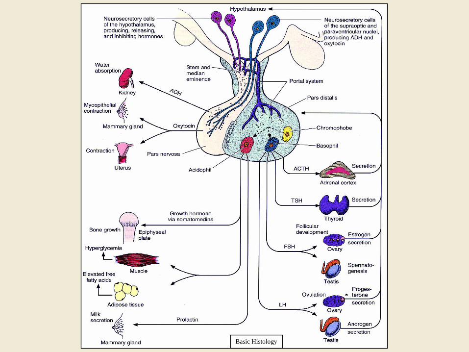
Basic Histology
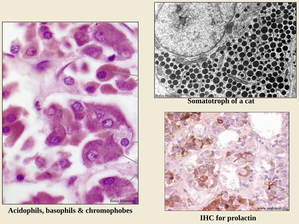
Acidophils, basophils & chromophobes
Basic Histology
Somatotroph of a cat Basic Histology
IHC for prolactin
www.endotext.org
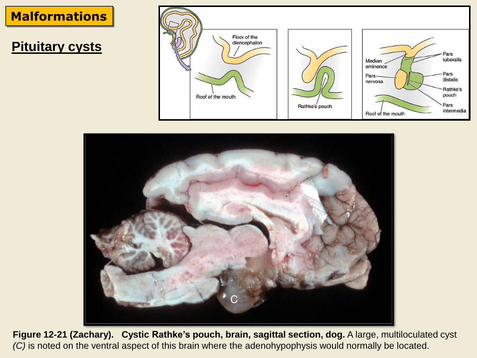
Pituitary cysts
Malformations
Figure 12-21 (Zachary). Cystic Rathke’s pouch, brain, sagittal section, dog. A large, multiloculated cyst
(C) is noted on the ventral aspect of this brain where the adenohypophysis would normally be located.
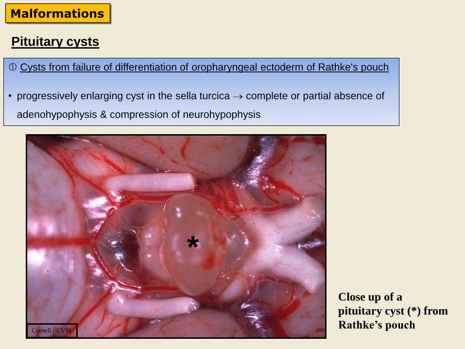
Pituitary cysts
Malformations
Cornell - CVM
Close up of a
pituitary cyst (*) from
Rathke’s pouch
*
Cysts from failure of differentiation of oropharyngeal ectoderm of Rathke's pouch
• progressively enlarging cyst in the sella turcica complete or partial absence of
adenohypophysis & compression of neurohypophysis
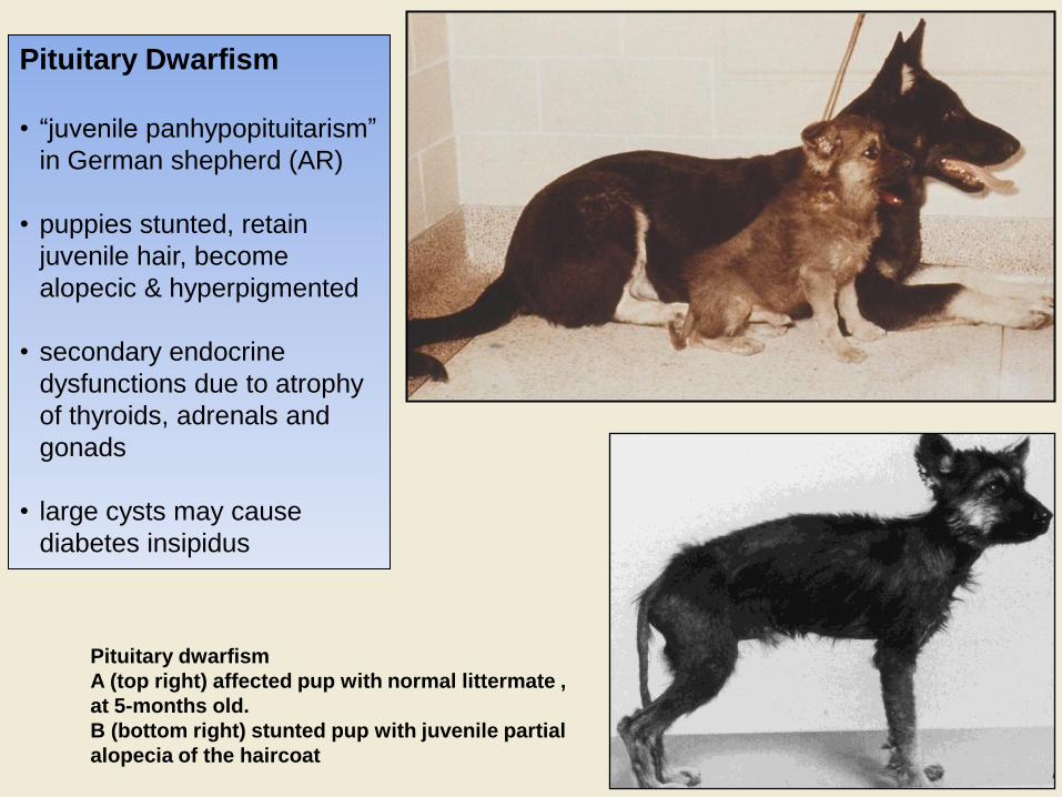
Pituitary dwarfism
A (top right) affected pup with normal littermate ,
at 5-months old.
B (bottom right) stunted pup with juvenile partial
alopecia of the haircoat
Pituitary Dwarfism
• “juvenile panhypopituitarism”
in German shepherd (AR)
• puppies stunted, retain
juvenile hair, become
alopecic & hyperpigmented
• secondary endocrine
dysfunctions due to atrophy
of thyroids, adrenals and
gonads
• large cysts may cause
diabetes insipidus
Inflammation
1) Pituitary Abscesses
• sporadically in ruminants and swine
• caused by bacteria or mycotic agents
• neurological signs due to local extension of the inflammation
2) Infiltration of mononuclear inflammatory cells
• in some viral and protozoan diseases as part of encephalitis or meningitis
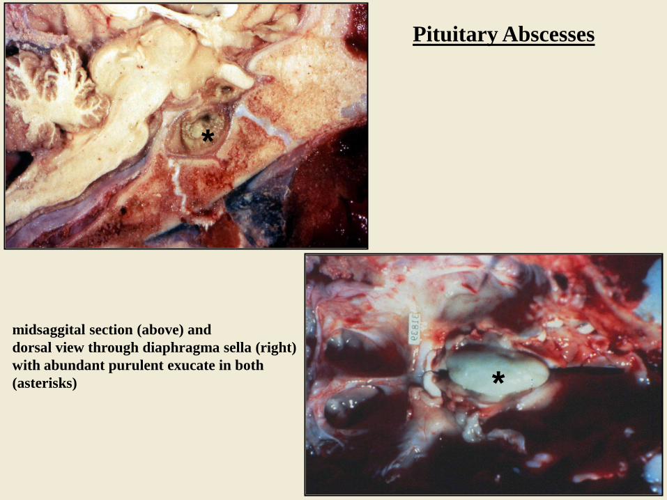
Pituitary Abscesses
midsaggital section (above) and
dorsal view through diaphragma sella (right)
with abundant purulent exucate in both
(asterisks)
*
*
Basic Histology
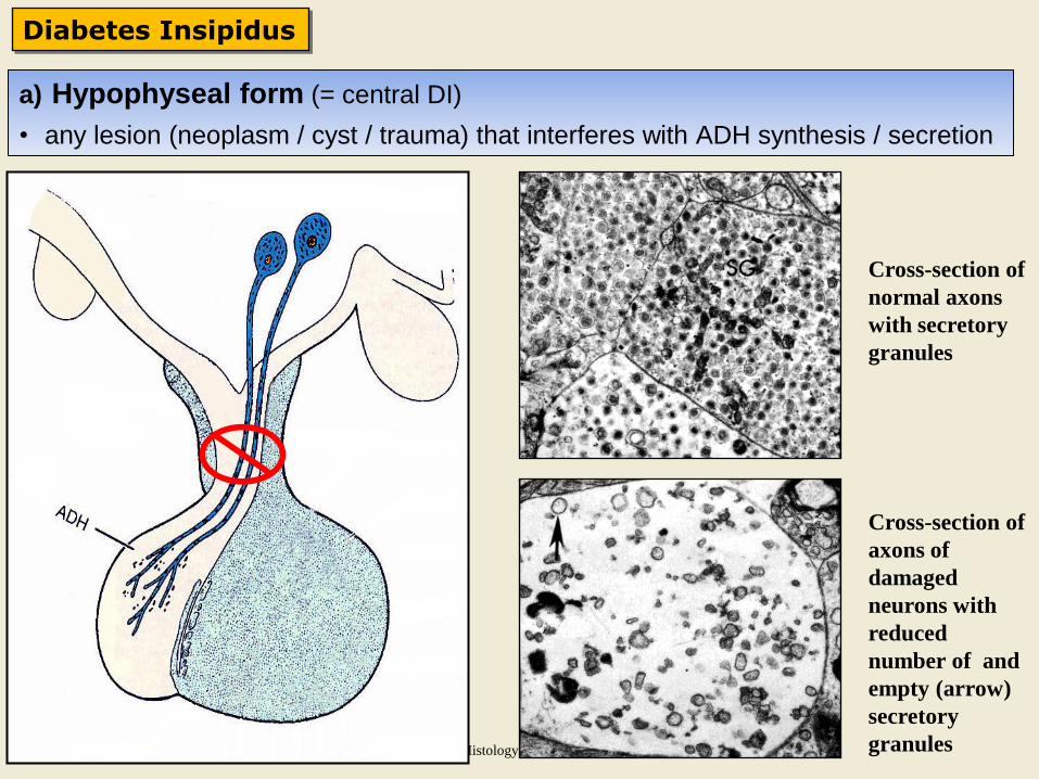
Diabetes Insipidus
Cross-section of
normal axons
with secretory
granules
Cross-section of
axons of
damaged
neurons with
reduced
number of and
empty (arrow)
secretory
granules
a) Hypophyseal form (= central DI)
• any lesion (neoplasm / cyst / trauma) that interferes with ADH synthesis / secretion
Diabetes Insipidus
Note: There can be genetic defects in ADH (=V2)
receptor or less frequently defects in the aquaporin 2
water channel.
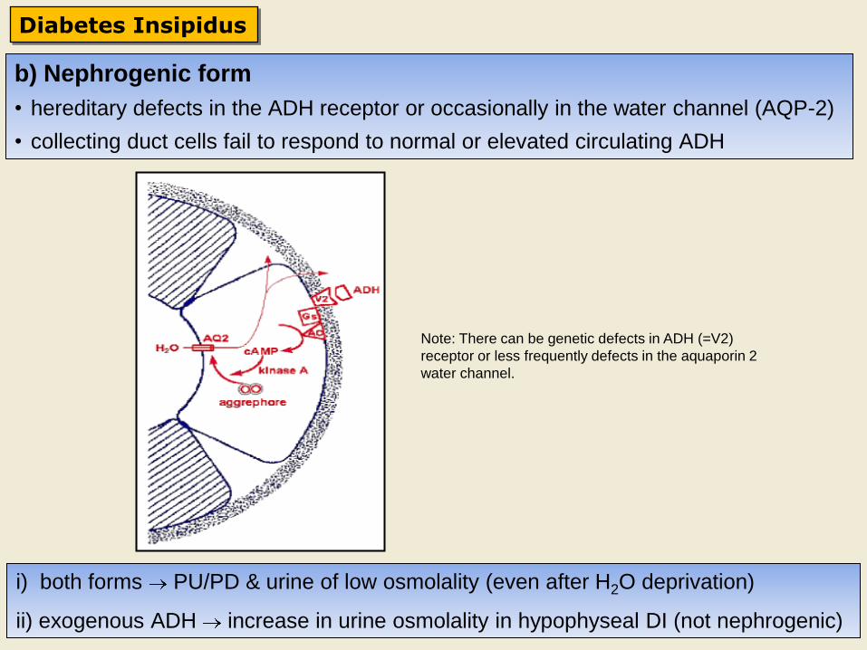
b) Nephrogenic form
• hereditary defects in the ADH receptor or occasionally in the water channel (AQP-2)
• collecting duct cells fail to respond to normal or elevated circulating ADH
i) both forms PU/PD & urine of low osmolality (even after H2O deprivation)
ii) exogenous ADH increase in urine osmolality in hypophyseal DI (not nephrogenic)
Hyperplasia / Neoplasia
• neoplasms may be functional (stimulate target organ)
or nonfunctional (destructive to adjacent structures)
• nodular hyperplasia vs adenomas (larger, capsule, compression)
Adenomas of Pars Distalis
www.endotext.org
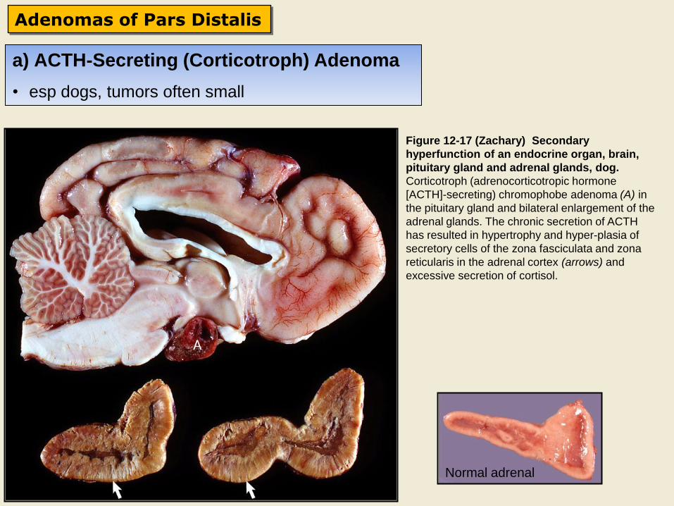
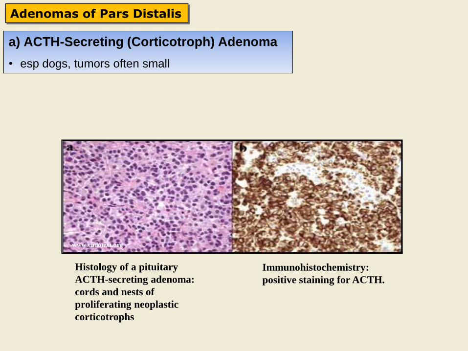
a) ACTH-Secreting (Corticotroph) Adenoma
• esp dogs, tumors often small
Figure 12-17 (Zachary) Secondary
hyperfunction of an endocrine organ, brain,
pituitary gland and adrenal glands, dog.
Corticotroph (adrenocorticotropic hormone
[ACTH]-secreting) chromophobe adenoma (A) in
the pituitary gland and bilateral enlargement of the
adrenal glands. The chronic secretion of ACTH
has resulted in hypertrophy and hyperplasia of
secretory cells of the zona fasciculata and zona
reticularis in the adrenal cortex (arrows) and
excessive secretion of cortisol.
Normal adrenal
Adenomas of Pars Distalis
Histology of a pituitary
ACTH-secreting adenoma:
cords and nests of
proliferating neoplastic
corticotrophs
Immunohistochemistry:
positive staining for ACTH.
www.endotext.org
a) ACTH-Secreting (Corticotroph) Adenoma
• esp dogs, tumors often small
Adenomas of Pars Distalis
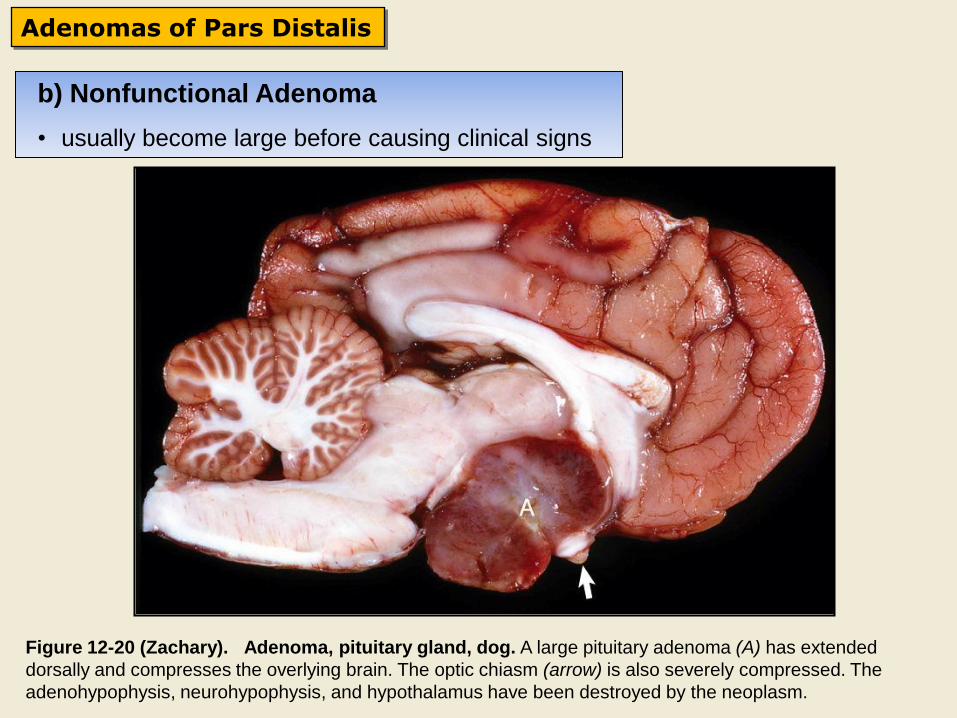
Figure 12-20 (Zachary). Adenoma, pituitary gland, dog. A large pituitary adenoma (A) has extended
dorsally and compresses the overlying brain. The optic chiasm (arrow) is also severely compressed. The
adenohypophysis, neurohypophysis, and hypothalamus have been destroyed by the neoplasm.
b) Nonfunctional Adenoma
• usually become large before causing clinical signs
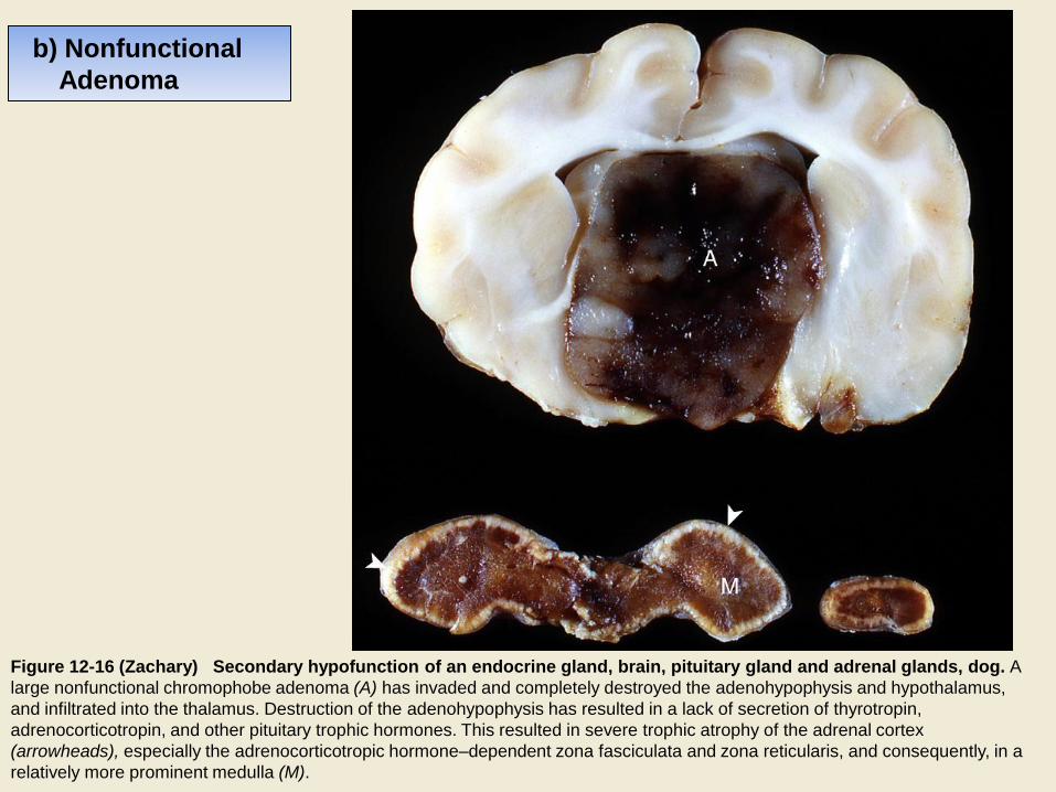
Figure 12-16 (Zachary) Secondary hypofunction of an endocrine gland, brain, pituitary gland and adrenal glands, dog. A
large nonfunctional chromophobe adenoma (A) has invaded and completely destroyed the adenohypophysis and hypothalamus,
and infiltrated into the thalamus. Destruction of the adenohypophysis has resulted in a lack of secretion of thyrotropin,
adrenocorticotropin, and other pituitary trophic hormones. This resulted in severe trophic atrophy of the adrenal cortex
(arrowheads), especially the adrenocorticotropic hormone–dependent zona fasciculata and zona reticularis, and consequently, in a
relatively more prominent medulla (M).
b) Nonfunctional
Adenoma
www.endotext.org
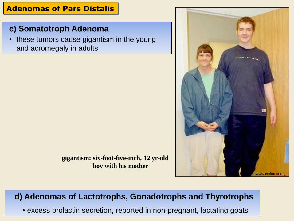
Adenomas of Pars Distalis
gigantism: six-foot-five-inch, 12 yr-old
boy with his mother
c) Somatotroph Adenoma
• these tumors cause gigantism in the young
and acromegaly in adults
d) Adenomas of Lactotrophs, Gonadotrophs and Thyrotrophs
• excess prolactin secretion, reported in non-pregnant, lactating goats
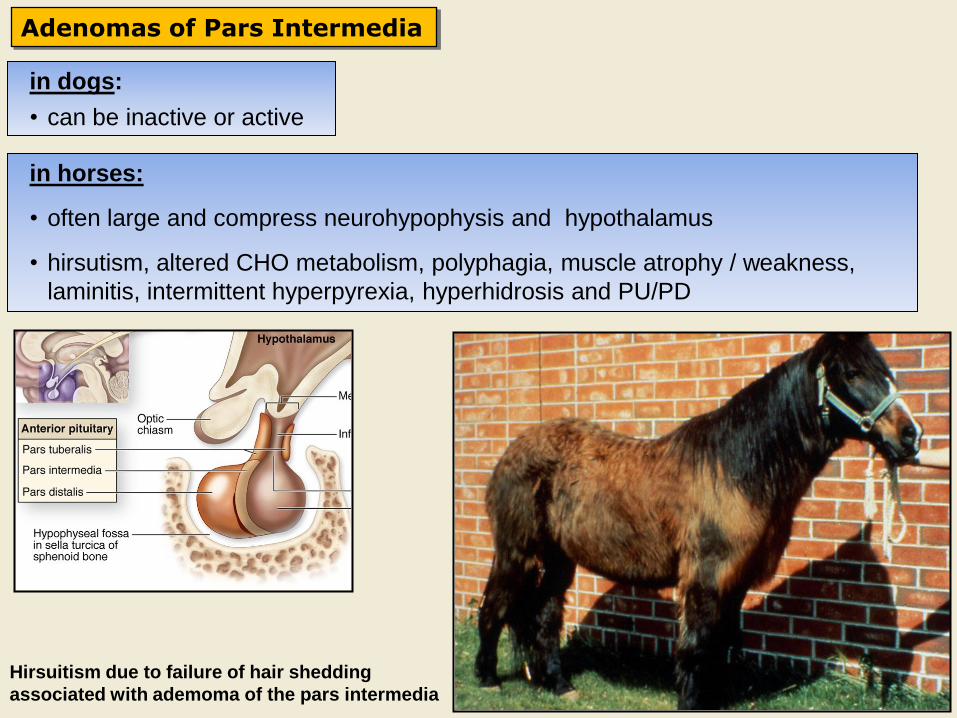
Adenomas of Pars Intermedia
Hirsuitism due to failure of hair shedding
associated with ademoma of the pars intermedia
in dogs:
• can be inactive or active
in horses:
• often large and compress neurohypophysis and hypothalamus
• hirsutism, altered CHO metabolism, polyphagia, muscle atrophy / weakness,
laminitis, intermittent hyperpyrexia, hyperhidrosis and PU/PD
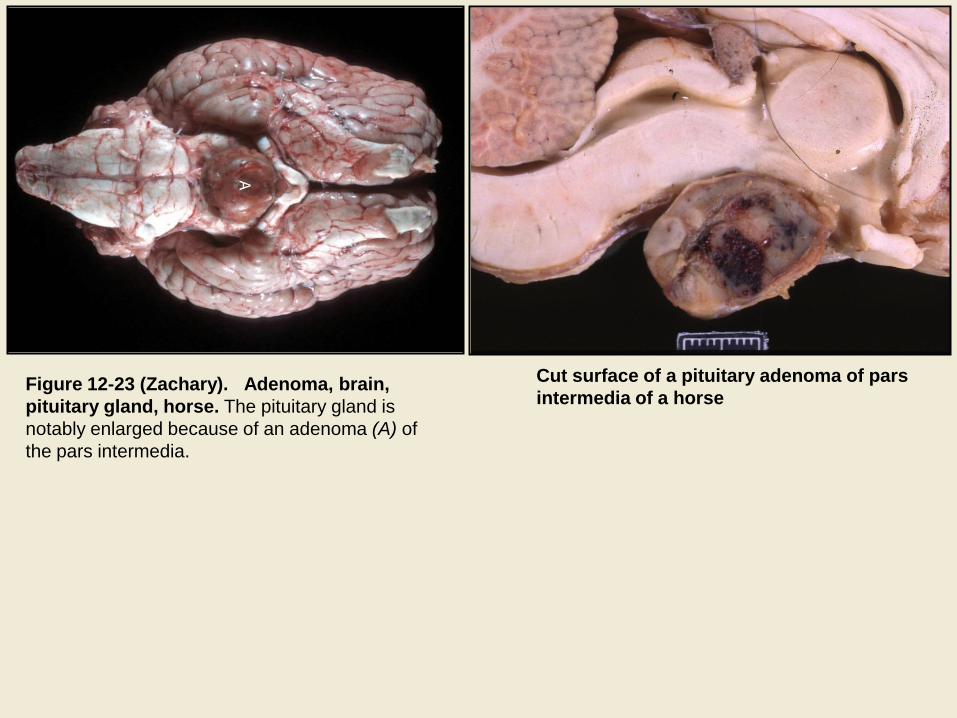
Cut surface of a pituitary adenoma of pars
intermedia of a horse Figure 12-23 (Zachary). Adenoma, brain,
pituitary gland, horse. The pituitary gland is
notably enlarged because of an adenoma (A) of
the pars intermedia.
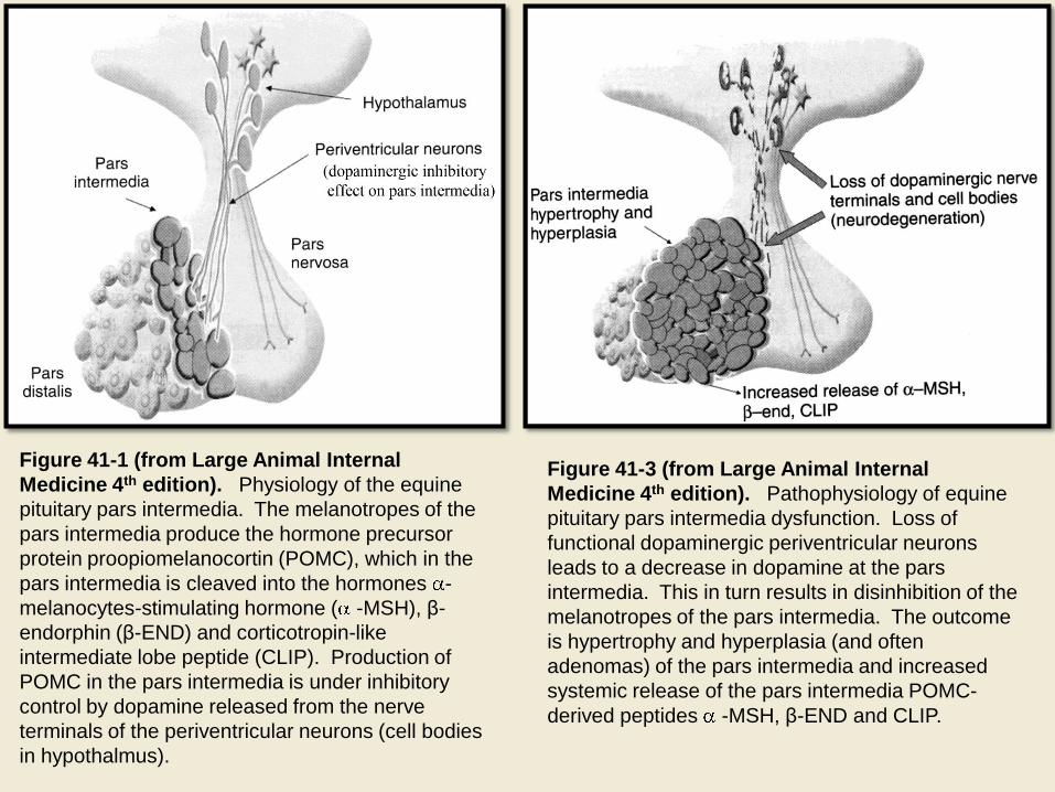
Figure 41-1 (from Large Animal Internal
Medicine 4th edition). Physiology of the equine
pituitary pars intermedia. The melanotropes of the
pars intermedia produce the hormone precursor
protein proopiomelanocortin (POMC), which in the
pars intermedia is cleaved into the hormones -
melanocytes-stimulating hormone ( -MSH), β-
endorphin (β-END) and corticotropin-like
intermediate lobe peptide (CLIP). Production of
POMC in the pars intermedia is under inhibitory
control by dopamine released from the nerve
terminals of the periventricular neurons (cell bodies
in hypothalmus).
Figure 41-3 (from Large Animal Internal
Medicine 4th edition). Pathophysiology of equine
pituitary pars intermedia dysfunction. Loss of
functional dopaminergic periventricular neurons
leads to a decrease in dopamine at the pars
intermedia. This in turn results in disinhibition of the
melanotropes of the pars intermedia. The outcome
is hypertrophy and hyperplasia (and often
adenomas) of the pars intermedia and increased
systemic release of the pars intermedia POMC-
derived peptides -MSH, β-END and CLIP.
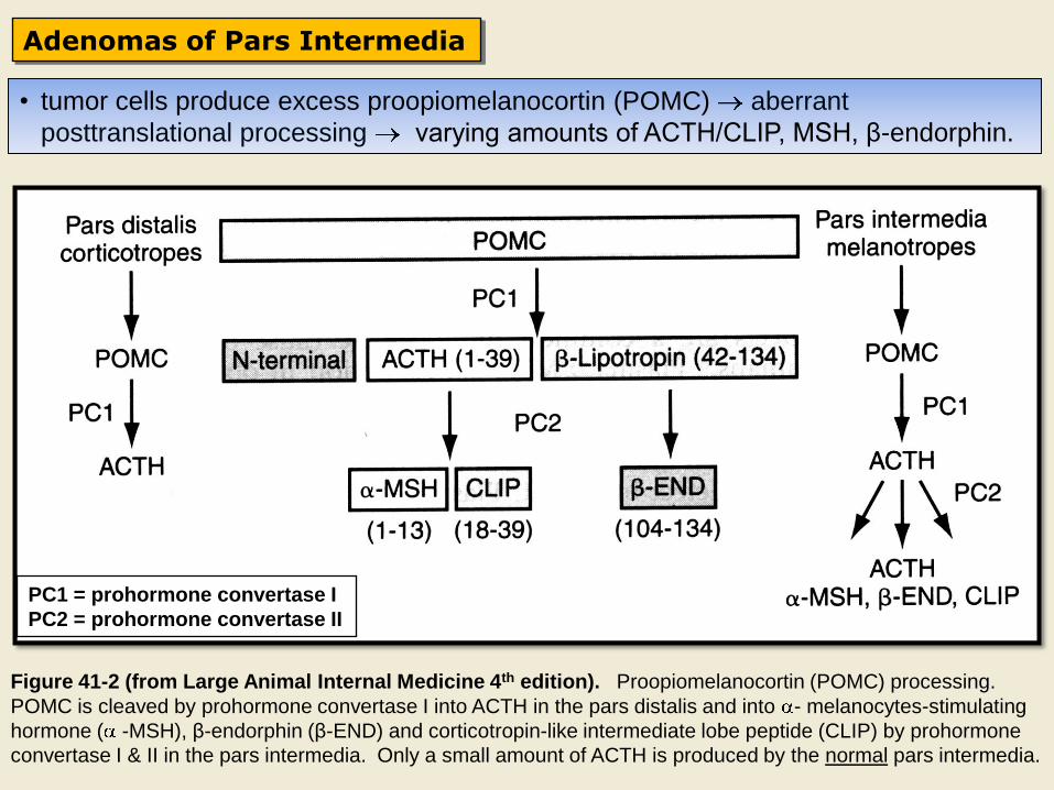
PC1 = prohormone convertase I
PC2 = prohormone convertase II
Figure 41-2 (from Large Animal Internal Medicine 4th edition). Proopiomelanocortin (POMC) processing.
POMC is cleaved by prohormone convertase I into ACTH in the pars distalis and into - melanocytes-stimulating
hormone ( -MSH), β-endorphin (β-END) and corticotropin-like intermediate lobe peptide (CLIP) by prohormone
convertase I & II in the pars intermedia. Only a small amount of ACTH is produced by the normal pars intermedia.
Adenomas of Pars Intermedia
• tumor cells produce excess proopiomelanocortin (POMC) aberrant
posttranslational processing varying amounts of ACTH/CLIP, MSH, β-endorphin.
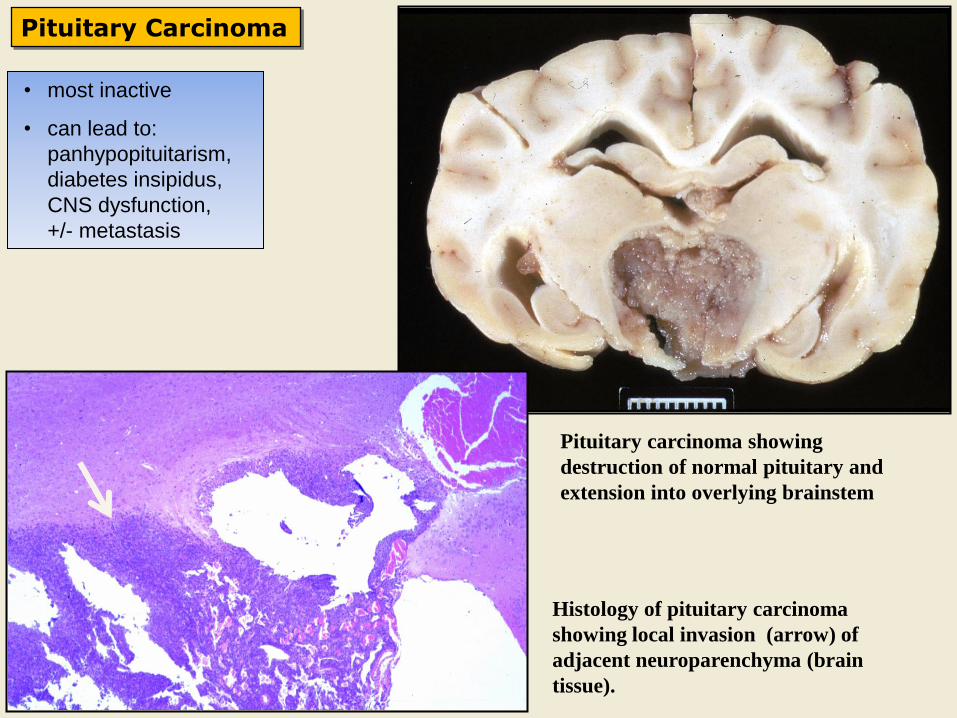
Pituitary Carcinoma
Pituitary carcinoma showing
destruction of normal pituitary and
extension into overlying brainstem
Histology of pituitary carcinoma
showing local invasion (arrow) of
adjacent neuroparenchyma (brain
tissue).
• most inactive
• can lead to:
panhypopituitarism,
diabetes insipidus,
CNS dysfunction,
+/- metastasis
Metastatic Tumors
• tumors metastasizing to the pituitary would be uncommon, but could also lead to
panhypopituitarism, CNS signs, etc


![Endocrine Pathology, 4E (2014) [UnitedVRG]](https://img.pdfslide.us/doc/110x75/577c7de31a28abe054a00b57/endocrine-pathology-4e-2014-pdf-unitedvrg.jpg)