Embed Size (px)
Citation preview
Systemic ModuleCNS1
“Anatomy”
Sensory TractsDr. Ayman Alzubi
Faculty of Medicine, Yarmouk University
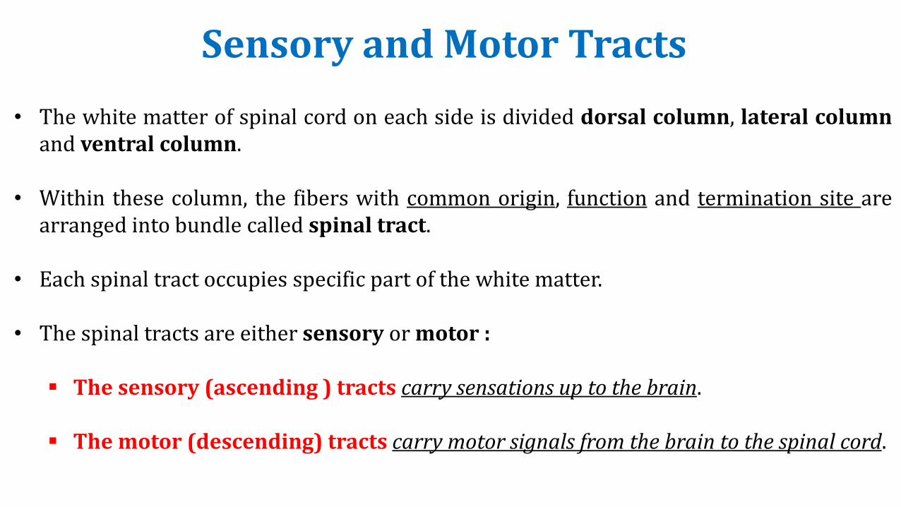
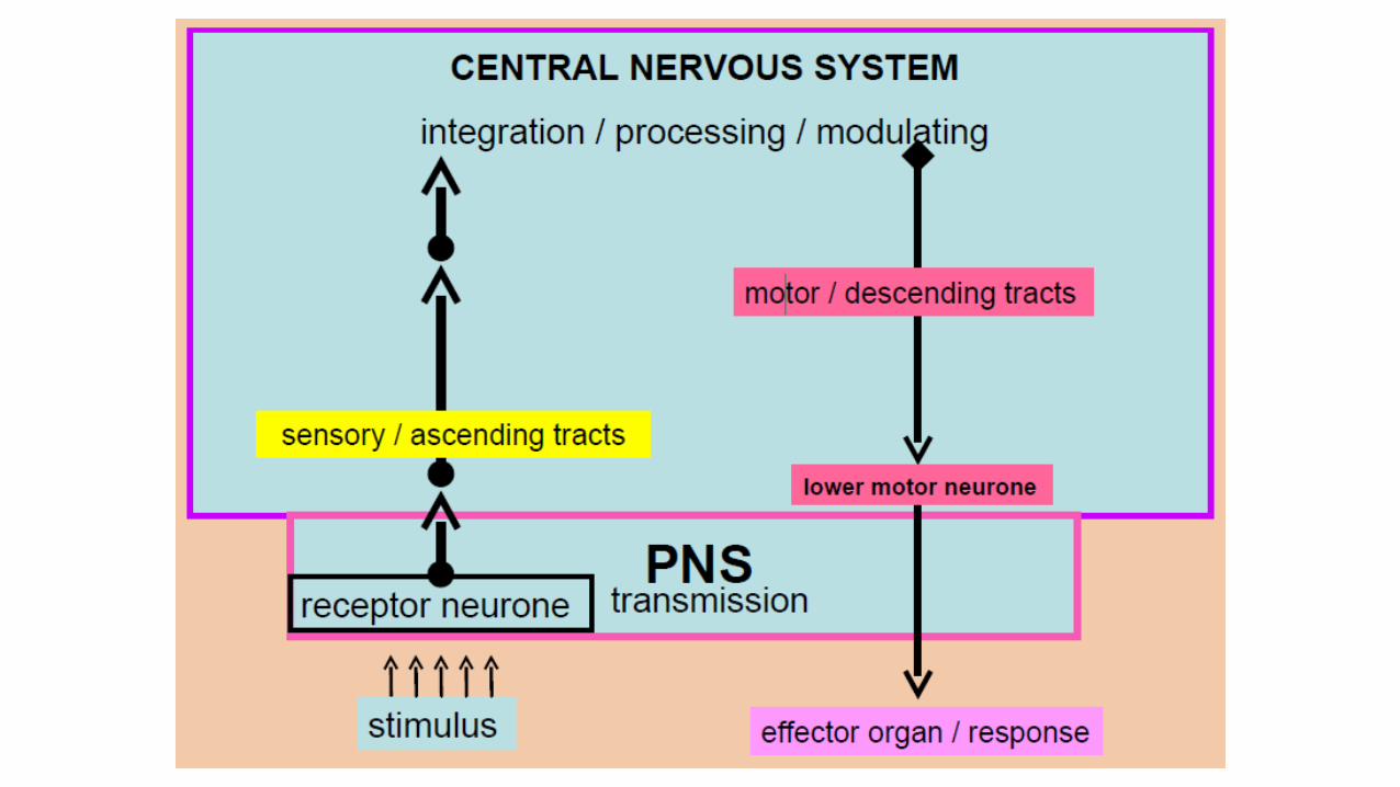
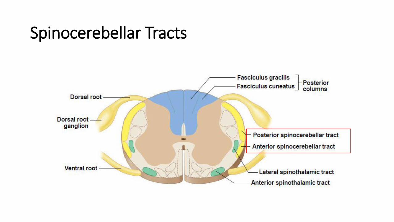
• The white matter of spinal cord on each side is divided dorsal column, lateral columnand ventral column.
• Within these column, the fibers with common origin, function and termination site arearranged into bundle called spinal tract.
• Each spinal tract occupies specific part of the white matter.
• The spinal tracts are either sensory or motor :
▪ The sensory (ascending ) tracts carry sensations up to the brain.
▪ The motor (descending) tracts carry motor signals from the brain to the spinal cord.
Sensory and Motor Tracts
Sensory Tracts
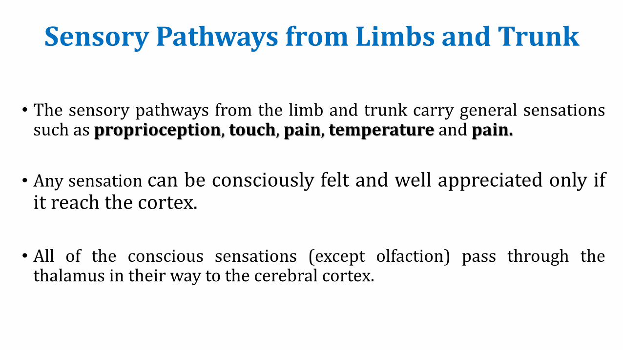
Sensory Pathways from Limbs and Trunk
• The sensory pathways from the limb and trunk carry general sensationssuch as proprioception, touch, pain, temperature and pain.
• Any sensation can be consciously felt and well appreciated only ifit reach the cortex.
• All of the conscious sensations (except olfaction) pass through thethalamus in their way to the cerebral cortex.
Sensory Tracts
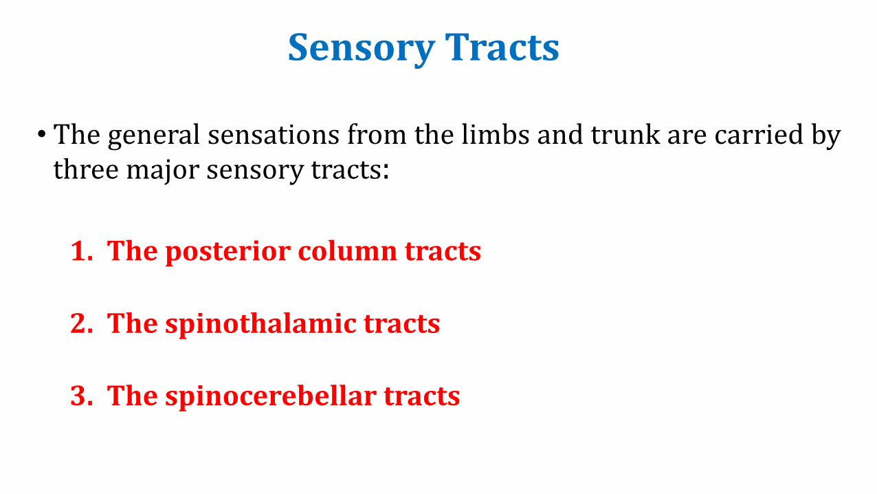
• The general sensations from the limbs and trunk are carried bythree major sensory tracts:
1. The posterior column tracts
2. The spinothalamic tracts
3. The spinocerebellar tracts
Sensory Tracts
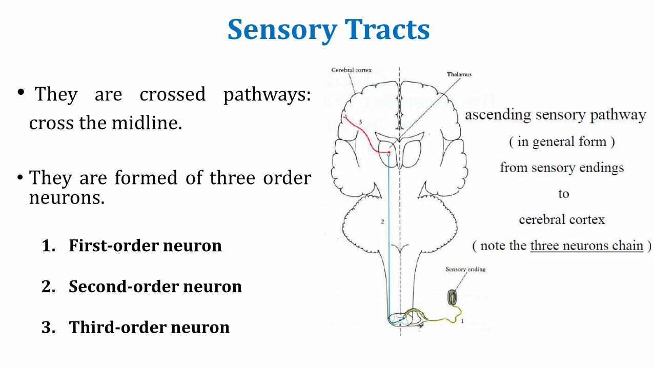
• They are crossed pathways:
cross the midline.
• They are formed of three orderneurons.
1. First-order neuron
2. Second-order neuron
3. Third-order neuron
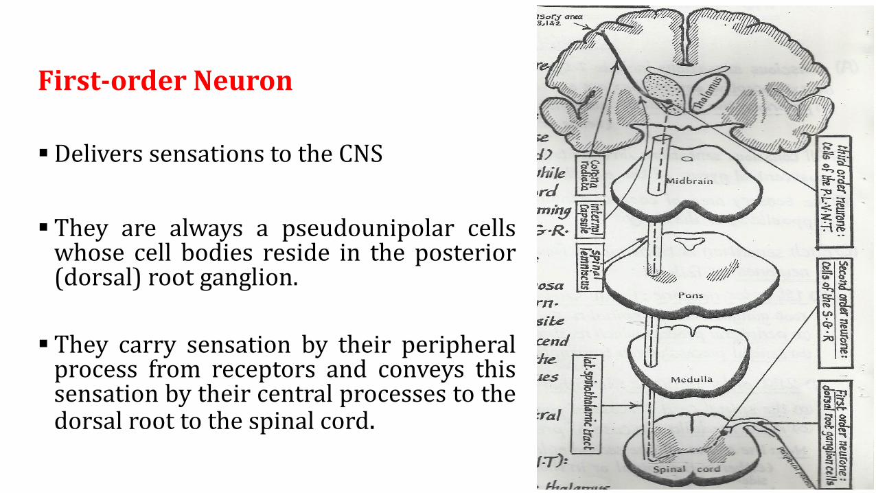
First-order Neuron
▪ Delivers sensations to the CNS
▪ They are always a pseudounipolar cellswhose cell bodies reside in the posterior(dorsal) root ganglion.
▪ They carry sensation by their peripheralprocess from receptors and conveys thissensation by their central processes to thedorsal root to the spinal cord.

FIRST ORDER NEURON
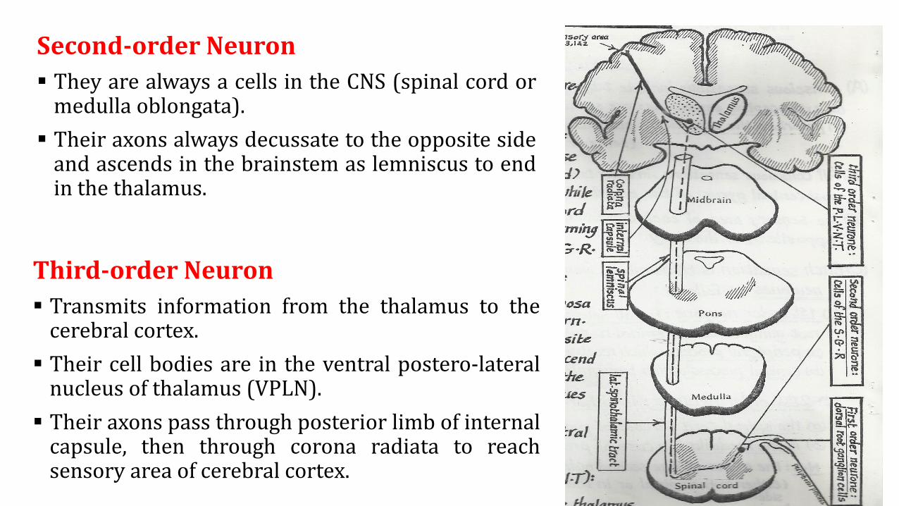
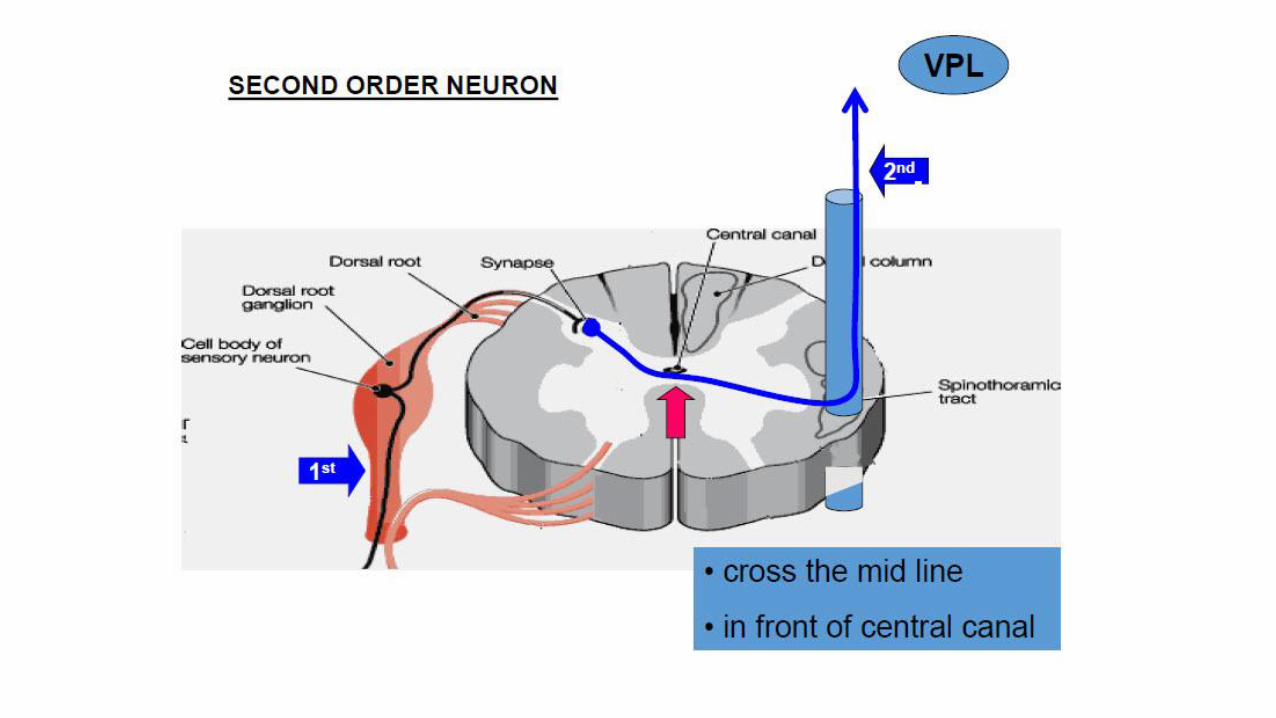
Second-order Neuron
▪ They are always a cells in the CNS (spinal cord ormedulla oblongata).
▪ Their axons always decussate to the opposite sideand ascends in the brainstem as lemniscus to endin the thalamus.
Third-order Neuron
▪ Transmits information from the thalamus to thecerebral cortex.
▪ Their cell bodies are in the ventral postero-lateralnucleus of thalamus (VPLN).
▪ Their axons pass through posterior limb of internalcapsule, then through corona radiata to reachsensory area of cerebral cortex.
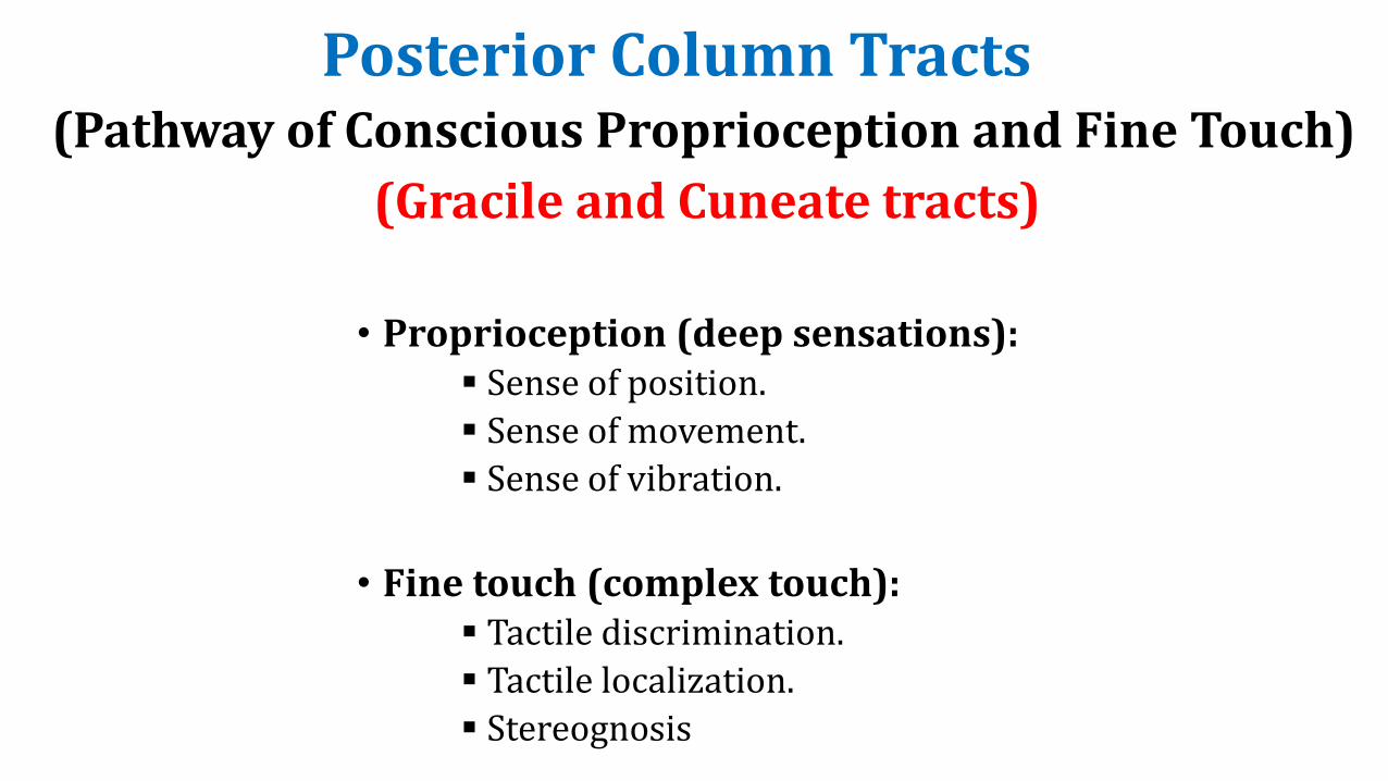
Posterior Column Tracts
• Proprioception (deep sensations):▪ Sense of position.
▪ Sense of movement.
▪ Sense of vibration.
• Fine touch (complex touch):▪ Tactile discrimination.
▪ Tactile localization.
▪ Stereognosis
(Pathway of Conscious Proprioception and Fine Touch)
(Gracile and Cuneate tracts)
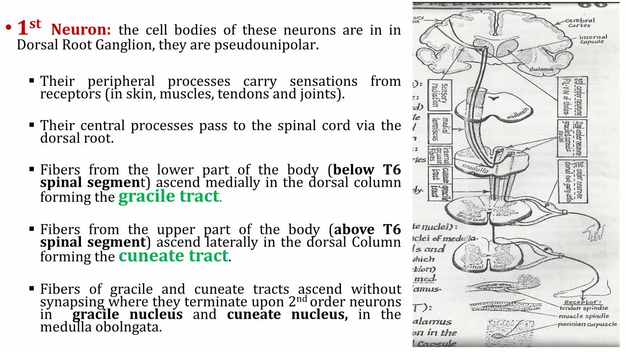
• 1st Neuron: the cell bodies of these neurons are in inDorsal Root Ganglion, they are pseudounipolar.
▪ Their peripheral processes carry sensations fromreceptors (in skin, muscles, tendons and joints).
▪ Their central processes pass to the spinal cord via thedorsal root.
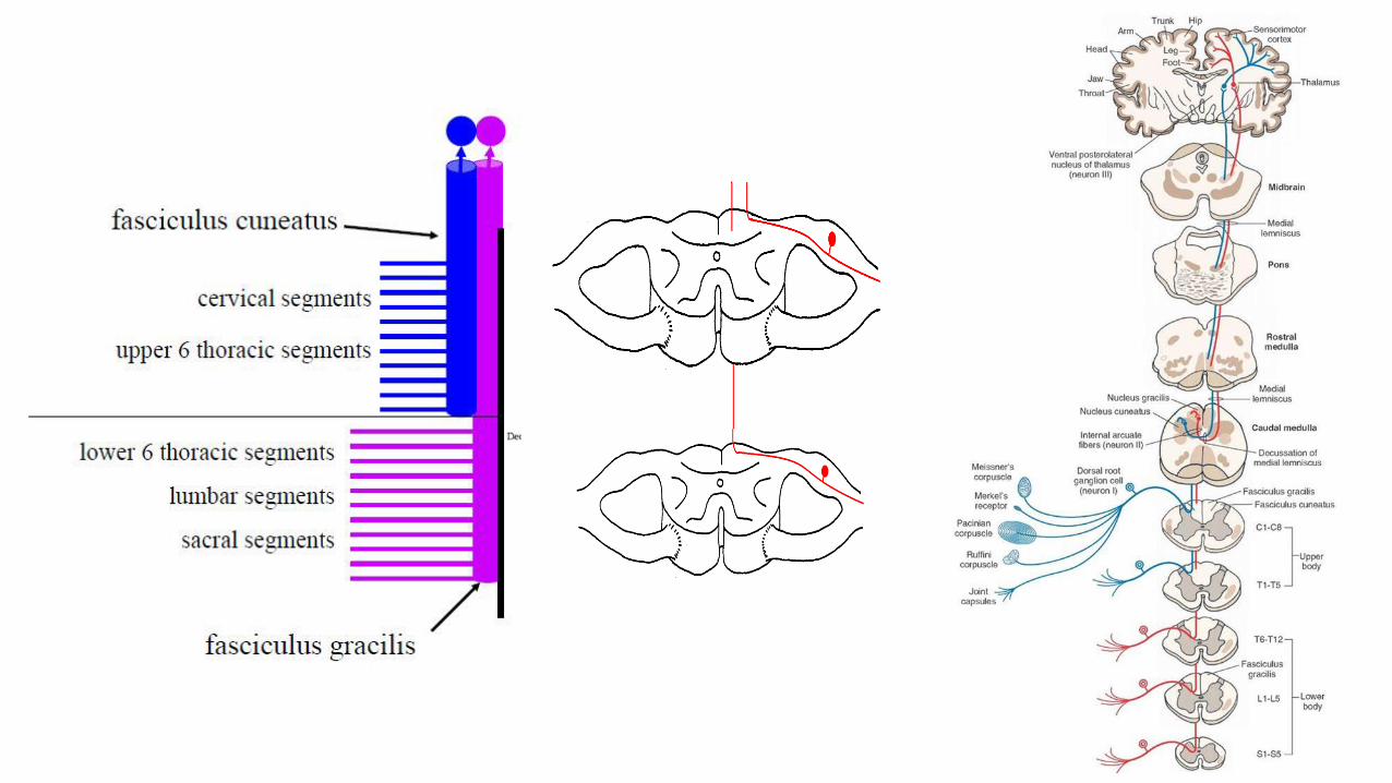
▪ Fibers from the lower part of the body (below T6spinal segment) ascend medially in the dorsal columnforming the gracile tract.
▪ Fibers from the upper part of the body (above T6spinal segment) ascend laterally in the dorsal Columnforming the cuneate tract.
▪ Fibers of gracile and cuneate tracts ascend withoutsynapsing where they terminate upon 2nd order neuronsin gracile nucleus and cuneate nucleus, in themedulla obolngata.
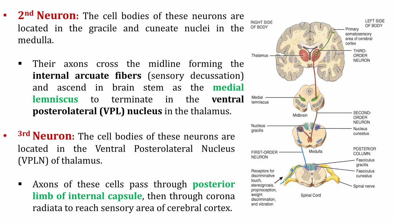
• 2nd Neuron: The cell bodies of these neurons arelocated in the gracile and cuneate nuclei in themedulla.
▪ Their axons cross the midline forming theinternal arcuate fibers (sensory decussation)and ascend in brain stem as the mediallemniscus to terminate in the ventralposterolateral (VPL) nucleus in the thalamus.
• 3rd Neuron: The cell bodies of these neurons arelocated in the Ventral Posterolateral Nucleus(VPLN) of thalamus.
▪ Axons of these cells pass through posteriorlimb of internal capsule, then through coronaradiata to reach sensory area of cerebral cortex.
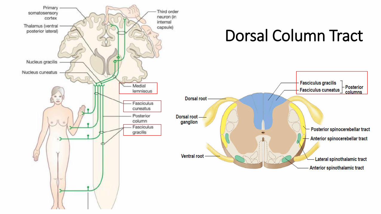
Dorsal Column Tract
Clinical Application
• Lesion in the pathway of discriminative touch and proprioception at any level produce thefollowing :
1. Loss of tactile discrimination: astereognosis and agraphesthesia2. Sensory ataxia*
• The body part affected by the lesion in these pathways depend on the site and the side ofthe lesion:
1. Lesion in the medial lemniscus or internal capsule (after sensory decussation) causesloss of sensation on the opposite side of the body
2. Lesion in the dorsal column of the spinal cord (before sensory decussation) causesloss of sensation on the same side of the body from the level of the lesion and below.
Lesion in Posterior Column Tract
• loss of movement coordination caused by the loss of proprioception.
▪ To avoid falling, the patient stands and walk on a wide base, and usually watch hislower limb to see where they are.
▪ His body sways from side to side when standing with feet close together and eyes closed(positive Romberg’s sign). The body swaying improve when the eye are openedbecause the vision help the patient to recognize the position of his feet.
▪ The patient has also stamping gait, in taking a step the patient lift the advancing legsuddenly and too high and then bring down his leg strongly on the floor.
• The sensory ataxia can be tested by Romberg test, the finger to nose test, and the heel to
knee test. Patient with sensory ataxia perform these tests better with hiseyes opened.
Sensory Ataxia
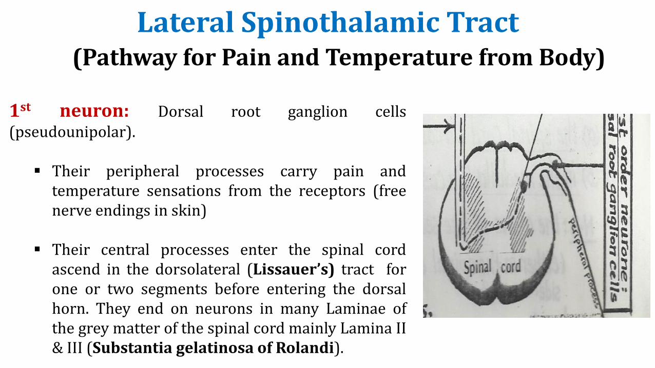
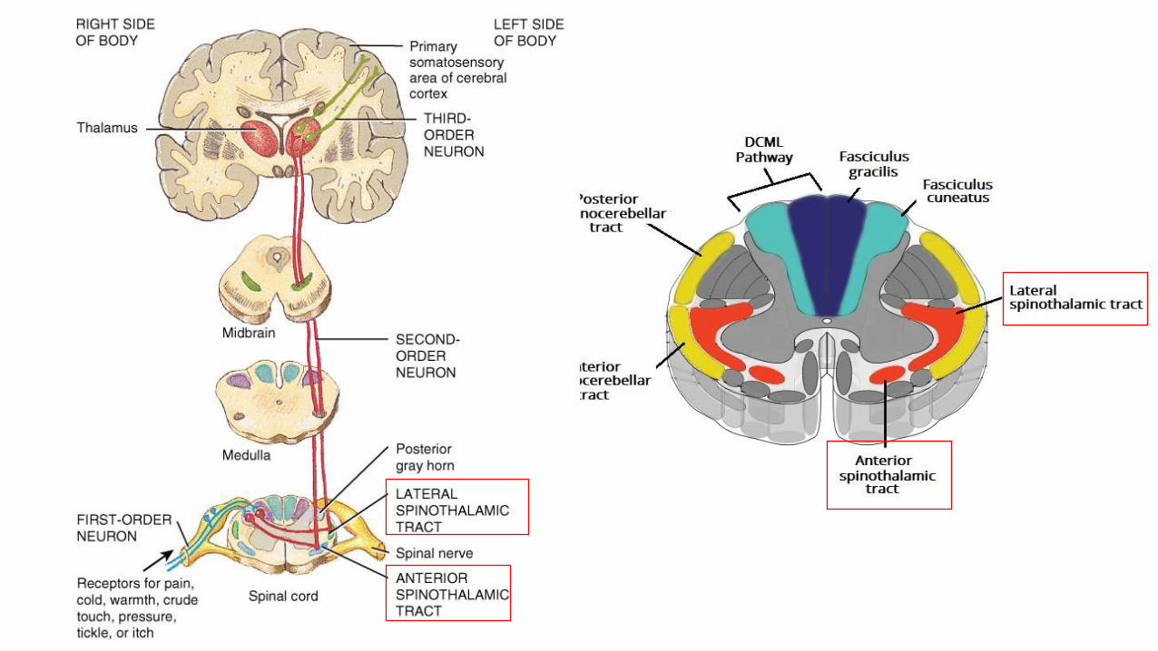
Lateral Spinothalamic Tract(Pathway for Pain and Temperature from Body)
1st neuron: Dorsal root ganglion cells(pseudounipolar).
▪ Their peripheral processes carry pain andtemperature sensations from the receptors (freenerve endings in skin)
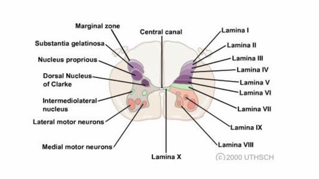
▪ Their central processes enter the spinal cordascend in the dorsolateral (Lissauer’s) tract forone or two segments before entering the dorsalhorn. They end on neurons in many Laminae ofthe grey matter of the spinal cord mainly Lamina II& III (Substantia gelatinosa of Rolandi).
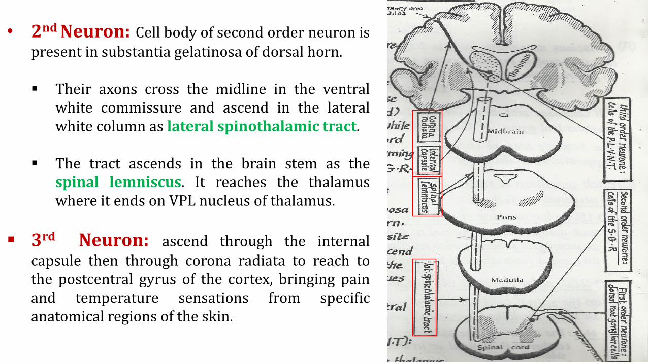
• 2nd Neuron: Cell body of second order neuron ispresent in substantia gelatinosa of dorsal horn.
▪ Their axons cross the midline in the ventralwhite commissure and ascend in the lateralwhite column as lateral spinothalamic tract.
▪ The tract ascends in the brain stem as thespinal lemniscus. It reaches the thalamuswhere it ends on VPL nucleus of thalamus.
▪ 3rd Neuron: ascend through the internalcapsule then through corona radiata to reach tothe postcentral gyrus of the cortex, bringing painand temperature sensations from specificanatomical regions of the skin.
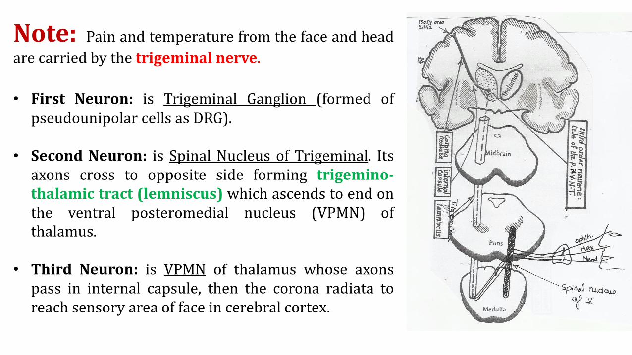
Note: Pain and temperature from the face and head
are carried by the trigeminal nerve.
• First Neuron: is Trigeminal Ganglion (formed ofpseudounipolar cells as DRG).
• Second Neuron: is Spinal Nucleus of Trigeminal. Itsaxons cross to opposite side forming trigemino-thalamic tract (lemniscus) which ascends to end onthe ventral posteromedial nucleus (VPMN) ofthalamus.
• Third Neuron: is VPMN of thalamus whose axonspass in internal capsule, then the corona radiata toreach sensory area of face in cerebral cortex.
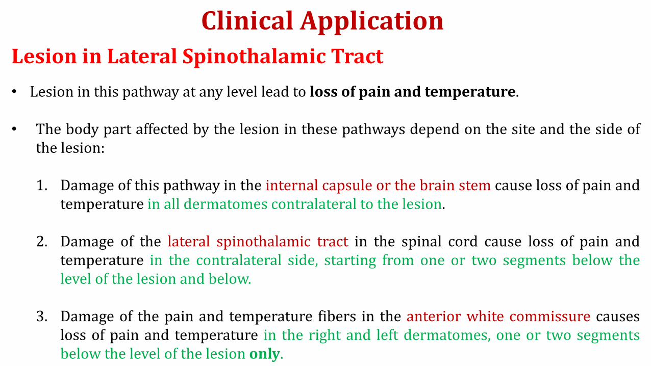
Clinical Application
• Lesion in this pathway at any level lead to loss of pain and temperature.
• The body part affected by the lesion in these pathways depend on the site and the side ofthe lesion:
1. Damage of this pathway in the internal capsule or the brain stem cause loss of pain andtemperature in all dermatomes contralateral to the lesion.
2. Damage of the lateral spinothalamic tract in the spinal cord cause loss of pain andtemperature in the contralateral side, starting from one or two segments below thelevel of the lesion and below.
3. Damage of the pain and temperature fibers in the anterior white commissure causesloss of pain and temperature in the right and left dermatomes, one or two segmentsbelow the level of the lesion only.
Lesion in Lateral Spinothalamic Tract
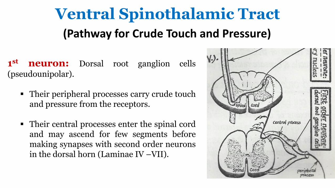
Ventral Spinothalamic Tract(Pathway for Crude Touch and Pressure)
1st neuron: Dorsal root ganglion cells
(pseudounipolar).
▪ Their peripheral processes carry crude touchand pressure from the receptors.
▪ Their central processes enter the spinal cordand may ascend for few segments beforemaking synapses with second order neuronsin the dorsal horn (Laminae IV –VII).
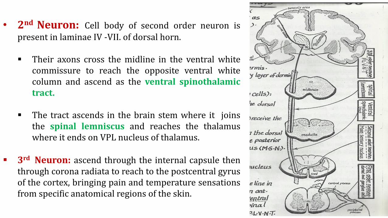
• 2nd Neuron: Cell body of second order neuron ispresent in laminae IV -VII. of dorsal horn.
▪ Their axons cross the midline in the ventral whitecommissure to reach the opposite ventral whitecolumn and ascend as the ventral spinothalamictract.
▪ The tract ascends in the brain stem where it joinsthe spinal lemniscus and reaches the thalamuswhere it ends on VPL nucleus of thalamus.
▪ 3rd Neuron: ascend through the internal capsule thenthrough corona radiata to reach to the postcentral gyrusof the cortex, bringing pain and temperature sensationsfrom specific anatomical regions of the skin.
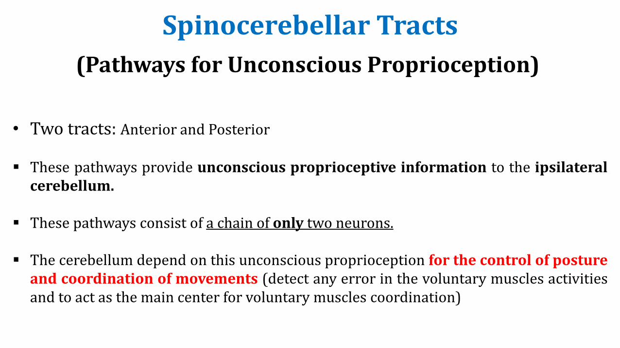
(Pathways for Unconscious Proprioception)
• Two tracts: Anterior and Posterior
▪ These pathways provide unconscious proprioceptive information to the ipsilateralcerebellum.
▪ These pathways consist of a chain of only two neurons.
▪ The cerebellum depend on this unconscious proprioception for the control of postureand coordination of movements (detect any error in the voluntary muscles activitiesand to act as the main center for voluntary muscles coordination)
Spinocerebellar Tracts
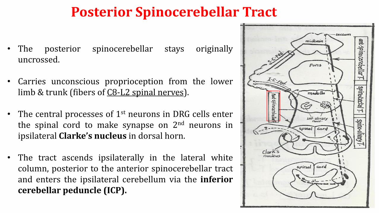
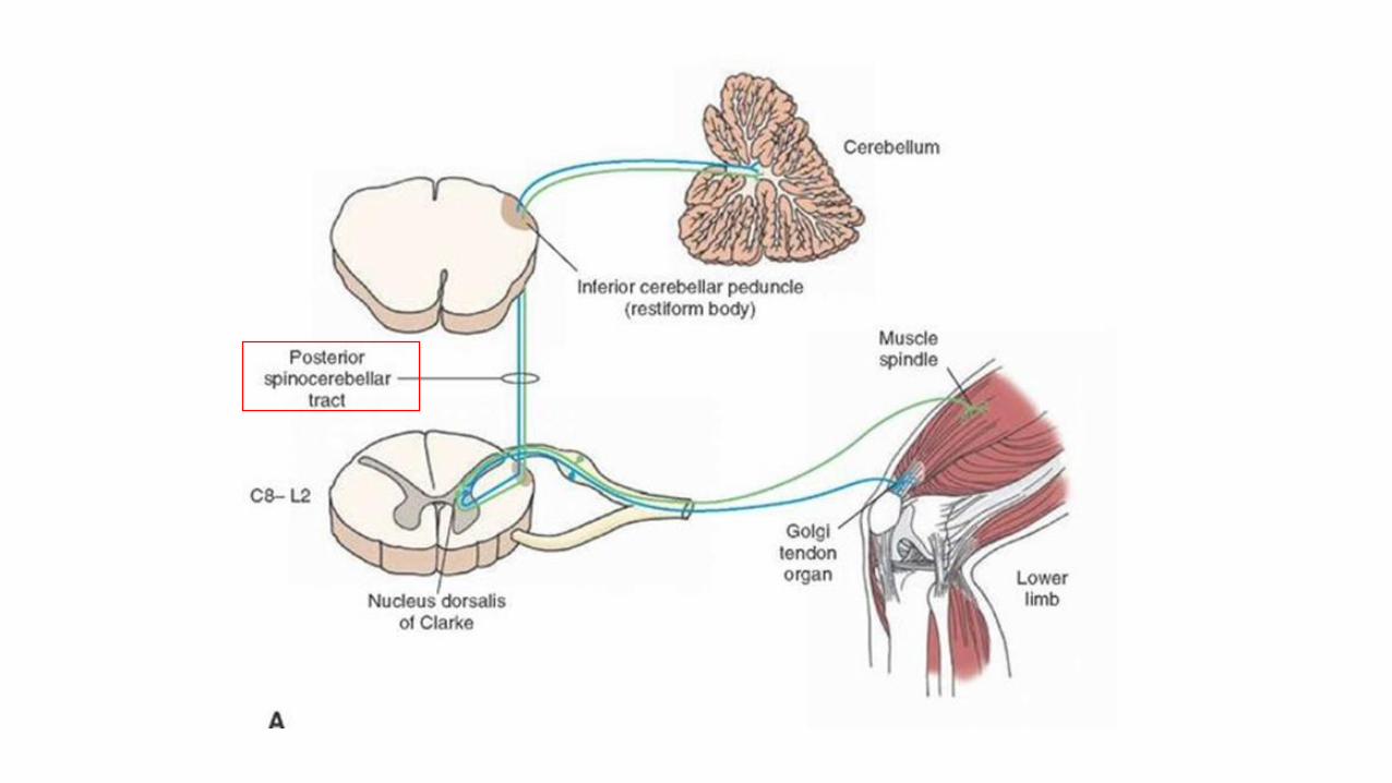
Posterior Spinocerebellar Tract
• The posterior spinocerebellar stays originallyuncrossed.
• Carries unconscious proprioception from the lowerlimb & trunk (fibers of C8-L2 spinal nerves).
• The central processes of 1st neurons in DRG cells enterthe spinal cord to make synapse on 2nd neurons inipsilateral Clarke’s nucleus in dorsal horn.
• The tract ascends ipsilaterally in the lateral whitecolumn, posterior to the anterior spinocerebellar tractand enters the ipsilateral cerebellum via the inferiorcerebellar peduncle (ICP).
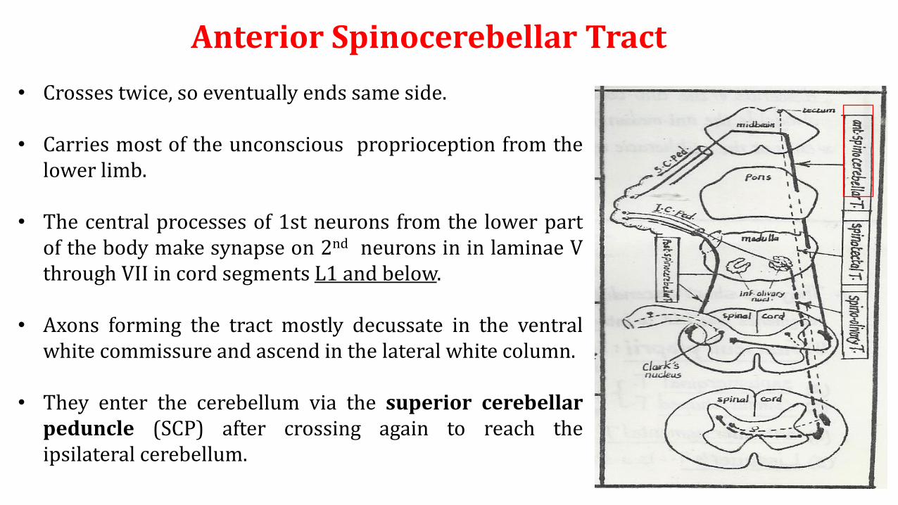
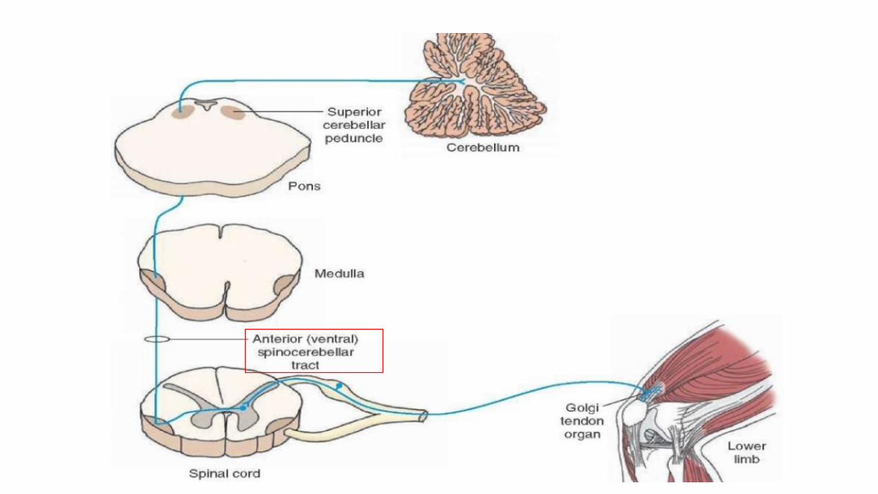
Anterior Spinocerebellar Tract
• Crosses twice, so eventually ends same side.
• Carries most of the unconscious proprioception from thelower limb.
• The central processes of 1st neurons from the lower partof the body make synapse on 2nd neurons in in laminae Vthrough VII in cord segments L1 and below.
• Axons forming the tract mostly decussate in the ventralwhite commissure and ascend in the lateral white column.
• They enter the cerebellum via the superior cerebellarpeduncle (SCP) after crossing again to reach theipsilateral cerebellum.
Spinocerebellar Tracts
Clinical Application
• Lesion in pathways for unconscious proprioception causes disturbance in
balance and movement coordination (Cerebellar Ataxia).
Lesion in Posterior and Anterior Spinocerebellar Tracts
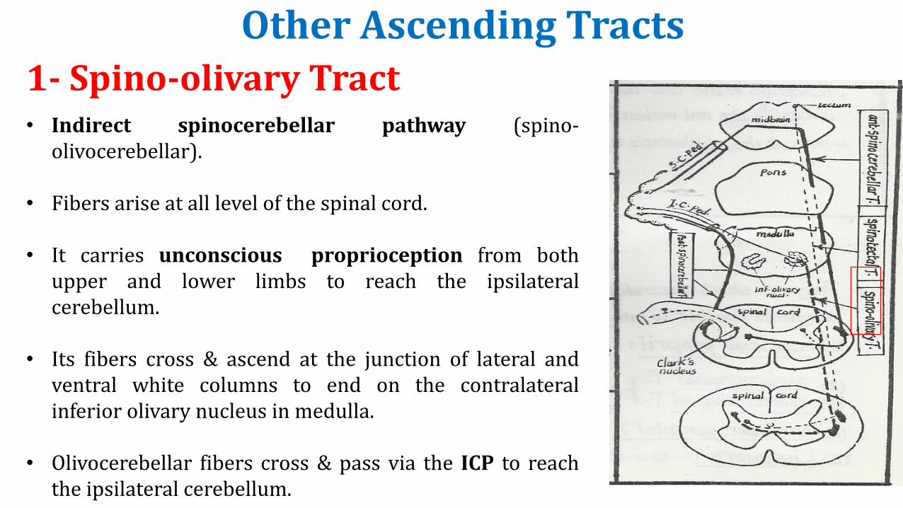
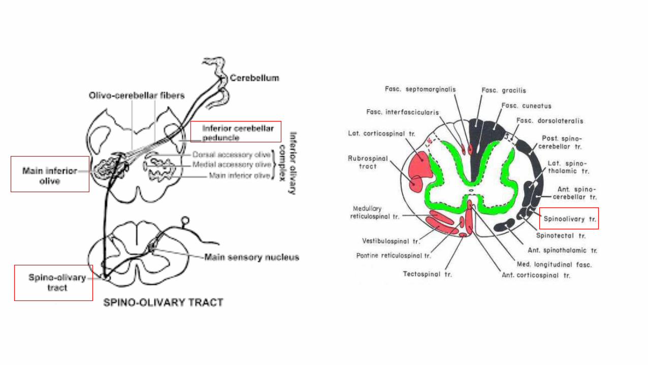
1- Spino-olivary Tract• Indirect spinocerebellar pathway (spino-
olivocerebellar).
• Fibers arise at all level of the spinal cord.
• It carries unconscious proprioception from bothupper and lower limbs to reach the ipsilateralcerebellum.
• Its fibers cross & ascend at the junction of lateral andventral white columns to end on the contralateralinferior olivary nucleus in medulla.
• Olivocerebellar fibers cross & pass via the ICP to reachthe ipsilateral cerebellum.
Other Ascending Tracts
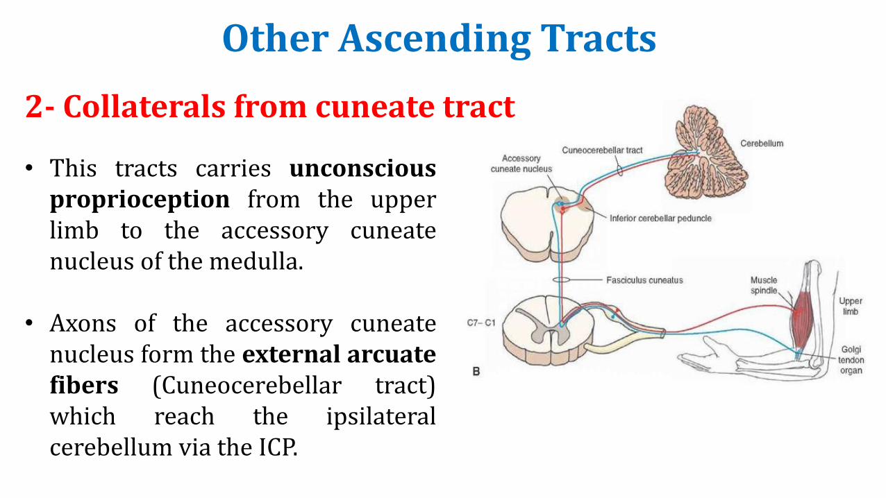
• This tracts carries unconsciousproprioception from the upperlimb to the accessory cuneatenucleus of the medulla.
• Axons of the accessory cuneatenucleus form the external arcuatefibers (Cuneocerebellar tract)which reach the ipsilateralcerebellum via the ICP.
2- Collaterals from cuneate tract
Other Ascending Tracts
Other Ascending Tracts
• Its fibers ascend in the lateral & ventral white columns where it isintermingled with the spinothalamic tracts.
• Most fibers cross to the opposite side and ascend to end on neurons ofthe ponto-medullary reticular formation.
• A spino-reticulo–thalamo-cortical pathway was suggested as a route forslow dull-aching pain sensation.
3- Spino-reticular Tract
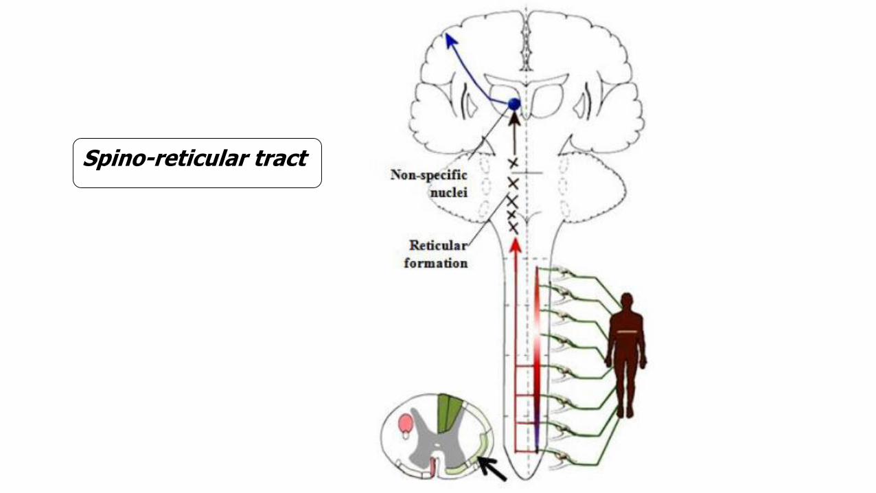
Spino-reticular tract
Other Ascending Tracts
• Most fibers cross to the opposite side and ascend in the lateral whitecolumn to end in the superior colliculi of the midbrain.
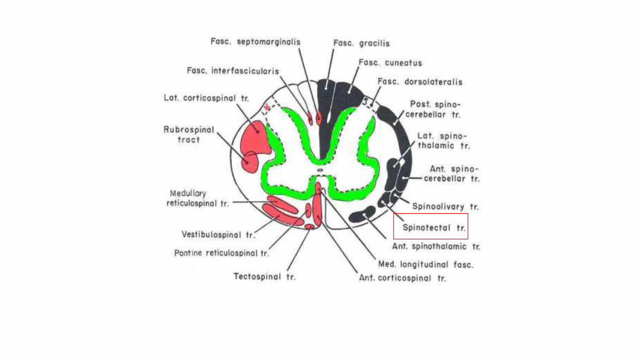
• The spino-tectal tract is concerned with spino-visual reflexes (headturning towards source of cutaneous stimulus).
4- Spino-tectal Tract