Embed Size (px)
Citation preview

Systemic Lupus Erythematosus
B.GANGADHAR, B.RANGAIAH

Systemic lupus erythematosus (SLE)
• SLE is a multiorgan, multisystem autoimmune disease.
• People of both sexes, all ages, and all ethnic groups are susceptible.
• Prevalence of SLE in the United States is 15–50 per 100,000.
• SLE predominantly occurs in women, with a gender ratio of 9:1.
• Onset is usually after puberty, typically in the 20s and 30s. • It is more common in African Americans than in whites. • The incidence in white females is 3.9 per 100,000 and in white males is 0.4
per 100,000. The prevalence in white females is 130 per 100,000

Pathogenesis and Etiology
Pathogenesis and Etiology
• Homozygous deficiencies of early components of complement (C1q,r,s; C2; C4) confer strong predisposition to SLE, but such deficiencies are rare.
• Some gene alleles probably contribute to disease susceptibility by influencing clearance of apoptotic cells (C1q, MBL) or immune complexes (FcR 2A and 3A), antigen presentation (HLA-DR2,3,8), B cell maturation (IL-10), T cell activation (PTPN22), or chemotaxis (MCP-1).
• Female sex is permissive for SLE; • Women exposed to estrogen-containing oral contraceptives or hormone
replacement have an increased risk of developing SLE (1.2- to 2-fold). • Exposure to ultraviolet light causes flares of SLE in approximately 70% of
patients. • Most SLE patients have autoantibodies for 3 years or more before the first
symptoms of disease
Pathogenesis and Etiology
• Epstein-Barr virus (EBV) may be one infectious agent that can trigger SLE in susceptible individuals.
• Children and adults with SLE are more likely to be infected by EBV.
• EBV activates and infects B lymphocytes and survives in those cells for decades; it also contains amino acid sequences that mimic sequences on human spliceosomes.
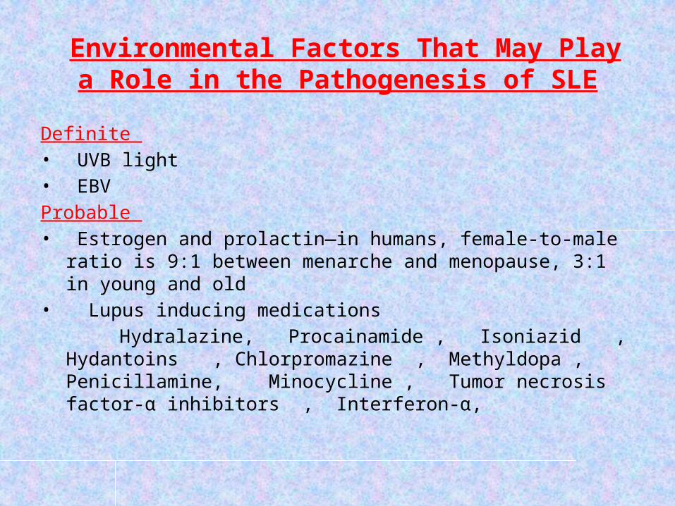
Environmental Factors That May Play a Role in the Pathogenesis of SLE
Definite • UVB light • EBV Probable • Estrogen and prolactin—in humans, female-to-male ratio is 9:1 between
menarche and menopause, 3:1 in young and old • Lupus inducing medications Hydralazine, Procainamide , Isoniazid , Hydantoins , Chlorpromazine
, Methyldopa , Penicillamine, Minocycline , Tumor necrosis factor-α inhibitors , Interferon-α,

Environmental Factors That May Play a Role in the Pathogenesis of SLE
Possible Dietary factors Alfalfa sprouts and related sprouting foods containing Canavanine
Pristane and similar substances Infectious agents other than EBV Bacterial DNA Human retroviruses Endotoxins, bacterial lipopolysaccharides

Auto antibodies in SLE

Correlation among Clinical Manifestations of SLE and Autoantibodies, Immune Complexes, and T Cells
ACR Revised Classification Criteria for SLE
Overview and Systemic Manifestations
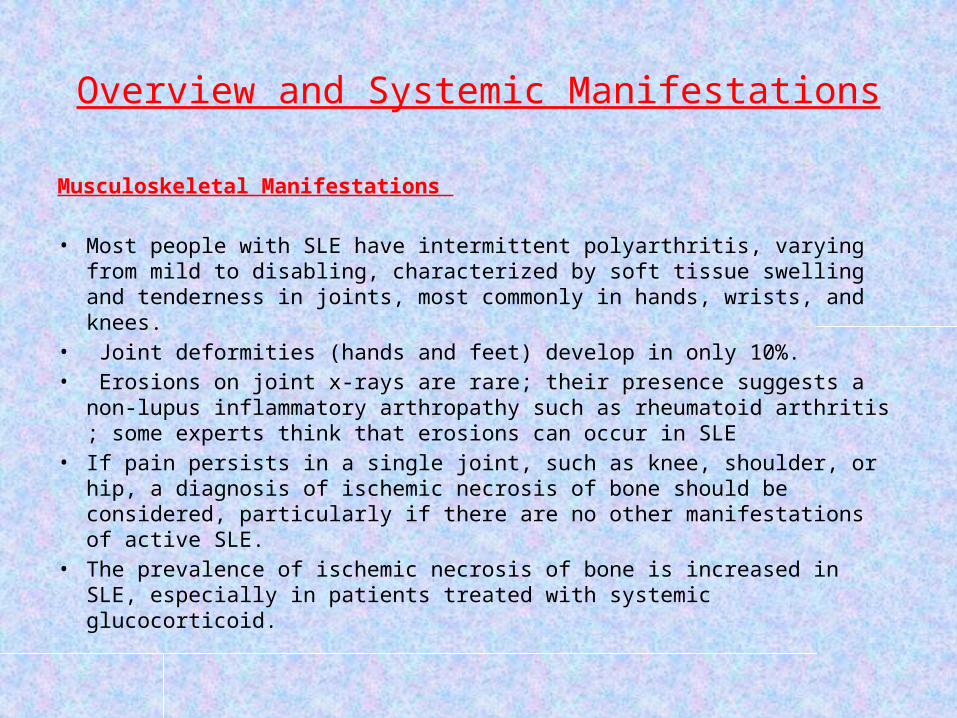
Musculoskeletal Manifestations
• Most people with SLE have intermittent polyarthritis, varying from mild to disabling, characterized by soft tissue swelling and tenderness in joints, most commonly in hands, wrists, and knees.
• Joint deformities (hands and feet) develop in only 10%.• Erosions on joint x-rays are rare; their presence suggests a non-lupus
inflammatory arthropathy such as rheumatoid arthritis ; some experts think that erosions can occur in SLE
• If pain persists in a single joint, such as knee, shoulder, or hip, a diagnosis of ischemic necrosis of bone should be considered, particularly if there are no other manifestations of active SLE.
• The prevalence of ischemic necrosis of bone is increased in SLE, especially in patients treated with systemic glucocorticoid.
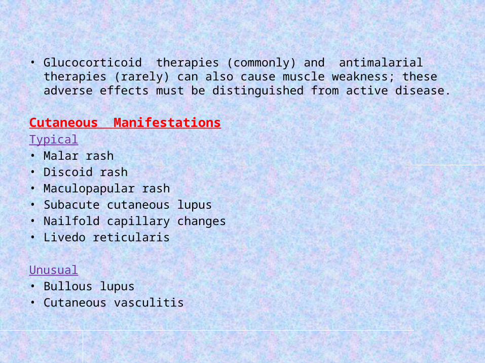
• Glucocorticoid therapies (commonly) and antimalarial therapies (rarely) can also cause muscle weakness; these adverse effects must be distinguished from active disease.
Cutaneous ManifestationsTypical• Malar rash• Discoid rash• Maculopapular rash• Subacute cutaneous lupus• Nailfold capillary changes• Livedo reticularis
Unusual• Bullous lupus• Cutaneous vasculitis
Cutaneous Manifestations
• Lupus dermatitis can be classified as discoid lupus erythematosus (DLE), systemic rash, subacute cutaneous lupus erythematosus (SCLE), or "other.“
• Discoid lesions are roughly circular with slightly raised, scaly hyperpigmented erythematous rims and depigmented, atrophic centers in which all dermal appendages are permanently destroyed.
• Lesions can be disfiguring, particularly on the face and scalp. • Treatment consists primarily of topical or locally injected glucocorticoids
and systemic antimalarials. • Only 5% of people with DLE have SLE (although half have positive ANA);
however, among individuals with SLE, as many as 20% have DLE
Cutaneous Manifestations
• The most common SLE rash is a photosensitive, slightly raised erythema, occasionally scaly, on the face (particularly the cheeks and nose—the "butterfly" rash), ears, chin, V region of the neck, upper back, and extensor surfaces of the arms.
• Worsening of this rash often accompanies flare of systemic disease. • SCLE consists of scaly red patches similar to psoriasis or circular flat red-
rimmed lesions.• Patients with these manifestations are exquisitely photosensitive; most
have antibodies to Ro (SS-A). • Other SLE rashes include recurring urticaria, lichen planus–like dermatitis,
bullae, and panniculitis ("lupus profundus").• Small ulcerations on the oral or nasal mucosa are common in SLE; the
lesions resemble aphthous ulcers.
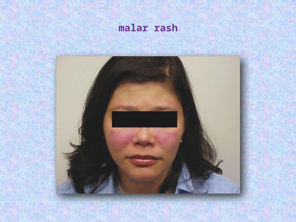
malar rash
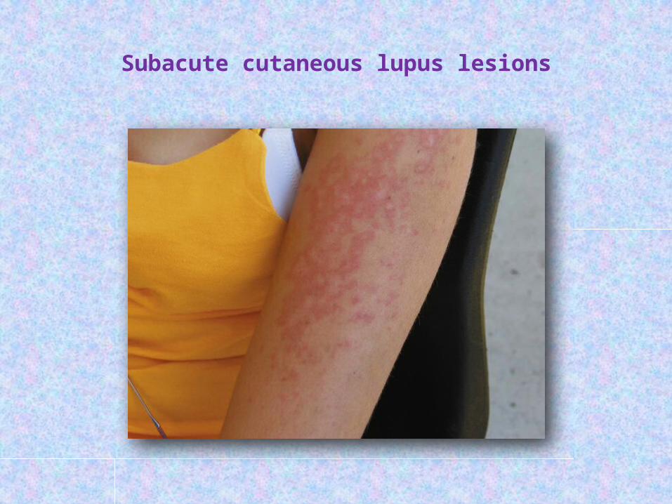
Subacute cutaneous lupus lesions

Discoid lupus erythematosus

Renal Manifestations
• Nephritis is usually the most serious manifestation of SLE
• Since nephritis is asymptomatic in most lupus patients, urinalysis should be ordered in any person suspected of having SLE.

Classification of Lupus Nephritis (International Society of Nephrology and Renal Pathology Society)
Class I: Minimal Mesangial Lupus Nephritis
• Normal glomeruli by light microscopy, but mesangial immune deposits by immunofluorescence.
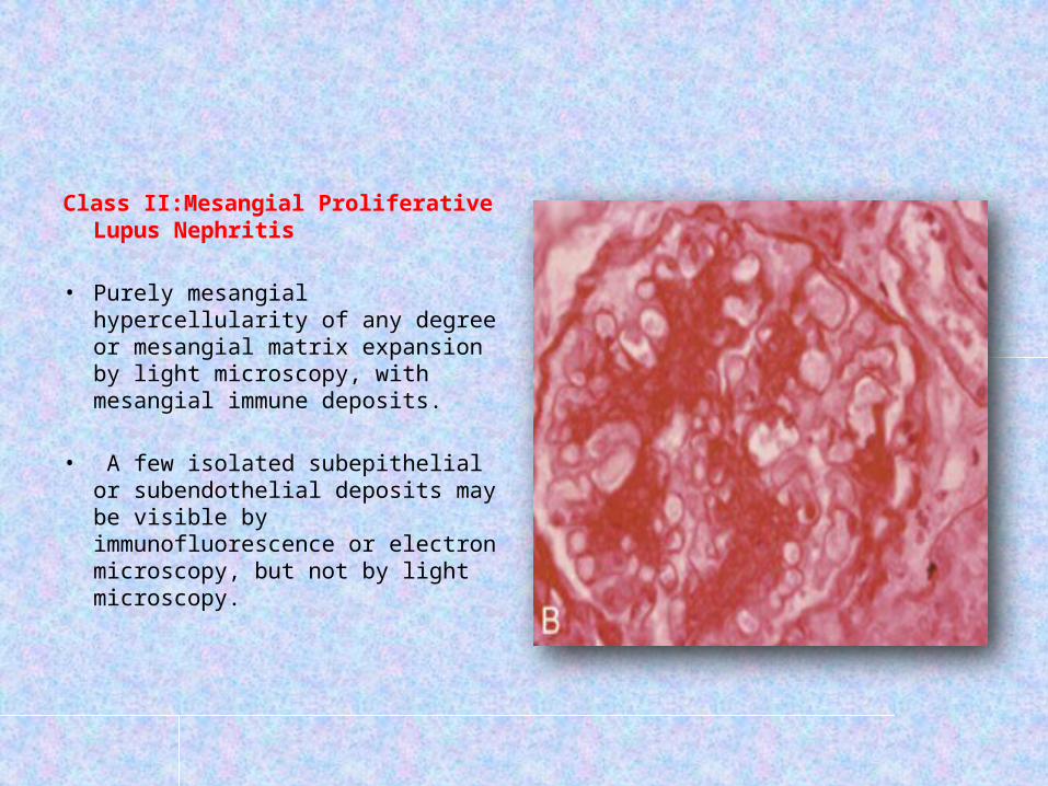
Class II:Mesangial Proliferative Lupus Nephritis
• Purely mesangial hypercellularity of any degree or mesangial matrix expansion by light microscopy, with mesangial immune deposits.
• A few isolated subepithelial or subendothelial deposits may be visible by immunofluorescence or electron microscopy, but not by light microscopy.

Class III: Focal Lupus Nephritis Active or inactive focal, segmental or global endo- or extracapillary
glomerulonephritis involving <50% of all glomeruli, typically with focal subendothelial immune deposits, with or without mesangial alterations.
Class III (A): Active lesions—focal proliferative lupus nephritis
Class III (A/C): Active and chronic lesions—focal proliferative and sclerosing lupus nephritis Class III (C): Chronic inactive lesions with glomerular scars—focal sclerosing lupus nephritis
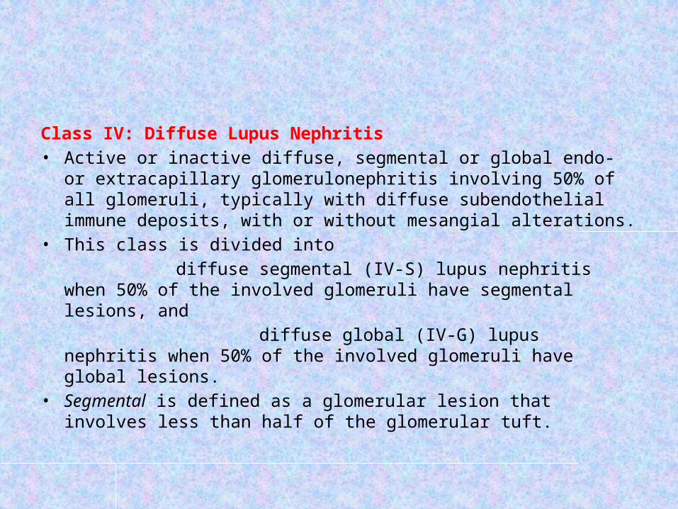
Class IV: Diffuse Lupus Nephritis • Active or inactive diffuse, segmental or global endo- or extracapillary
glomerulonephritis involving 50% of all glomeruli, typically with diffuse subendothelial immune deposits, with or without mesangial alterations.
• This class is divided into diffuse segmental (IV-S) lupus nephritis when 50% of the involved
glomeruli have segmental lesions, and diffuse global (IV-G) lupus nephritis when 50% of the involved
glomeruli have global lesions. • Segmental is defined as a glomerular lesion that involves less than half of
the glomerular tuft.
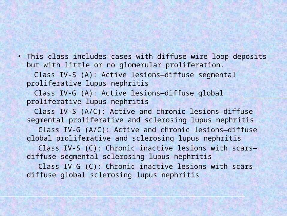
• This class includes cases with diffuse wire loop deposits but with little or no glomerular proliferation.
Class IV-S (A): Active lesions—diffuse segmental proliferative lupus nephritis
Class IV-G (A): Active lesions—diffuse global proliferative lupus nephritis Class IV-S (A/C): Active and chronic lesions—diffuse segmental proliferative
and sclerosing lupus nephritis Class IV-G (A/C): Active and chronic lesions—diffuse global proliferative
and sclerosing lupus nephritis Class IV-S (C): Chronic inactive lesions with scars—diffuse segmental
sclerosing lupus nephritis Class IV-G (C): Chronic inactive lesions with scars—diffuse global sclerosing
lupus nephritis

Class V: Membranous Lupus Nephritis • Global or segmental subepithelial
immune deposits or their morphologic sequelae by light microscopy and by immunofluorescence or electron microscopy, with or without mesangial alterations.
• Class V lupus nephritis may occur in combination with class III or IV, in which case both will be diagnosed. Class V lupus nephritis may show advanced sclerosis.
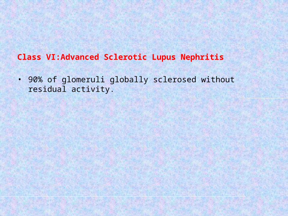
Class VI:Advanced Sclerotic Lupus Nephritis • 90% of glomeruli globally sclerosed without residual activity.

Renal Manifestations
• Renal biopsy is useful in planning current and near-future therapies.• Patients with dangerous proliferative forms of glomerular damage (ISN III
and IV) usually have microscopic hematuria and proteinuria (>500 mg per 24 h);
approximately one-half develop nephrotic syndrome, and most develop hypertension.
• If diffuse proliferative glomerulonephritis (DPGN) is untreated, virtually all patients develop ESRD within 2 years of diagnosis.
• Therefore, aggressive immunosuppression is indicated (usually systemic glucocorticoids plus a cytotoxic drug), unless damage is irreversible

Renal Manifestations
• African Americans are more likely to develop ESRD than are Caucasians, even with the most current therapies.
• Overall in the United States, ~20% of individuals with lupus DPGN die or develop ESRD within 10 years of diagnosis.
• A small proportion of SLE patients with proteinuria (usually nephrotic) have membranous glomerular changes without proliferation on renal biopsy.
Their outcome is better than for those with DPGN, but proteinuria is less likely to improve on lupus nephritis immunosuppressive therapies.
• Lupus nephritis tends to be an ongoing disease, with flares requiring re-treatment over many years.
• For most people with lupus nephritis, accelerated atherosclerosis becomes important after several years of disease; attention must be given to control of blood pressure, hyperlipidemia, and hyperglycemia.
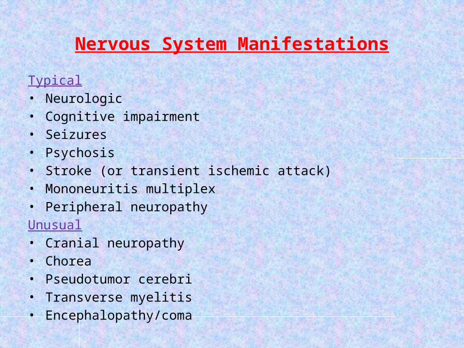
Nervous System Manifestations
Typical• Neurologic• Cognitive impairment• Seizures• Psychosis• Stroke (or transient ischemic attack)• Mononeuritis multiplex• Peripheral neuropathyUnusual• Cranial neuropathy• Chorea• Pseudotumor cerebri• Transverse myelitis• Encephalopathy/coma
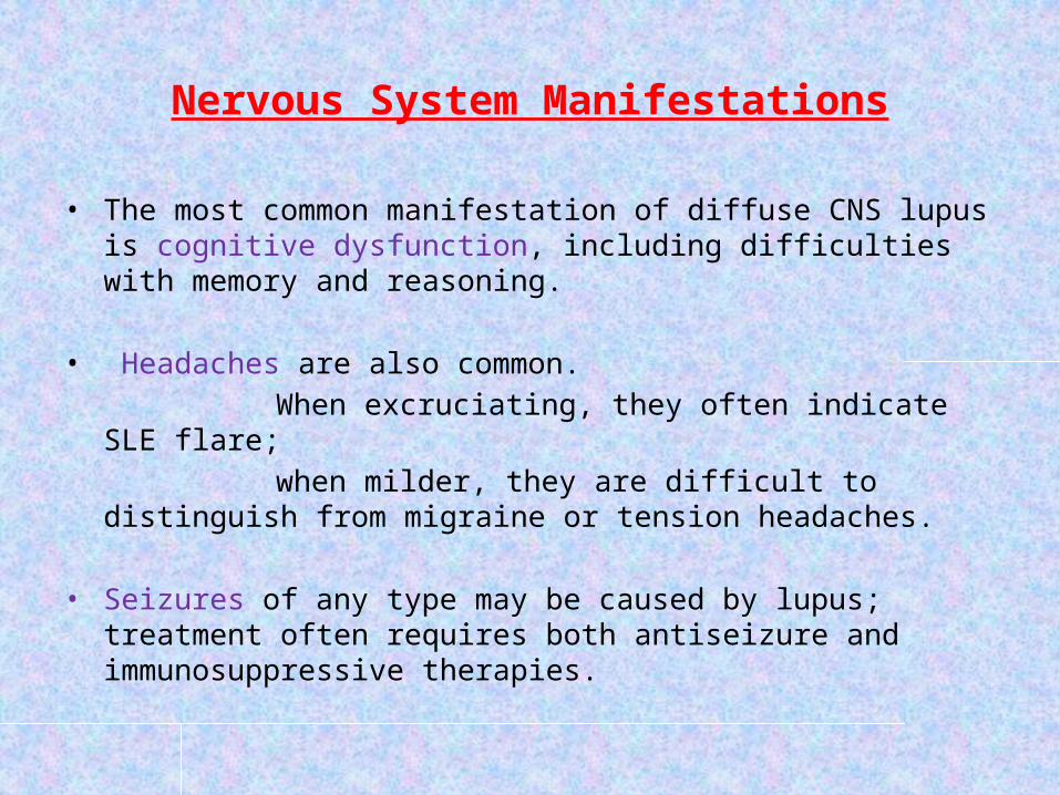
Nervous System Manifestations
• The most common manifestation of diffuse CNS lupus is cognitive dysfunction, including difficulties with memory and reasoning.
• Headaches are also common. When excruciating, they often indicate SLE flare; when milder, they are difficult to distinguish from migraine or tension
headaches.
• Seizures of any type may be caused by lupus; treatment often requires both antiseizure and immunosuppressive therapies.
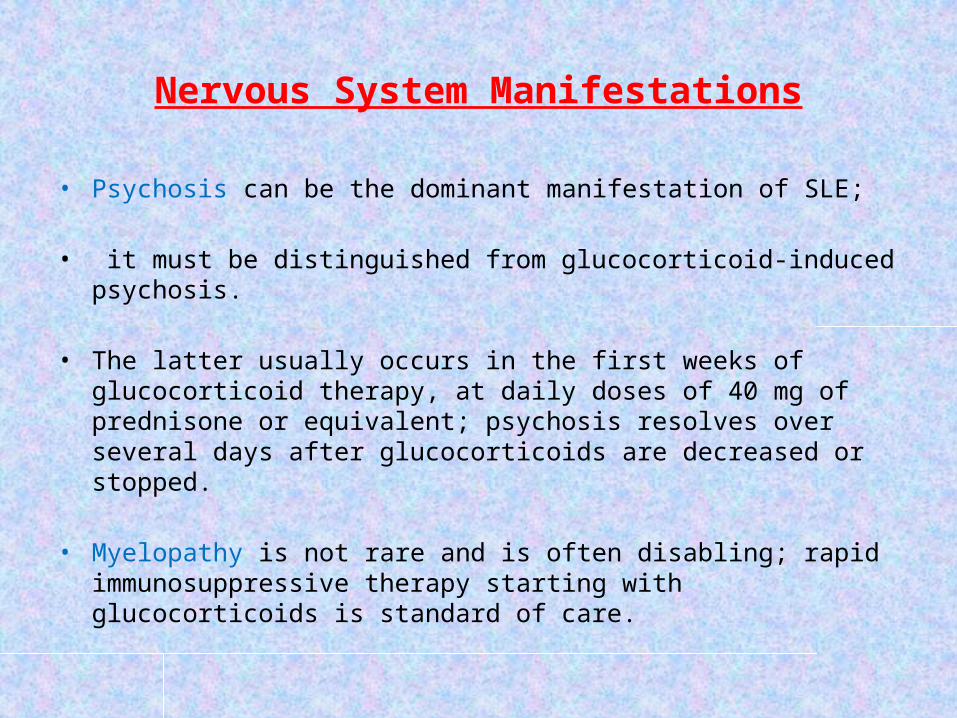
Nervous System Manifestations
• Psychosis can be the dominant manifestation of SLE;
• it must be distinguished from glucocorticoid-induced psychosis.
• The latter usually occurs in the first weeks of glucocorticoid therapy, at daily doses of 40 mg of prednisone or equivalent; psychosis resolves over several days after glucocorticoids are decreased or stopped.
• Myelopathy is not rare and is often disabling; rapid immunosuppressive therapy starting with glucocorticoids is standard of care.

Vascular Occlusions
• The prevalence of transient ischemic attacks, strokes, and myocardial infarctions is increased in patients with SLE.
• These vascular events are increased in, but not exclusive to, SLE patients with antibodies to phospholipids (aPL).
• It is likely that antiphospholipid antibodies are associated with hypercoagulability and acute thrombotic events, whereas chronic disease is associated with accelerated atherosclerosis.
• Ischemia in the brain can be caused by focal occlusion (either noninflammatory or associated with vasculitis) or by embolization from carotid artery plaque or from fibrinous vegetations of Libman-Sacks endocarditis
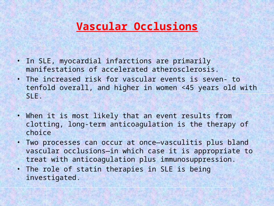
Vascular Occlusions
• In SLE, myocardial infarctions are primarily manifestations of accelerated atherosclerosis.
• The increased risk for vascular events is seven- to tenfold overall, and higher in women <45 years old with SLE.
• When it is most likely that an event results from clotting, long-term anticoagulation is the therapy of choice
• Two processes can occur at once—vasculitis plus bland vascular occlusions—in which case it is appropriate to treat with anticoagulation plus immunosuppression.
• The role of statin therapies in SLE is being investigated.

Pulmonary Manifestations
• Pleurisy/pleural effusion• Pericarditis/pericardial effusion• Interstitial pneumonitis (acute or chronic)• Pulmonary hypertension• Pulmonary hemorrhage
• The most common pulmonary manifestation of SLE is pleuritis with or without pleural effusion.
This manifestation, when mild, may respond to treatment with NSAIDs; when more severe, patients require a brief course of glucocorticoid therapy.

Pulmonary Manifestations
• Pulmonary infiltrates also occur as a manifestation of active SLE and are difficult to distinguish from infection on imaging studies.
• Life-threatening pulmonary manifestations include interstitial inflammation leading to fibrosis, shrinking lung syndrome, and intraalveolar hemorrhage; • all of these probably require early aggressive immunosuppressive therapy
as well as supportive care.

Cardiac Manifestations
• Pericarditis is the most frequent cardiac manifestation; it usually responds to anti-inflammatory therapy and infrequently leads to tamponade.
• More serious cardiac manifestations are myocarditis and fibrinous endocarditis of Libman-Sacks.
• The endocardial involvement can lead to valvular insufficiencies, most commonly of the mitral or aortic valves, or to embolic events.
• It has not been proven that glucocorticoid or other immunosuppressive therapies lead to improvement of lupus myocarditis or endocarditis,
but it is usual practice to administer a trial of high-dose steroids along with appropriate supportive therapy for heart failure, arrhythmia, or embolic events.
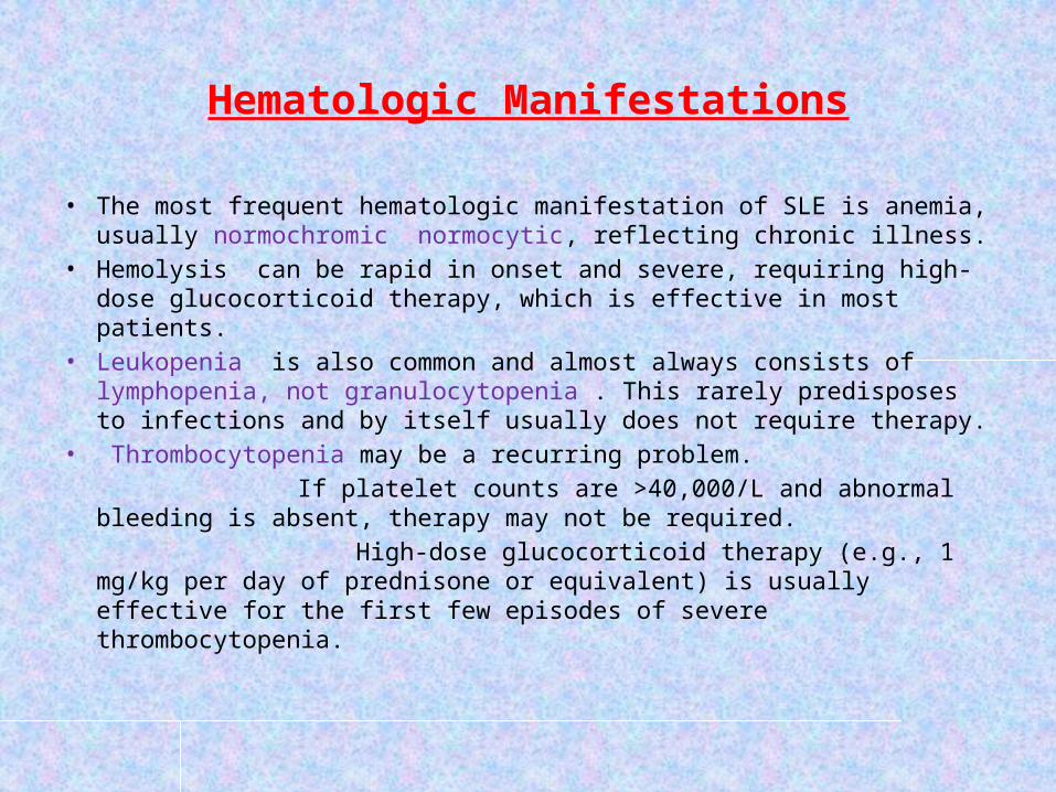
Hematologic Manifestations
• The most frequent hematologic manifestation of SLE is anemia, usually normochromic normocytic, reflecting chronic illness.
• Hemolysis can be rapid in onset and severe, requiring high-dose glucocorticoid therapy, which is effective in most patients.
• Leukopenia is also common and almost always consists of lymphopenia, not granulocytopenia . This rarely predisposes to infections and by itself usually does not require therapy.
• Thrombocytopenia may be a recurring problem. If platelet counts are >40,000/L and abnormal bleeding is absent,
therapy may not be required. High-dose glucocorticoid therapy (e.g., 1 mg/kg per day of
prednisone or equivalent) is usually effective for the first few episodes of severe thrombocytopenia.

Gastrointestinal Manifestations
Typical• Esophageal dysmotility• Hepatomegaly• Splenomegaly• Elevated liver function testsUnusual• Mesenteric vasculitis (with or without infarcts)• Colitis• Protein-losing enteropathy• Primary biliary cirrhosis• Budd-Chiari syndrome• Ascites

Gastrointestinal Manifestations
• Nausea, sometimes with vomiting, and diarrhea can be manifestations of an SLE flare.
• Increases in serum aspartate aminotransferase (AST) and alanine aminotransferase (ALT) are common when SLE is active.
• These manifestations usually improve promptly during systemic glucocorticoid therapy.
• Vasculitis involving the intestine may be life-threatening; perforations, ischemia, bleeding, and sepsis are frequent complications.
• Aggressive immunosuppressive therapy with high-dose glucocorticoids is recommended for short-term control.
evidence of recurrence is an indication for additional therapies.
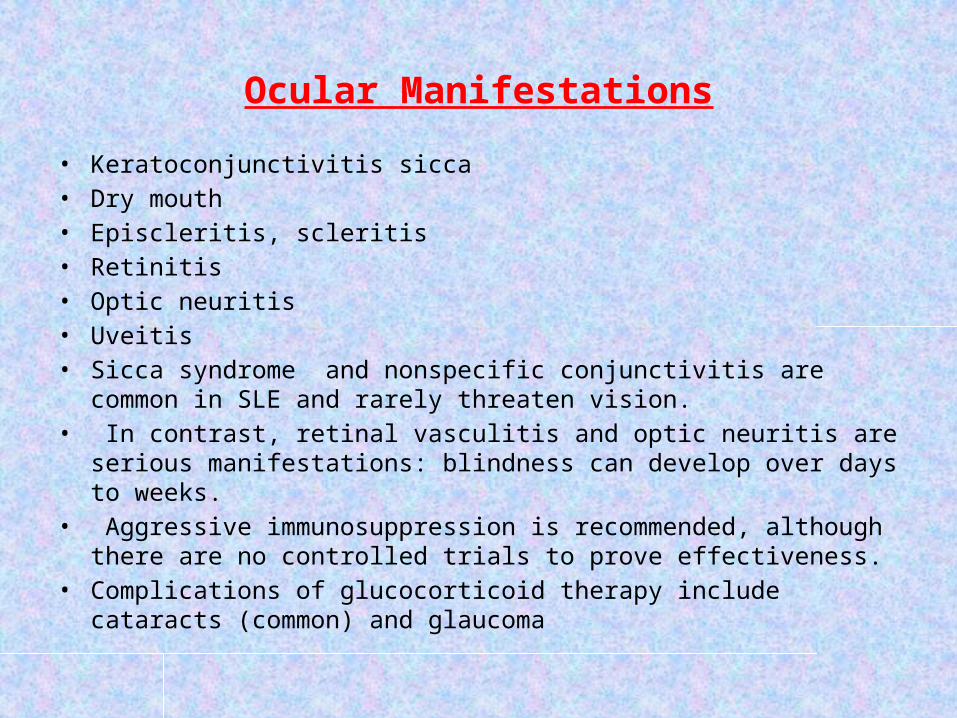
Ocular Manifestations
• Keratoconjunctivitis sicca• Dry mouth• Episcleritis, scleritis• Retinitis• Optic neuritis• Uveitis• Sicca syndrome and nonspecific conjunctivitis are common in SLE and
rarely threaten vision.• In contrast, retinal vasculitis and optic neuritis are serious manifestations:
blindness can develop over days to weeks.• Aggressive immunosuppression is recommended, although there are no
controlled trials to prove effectiveness. • Complications of glucocorticoid therapy include cataracts (common) and
glaucoma
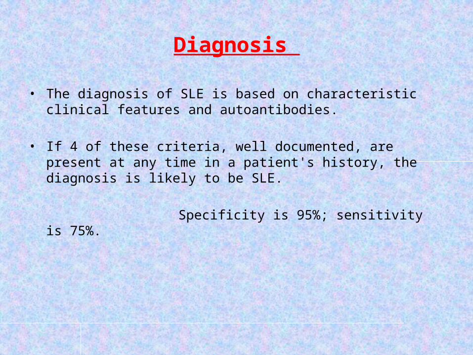
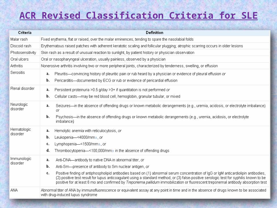
Diagnosis
• The diagnosis of SLE is based on characteristic clinical features and autoantibodies.
• If 4 of these criteria, well documented, are present at any time in a patient's history, the diagnosis is likely to be SLE.
Specificity is 95%; sensitivity is 75%.
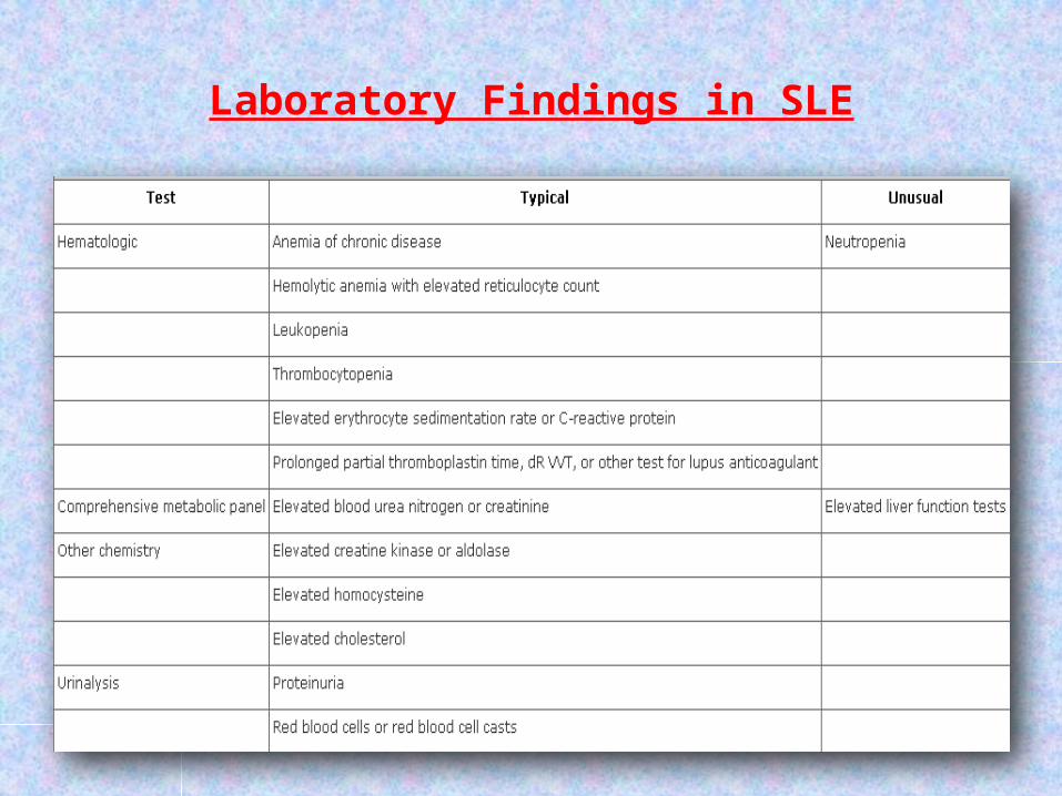
Laboratory Findings in SLE
Diagnosis
Auto antibodies• Most (96% or more) SLE patients have a positive ANA test result.
Because up to 20% of healthy young women also have a positive ANA, the presence of an ANA alone is not given much weight.
Titers of 1:640 or higher are more indicative of a connective tissue disease of some sort.
• Some autoantibodies are very specific for lupus, such as anti-dsDNA (which occurs in about 30%) or anti-Sm .
• Other autoantibodies, such as anti-Ro/SS-A, anti-La/SS-B, and anti-ribonucleoprotein, occur in SLE but also in rheumatoid arthritis and in Sjögren syndrome.
Diagnosis
• Antiphospholipid antibodies (lupus anticoagulant, anticardiolipin, and anti-b2 glycoprotein-1) are found in about 50% of SLE patients during the course of disease.
They are associated with an increased risk of thrombosis and pregnancy loss.
Complement
• Reduction in the complement components C3 and C4 or in total hemolytic complement occurs frequently, but is not specific for lupus.

TREATMENT
PRINCIPLES OF MANAGEMENT
• Patient education and multidisciplinary interventions, particularly in newly diagnosed patients, are an important aspect of the management of SLE.
• Generally, the management of the disease is divided between the management of disease with nonmajor organ (or nonvisceral) involvement and disease with major organ (or visceral) involvement.
MANAGEMENT OF MILD SLE WITHOUT MAJOR ORGAN INVOLVEMENT
• NSAIDs, antimalarials , glucocorticoids , and, in severe, refractory cases, immunosuppressive agents (azathioprine, mycophenolate mofetil, methotrexate) are used in the treatment of SLE patients without major organ involvement.
• NSAIDs are believed to be effective in the treatment of musculoskeletal disorders and complaints in SLE patients.
• Antimalarials—mainly hydroxychloroquine—are widely used for musculoskeletal and cutaneous manifestations of lupus.
The drug has a long-term effect in preventing major flares in SLE
MANAGEMENT OF MILD SLE WITHOUT MAJOR ORGAN INVOLVEMENT
• Several studies indicate beneficial effects of methotrexate on disease activity and articular and cutaneous manifestations in SLE.
• In SLE patients without CNS or renal involvement, azathioprine therapy has been associated with fewer hospitalizations but no decrease in prednisone maintenance requirement.
• Mycophenolate mofetil also has been used in the treatment of SLE without major organ involvement in a few uncontrolled case studies
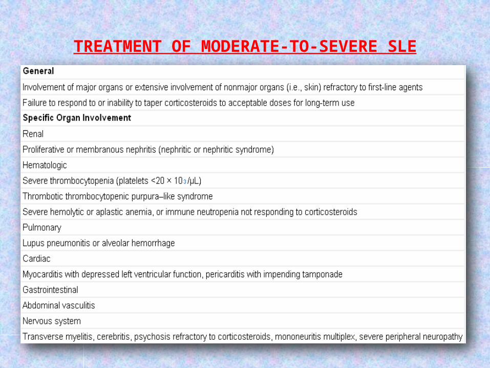
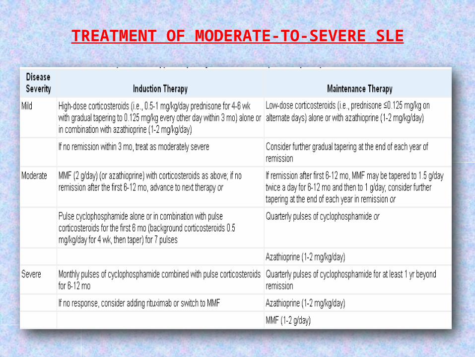
TREATMENT OF MODERATE-TO-SEVERE SLE
TREATMENT OF MODERATE-TO-SEVERE SLE
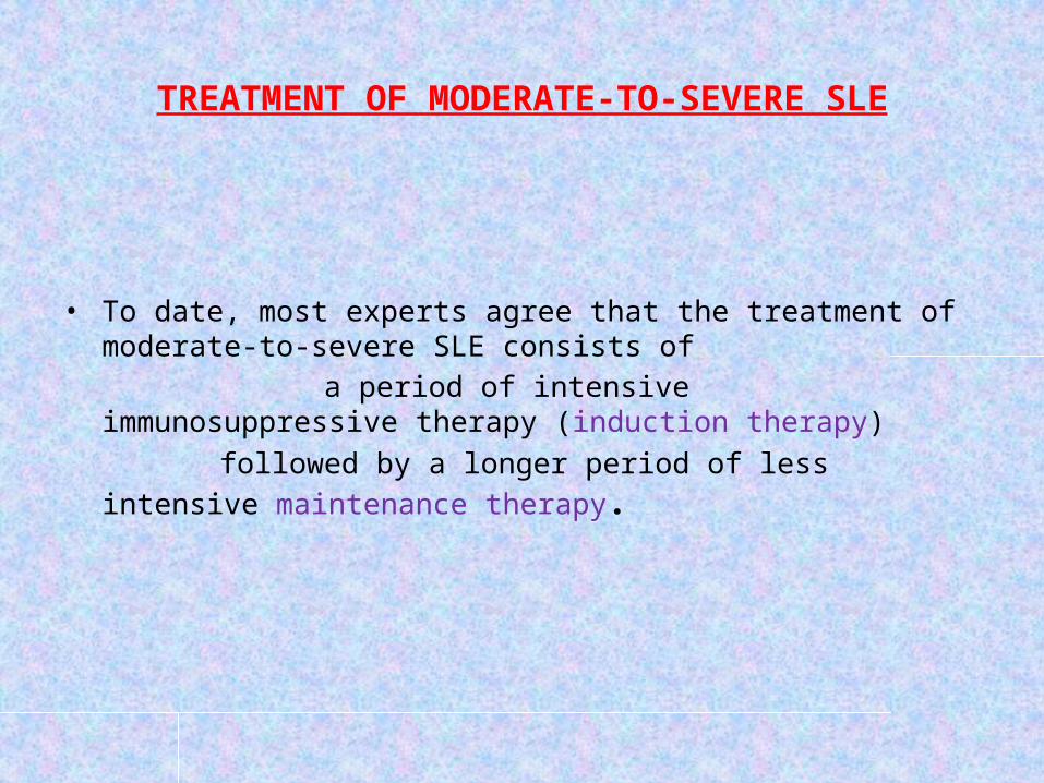
• To date, most experts agree that the treatment of moderate-to-severe SLE consists of
a period of intensive immunosuppressive therapy (induction therapy)
followed by a longer period of less intensive maintenance therapy.

TREATMENT OF MODERATE-TO-SEVERE SLE
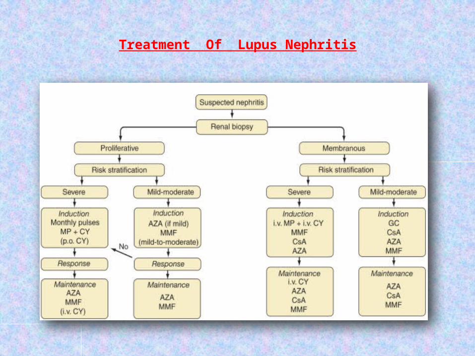
Treatment Of Lupus Nephritis

Intravenous Gamma Gobulin
• used for the treatment of a variety of severe SLE manifestations
• Intravenous gamma globulin is administered in doses of 400 mg/kg/day for 5 consecutive days and is most commonly used for the treatment of severe, refractory thrombocytopenia, usually achieving a rapid increase in the number of platelets within hours of administration.
• Nephritis, arthritis, fever, rashes, and immunologic parameters improve with intravenous gamma globulin.
• Side effects of intravenous gamma globulin include fever, myalgia, headache, arthralgia, and, rarely, aseptic meningitis.
• The drug is contraindicated in cases of known IgA deficiency.
Special Conditions in SLE
Pregnancy and Lupus• Fertility rates for men and women with SLE are probably normal. However,
rate of fetal loss is increased (approximately 2-3 fold) in women with SLE.• Fetal demise is higher in mothers with high disease activity,
antiphospholipid antibodies, and/or nephritis • Suppression of disease activity can be achieved by administration of
systemic glucocorticoids. • A placental enzyme, 11--dehydrogenase 2, deactivates glucocorticoids; it is
more effective in deactivating prednisone and prednisolone than the fluorinated glucocorticoids dexamethasone and betamethasone.
• Therefore, maternal SLE should be controlled with prednisone/prednisolone at the lowest effective doses for the shortest time required.

Pregnancy and Lupus
• In SLE patients with aPL (on at least 2 occasions) and prior fetal losses, treatment with heparin (standard or low-molecular-weight) plus low-dose aspirin has been shown in prospective controlled trials to increase significantly the proportion of live births.
• An additional potential problem for the fetus is the presence of antibodies to Ro, sometimes associated with neonatal lupus consisting of rash and congenital heart block.
It can be life-threatening. Therefore the presence of anti-Ro requires vigilant monitoring of fetal heart rates with prompt intervention (delivery if possible) if distress occurs

Pregnancy and Lupus
• Women with SLE usually tolerate pregnancy without disease flares.
• However, a small proportion develops severe flares requiring aggressive glucocorticoid therapy or early delivery.
• Poor maternal outcomes are highest in women with active nephritis or irreversible organ damage in kidneys, brain, or heart.

Pregnancy and Lupus
Lupus and Antiphospholipid Antibody Syndrome
• Patients with SLE who have venous or arterial clotting, and/or repeated fetal losses, and at least two positive tests for aPL have APS and should be managed with long-term anticoagulation.
• A target international normalized ratio (INR) of 2.0–2.5 is recommended for patients with one episode of venous clotting.
• An INR of 3.0–3.5 is recommended for patients with recurring clots or arterial clotting, particularly in the central nervous system

Microvascular Thrombotic Crisis (TTP,HUS)
• This syndrome of hemolysis, thrombocytopenia, and microvascular thrombosis in kidneys, brain, and other tissues carries a high mortality rate and occurs most commonly in young individuals with lupus nephritis.
• The most useful laboratory tests are identification of schistocytes on peripheral blood smears and elevated serum levels of lactate dehydrogenase.
• Plasma exchange or extensive plasmapheresis is usually life-saving;
• There is no evidence that cytotoxic drugs are effective.
Lupus Dermatitis
• Patients with any form of lupus dermatitis should minimize exposure to ultraviolet light, employing appropriate clothing and sunscreens with a sun protection factor of at least 15.
• Topical glucocorticoids and antimalarials (such as hydroxychloroquine ) are effective in reducing lesion severity in most patients .
• Systemic treatment with retinoic acid is a useful strategy in patients with inadequate improvement on topical glucocorticoids and antimalarials;
adverse effects are potentially severe (particularly fetal abnormalities).

Lupus Dermatitis
• Extensive, pruritic, bullous, or ulcerating dermatitides usually improve promptly after institution of systemic glucocorticoids.
Tapering may be accompanied by flare of lesions, thus necessitating use of a second medication such as hydroxychloroquine, retinoids, or cytotoxic medications such as methotrexate or azathioprine.
• In Therapy-resistant lupus dermatitis there are reports of success with topical tacrolimus or with systemic dapsone or thalidomide.

Prognosis and Survival
• Survival in patients with SLE in the United States is approximately 95% at 5 years, 90% at 10 years, and 78% at 20 years.
• Poor prognosis (~50% mortality in 10 years) in most series is associated with (at the time of diagnosis)
high serum creatinine levels (>1.4 mg/dL), hypertension, nephrotic syndrome (24-h urine protein excretion >2.6 g), anemia [hemoglobin <12.4 g/dL], hypoalbuminemia, hypocomplementemia, aPL, male sex, and ethnicity (African American, Hispanic, and mestizo heritage)

REFERENCES
Harrison's principles of internal medicine ,17th E
Current Rheumatology Diagnosis and Treatment ,2E
Kelley's Textbook of Rheumatology, 8th ed.

THANK YOU