Embed Size (px)
Citation preview
ORIGINAL RESEARCH ARTICLEpublished: 12 February 2013
doi: 10.3389/fphys.2013.00016
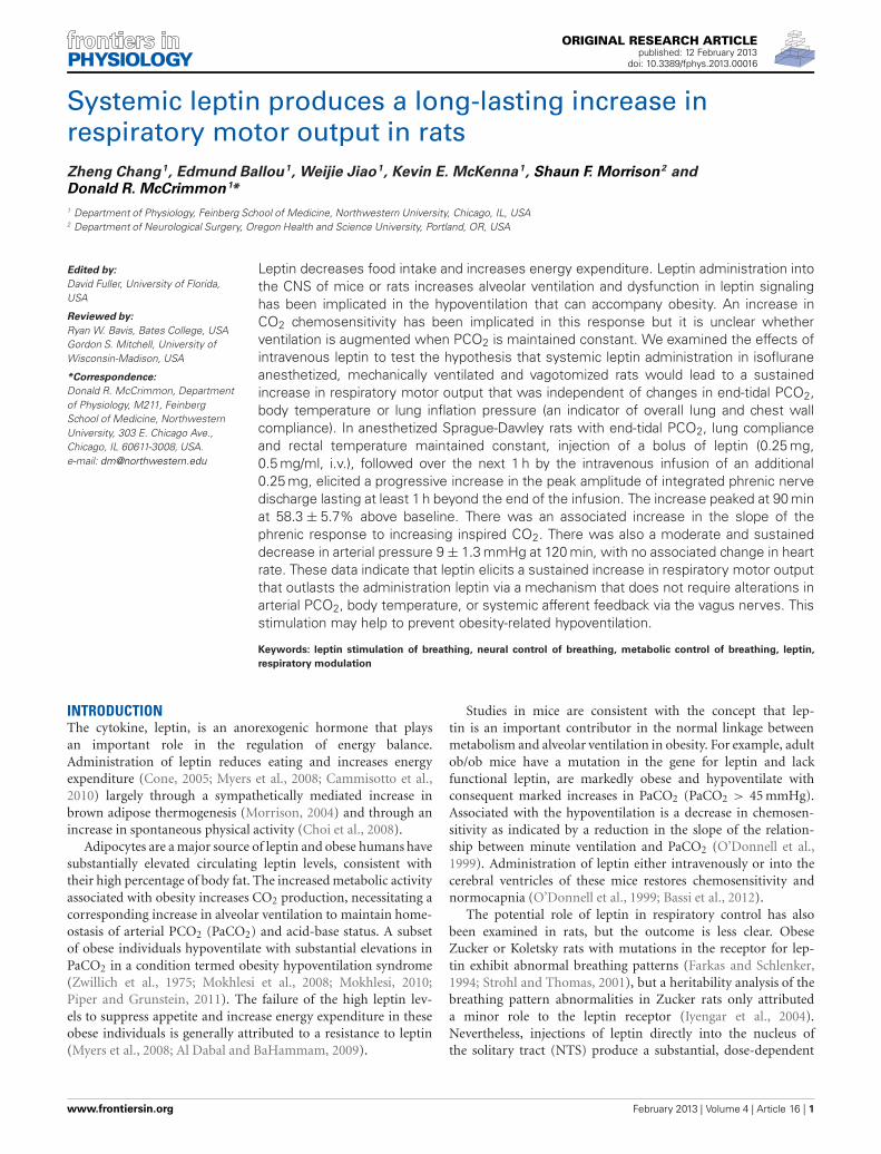
Systemic leptin produces a long-lasting increase inrespiratory motor output in ratsZheng Chang1, Edmund Ballou1, Weijie Jiao1, Kevin E. McKenna1, Shaun F. Morrison2 andDonald R. McCrimmon 1*
1 Department of Physiology, Feinberg School of Medicine, Northwestern University, Chicago, IL, USA2 Department of Neurological Surgery, Oregon Health and Science University, Portland, OR, USA
Edited by:
David Fuller, University of Florida,USA
Reviewed by:
Ryan W. Bavis, Bates College, USAGordon S. Mitchell, University ofWisconsin-Madison, USA
*Correspondence:
Donald R. McCrimmon, Departmentof Physiology, M211, FeinbergSchool of Medicine, NorthwesternUniversity, 303 E. Chicago Ave.,Chicago, IL 60611-3008, USA.e-mail: [email protected]
Leptin decreases food intake and increases energy expenditure. Leptin administration intothe CNS of mice or rats increases alveolar ventilation and dysfunction in leptin signalinghas been implicated in the hypoventilation that can accompany obesity. An increase inCO2 chemosensitivity has been implicated in this response but it is unclear whetherventilation is augmented when PCO2 is maintained constant. We examined the effects ofintravenous leptin to test the hypothesis that systemic leptin administration in isofluraneanesthetized, mechanically ventilated and vagotomized rats would lead to a sustainedincrease in respiratory motor output that was independent of changes in end-tidal PCO2,body temperature or lung inflation pressure (an indicator of overall lung and chest wallcompliance). In anesthetized Sprague-Dawley rats with end-tidal PCO2, lung complianceand rectal temperature maintained constant, injection of a bolus of leptin (0.25 mg,0.5 mg/ml, i.v.), followed over the next 1 h by the intravenous infusion of an additional0.25 mg, elicited a progressive increase in the peak amplitude of integrated phrenic nervedischarge lasting at least 1 h beyond the end of the infusion. The increase peaked at 90 minat 58.3 ± 5.7% above baseline. There was an associated increase in the slope of thephrenic response to increasing inspired CO2. There was also a moderate and sustaineddecrease in arterial pressure 9 ± 1.3 mmHg at 120 min, with no associated change in heartrate. These data indicate that leptin elicits a sustained increase in respiratory motor outputthat outlasts the administration leptin via a mechanism that does not require alterations inarterial PCO2, body temperature, or systemic afferent feedback via the vagus nerves. Thisstimulation may help to prevent obesity-related hypoventilation.
Keywords: leptin stimulation of breathing, neural control of breathing, metabolic control of breathing, leptin,
respiratory modulation
INTRODUCTIONThe cytokine, leptin, is an anorexogenic hormone that playsan important role in the regulation of energy balance.Administration of leptin reduces eating and increases energyexpenditure (Cone, 2005; Myers et al., 2008; Cammisotto et al.,2010) largely through a sympathetically mediated increase inbrown adipose thermogenesis (Morrison, 2004) and through anincrease in spontaneous physical activity (Choi et al., 2008).
Adipocytes are a major source of leptin and obese humans havesubstantially elevated circulating leptin levels, consistent withtheir high percentage of body fat. The increased metabolic activityassociated with obesity increases CO2 production, necessitating acorresponding increase in alveolar ventilation to maintain home-ostasis of arterial PCO2 (PaCO2) and acid-base status. A subsetof obese individuals hypoventilate with substantial elevations inPaCO2 in a condition termed obesity hypoventilation syndrome(Zwillich et al., 1975; Mokhlesi et al., 2008; Mokhlesi, 2010;Piper and Grunstein, 2011). The failure of the high leptin lev-els to suppress appetite and increase energy expenditure in theseobese individuals is generally attributed to a resistance to leptin(Myers et al., 2008; Al Dabal and BaHammam, 2009).
Studies in mice are consistent with the concept that lep-tin is an important contributor in the normal linkage betweenmetabolism and alveolar ventilation in obesity. For example, adultob/ob mice have a mutation in the gene for leptin and lackfunctional leptin, are markedly obese and hypoventilate withconsequent marked increases in PaCO2 (PaCO2 > 45 mmHg).Associated with the hypoventilation is a decrease in chemosen-sitivity as indicated by a reduction in the slope of the relation-ship between minute ventilation and PaCO2 (O’Donnell et al.,1999). Administration of leptin either intravenously or into thecerebral ventricles of these mice restores chemosensitivity andnormocapnia (O’Donnell et al., 1999; Bassi et al., 2012).
The potential role of leptin in respiratory control has alsobeen examined in rats, but the outcome is less clear. ObeseZucker or Koletsky rats with mutations in the receptor for lep-tin exhibit abnormal breathing patterns (Farkas and Schlenker,1994; Strohl and Thomas, 2001), but a heritability analysis of thebreathing pattern abnormalities in Zucker rats only attributeda minor role to the leptin receptor (Iyengar et al., 2004).Nevertheless, injections of leptin directly into the nucleus ofthe solitary tract (NTS) produce a substantial, dose-dependent
www.frontiersin.org February 2013 | Volume 4 | Article 16 | 1
Chang et al. Leptin induces sustained breathing increase
stimulation of breathing, primarily through an increase in themagnitude of phrenic nerve bursts, the neural equivalent oftidal volume (Inyushkin et al., 2009; Inyushkina et al., 2010).The reasons are not clear for this apparent discrepancy in thelinkage between leptin and alveolar ventilation. One possibilityis that obese Zucker and Koletsky rats develop compensatorychanges in respiratory control that make them less dependenton leptin for respiratory homeostasis. Additionally, a numberof factors are difficult to control for, such as the impact ofobesity on pulmonary mechanics, metabolism, and thermoregu-lation and on the regulation of arterial blood gases and acid-basebalance.
The current study was designed to test the hypothesis thatsystemic leptin administration in rats would lead to a sus-tained increase in respiratory motor output that was inde-pendent of leptin-induced changes in CO2 production, bodytemperature or overall lung mechanics. The findings supportthe concept that when end-tidal PCO2, body temperature,and overall lung mechanics are maintained within narrowlimits, intravenous leptin administration produces a time-dependent and sustained increase in respiratory motor outputthat outlasts the period of leptin administration by at least60 min.
MATERIALS AND METHODSANIMALSExperiments were performed on adult male Sprague-Dawley rats(Charles River, Wilmington, MA, USA) weighing 300–500 g. Allsurgeries were performed using sterile procedures adapted forsmall rodents, in accordance with guidelines recommended bythe NIH and by the Society for Neuroscience. The NorthwesternUniversity Animal Care and Use Committee approved allprocedures.
SURGERYAnesthesia was induced with 5% isoflurane (in 50% O2, bal-ance N2) in an induction chamber. Animals were then rapidlyswitched from the induction chamber to a nose cone attachedto a stereotaxic frame where the animal was allowed to freelybreathe isoflurane (2.5–3%) for maintenance. The depth of anes-thesia was frequently assessed (10–15 min intervals) and judgedby the absence of retraction responses to a strong noxious pawpinch, and by the absence of changes in heart rate or breathingpattern in response to the noxious stimulation. Rectal tempera-ture was monitored and maintained at 37.5 ± 0.5◦C by meansof a thermistor-controlled heat lamp. The ECG was continu-ously monitored using transcutaneous needle electrodes placedon the caudal thorax with a ground wire placed laterally onthe abdomen. These electrodes also recorded diaphragm EMGprior to paralyzing the animals (see below). Oxygen satura-tion as well as heart rate and respiratory rate were monitoredvia a pulse oximeter (Mouse Ox, Starr Life Sciences, Oakmont,PA, USA).
Vagi were sectioned bilaterally in the neck. The trachea wascannulated high in the neck and rats were mechanically venti-lated (2.0–2.5 ml, ∼70 min−1) and airway pressure was contin-uously monitored. End-tidal PCO2 was continuously sampled
at the mouth using a rapidly responding infrared CO2 monitor(Puritan–Bennett Co., Datex 223) modified to minimize the deadspace volume. The tidal volume was increased for 2–3 breathsevery 10–15 min to prevent atelectasis.
With these ventilator settings, baseline ventilation was suffi-cient to lower PCO2 below the apneic threshold and CO2 wasadded to the inspired port of the ventilator to raise the end-tidal PCO2 3–5 mmHg above the apneic threshold. The levelof inspired CO2 was then continuously adjusted to maintainend-tidal PCO2 within 1 mmHg of this initial level. Since theadministration of leptin increases brown adipose thermogene-sis and hence PCO2 (Morrison, 2004), for rats receiving leptinthe level of inspired CO2 was decreased to maintain a constantend-tidal PCO2.
The left femoral artery was cannulated (PE-50) for record-ing arterial pressure. The femoral veins were cannulated (PE-10)bilaterally. One vein was used for infusion of fluids (lactatedRinger’s, 3–8 ml/h) and administration of a paralytic, eitherpancuronium bromide (1 mg/kg/h) or succinyl choline chloride(5 mg/kg/h). The contralateral vein was used for infusion ofleptin.
Animals were placed in a stereotaxic frame and a phrenic nervewas exposed in the neck, cleaned of connective tissue, placed ona bipolar silver hook recording electrode and immersed in min-eral oil to prevent drying. Nerve activity was amplified (bandpassfiltered, 0.3–3 kHz), digitized at 10 kHz and recorded on a labo-ratory computer using Spike 2 software (CED Ltd., Cambridge,England).
EXPERIMENTAL PROTOCOLAt least 1 h following surgery, baseline levels of integrated phrenicnerve activity, arterial pressure, heart rate, end-tidal PCO2, airwaypressure, and rectal temperature were recorded for 10–30 min.Leptin was dissolved in lactated Ringer’s solution and adminis-tered using a protocol similar to that reported by Haynes et al.(1997) with the total dose being within the range that has previ-ously been shown to induce Fos-immunoreactivity in the CNS ofadult rats (Elias et al., 2000). Separate groups of rats received lep-tin (n = 6) or lactated Ringer’s solution (n = 4) intravenously. Inthe leptin group, an initial bolus of leptin (0.25 mg, 0.5 mg/ml)was administered, followed by the continuous intravenous infu-sion of an additional 0.25 mg over the following 60 min. Thecontrol group received lactated Ringer’s (the leptin vehicle) deliv-ered as a 0.5 ml bolus and subsequent 0.5 ml infusion over a1 h period. Phrenic nerve activity, arterial pressure, heart rate,end-tidal PCO2, rectal temperature, and airway pressure werecontinuously monitored during the pre-injection control andfor an additional 1 h following the termination of the infu-sion. At the end of this recording period, a CO2 response curvewas generated by increasing the fraction of CO2 in the inspiredgas. CO2 was elevated in two steps; the first was 20–30 mmHgabove baseline while the 2nd raised end-tidal PCO2 to about100 mmHg. Each step was maintained for about 4 min withquasi steady-state measurements made during the last 30 s ofeach step.
Rats were then sacrificed by increasing the inspired isofluraneto 5% followed by a bilateral pneumothorax or decapitation.
Frontiers in Physiology | Respiratory Physiology February 2013 | Volume 4 | Article 16 | 2
Chang et al. Leptin induces sustained breathing increase
DATA ANALYSISData obtained during control measurements and for the 2 h testof the effect of leptin administration were analyzed using a nestedanalysis of variance model with group as the between-rat factorand time as the within rat factor. The group by time interac-tion term was also tested. Post-hoc comparisons were made usingBonferroni-corrected t-tests.
Slopes of the CO2 response curves for each rat were calculatedusing a least squares regression through the three levels of CO2.Slopes for leptin treated vs. control rats were then compared usinga one-tailed T-test.
The possible correlation between any change in phrenic nerveactivity and a corresponding change in arterial pressure was deter-mined using a Spearman’s rank correlation coefficient. Valuesof phrenic nerve discharge and mean arterial pressure (MAP)during control and at 15 min intervals for 2 h after the initialadministration of leptin were compared.
RESULTSIntravenous administration of murine leptin to anesthetized adultrats (n = 6) elicited a sustained increase in phrenic nerve dis-charge and a modest decrease in arterial pressure. Over the courseof an experiment, homeostatic parameters that could influenceeither gas exchange or cardiorespiratory control were main-tained within narrow limits for both leptin and control groupsof animals. Specifically, no significant changes were recorded inend-tidal PCO2, rectal temperature (Figure 1, Table 1), or air-way pressure (not shown) over the recording period. Althoughthere was an initial difference in PCO2 between the groups, thePCO2 of individual animals was maintained within 1 mmHg ofits starting value and therefore did not significantly contributeto the progressive increase in phrenic nerve discharge seen inthe leptin group (see below). At the beginning of each exper-iment the ventilator was set at a tidal volume of ∼2.5 ml and70 cycles/min. For both groups CO2 was added to the inspiredgas mixture to bring the end-tidal PCO2 to 3–5 mmHg above theapneic threshold.
RESPIRATORY MOTOR RESPONSES TO INTRAVENOUS LEPTINInjection of a bolus of leptin (250 µg, i.v.), followed over the nexthour by the intravenous infusion of an additional 250 µg, eliciteda progressive increase in phrenic nerve discharge (Figures 1A, 2;n = 5 − 6; p < 0.05) lasting at least 1 h beyond the end of theinfusion. In contrast, intravenous injection of the vehicle (lactatedRinger’s containing phosphate buffered saline; Figures 1B, 2;n = 2 − 4) elicited no significant change in respiratory motoroutput.
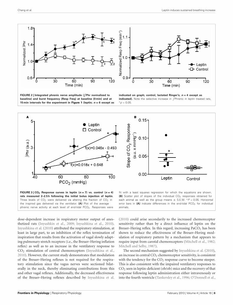
The increase in phrenic nerve discharge in leptin treated ratswas primarily seen as an increase in the peak amplitude ofthe integrated phrenic nerve discharge (Figure 2). The increasebegan within 15 min of the initial leptin injection and was sig-nificant at 60 min following the initial injection, at which timethe increase averaged 39.2 ± 9.3% above baseline. The increasepeaked at 90 min (i.e., 30 min after terminating the leptin infu-sion) when it averaged 58.3 ± 5.7% above baseline. One hourafter terminating the leptin infusion, the peak amplitude of inte-grated phrenic nerve activity was still elevated 47.5 ± 6.7% above
baseline. The neural equivalent of minute ventilation (phrenicburst frequency x peak amplitude of each phrenic burst) was alsosignificantly increased from baseline at 75, 90, and 120 min afterthe initiation of the leptin injection (not shown), although therewas no significant change in the phrenic nerve burst frequency(Figures 1A, 2).
To test the aggregate sensitivity of central and peripheralchemoreceptors, a CO2 response curve was generated at the endof the experiment in both the leptin and control groups of ani-mals. The average slope of the CO2 response curve in leptintreated rats (Figure 3; n = 7) was more than twice that of controlanimals (P < 0.05; n = 4).
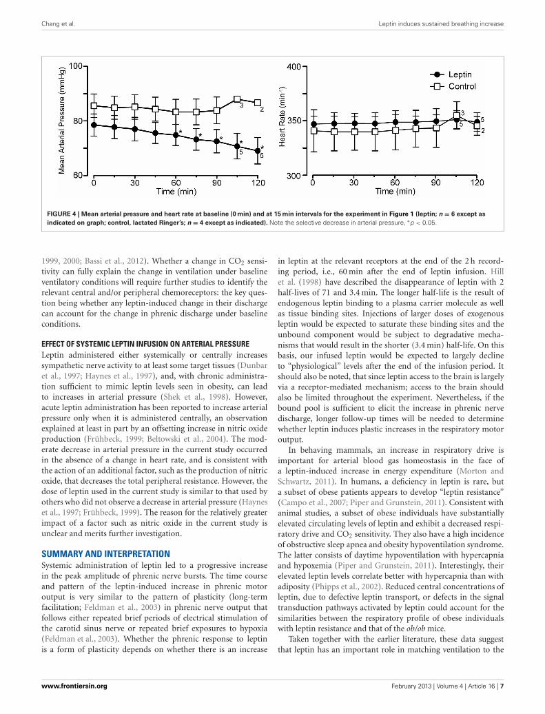
CARDIOVASCULAR RESPONSES TO INTRAVENOUS LEPTINLeptin treatment elicited a progressive decrease in MAP overthe 120 min following the initiation of leptin administration.At 120 min, MAP had decreased 10 ± 1.3 mmHg from a con-trol of 79 ± 4.1 mmHg (Figures 1, 4; p < 0.05). Both systolicand diastolic pressures exhibited similar decreases. There wasno significant change in MAP of control animals; compared toan initial value of 86 ± 4.3 mmHg, MAP was 84 ± 4.9 mmHg at90 min, (n = 4) and in the two rats measured at 120 min it was87 ± 1.0 mmHg (Figures 1, 4). Despite the decrease in MAP inleptin treated rats, there was no change in heart rate over thecourse of the experiment in either the leptin or control group ofanimals (Figures 1, 4).
CORRELATION BETWEEN THE INCREASE IN PHRENIC DISCHARGE ANDDECREASE IN MEAN ARTERIAL PRESSUREThe Spearman’s rank correlation coefficient revealed a signifi-cant correlation between the increase in phrenic discharge anddecrease in MAP (−0.8833, P < 0.05). While the decrease inMAP could contribute to the increase in phrenic discharge, thedecrease was relatively small, averaging 10 ± 1.3 mmHg. At theend of the experiment, the MAP was still 69 ± 4.8 mmHg inleptin-treated rats. This is still above the value generally taken asthe lower limit for autoregulation (e.g., cerebral autoregulatoryrange: 60–150 mmHg; Paulson et al., 1990). Thus, a significantdecrease in organ perfusion, including the brain, would not beanticipated. Further, equivalent decreases in MAP induced withi.v. sodium nitroprusside in a separate group of rats did not elicitsignificant increases in phrenic burst amplitude (not shown).Perhaps more importantly, is the progressive nature of thechanges in both MAP and phrenic burst amplitude. For instance,if there was a causal relationship, the small decreases in MAPat 30 min (−1.5 ± 0.7 mmHg) and 60 min (−3.7 ± 0.5 mmHg)would have been responsible for 31.7 ± 10.0% and 39.4 ± 8.2%increases in phrenic nerve discharge, respectively. Thus, althougha contribution of the decrease in arterial pressure to the increasein the increase in phrenic nerve discharge cannot be completelyruled out, a direct effect of leptin on central respiratory controlseems likely to be more important.
DISCUSSIONThe major finding of the current investigation was that sys-temic leptin infusion causes a progressive and sustained increasein respiratory motor output in isoflurane-anesthetized rats.
www.frontiersin.org February 2013 | Volume 4 | Article 16 | 3
Chang et al. Leptin induces sustained breathing increase
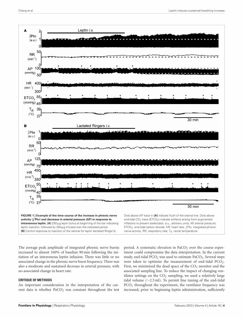
FIGURE 1 | Example of the time course of the increase in phrenic nerve
activity (∫
Phr) and decrease in arterial pressure (AP) in response to
intravenous leptin. (A) 250 µg leptin bolus at beginning of the bar indicatingleptin injection, followed by 250µg infused over the indicated period.(B) Control response to injection of the vehicle for leptin (lactated Ringer’s).
Dots above AP trace in (A) indicate flush of the arterial line. Dots aboveend-tidal CO2 trace (ETCO2) indicate artifacts arising from augmentedinflations to prevent atelectasis. a.u., arbitrary units; AP, arterial pressure;ETCO2, end-tidal carbon dioxide; HR, heart rate;
∫Phr, integrated phrenic
nerve activity; RR, respiratory rate; TR, rectal temperature.
The average peak amplitude of integrated phrenic nerve burstsincreased to almost 160% of baseline 90 min following the ini-tiation of an intravenous leptin infusion. There was little or noassociated change in the phrenic nerve burst frequency. There wasalso a moderate and sustained decrease in arterial pressure, withno associated change in heart rate.
CRITIQUE OF METHODSAn important consideration in the interpretation of the cur-rent data is whether PaCO2 was constant throughout the test
period. A systematic elevation in PaCO2 over the course exper-iment could compromise the data interpretation. In the currentstudy, end-tidal PCO2 was used to estimate PaCO2. Several stepswere taken to optimize the measurement of end-tidal PCO2.First, we minimized the dead space of the CO2 monitor and theassociated sampling line. To reduce the impact of changing ven-tilator settings on the CO2 sampling, we used a relatively largetidal volume (∼2.5 ml). To permit fine tuning of the end-tidalPCO2 throughout the experiment, the ventilator frequency wasincreased, prior to beginning leptin administration, sufficiently
Frontiers in Physiology | Respiratory Physiology February 2013 | Volume 4 | Article 16 | 4
Chang et al. Leptin induces sustained breathing increase
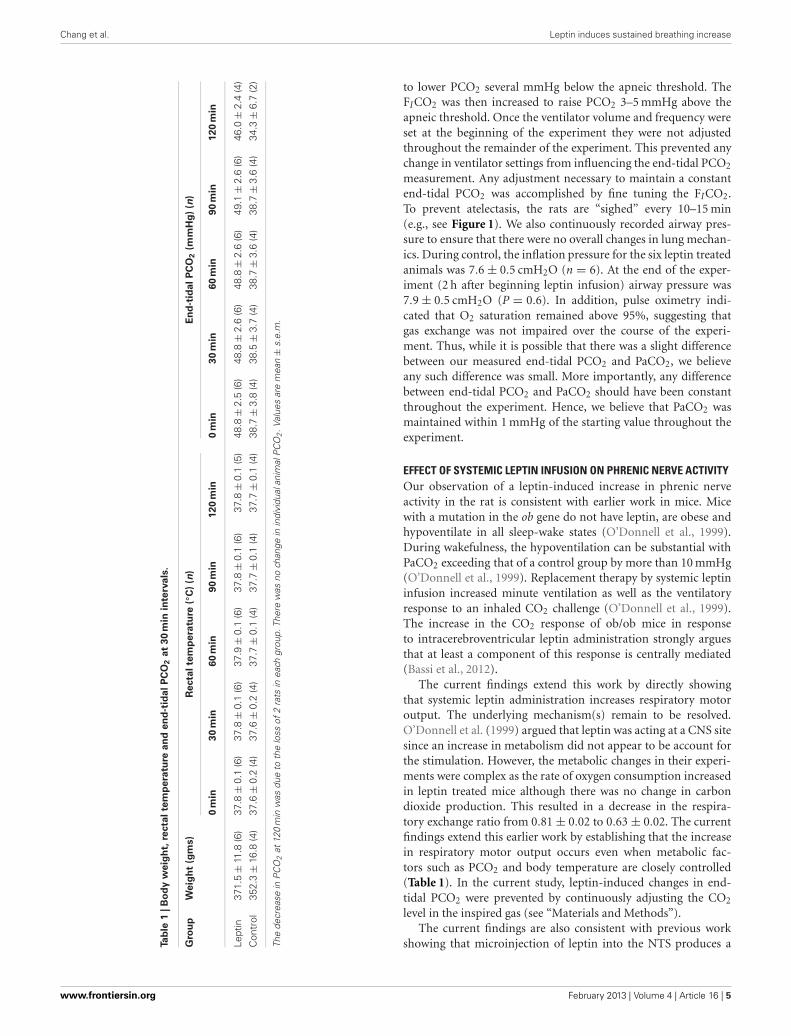
Tab
le1
|B
od
yw
eig
ht,
recta
lte
mp
era
ture
an
de
nd
-tid
al
PC
O2
at
30
min
inte
rva
ls.
Gro
up
We
igh
t(g
ms)
Re
cta
lte
mp
era
ture
(◦C
)(n
)E
nd
-tid
al
PC
O2
(mm
Hg
)(n
)
0m
in3
0m
in6
0m
in9
0m
in1
20
min
0m
in3
0m
in6
0m
in9
0m
in1
20
min
Lept
in37
1.5
±11
.8(6
)37
.8±
0.1
(6)
37.8
±0.
1(6
)37
.9±
0.1
(6)
37.8
±0.
1(6
)37
.8±
0.1
(5)
48.8
±2.
5(6
)48
.8±
2.6
(6)
48.8
±2.
6(6
)49
.1±
2.6
(6)
46.0
±2.
4(4
)
Con
trol
352.
3±
16.8
(4)
37.6
±0.
2(4
)37
.6±
0.2
(4)
37.7
±0.
1(4
)37
.7±
0.1
(4)
37.7
±0.
1(4
)38
.7±
3.8
(4)
38.5
±3.
7(4
)38
.7±
3.6
(4)
38.7
±3.
6(4
)34
.3±
6.7
(2)
The
decr
ease
inP
CO
2at
120
min
was
due
toth
elo
ssof
2ra
tsin
each
grou
p.Th
ere
was
noch
ange
inin
divi
dual
anim
alP
CO
2.V
alue
sar
em
ean
±s.
e.m
.
to lower PCO2 several mmHg below the apneic threshold. TheFICO2 was then increased to raise PCO2 3–5 mmHg above theapneic threshold. Once the ventilator volume and frequency wereset at the beginning of the experiment they were not adjustedthroughout the remainder of the experiment. This prevented anychange in ventilator settings from influencing the end-tidal PCO2
measurement. Any adjustment necessary to maintain a constantend-tidal PCO2 was accomplished by fine tuning the FICO2.To prevent atelectasis, the rats are “sighed” every 10–15 min(e.g., see Figure 1). We also continuously recorded airway pres-sure to ensure that there were no overall changes in lung mechan-ics. During control, the inflation pressure for the six leptin treatedanimals was 7.6 ± 0.5 cmH2O (n = 6). At the end of the exper-iment (2 h after beginning leptin infusion) airway pressure was7.9 ± 0.5 cmH2O (P = 0.6). In addition, pulse oximetry indi-cated that O2 saturation remained above 95%, suggesting thatgas exchange was not impaired over the course of the experi-ment. Thus, while it is possible that there was a slight differencebetween our measured end-tidal PCO2 and PaCO2, we believeany such difference was small. More importantly, any differencebetween end-tidal PCO2 and PaCO2 should have been constantthroughout the experiment. Hence, we believe that PaCO2 wasmaintained within 1 mmHg of the starting value throughout theexperiment.
EFFECT OF SYSTEMIC LEPTIN INFUSION ON PHRENIC NERVE ACTIVITYOur observation of a leptin-induced increase in phrenic nerveactivity in the rat is consistent with earlier work in mice. Micewith a mutation in the ob gene do not have leptin, are obese andhypoventilate in all sleep-wake states (O’Donnell et al., 1999).During wakefulness, the hypoventilation can be substantial withPaCO2 exceeding that of a control group by more than 10 mmHg(O’Donnell et al., 1999). Replacement therapy by systemic leptininfusion increased minute ventilation as well as the ventilatoryresponse to an inhaled CO2 challenge (O’Donnell et al., 1999).The increase in the CO2 response of ob/ob mice in responseto intracerebroventricular leptin administration strongly arguesthat at least a component of this response is centrally mediated(Bassi et al., 2012).
The current findings extend this work by directly showingthat systemic leptin administration increases respiratory motoroutput. The underlying mechanism(s) remain to be resolved.O’Donnell et al. (1999) argued that leptin was acting at a CNS sitesince an increase in metabolism did not appear to be account forthe stimulation. However, the metabolic changes in their experi-ments were complex as the rate of oxygen consumption increasedin leptin treated mice although there was no change in carbondioxide production. This resulted in a decrease in the respira-tory exchange ratio from 0.81 ± 0.02 to 0.63 ± 0.02. The currentfindings extend this earlier work by establishing that the increasein respiratory motor output occurs even when metabolic fac-tors such as PCO2 and body temperature are closely controlled(Table 1). In the current study, leptin-induced changes in end-tidal PCO2 were prevented by continuously adjusting the CO2
level in the inspired gas (see “Materials and Methods”).The current findings are also consistent with previous work
showing that microinjection of leptin into the NTS produces a
www.frontiersin.org February 2013 | Volume 4 | Article 16 | 5
Chang et al. Leptin induces sustained breathing increase
FIGURE 2 | Integrated phrenic nerve amplitude (∫
Phr normalized to
baseline) and burst frequency (Resp Freq) at baseline (0 min) and at
15 min intervals for the experiment in Figure 1 (leptin; n = 6 except as
indicated on graph; control, lactated Ringer’s; n = 4 except as
indicated). Note the selective increase in∫
Phrenic in leptin treated rats,∗p < 0.05.
FIGURE 3 | CO2 Response curves in leptin (n = 7) vs. control (n = 4)
rats measured 2–2.5 h following the initial bolus injection of leptin.
Three levels of CO2 were delivered via altering the fraction of CO2 inthe inspired gas delivered via the ventilator. (A) Plot of the averagephrenic nerve activity at each level of end-tidal PCO2. Responses were
fit with a least squares regression for which the equations are shown.(B) Scatter plot of slopes of the individual CO2 responses obtained foreach animal as well as the group means ± S.E.M. ∗P < 0.05. Horizontalerror bars in (A) indicate differences in the end-tidal PCO2 for individualanimals.
dose-dependent increase in respiratory motor output of anes-thetized rats (Inyushkin et al., 2009; Inyushkina et al., 2010).Inyushkina et al. (2010) attributed the respiratory stimulation, atleast in large part, to an inhibition of the reflex termination ofinspiration that results from the activation of vagal slowly adapt-ing pulmonary stretch receptors (i.e., the Breuer–Hering inflationreflex) as well as to an increase in the ventilatory response toCO2 stimulation of central chemoreceptors (Inyushkina et al.,2010). However, the current study demonstrates that modulationof the Breuer–Hering reflexes is not required for the respira-tory stimulation since the vagus nerves were sectioned bilat-erally in the neck, thereby eliminating contributions from thisand other vagal reflexes. Additionally, the decreased effectivenessof the Breuer–Hering reflexes described by Inyushkina et al.
(2010) could arise secondarily to the increased chemoreceptorsensitivity rather than by a direct influence of leptin on theBreuer–Hering reflex. In this regard, increasing PaCO2 has beenshown to reduce the effectiveness of the Breuer–Hering mod-ulation of respiratory pattern by a mechanism that appears torequire input from carotid chemoreceptors (Mitchell et al., 1982;Mitchell and Selby, 1987).
The second mechanism suggested by Inyushkina et al. (2010),an increase in central CO2 chemoreceptor sensitivity, is consistentwith the tendency for the CO2 response curve to become steeper.This is also consistent with the impaired ventilatory responses toCO2 seen in leptin-deficient (ob/ob) mice and the recovery of thatresponse following leptin administration either intravenously orinto the fourth ventricle (Tankersley et al., 1996; O’Donnell et al.,
Frontiers in Physiology | Respiratory Physiology February 2013 | Volume 4 | Article 16 | 6
Chang et al. Leptin induces sustained breathing increase
FIGURE 4 | Mean arterial pressure and heart rate at baseline (0 min) and at 15 min intervals for the experiment in Figure 1 (leptin; n = 6 except as
indicated on graph; control, lactated Ringer’s; n = 4 except as indicated). Note the selective decrease in arterial pressure, ∗p < 0.05.
1999, 2000; Bassi et al., 2012). Whether a change in CO2 sensi-tivity can fully explain the change in ventilation under baselineventilatory conditions will require further studies to identify therelevant central and/or peripheral chemoreceptors: the key ques-tion being whether any leptin-induced change in their dischargecan account for the change in phrenic discharge under baselineconditions.
EFFECT OF SYSTEMIC LEPTIN INFUSION ON ARTERIAL PRESSURELeptin administered either systemically or centrally increasessympathetic nerve activity to at least some target tissues (Dunbaret al., 1997; Haynes et al., 1997), and, with chronic administra-tion sufficient to mimic leptin levels seen in obesity, can leadto increases in arterial pressure (Shek et al., 1998). However,acute leptin administration has been reported to increase arterialpressure only when it is administered centrally, an observationexplained at least in part by an offsetting increase in nitric oxideproduction (Frühbeck, 1999; Beltowski et al., 2004). The mod-erate decrease in arterial pressure in the current study occurredin the absence of a change in heart rate, and is consistent withthe action of an additional factor, such as the production of nitricoxide, that decreases the total peripheral resistance. However, thedose of leptin used in the current study is similar to that used byothers who did not observe a decrease in arterial pressure (Hayneset al., 1997; Frühbeck, 1999). The reason for the relatively greaterimpact of a factor such as nitric oxide in the current study isunclear and merits further investigation.
SUMMARY AND INTERPRETATIONSystemic administration of leptin led to a progressive increasein the peak amplitude of phrenic nerve bursts. The time courseand pattern of the leptin-induced increase in phrenic motoroutput is very similar to the pattern of plasticity (long-termfacilitation; Feldman et al., 2003) in phrenic nerve output thatfollows either repeated brief periods of electrical stimulation ofthe carotid sinus nerve or repeated brief exposures to hypoxia(Feldman et al., 2003). Whether the phrenic response to leptinis a form of plasticity depends on whether there is an increase
in leptin at the relevant receptors at the end of the 2 h record-ing period, i.e., 60 min after the end of leptin infusion. Hillet al. (1998) have described the disappearance of leptin with 2half-lives of 71 and 3.4 min. The longer half-life is the result ofendogenous leptin binding to a plasma carrier molecule as wellas tissue binding sites. Injections of larger doses of exogenousleptin would be expected to saturate these binding sites and theunbound component would be subject to degradative mecha-nisms that would result in the shorter (3.4 min) half-life. On thisbasis, our infused leptin would be expected to largely declineto “physiological” levels after the end of the infusion period. Itshould also be noted, that since leptin access to the brain is largelyvia a receptor-mediated mechanism; access to the brain shouldalso be limited throughout the experiment. Nevertheless, if thebound pool is sufficient to elicit the increase in phrenic nervedischarge, longer follow-up times will be needed to determinewhether leptin induces plastic increases in the respiratory motoroutput.
In behaving mammals, an increase in respiratory drive isimportant for arterial blood gas homeostasis in the face ofa leptin-induced increase in energy expenditure (Morton andSchwartz, 2011). In humans, a deficiency in leptin is rare, buta subset of obese patients appears to develop “leptin resistance”(Campo et al., 2007; Piper and Grunstein, 2011). Consistent withanimal studies, a subset of obese individuals have substantiallyelevated circulating levels of leptin and exhibit a decreased respi-ratory drive and CO2 sensitivity. They also have a high incidenceof obstructive sleep apnea and obesity hypoventilation syndrome.The latter consists of daytime hypoventilation with hypercapniaand hypoxemia (Piper and Grunstein, 2011). Interestingly, theirelevated leptin levels correlate better with hypercapnia than withadiposity (Phipps et al., 2002). Reduced central concentrations ofleptin, due to defective leptin transport, or defects in the signaltransduction pathways activated by leptin could account for thesimilarities between the respiratory profile of obese individualswith leptin resistance and that of the ob/ob mice.
Taken together with the earlier literature, these data suggestthat leptin has an important role in matching ventilation to the
www.frontiersin.org February 2013 | Volume 4 | Article 16 | 7
Chang et al. Leptin induces sustained breathing increase
metabolic requirements imposed by adipose tissue. The mecha-nisms by which PaCO2 is maintained within narrow limits despitemarked changes in metabolism are largely a mystery. Our resultsdemonstrating leptin modulation of breathing begin to addressthis question. Further elucidation will require identification of thecentral pathways and molecular mechanisms by which activationof central leptin receptors alters breathing.
ACKNOWLEDGMENTSWe thank biostatistician Dr. Alfred W. Rademaker for perform-ing the statistical analyses, Dr. George F. Alheid for suggestingwe examine the role of leptin in respiratory control, and Drs.Marco Martina and George F. Alheid for helpful discussions andcomments on the manuscript. This work was supported by NIHgrants HL088580 and HL095731.
REFERENCESAl Dabal, L., and BaHammam, A.
S. (2009). Obesity hypoventilationsyndrome. Ann. Thorac. Med. 4,41–49.
Bassi, M., Giusti, H., Leite, C. M.,Anselmo-Franci, J. A., do Carmo,J. M., da Silva, A. A., et al.(2012). Central leptin replace-ment enhances chemorespiratoryresponses in leptin-deficient miceindependent of changes in bodyweight. Pflugers Arch. 464, 145–153.
Beltowski, J., Jochem, J., Wojcicka, G.,and Zwirska-Korczala, K. (2004).Influence of intravenously adminis-tered leptin on nitric oxide produc-tion, renal hemodynamics and renalfunction in the rat. Regul. Pept. 120,59–67.
Cammisotto, P. G., Levy, E.,Bukowiecki, L. J., and Bendayan, M.(2010). Cross-talk between adiposeand gastric leptins for the control offood intake and energy metabolism.Prog. Histochem. Cytochem. 45,143–200.
Campo, A., Fruhbeck, G., Zulueta, J. J.,Iriarte, J., Seijo, L. M., Alcaide, A.B., et al. (2007). Hyperleptinaemia,respiratory drive and hypercapnicresponse in obese patients. Eur.Respir. J. 30, 223–231.
Choi, Y. H., Li, C., Hartzell, D. L.,Little, D. E., Della-Fera, M. A.,and Baile, C. A. (2008). ICV lep-tin effects on spontaneous physicalactivity and feeding behavior in rats.Behav. Brain Res. 188, 100–108.
Cone, R. D. (2005). Anatomy and reg-ulation of the central melanocortinsystem. Nat. Neurosci. 8, 571–578.
Dunbar, J. C., Hu, Y., and Lu, H.(1997). Intracerebroventricular lep-tin increases lumbar and renal sym-pathetic nerve activity and bloodpressure in normal rats. Diabetes 46,2040–2043.
Elias, C. F., Kelly, J. F., Lee, C. E., Ahima,R. S., Drucker, D. J., Saper, C. B.,et al. (2000). Chemical characteriza-tion of leptin-activated neurons in
the rat brain. J. Comp. Neurol. 423,261–281.
Farkas, G. A., and Schlenker, E. H.(1994). Pulmonary ventilationand mechanics in morbidly obeseZucker rats. Am. J. Respir. Crit. CareMed. 150, 356–362.
Feldman, J. L., Mitchell, G. S., andNattie, E. E. (2003). Breathing:rhythmicity, plasticity, chemosen-sitivity. Annu. Rev. Neurosci. 26,239–266.
Frühbeck, G. (1999). Pivotal role ofnitric oxide in the control of bloodpressure after leptin administration.Diabetes 48, 903–908.
Haynes, W. G., Morgan, D. A., Walsh,S. A., Mark, A. L., and Sivitz,W. I. (1997). Receptor-mediatedregional sympathetic nerve activa-tion by leptin. J. Clin. Invest. 100,270–278.
Hill, R. A., Margetic, S., Pegg, G. G.,and Gazzola, C. (1998). Leptin: itspharmacokinetics and tissue distri-bution. Int. J. Obes. Relat. Metab.Disord. 22, 765–770.
Inyushkin, A. N., Inyushkina, E. M.,and Merkulova, N. A. (2009).Respiratory responses to microin-jections of leptin into the solitarytract nucleus. Neurosci. Behav.Physiol. 39, 231–240.
Inyushkina, E. M., Merkulova, N.A., and Inyushkin, A. N. (2010).Mechanisms of the respiratoryactivity of leptin at the level of thesolitary tract nucleus. Neurosci.Behav. Physiol. 40, 707–713.
Iyengar, S. K., Stein, C. M., Russo,K., Erokwu, B. O., and Strohl,K. P. (2004). The fa leptin recep-tor mutation and the heritabil-ity of respiratory frequency in aBrown Norway and Zucker inter-cross. J. Appl. Physiol. 97, 811–820.
Mitchell, G. S., Cross, B. A.,Hiramoto, T., and Scheid, P.(1982). Interactions between lungstretch and PaCO2 in modulatingventilatory activity in dogs. J. Appl.Physiol. 53, 185–191.
Mitchell, G. S., and Selby, B. D. (1987).Effects of carotid denervation oninteractions between lung inflationand PaCO2 in modulating phrenicactivity. Respir. Physiol. 67, 367–378.
Mokhlesi, B. (2010). Obesity hypoven-tilation syndrome: a state-of-the-artreview. Respir. Care 55, 1347–1362.
Mokhlesi, B., Kryger, M. H., andGrunstein, R. R. (2008). Assessmentand management of patients withobesity hypoventilation syndrome.Proc. Am. Thorac. Soc. 5, 218–225.
Morrison, S. F. (2004). Activation of 5-HT1A receptors in raphe pallidusinhibits leptin-evoked increases inbrown adipose tissue thermogen-esis. Am. J. Physiol. Regul. Integr.Comp. Physiol. 286, R832–R837.
Morton, G. J., and Schwartz, M.W. (2011). Leptin and the cen-tral nervous system control of glu-cose metabolism. Physiol. Rev. 91,389–411.
Myers, M. G., Cowley, M. A., andMunzberg, H. (2008). Mechanismsof leptin action and leptin resis-tance. Annu. Rev. Physiol. 70,537–556.
O’Donnell, C. P., Schaub, C. D., Haines,A. S., Berkowitz, D. E., Tankersley,C. G., Schwartz, A. R., et al. (1999).Leptin prevents respiratory depres-sion in obesity. Am. J. Respir. Crit.Care Med. 159, 1477–1484.
O’Donnell, C. P., Tankersley, C. G.,Polotsky, V. P., Schwartz, A. R.,and Smith, P. L. (2000). Leptin,obesity, and respiratory function.Respir. Physiol. 119, 163–170.
Paulson, O. B., Strandgaard, S., andEdvinsson, L. (1990). Cerebralautoregulation. Cerebrovasc. BrainMetab. Rev. 2, 161–192.
Phipps, P. R., Starritt, E., Caterson,I., and Grunstein, R. R. (2002).Association of serum leptin withhypoventilation in human obesity.Thorax 57, 75–76.
Piper, A. J., and Grunstein, R. R. (2011).Obesity hypoventilation syndrome:mechanisms and management.
Am. J. Respir. Crit. Care Med. 183,292–298.
Shek, E. W., Brands, M. W., andHall, J. E. (1998). Chronic leptininfusion increases arterial pressure.Hypertension 31, 409–414.
Strohl, K. P., and Thomas, A. J.(2001). Ventilatory behavior andmetabolism in two strains of obeserats. Respir. Physiol. 124, 85–93.
Tankersley, C., Kleeberger, S., Russ,B., Schwartz, A., and Smith, P.(1996). Modified control of breath-ing in genetically obese (ob/ob)mice. J. Appl. Physiol. 81, 716–723.
Zwillich, C. W., Sutton, F. D., Pierson,D. J., Greagh, E. M., and Weil,J. V. (1975). Decreased hypoxicventilatory drive in the obesity-hypoventilation syndrome. Am. J.Med. 59, 343–348.
Conflict of Interest Statement: Theauthors declare that the researchwas conducted in the absence of anycommercial or financial relationshipsthat could be construed as a potentialconflict of interest.
Received: 19 November 2012; accepted:22 January 2013; published online: 12February 2013.Citation: Chang Z, Ballou E, JiaoW, McKenna KE, Morrison SF andMcCrimmon DR (2013) Systemic lep-tin produces a long-lasting increasein respiratory motor output in rats.Front. Physio. 4:16. doi: 10.3389/fphys.2013.00016This article was submitted to Frontiersin Respiratory Physiology, a specialty ofFrontiers in Physiology.Copyright © 2013 Chang, Ballou, Jiao,McKenna, Morrison and McCrimmon.This is an open-access article dis-tributed under the terms of the CreativeCommons Attribution License, whichpermits use, distribution and reproduc-tion in other forums, provided the origi-nal authors and source are credited andsubject to any copyright notices concern-ing any third-party graphics etc.
Frontiers in Physiology | Respiratory Physiology February 2013 | Volume 4 | Article 16 | 8