Embed Size (px)
Citation preview

Systematic screen of chemotherapeutics in Drosophilastem cell tumorsMichele Marksteina,b,1, Samantha Dettorrea,b, Julio Chob, Ralph A. Neumüllerb, Sören Craig-Müllerb,and Norbert Perrimonb,c,1
aDepartment of Biology, University of Massachusetts, Amherst, MA 01003; and bDepartment of Genetics and cHoward Hughes Medical Institute, HarvardMedical School, Boston, MA 02115
Contributed by Norbert Perrimon, January 21, 2014 (sent for review December 19, 2013)
Here we report the development of an in vivo system to study theinteraction of stem cells with drugs using a tumor model in theadult Drosophila intestine. Strikingly, we find that some Food andDrug Administration-approved chemotherapeutics that can inhibitthe growth of Drosophila tumor stem cells can paradoxically pro-mote the hyperproliferation of their wild-type counterparts. Theseresults reveal an unanticipated side effect on stem cells that maycontribute to tumor recurrence. We propose that the same sideeffect may occur in humans based on our finding that it is driven inDrosophila by the evolutionarily conserved Janus kinase-signaltransducers and activators of transcription (JAK-STAT) pathway.An immediate implication of our findings is that supplementingtraditional chemotherapeutics with anti-inflammatories may reducetumor recurrence.
cancer stem cell | drug screening | Drosophila intestinal stem cell |whole-animal screening
Avexing problem in cancer therapeutics is tumor recurrence:tumors that initially respond to chemotherapy ultimately
return resistant to chemotherapy. Drug-resistant tumors emergebecause drugs select for the survival of cells with either preexistingor newly acquired drug resistance properties (1). For example,tumors that recur in the wake of treatment with the ABL-BCRinhibitor Gleevec typically have mutations in ABL-BCR that pre-vent Gleevec from inhibiting it (2). Sometimes these mutations existin small populations of tumor cells before drug treatment, whereasother times they arise spontaneously in cells during treatment.Another form of drug resistance comes from “bypass” mutationsthat activate multiple oncogenes, thereby rendering drug inhibitionof a specific oncogene inconsequential (3, 4). This form of re-sistance is believed to be prevalent due to the genomic instability ofmost tumors (5). Additionally, there is mounting evidence that se-lection may act on yet a third level, in which selection is not forspecific mutations or oncogenes, but instead for a class of cells withstem cell properties, called cancer stem cells (CSCs) (6, 7). CSCs,like wild-type (WT) stem cells, are defined by their ability to giverise to all of the cell types in a tissue, which in the case of CSCs areall of the cell types of its cognate tumor. CSCs have been identifiedas rare populations of cells in several cancers including breast, brain,and colorectal cancers. Based on similarities between the biology ofCSCs and WT stem cells, including drug resistance (8–12), a rela-tively new field is emerging to identify small molecules that cantarget the underlying biology of “stemness.”To date, screens for drugs that target stemness have been largely
in vitro using either cultured cell lines induced to become stem cellsor stem cells isolated from freshly dissected tissue and cultured inconditions that permit stem cell survival (13). These approacheshave identified stem-cell–selective drugs such as salinomycin (14)and metformin (15) for breast CSCs and neurotransmitter inhibitorsthat suppress neuronal CSCs (16, 17). However, although in vitrostem cell screens have proven successful in identifying drugs thatdirectly act on stem cells, they cannot in their present form identifydrugs that act on the stem cell microenvironment (18). Becausestem cells rely on their microenvironment for cues to divide,
differentiate, and die, this omission from drug screens could missthe identification of drugs with potent effects on stem cells.However, to include the stem cell microenvironment in chemical
screens requires methods to culture stem cells in entirely newways. Current methods that enable stem cells to be culturedeither supply niche signals in lieu of the niche itself or use stem-like cells engineered to retain stem cell characteristics autono-mously. Efforts are underway to more precisely culture and screenstem cells cocultured with their niche (19, 20). However, an alter-native approach that is more immediately available is to use theready-made stem cell microenvironments found in living animals,which can be probed by performing whole-animal screens (21).We set out to develop a whole-animal approach to screen for
drugs affecting stemness, using the fruit fly Drosophila melanogaster.A strength of Drosophila as an organism for whole-animal screeningis its small size: adults can fit into the wells of 96-well plates,opening the possibility of using flies to identify drugs that affectadult stem cells in vivo. To take full advantage of this feature, wedeveloped methods to handle flies in 96-well plates. First, we de-veloped a method to house and feed flies drugs in 1-mL deep 96-well plates, containing as little as 100 μL of food. In addition, wedeveloped a method to score the size of stem cell tumors withinadults using a luciferase reporter assay adapted to 96-well plates(see below). Together, these methods make it possible to performchemical screens in adult Drosophila for stem-cell–modifying drugs.We chose to focus on drug interactions with the stem cells of the
adult Drosophila intestine because they have molecular, physio-logical, and cellular properties in common with their mammaliancounterparts (22–24). Moreover, because they line the digestive
Significance
In this article we report a large-scale chemical screen in adultDrosophila to find inhibitors of stem-cell–derived tumors. Toour surprise, we found that some Food and Drug Administra-tion-approved chemotherapy drugs have the dual property ofreducing growth of stem-cell–derived tumors while also stimu-lating hyperproliferation of their wild-type counterparts. Sincehyperproliferation is one of the hallmarks of cancer cells, thisside effect could contribute to refueling the growth of the verytumors that these chemotherapeutics are intended to inhibit.We show that this side effect is driven by the evolutionarilyconserved Janus kinase-signal transducers and activators oftranscription (JAK-STAT) inflammatory pathway, raising thepossibility that the JAK-STAT pathway may also be activated inhumans who are treated with some chemotherapeutics.
Author contributions: M.M. designed research; M.M., S.D., J.C., R.A.N., and S.C.-M. per-formed research; M.M., J.C., R.A.N., and N.P. analyzed data; and M.M. wrote the paper.
The authors declare no conflict of interest.1To whom correspondence may be addressed. E-mail: [email protected] or [email protected].
This article contains supporting information online at www.pnas.org/lookup/suppl/doi:10.1073/pnas.1401160111/-/DCSupplemental.
4530–4535 | PNAS | March 25, 2014 | vol. 111 | no. 12 www.pnas.org/cgi/doi/10.1073/pnas.1401160111
Dow
nloa
ded
by g
uest
on
Janu
ary
18, 2
020

track, they are optimally placed to come into direct contact withingested drugs.Several features make Drosophila intestinal stem cells (ISCs)
a compelling model for mammalian ISCs: they are multipotent,giving rise to cell types similar to those in mammals, large absorptiveenterocytes (ECs) and a wide array of secretory cell types (25, 26)(Fig. 1A); they are similarly situated in a single-layered epitheliumthat abuts the muscle layer; they differentiate based on stochasticcompetition (29); and they use evolutionarily conserved pathwaysfor similar processes—the Wnt pathway for stem cell proliferation(30, 31) and the Notch pathway for stem cell differentiation (25,26, 28). Additional evolutionarily conserved pathways, includingthe EGFR, Hippo, AKT, and Janus kinase-signal transducers andactivators of transcription (JAK-STAT) pathways, are at play inDrosophila ISCs, each of which are linked to human cancers (24,32). Thus, Drosophila ISCs provide both a model for their mam-malian counterparts and a multicellular context in which to dis-sect the interplay of drugs with human oncogenic pathways.
ResultsBuilding an in Vivo Stem-Cell–Derived Tumor Model. Based on theparallels between Drosophila and mammalian ISCs, we builta “screenable” tumor model using the ISC-expressed esg-Gal4transcription factor to express transgenes engineered with up-stream Gal4-binding sites called upstream activating sequence(UAS) sites (33). We constructed flies to simultaneously expressthree UAS transgenes under control of the esg-Gal4 transcriptionfactor: UAS-human RAFgof (gain-of-function allele of the serine-threonine kinase Raf) to hyperactivate the downstream oncogenicMAPK pathway (34), UAS-luciferase to estimate tumor size fromwhole-animal homogenates (35), and UAS-GFP to visualize stem-cell–initiated tumors in dissected intestinal tissue (36) (Fig. 1B).We found that expression of human RAFgof drives the forma-
tion of large heterogeneous tumors characterized by the persistentexpression of the progenitor marker esg and continued activationof the MAPK pathway, which we detected with a phospho-specificantibody against the active di-phosphorlyated form of dpERK,a downstream kinase that mediates MAPK signaling (Fig. 1C).These tumors, which we refer to as RAFgof ISC tumors, compriseboth mitotic stem-like cells that express the Notch ligand Deltaand nondividing daughter cells with large polyploid nuclei (Fig.1C). We observed that only the Delta+ cells stain positively forthe mitotic marker phospho-histone 3 (PH3), suggesting that they
alone drive proliferation of the tumors (Fig. S1). These results areconsistent with observations reported by Jiang et al. (27). In ad-dition, we found that these cells have a great regenerative ca-pacity; they not only maintain the tumor, but also, when wetransplanted the tumors to wild-type hosts by injection into thehost abdomen, about 10% of the time (n > 100 transplants) thetumors continued growing until they filled the abdomen and killedthe host (Fig. 1D and Fig. S2).Although the tumors are heterogeneous, luciferase expression
from the tumors is largely consistent from animal to animal,comprising about two-thirds of the total luciferase signal in whole-animal homogenates (Fig. 1E). Consequently, changes in luciferaseexpression can be used to approximate changes in tumor growth.For example, feeding flies the mitotic inhibitor colchicine results incomplete tumor loss (Fig. 1F) and a corresponding loss of tumor-expressed luciferase activity that can be readily measured in whole-animal homogenates (Fig. 1G). These results demonstrate thatexpressing RAFgof in the ISCs creates heterogeneous tumors thatare amenable to screening with the luciferase assay.
Screen of Food and Drug Administration-Approved ChemotherapeuticsIdentifies Drugs That Inhibit Drosophila Tumors. With the screenableRAFgof ISC tumor model in hand we systematically screened theeffects of 88 Food and Drug Administration (FDA)-approvedchemotherapy drugs [National Cancer Institute (NCI) DrugTherapeutics Program set] to determine the sensitivity of Dro-sophila ISCs to human drugs (Fig. 2A). We diluted each drug ina food we developed for mixing chemicals, which we call low-meltfly food (Materials and Methods). We decided to test drugs at thehighest concentration feasible, 100 μM, to maximize our ability todetect drugs with putative antitumorigenic activity. To determineif the flies would consume the drugs, we added human-grade redfood coloring to the low-melt fly food. We found that within 1 h offeeding the dye could be visualized through the abdomen of eachfly, indicating that the low-melt fly food and chemotherapeuticswere palatable to the flies. However, because dyes in food coloringcan have deleterious effects in humans (37), we omitted themfrom the food used in the screens.Because mammalian stem cells and their CSC equivalents are
largely resistant to chemotherapy (8–12), we expected that theDrosophila WT ISCs would likewise be resistant. However, be-cause mammalian stem cells can be sensitized to chemotherapywhen induced to actively proliferate (16, 38), we expected that
Fig. 1. Characterization of the screenable stem celltumor model. (A) Diagram of intestinal stem cell(ISC) lineage showing polyploid enterocytes (EC)and diploid enteroendocrine cells (EE). (B) Genotypeof the screenable tumor model showing the esg-Gal4 transcription factor driving the expression ofGFP, luciferase, and RAFgof UAS-linked transgenes.(C) WT ISCs and RAFgof ISC tumors. ISCs are labeledby esg-Gal4 driving UAS-GFP (green). Nuclei arevisualized with the DNA dye DAPI (blue). WT ISCsexpress dpERK (27) (Upper: red cytoplasmic staining)and Delta (28) (Lower: red membrane staining).Expression of UAS-RAFgof with the esg-Gal4 driverincreases dpERK and proliferation of ISC-like Delta-expressing cells. (D) Fate of WT and RAFgof intestinalfragments injected into WT hosts (n > 100). The esg+cells within the injected intestines are marked withGFP (green). (E) Measurements of luciferase activityfrom individually dissected flies shows that gut tu-mors contribute about 66% of the total luciferaseactivity in each animal, which correlates with theamount that is absent from colchicine-treated ani-mals (F and G).
Markstein et al. PNAS | March 25, 2014 | vol. 111 | no. 12 | 4531
MED
ICALSC
IENCE
S
Dow
nloa
ded
by g
uest
on
Janu
ary
18, 2
020

rapidly dividing RAFgof ISC tumors would be sensitive to at leastsome chemotherapy drugs.Consistent with the expectation that RAFgof ISC tumors would
be sensitive to human chemotherapy drugs, we identified 14drugs from the luciferase screen with putative tumor suppressoractivity. These drugs, when fed to flies with RAFgof ISC tumors,resulted in a 50% or greater loss of luciferase activity in whole-animal lysates compared with DMSO controls (rank sum P <0.001) (Fig. 2B). To validate the luciferase results, we dissectedthe intestines from flies treated with these drugs to visualizeGFP-expressing tumor cells. We also dissected and visualized theintestines of flies treated with drugs that did not reduce lucif-erase expression. In each case, the GFP observations validatedthe luciferase results: the drugs that scored as hits in the lucif-erase screen each reduced tumor burden, whereas drugs, likebleomycin, that failed to score as a hit had no apparent effect onthe tumors (Fig. 2C). The tumor inhibitors constitute a wide-spectrum of cytotoxic cell cycle inhibitors, including S-phaseinhibitors and the pathway-specific mTOR inhibitor rapamycin(39) (Fig. 2C, Upper), and transcriptional, proteasome, and mitoticinhibitors, as well as inducers of DNA damage (Fig. 2C, Lower).Together, these results establish thatDrosophilaRAFgof ISC tumorsare sensitive to a broad range of compounds of clinical significance.
Side Effect of Class II Drugs Drives Stem Cell Hyperproliferation. Wenext tested the effects of the 14 RAFgof ISC tumor inhibitors onWT ISCs. Our expectation was that the WT ISCs, like mammalianWT stem cells and mammalian CSCs, would be resistant to tra-ditional chemotherapy drugs. Indeed, under the same conditionsas the screen, none of the drugs had obvious inhibitory effects on
the WT ISCs (Fig. 2D). Because WT ISCs inDrosophila, as well asin mammals, divide on average once a day (22), this result is notdue to stem cell quiescence. At least within the parameters of ourexperiment, WT ISCs are less susceptible than their tumorcounterparts to the destructive effects of the chemotherapydrugs that we tested.Although we did not observe inhibitory effects of the drugs on
WT ISCs, to our surprise, we found that a diverse spectrum ofthe drugs induced overgrowth of WT ISCs, including the tran-scriptional inhibitor actinomycin; the proteasome inhibitorbortezomib; the mitotic inhibitors paclitaxel, vinblastine, and vin-cristine; and two inducers of DNA damage, mitomycin, and dau-norubicin (Fig. 2D, Lower). The overgrowth of WT ISCs was alsoobserved with bleomycin, as previously reported (40) (Fig. 2D).To determine whether the overgrowth of WT ISCs was due to
an increase in proliferation, we stained dissected intestines withantibodies against the mitotic marker PH3. We observed PH3+staining in the ISCs in both DMSO-treated and drug-treatedanimals, as seen, for example, in a cluster of esg+ cells aftertreatment with bortezomib (Fig. 3A). PH3+ staining was specificto these cells, evident by focusing on the surface of the intestinewhere the ISC nuclei are in focus, and it was absent from the ECnuclei, evident by focusing about 1 μm below the ISC nuclei,where the EC nuclei are in focus.Consistent with the possibility that the increase in WT ISCs
could be due to an increase in cell proliferation, we observeda statistically significant increase in the number of PH3+ cellsper gut between the cohort of animals treated with DMSO (46flies) and animals treated with drugs that increased WT ISCs(ranging from 6 to 24 flies per cohort; rank sum <0.001) (Fig. 3B).
Fig. 2. Screen of 88 FDA-approved oncology drugs identifies two classes of drugs that inhibit Drosophila RAFgof tumors. (A) Schematic of drug screen. (B)Replicate whole-animal luciferase assays from flies fed either DMSO controls or drugs identified as hits from the screen. Bleomycin did not score as a hit and isincluded as a negative control. Each bar represents the average of 12 biological replicates; error bar = 1 SD; P < 0.001 by rank-sum analysis. (C) Confocalimages of posterior midguts dissected from RAFgof flies treated with the compounds that scored as hits, with bleomycin included as a negative control. (D)Confocal images of posterior midguts dissected from WT flies fed with drugs that scored as hits from the drug screen. (Upper) Drugs that have no effect andare termed class I drugs. (Lower) Drugs that induce an increase in esg+ cells (green) and are termed class II drugs.
4532 | www.pnas.org/cgi/doi/10.1073/pnas.1401160111 Markstein et al.
Dow
nloa
ded
by g
uest
on
Janu
ary
18, 2
020

Conversely, we did not see a statistically significant difference inthe number of PH3+ cells/gut between the cohorts of animalstreated with DMSO and animals treated with drugs that did notincrease ISCs (ranging from 6 to 10 flies/cohort) (Fig. 3D).Our results thus distinguish two classes of chemotherapy drugs:
class I drugs inhibit the growth of RAFgof ISC tumors but do notaffect WT ISCs, whereas class II drugs not only inhibit growth ofRAFgof ISC tumors but also paradoxically increase growth of theirwild-type counterparts (Table 1). Because class II drugs inhibitedgrowth of ISC-induced tumors, we did not expect them to alsoinduce the hyperproliferation of ISCs. Indeed, the finding thatdrugs that can kill rapidly dividing stem cells can paradoxically alsoinduce them to hyperproliferate was, to our knowledge, un-precedented. However, as noted in the discussion, similar resultswere recently reported in mammals, thus highlighting the gener-ality of our findings (41).
Class II Side Effect Is Mediated by the Stem Cell Microenvironment. Inprevious studies, ISCs had been shown to proliferate in responseto damage to their EC daughter cells, the major constituency ofthe ISC microenvironment. We thus explored the possibility thathyperproliferation was a “side effect” of chemotherapy effects onthe microenvironment.We envisioned that the class II side effect might be mediated
by the JAK-STAT–signaling pathway because this pathway hasbeen shown to mediate ISC proliferation in response to a varietyof agents that can damage the EC daughter cells, including ge-netically induced apoptosis and stress, bacterial infection, andtreatment with the DNA-damaging drug bleomycin (40, 42–46).Interestingly, bleomycin had failed to inhibit the growth of RAFgof
ISC tumors in our screen (Fig. 2 B and C). This result thus showsthat induction of the JAK-STAT pathway, although sufficient toinduce ISC proliferation, is not sufficient to kill RAFgof ISCtumors. Thus, the possibility that the JAK-STAT pathway mightunderlie the ability of class II drugs to induce ISC proliferation wasan appealing prospect because it would indicate that class II drugselicit not only a side effect in the ECs, but also a side effect that ismechanistically separable from their ability to kill the tumor.In response to bacterial infection, genetically induced stress,
and cell death, ECs have been shown to express Unpaired(Upds), IL-6–like cytokines that activate the JAK-STAT path-way (42). To investigate whether the same mechanism is trig-gered by treatment with class II chemotherapy drugs, we used anUpd-3 Gal4 enhancer trap (47) to track expression of Upd-3. Wefound that Upd-3 expression correlated precisely with the effectsof class I and class II chemotherapy drugs on WT proliferation:none of the class I drugs induced Upd-3 expression whereas eachof the class II drugs did induce EC expression of Upd-3 (Fig. 4A).
Similarly, we found that bleomycin, which was previously shownto induce Stat activation in the ISCs, stimulated Upd-3 expressionin the ECs (Fig. 4A). In all cases, Upd-3 induction was specificto the EC cells, evident by focusing on either the surface of theintestine where the diploid ISC nuclei are in focus or by focusing1 μM down, at the “subsurface” layer where the EC nuclei are infocus (Fig. S3).In addition to observing the expression of Upd-3 in the ECs,
we found that activation of the JAK-STAT–signaling cascadewithin ISCs was required for their proliferation. For example,when we reduced JAK-STAT signaling in ISCs, either by RNAiagainst the Upd-3 receptor, domeless, or by overexpression ofSocs36E, a repressor that acts downstream of domeless, we foundthat the hyperproliferation response was reduced when treatedwith one of the strongest class II drugs, bortezomib (Fig. 4B).These results indicate that the JAK-STAT pathway is requiredspecifically in the ISC hyperproliferating cells.Collectively, these results demonstrate that class II drugs
stimulate expression of Upd-3 in the EC daughter cells, culmi-nating in JAK-STAT–mediated proliferation in WT ISC cells.Our finding that bleomycin induces ISC proliferation by thesame mechanism and yet fails to kill RAFgof ISC tumors indi-cates that the neither the induction of Upd-3 from the ECs northe stimulation of JAK-STAT signaling in the ISCs is sufficientto kill the tumor. These results suggest that the side effect ofclass II drugs on the ISC microenvironment is mechanisticallyseparable from their ability to kill RAFgof ISC tumors.
Separation of Class II Tumor Inhibition from Tumor Initiation. Ourfinding that chemotherapy drugs that block tumor growth canalso induce growth of WT stem cells was not only an unforeseenside effect, but also possibly a deleterious side effect with thepotential to fuel tumor recurrence by multiple mechanisms. Forexample, in humans, the JAK-STAT pathway mediates the in-flammation response, which correlates strongly with the onset ofcancer (48). In addition, the induction of stem cell hyperprolif-eration could conceivably drive CSCs to regenerate tumors or evendrive the formation of de novo stem-cell–initiated tumors.Given that we identified the JAK-STAT pathway as driving
WT stem cell hyperproliferation, one mechanism to suppress thepotential for stem-cell–mediated tumor recurrence would be tocouple traditional chemotherapy with anti-inflammatory drugs.However, a complementary approach and one that may be morebroadly applicable would be to circumvent the side effect alto-gether. Along these lines, we tested class II drugs over a range ofconcentrations to identify a therapeutic window in which specificdoses could inhibit tumors without also stimulating stem cellproliferation. In no instance were we able to separate the tumorinhibitory effects from the stem cell proliferation side effect (Fig.S4). These results indicate that the therapeutic windows of classII drugs may not be easily separable from their side effect on WTISCs, suggesting that the best way to circumvent the side effect isto find drugs that avoid the side effect altogether.In our screen of FDA-approved chemotherapy drugs, we
identified seven drugs that can block RAFgof ICS tumors without
Fig. 3. Class II drugs increase stem cell proliferation. (A) PH3 staining (red)after treatment with the class II drug bortezomib. PH3 is evident in thenuclei of esg+ cells (green), visible in the surface view, and is absent from thenuclei of polyploid EC cells, visualized 1 μM below subsurface. (B) Box plotshowing the number of PH3+ cells/gut: the class I drugs are labeled in blue,the class II in red. Note: vinblastine, vincristine, and paclitaxel arrest the cellcycle in M-phase and are therefore expected to increase PH3+ cells regard-less of their effect on proliferation.
Table 1. Hits in screen of 6,100 compounds
Class I compounds Class II compounds New class I
Gemcitabine D-actinomycin HalcinonideMethotrexate Bortezomib Harmalol hydrochlorideThiotepa Paclitaxel SeneciphyllineTopotecan Vincristine HeliotrineRapamycin Vinblastine Chinese medicinal herbs (3)
Mitomycin Fungal extracts (3)Daunorubicin
Markstein et al. PNAS | March 25, 2014 | vol. 111 | no. 12 | 4533
MED
ICALSC
IENCE
S
Dow
nloa
ded
by g
uest
on
Janu
ary
18, 2
020
also stimulating WT ISC hyperproliferation. However, thesedrugs constitute only a narrow slice of the drug spectrum: six areinhibitors of DNA synthesis, and one, rapamycin, is an inhibitorof the TOR pathway. To expand the repertoire of anticancerdrugs lacking the proliferation side effect, we screened a libraryof 6,100 small molecules for inhibitors of RAFgof ISC tumors(Table S1). Based on the success of our original screen and thefinding that some clinically relevant drugs, such as vincristine,inhibit tumors only when supplied at high doses (Fig. S4), wedecided to screen the drugs at 100 μM. The screen identified 35compounds that reduced luciferase activity by 50% or more in atleast two of three biological replicates (Dataset S1). Of these, sixwere previously identified and confirmed as tumor suppressors inour FDA drug screen. We obtained material to retest 22 of theremaining hits and found that 10 validated as bona fide inhibitorsof the RAFgof ISC tumors, and, moreover, they did not inducethe WT stem cell side effect (Table 1). These newly identifiedclass I compounds include synthetic kinase inhibitors, knowncytotoxics, and unclassified natural products extracted from fungiand Chinese medicinal herbs. These hits thus expand the rep-ertoire of small molecules that in our model can block tumorswithout inducing WT stem cell hyperproliferation.
DiscussionHere we have established the use of Drosophila as an organismfor large-scale drug screening of stem cell tumors. Drosophila hasnot yet achieved the status of a conventional organism for drugscreening, but it is emerging as one based on a growing listof successful screens (49, 50). Our systematic study of FDA-approved chemotherapy drugs shows that Drosophila stem celltumors are sensitive to a wide range of clinically relevant drugs,including 14 of the 88 tested chemotherapy drugs and an addi-tional 10 uncharacterized compounds from our screen of 6,100small molecules. Our finding that these drugs suppress growth ofrapidly dividing RAFgof ISC tumors but not of their wild-type
counterparts is consistent with the antiproliferative activity thattypifies classical chemotherapy drugs: they are potent againstrapidly dividing tumor cells, which march through the cell cyclein an unregulated fashion, but not against normally dividing cells,which keep regulatory checkpoints intact.A strength of using Drosophila for in vivo drug screening is
that it enables the study of stem cells in the context of theirnatural microenvironment. The importance of this feature ishighlighted by our discovery that a subset of classic chemother-apy drugs that inhibit growth of RAFgof ISC tumors also elicitsa side effect on the stem cell microenvironment, driving wild-type stem cells to hyperproliferate. Because hyperproliferation isa hallmark of tumorigenesis, this side effect is potentially detri-mental and may possibly contribute to tumor recurrence in thewake of chemotherapy (Fig. 5). Indeed, another group reporteda similar side effect in mammals in which anticancer drugs in-duced cells in the microenvironment to express TNF-α, whichthen fed back to the tumor cells to increase tumorigenesis (41).This result in mammals supports the generality of our findings,showing that the impact of a chemotherapy drug on the stem cellmicroenvironment is just as important as its impact on the stemcell itself.Here we have identified a negative consequence of drug effects
on the microenvironment; however, we anticipate that positiveconsequences will be identified as well, as more drugs are studiedusing whole-animal models.
Materials and MethodsDrosophila Stocks. Drosophila stocks were raised on standard cornmeal me-dia at 18–25 °C. Fly genotypes are detailed in SI Materials and Methods.
Tumor Transplantation. Transplants were conducted with an EppendorfFemtoJet Injection System as detailed in SI Materials and Methods.
Low-Melt Fly Food. We developed a fly food formula with optimal propertiesfor mixing drugs in low volumes, which we call low-melt fly food. It containslow-melt agarose and standard agarose in place of agar. Low-melt fly foodwas developed with distilled water containing 2% (wt/vol) autoclaved yeast,7% (vol/vol) corn syrup, and 1.5% (wt/vol) agarose (composed of 1 partstandard agarose to 11 parts low-melt agarose). The food was stored at 4 °C,boiled, and mixed as a liquid with drugs at 37 °C. The resulting food-plus-drug mixtures solidified at 30 °C into soft fly-edible gels. In initial experi-ments, low-melt fly food was labeled with human-grade red food coloring(McCormics) and mixed with a wide spectrum of known bioactives. Ingestionof the food was visible in the abdomen of flies within 1 h of feeding, in-dicating that low-melt fly food and likely most drugs are palatable to flies.
Drug Libraries. Two libraries were screened: (i): 88 FDA-approved chemotherapydrugs from the Drug Therapeutics Program of the NCI and (ii) 6,100 compoundsfrom the Harvard Institute for Chemistry and Cell Biology. Drugs were dissolvedin 100% DMSO at a concentration of 10 mM, stored at −20 °C, and screened ata concentration of 100 μM by diluting 1:100 in low-melt fly food.
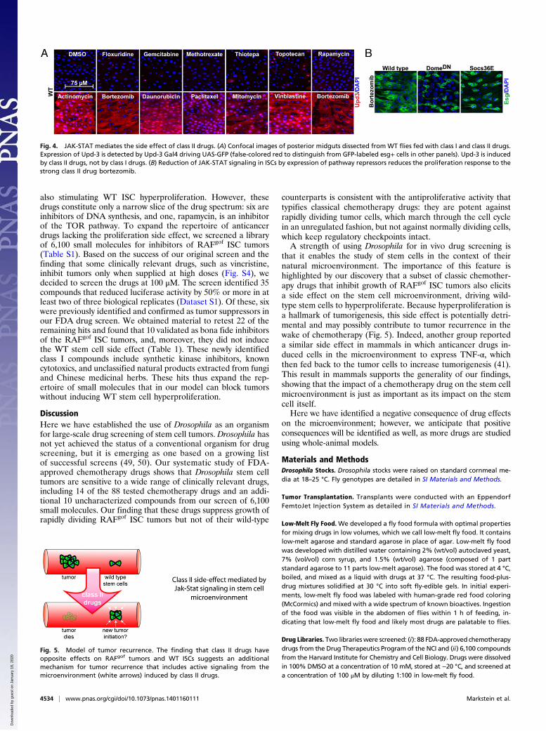
Fig. 4. JAK-STAT mediates the side effect of class II drugs. (A) Confocal images of posterior midguts dissected from WT flies fed with class I and class II drugs.Expression of Upd-3 is detected by Upd-3 Gal4 driving UAS-GFP (false-colored red to distinguish from GFP-labeled esg+ cells in other panels). Upd-3 is inducedby class II drugs, not by class I drugs. (B) Reduction of JAK-STAT signaling in ISCs by expression of pathway repressors reduces the proliferation response to thestrong class II drug bortezomib.
Fig. 5. Model of tumor recurrence. The finding that class II drugs haveopposite effects on RAFgof tumors and WT ISCs suggests an additionalmechanism for tumor recurrence that includes active signaling from themicroenvironment (white arrows) induced by class II drugs.
4534 | www.pnas.org/cgi/doi/10.1073/pnas.1401160111 Markstein et al.
Dow
nloa
ded
by g
uest
on
Janu
ary
18, 2
020

Drug Screening. The screen was conducted in 96-well plates as detailed in SIMaterials and Methods.
Fly Homogenates and Luciferase Assay. Flies were homogenized with a 96-wellplate multihomogenizer (Burkard Scientific, BAMH-96 1911101). Luciferaseassays were conducted as previously described (35) and are detailed in SIMaterials and Methods.
Immunostaining. Tissues were prepared as previously described (28). In-formation on antibodies used can be found in SI Materials and Methods.
Dose–Response Curves. The IC50 values for each drug were calculated byfitting the data to the four-parameter logistic sigmoidal dose–responsecurve, using the Prism6 software package (GraphPad Software, Inc.). Drugs
were tested from 100 to 0.1 μM. The IC50 dose was defined as the dose thatreduced luciferase by 50% in flies with RAFgof ISC tumors.
ACKNOWLEDGMENTS. We thank Chrysoula Pitsouli and Sara Cherry forinvaluable advice; Moritz Matthey, Ben Housden, Laura Quilter, and KendraFrederick for critical reading of the manuscript; Michael Schnall-Levin, JenniferNale, Claire Hu, and Jacob Mayfield for help with statistical analyses; RichardBinari, Djade Soumana, and Laura Holderbaum for technical support; CarolineShamu and David Wrobel (Harvard Institute of Chemistry and Cell Biology) forassistance with screening; the National Cancer Institute Drug TherapeuticsProgram for generously providing the set of 88 “approved oncology” drugs;and the Bloomington Drosophila Stock Center and the Harvard Transgenic RNAiProject for fly stocks. M.M. received support from the Jane Coffin Childs Memo-rial Fund, the Charles King Trust, and a Harvard University Research Enablinggrant. This work was supported by the National Institutes of Health and theHoward Hughes Medical Institute (HHMI). N.P. is an investigator of the HHMI.
1. Lederberg J, Lederberg EM (1952) Replica plating and indirect selection of bacterialmutants. J Bacteriol 63(3):399–406.
2. Ernst T, Hochhaus A (2012) Chronic myeloid leukemia: Clinical impact of BCR-ABL1mutations and other lesions associated with disease progression. Semin Oncol 39(1):58–66.
3. Tortora G, et al. (2007) Overcoming resistance to molecularly targeted anticancertherapies: Rational drug combinations based on EGFR and MAPK inhibition for solidtumours and haematologic malignancies. Drug Resist Updat 10(3):81–100.
4. Rexer BN, Engelman JA, Arteaga CL (2009) Overcoming resistance to tyrosine kinaseinhibitors: Lessons learned from cancer cells treated with EGFR antagonists. Cell Cycle8(1):18–22.
5. Cassidy LD, Venkitaraman AR (2012) Genome instability mechanisms and the structureof cancer genomes. Curr Opin Genet Dev 22(1):10–13.
6. Diehn M, Cho RW, Clarke MF (2009) Therapeutic implications of the cancer stem cellhypothesis. Semin Radiat Oncol 19(2):78–86.
7. Park CY, Tseng D, Weissman IL (2009) Cancer stem cell-directed therapies: Recent datafrom the laboratory and clinic. Mol Ther 17(2):219–230.
8. Hodgson GS, Bradley TR (1979) Properties of haematopoietic stem cells surviving5-fluorouracil treatment: Evidence for a pre-CFU-S cell? Nature 281(5730):381–382.
9. Lee J, et al. (2006) Tumor stem cells derived from glioblastomas cultured in bFGF andEGF more closely mirror the phenotype and genotype of primary tumors than doserum-cultured cell lines. Cancer Cell 9(5):391–403.
10. Dylla SJ, et al. (2008) Colorectal cancer stem cells are enriched in xenogeneic tumorsfollowing chemotherapy. PLoS ONE 3(6):e2428.
11. Li X, et al. (2008) Intrinsic resistance of tumorigenic breast cancer cells to chemo-therapy. J Natl Cancer Inst 100(9):672–679.
12. Matsui W, et al. (2008) Clonogenic multiple myeloma progenitors, stem cell proper-ties, and drug resistance. Cancer Res 68(1):190–197.
13. Winquist RJ, Furey BF, Boucher DM (2010) Cancer stem cells as the relevant biomassfor drug discovery. Curr Opin Pharmacol 10(4):385–390.
14. Gupta PB, et al. (2009) Identification of selective inhibitors of cancer stem cells byhigh-throughput screening. Cell 138(4):645–659.
15. Hirsch HA, Iliopoulos D, Tsichlis PN, Struhl K (2009) Metformin selectively targetscancer stem cells, and acts together with chemotherapy to block tumor growth andprolong remission. Cancer Res 69(19):7507–7511.
16. Diamandis P, et al. (2007) Chemical genetics reveals a complex functional groundstate of neural stem cells. Nat Chem Biol 3(5):268–273.
17. Pollard SM, et al. (2009) Glioma stem cell lines expanded in adherent culture havetumor-specific phenotypes and are suitable for chemical and genetic screens. CellStem Cell 4(6):568–580.
18. Lander AD, et al. (2012) What does the concept of the stem cell niche really meantoday? BMC Biol 10:19.
19. Ootani A, et al. (2009) Sustained in vitro intestinal epithelial culture within a Wnt-dependent stem cell niche. Nat Med 15(6):701–706.
20. Sato T, et al. (2011) Paneth cells constitute the niche for Lgr5 stem cells in intestinalcrypts. Nature 469(7330):415–418.
21. Markstein M (2013) Modeling colorectal cancer as a 3-dimensional disease in a dish:The case for drug screening using organoids, zebrafish, and fruit flies. Drug DiscovToday Technol 10(1):e73–e81.
22. Casali A, Batlle E (2009) Intestinal stem cells in mammals and Drosophila. Cell StemCell 4(2):124–127.
23. Jiang H, Edgar BA (2012) Intestinal stem cell function in Drosophila and mice. CurrOpin Genet Dev 22(4):354–360.
24. Takashima S, Hartenstein V (2012) Genetic control of intestinal stem cell specificationand development: A comparative view. Stem Cell Rev 8(2):597–608.
25. Micchelli CA, Perrimon N (2006) Evidence that stem cells reside in the adult Drosophilamidgut epithelium. Nature 439(7075):475–479.
26. Ohlstein B, Spradling A (2006) The adult Drosophila posterior midgut is maintained bypluripotent stem cells. Nature 439(7075):470–474.
27. Jiang H, Grenley MO, Bravo MJ, Blumhagen RZ, Edgar BA (2011) EGFR/Ras/MAPKsignaling mediates adult midgut epithelial homeostasis and regeneration in Dro-sophila. Cell Stem Cell 8(1):84–95.
28. Ohlstein B, Spradling A (2007) Multipotent Drosophila intestinal stem cells specifydaughter cell fates by differential notch signaling. Science 315(5814):988–992.
29. O’Brien LE, Soliman SS, Li X, Bilder D (2011) Altered modes of stem cell division driveadaptive intestinal growth. Cell 147(3):603–614.
30. Lin G, Xu N, Xi R (2008) Paracrine Wingless signalling controls self-renewal of Dro-sophila intestinal stem cells. Nature 455(7216):1119–1123.
31. Lee WC, Beebe K, Sudmeier L, Micchelli CA (2009) Adenomatous polyposis coli reg-ulates Drosophila intestinal stem cell proliferation. Development 136(13):2255–2264.
32. Jiang H, Edgar BA (2011) Intestinal stem cells in the adult Drosophila midgut. Exp CellRes 317(19):2780–2788.
33. Brand AH, Perrimon N (1993) Targeted gene expression as a means of altering cellfates and generating dominant phenotypes. Development 118(2):401–415.
34. Brand AH, Perrimon N (1994) Raf acts downstream of the EGF receptor to determinedorsoventral polarity during Drosophila oogenesis. Genes Dev 8(5):629–639.
35. Markstein M, Pitsouli C, Villalta C, Celniker SE, Perrimon N (2008) Exploiting positioneffects and the gypsy retrovirus insulator to engineer precisely expressed transgenes.Nat Genet 40(4):476–483.
36. Lee T, Luo L (1999) Mosaic analysis with a repressible cell marker for studies of genefunction in neuronal morphogenesis. Neuron 22(3):451–461.
37. Kobylewski S, Jacobson MF (2012) Toxicology of food dyes. Int J Occup Environ Health18(3):220–246.
38. Saito Y, et al. (2010) Induction of cell cycle entry eliminates human leukemia stemcells in a mouse model of AML. Nat Biotechnol 28(3):275–280.
39. Zoncu R, Efeyan A, Sabatini DM (2011) mTOR: From growth signal integration tocancer, diabetes and ageing. Nat Rev Mol Cell Biol 12(1):21–35.
40. Amcheslavsky A, Jiang J, Ip YT (2009) Tissue damage-induced intestinal stem cell di-vision in Drosophila. Cell Stem Cell 4(1):49–61.
41. Acharyya S, et al. (2012) A CXCL1 paracrine network links cancer chemoresistance andmetastasis. Cell 150(1):165–178.
42. Jiang H, et al. (2009) Cytokine/Jak/Stat signaling mediates regeneration and ho-meostasis in the Drosophila midgut. Cell 137(7):1343–1355.
43. Buchon N, Broderick NA, Chakrabarti S, Lemaitre B (2009) Invasive and indigenousmicrobiota impact intestinal stem cell activity through multiple pathways in Dro-sophila. Genes Dev 23(19):2333–2344.
44. Buchon N, Broderick NA, Poidevin M, Pradervand S, Lemaitre B (2009) Drosophilaintestinal response to bacterial infection: Activation of host defense and stem cellproliferation. Cell Host Microbe 5(2):200–211.
45. Apidianakis Y, Pitsouli C, Perrimon N, Rahme L (2009) Synergy between bacterialinfection and genetic predisposition in intestinal dysplasia. Proc Natl Acad Sci USA106(49):20883–20888.
46. Ren F, et al. (2010) Hippo signaling regulates Drosophila intestine stem cell pro-liferation through multiple pathways. Proc Natl Acad Sci USA 107(49):21064–21069.
47. Agaisse H, Petersen UM, Boutros M, Mathey-Prevot B, Perrimon N (2003) Signalingrole of hemocytes in Drosophila JAK/STAT-dependent response to septic injury. DevCell 5(3):441–450.
48. Grivennikov SI, Greten FR, Karin M (2010) Immunity, inflammation, and cancer. Cell140(6):883–899.
49. Edwards A, et al. (2011) Combinatorial effect of maytansinol and radiation in Dro-sophila and human cancer cells. Dis Model Mech 4(4):496–503.
50. Dar AC, Das TK, Shokat KM, Cagan RL (2012) Chemical genetic discovery of targetsand anti-targets for cancer polypharmacology. Nature 486(7401):80–84.
Markstein et al. PNAS | March 25, 2014 | vol. 111 | no. 12 | 4535
MED
ICALSC
IENCE
S
Dow
nloa
ded
by g
uest
on
Janu
ary
18, 2
020


![Integrin Targeted Delivery of Chemotherapeutics · Integrin Targeted Delivery of Chemotherapeutics ... Molecular Imaging Center, ... vealed an atomic basis for this interaction [43]](https://img.pdfslide.us/doc/110x75/5b5094a97f8b9a3e6e8ec427/integrin-targeted-delivery-of-integrin-targeted-delivery-of-chemotherapeutics.jpg)