Embed Size (px)
Citation preview
B.Sc. Hons Part I - Subsidiary Dept. of Botany Dr. R. K. Sinha
pg. 1
Systematic Position of Marchantia:
Structure & Life History of Marchantia:
Marchantia, the most important genus of family Marchantiaceae is represented by about 65
species. The name Marchantia was given in honour of Nicolas Merchant, director of botanical
garden of Gaston d’ Orleans in Blois, France.
All species are terrestrial and cosmopolitan in distribution. The species prefer to grow in moist
and shady places like wet open woodlands, banks of streams, wood rocks or on shaded stub
rocks. These grow best after the forest fire in the burnt soil. It is perhaps because of nitrification
of soil due to fire (Richard, 1958).
In India, Marchantia is represented by about 11 species (Chopra, 1943). Udar (1970) reported
only 6 species from different parts of the country. These species are commonly found growing’
in the Himalayan region at an altitude of 4000-8000 feet. Eastern Himalayan region particularly
supports the growth of these species.
Some species are also found growing in plains of Haryana, Punjab, Uttar Pradesh and hilly
regions of South India.
Some of the common Indian species are M. palmata, M. polymorpha, M. simlana etc. M.
polymorpha is most widely distributed species. M. polymorpha var aquatic grows submerged in
swampy meadows. The thalli with gemma cups are found throughout the year whereas the
thalli with sex organs occur abundantly during February to March in Himalayas and October to
November in hills of South India.
Gametophytic Phase of Marchantia:
External Features of Gametophyte:
The plant body is gametophytic, thalloid, flat, prostrate, plagiotropic, 2-10 cm. long and
dichotomously branched (Fig. 1 A).
B.Sc. Hons Part I - Subsidiary Dept. of Botany Dr. R. K. Sinha
pg. 2
Dorsal surface:
Dorsal surface is dark green. It has a conspicuous midrib and a number of polygonal areas called
areolae. The midrib is marked on the dorsal surface by a shallow groove and on the ventral
surface by a low ridge. Each polygonal area re-presents the underlying air chamber.
The boundaries of these areas represent the walls that separate each air chamber from the
next. Each air chamber has a central pore. The midrib ends in a depression at the apical region
forming an apical notch in which growing point is situated (Fig. 28 B).
Dorsal surface also bears the vegetative and sexual reproductive structures. The vegetative
reproductive structures are gemma cup and develop along the midrib. These are crescent
shaped with spiny or fimbriate margins and are about one eighth of a inch in diameter (Fig. I A,
15).
Sexual reproductive structures are borne on special Stalked structures called gametophores or
gamctangiophores. The gametophores bearing archegonia are called archegoniophores and
that bearing antheridia are called antheridiophores (Fig. 1 A, B).
Ventral surface:
The ventral surface of the thallus bears scales and rhizoids along the midrib. Scales are violet
coloured, multicellular, one cell thick and arranged in 2-4 rows (Fig. 1 C). Scales are of two
types:
(i) Simple or ligulate
(ii) Appendiculate.
Appendiculate (Fig- 1 C, D) scales form the inner row of the scales close with midrib. Ligulate
scales form the outer or marginal row and are smaller than the appendiculate scales (Fig. 1 C,
E).
Rhizoids are unicellular, branched and develop as prolongation of the lower epidermal cells.
They are of two types:
(i) Smooth-walled rhizoids,
(ii) Tuberculate rhizoids.
B.Sc. Hons Part I - Subsidiary Dept. of Botany Dr. R. K. Sinha
pg. 3
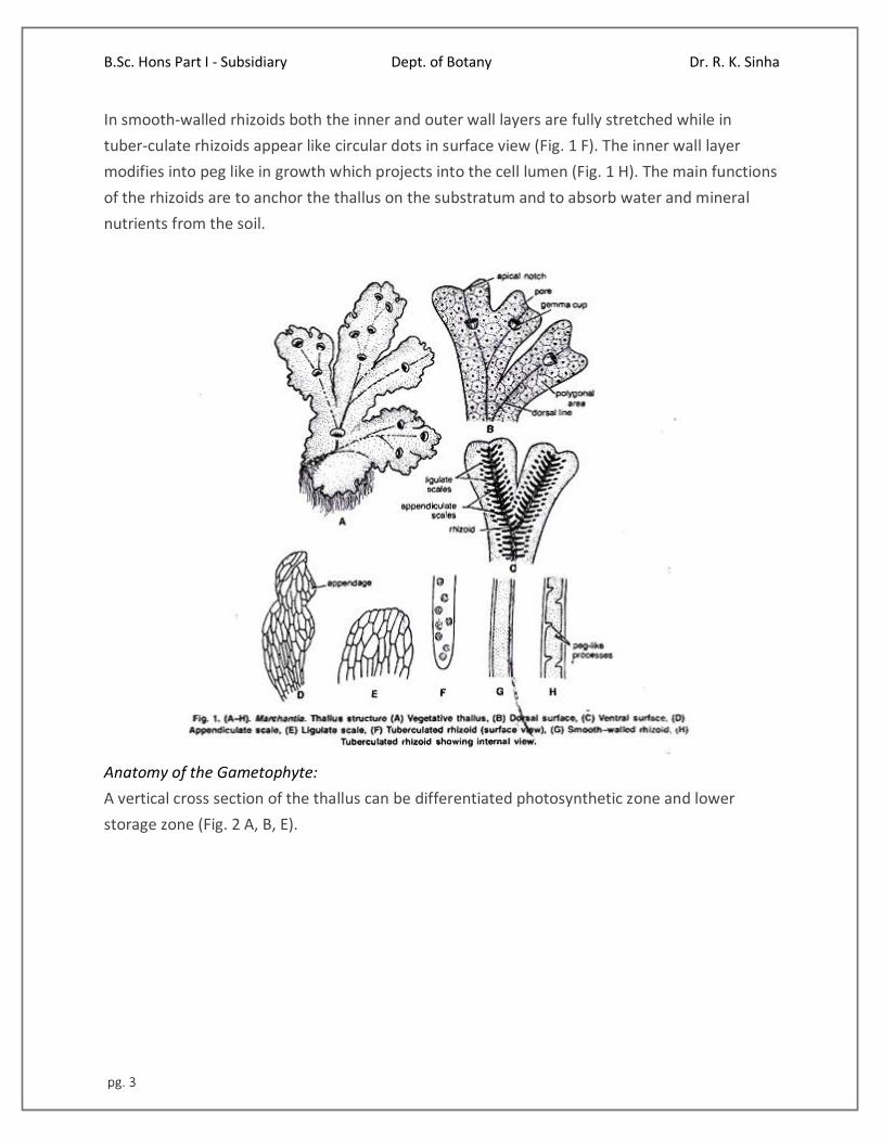
In smooth-walled rhizoids both the inner and outer wall layers are fully stretched while in
tuber-culate rhizoids appear like circular dots in surface view (Fig. 1 F). The inner wall layer
modifies into peg like in growth which projects into the cell lumen (Fig. 1 H). The main functions
of the rhizoids are to anchor the thallus on the substratum and to absorb water and mineral
nutrients from the soil.
Anatomy of the Gametophyte:
A vertical cross section of the thallus can be differentiated photosynthetic zone and lower
storage zone (Fig. 2 A, B, E).
B.Sc. Hons Part I - Subsidiary Dept. of Botany Dr. R. K. Sinha
pg. 4
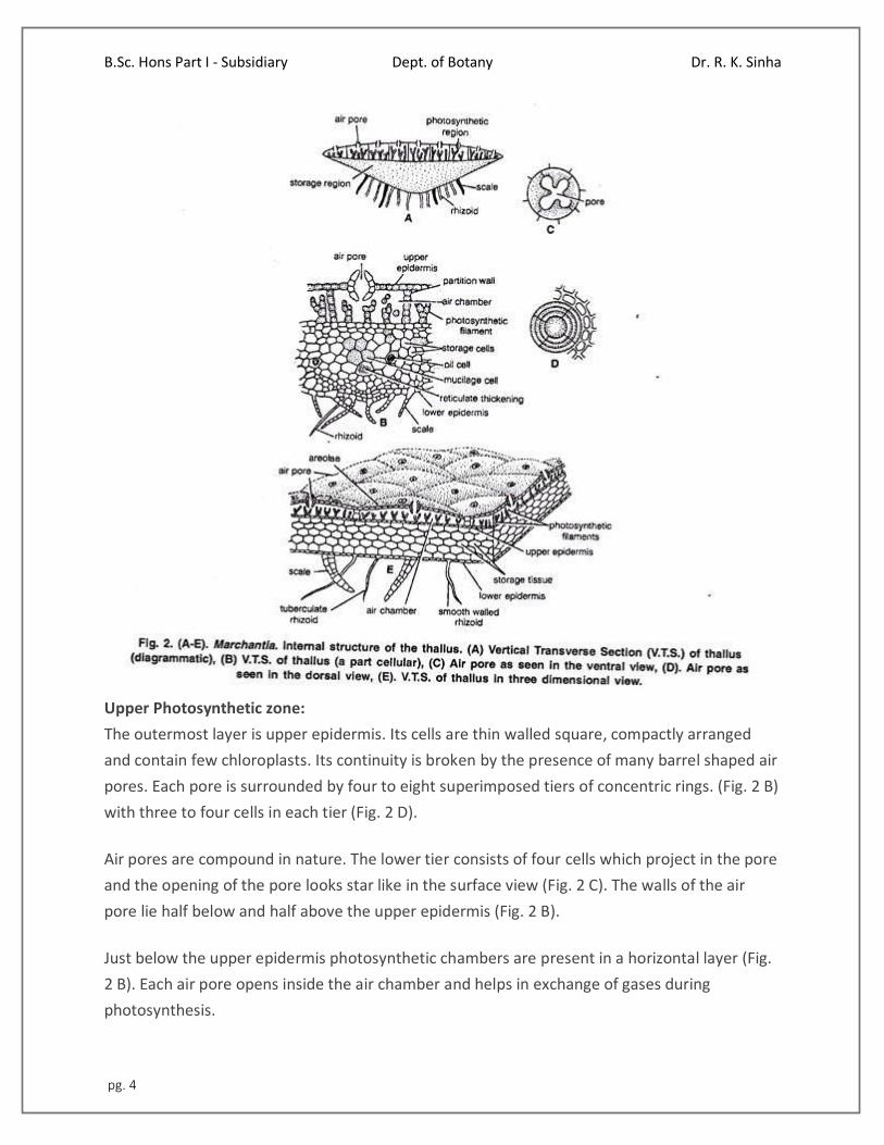
Upper Photosynthetic zone:
The outermost layer is upper epidermis. Its cells are thin walled square, compactly arranged
and contain few chloroplasts. Its continuity is broken by the presence of many barrel shaped air
pores. Each pore is surrounded by four to eight superimposed tiers of concentric rings. (Fig. 2 B)
with three to four cells in each tier (Fig. 2 D).
Air pores are compound in nature. The lower tier consists of four cells which project in the pore
and the opening of the pore looks star like in the surface view (Fig. 2 C). The walls of the air
pore lie half below and half above the upper epidermis (Fig. 2 B).
Just below the upper epidermis photosynthetic chambers are present in a horizontal layer (Fig.
2 B). Each air pore opens inside the air chamber and helps in exchange of gases during
photosynthesis.
B.Sc. Hons Part I - Subsidiary Dept. of Botany Dr. R. K. Sinha
pg. 5
These are chambers develop schizogenously (Vocalized separation of cells to form a cavity) and
are separated from each other by single layered partition walls. The partition walls are two to
four cells in height. Cells contain chloroplast. Many simple or branched photosynthetic
filaments arise from the base of the air chambers (Fig. 2 B).
Storage zone:
It lies below the air chambers. It is more thickened in the centre and gradually tapers towards
the margins. It consists of several lasers of compactly arranged, thin walled parenchymatous
isodiametric cells. Intercellular spaces are absent.
The cells of this zone contain starch. Some cells contain a single large oil body or filled with
mucilage. The cells of the midrib region possess reticulate thickenings. The lower most cell layer
of the zone forms the lower epidermis. Some cells of the middle layer of lower epidermis
extend to form both types of scales and rhizoids (Fig. 2 B).
Reproduction in Marchantia:
Marchantia reproduces by vegetative and sexual methods.
Vegetative Reproduction:
In Marchantia it is quite common and takes place by the following methods:
1. By Gemmae:
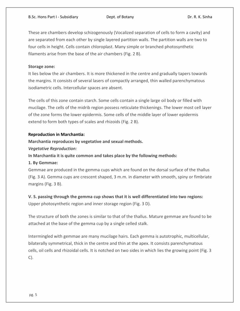
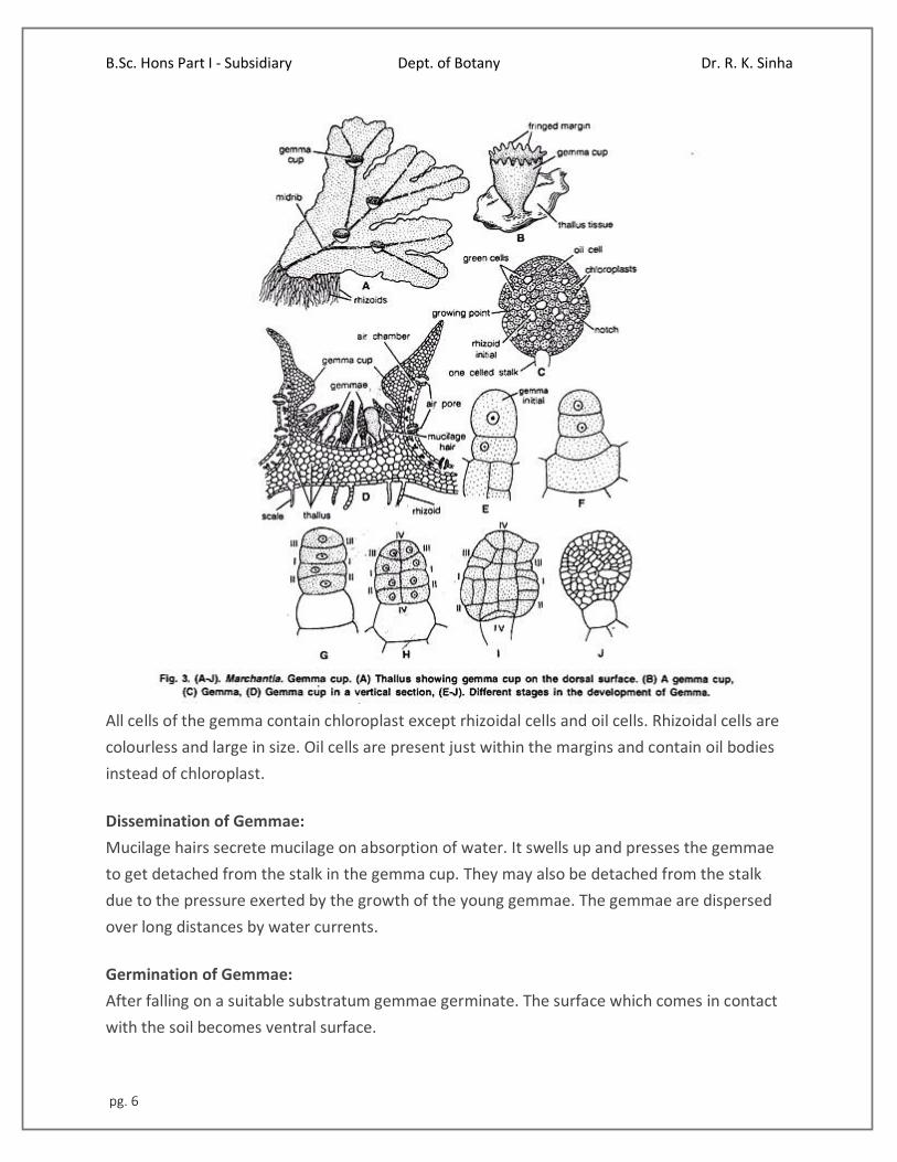
Gemmae are produced in the gemma cups which are found on the dorsal surface of the thallus
(Fig. 3 A). Gemma cups are crescent shaped, 3 m.m. in diameter with smooth, spiny or fimbriate
margins (Fig. 3 B).
V. S. passing through the gemma cup shows that it is well differentiated into two regions:
Upper photosynthetic region and inner storage region (Fig. 3 D).
The structure of both the zones is similar to that of the thallus. Mature gemmae are found to be
attached at the base of the gemma cup by a single celled stalk.
Intermingled with gemmae are many mucilage hairs. Each gemma is autotrophic, multicellular,
bilaterally symmetrical, thick in the centre and thin at the apex. It consists parenchymatous
cells, oil cells and rhizoidal cells. It is notched on two sides in which lies the growing point (Fig. 3
C).
B.Sc. Hons Part I - Subsidiary Dept. of Botany Dr. R. K. Sinha
pg. 6
All cells of the gemma contain chloroplast except rhizoidal cells and oil cells. Rhizoidal cells are
colourless and large in size. Oil cells are present just within the margins and contain oil bodies
instead of chloroplast.
Dissemination of Gemmae:
Mucilage hairs secrete mucilage on absorption of water. It swells up and presses the gemmae
to get detached from the stalk in the gemma cup. They may also be detached from the stalk
due to the pressure exerted by the growth of the young gemmae. The gemmae are dispersed
over long distances by water currents.
Germination of Gemmae:
After falling on a suitable substratum gemmae germinate. The surface which comes in contact
with the soil becomes ventral surface.
B.Sc. Hons Part I - Subsidiary Dept. of Botany Dr. R. K. Sinha
pg. 7
The rhizoidai cells develop into rhizoids. Meanwhile, the growing points in which lies the two
lateral notches form thalli in opposite directions. Thus, from a single gemmae two thalli are
formed. Gemmae which develop on the male thalli form the male plants and those on the
female thalli form the female plant.
Development of Gemma:
The gemma develops from a single superficial cell. It develops on the floor of a gemma cup. It is
papillate and called gemma initial (Fig. 3 E). It divides by a transverse division to form lower
stalk cell and upper cell (Fig. 3 F). The lower cell forms the single celled stalk.
The upper cell further divides by transverse division to form two cells. Both cells undergo by
similar divisions to form four cells (Fig. 3 G). These cells divide by vertical and horizontal division
to form a plate like structure with two marginal notches. It is called gemma (Fig. 3 H-J).
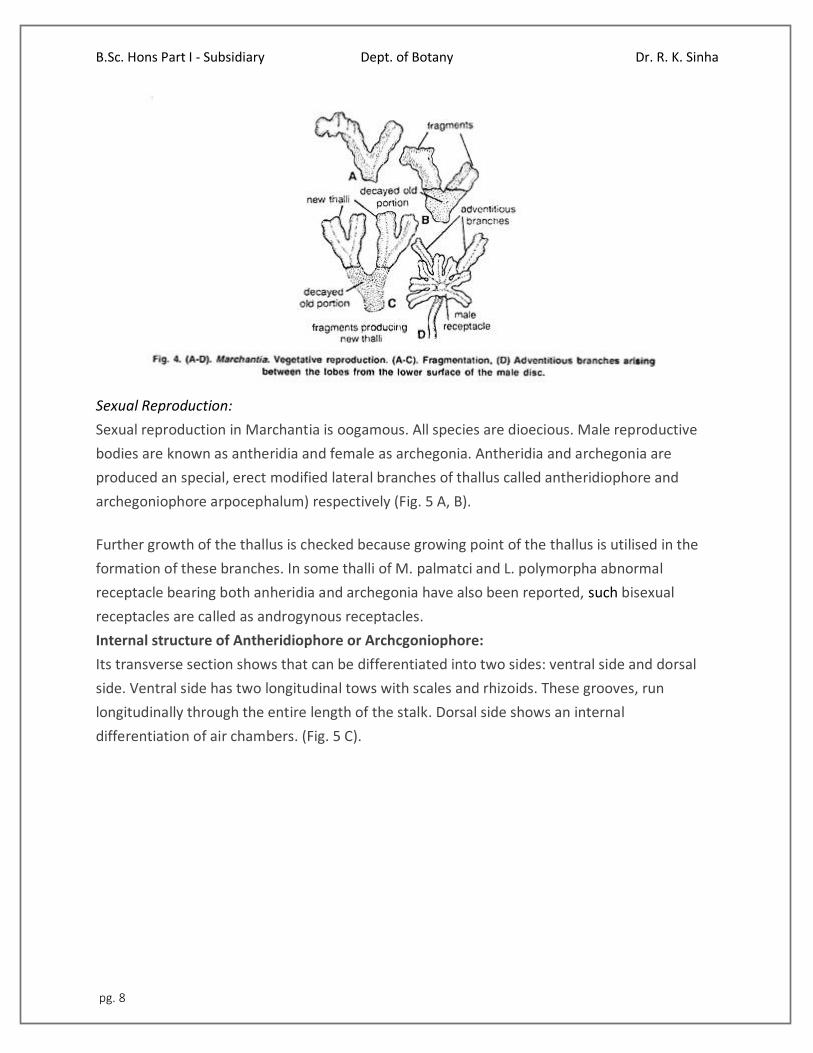
2. Death and decay of the older portion of the thallus or fragmentation:
The thallus is dichotomously branched. The basal part of the thallus rots and disintegrates due
to ageing. When this process reaches up to the place of dichotomy, the lobes of the thallus get
separated. The detached lobes or fragments develop into independent thalli by apical growth
(Fig. 4 A-C).
3. By adventitious branches:
The adventitious branches develop from any part of the thallus or the ventral surface of the
thallus or rarely from the stalk and disc of the archegoniophore in species like M. palmata
(Kashyap, 1919). On being detached, these branches develop into new thalli (Fie. 4 D).
B.Sc. Hons Part I - Subsidiary Dept. of Botany Dr. R. K. Sinha
pg. 8
Sexual Reproduction:
Sexual reproduction in Marchantia is oogamous. All species are dioecious. Male reproductive
bodies are known as antheridia and female as archegonia. Antheridia and archegonia are
produced an special, erect modified lateral branches of thallus called antheridiophore and
archegoniophore arpocephalum) respectively (Fig. 5 A, B).
Further growth of the thallus is checked because growing point of the thallus is utilised in the
formation of these branches. In some thalli of M. palmatci and L. polymorpha abnormal
receptacle bearing both anheridia and archegonia have also been reported, such bisexual
receptacles are called as androgynous receptacles.
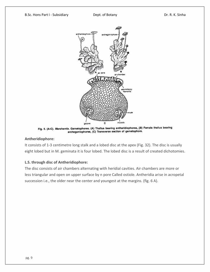
Internal structure of Antheridiophore or Archcgoniophore:
Its transverse section shows that can be differentiated into two sides: ventral side and dorsal
side. Ventral side has two longitudinal tows with scales and rhizoids. These grooves, run
longitudinally through the entire length of the stalk. Dorsal side shows an internal
differentiation of air chambers. (Fig. 5 C).
B.Sc. Hons Part I - Subsidiary Dept. of Botany Dr. R. K. Sinha
pg. 9
Antheridiophore:
It consists of 1-3 centimetre long stalk and a lobed disc at the apex (Fig. 32). The disc is usually
eight lobed but in M. geminata it is four lobed. The lobed disc is a result of created dichotomies.
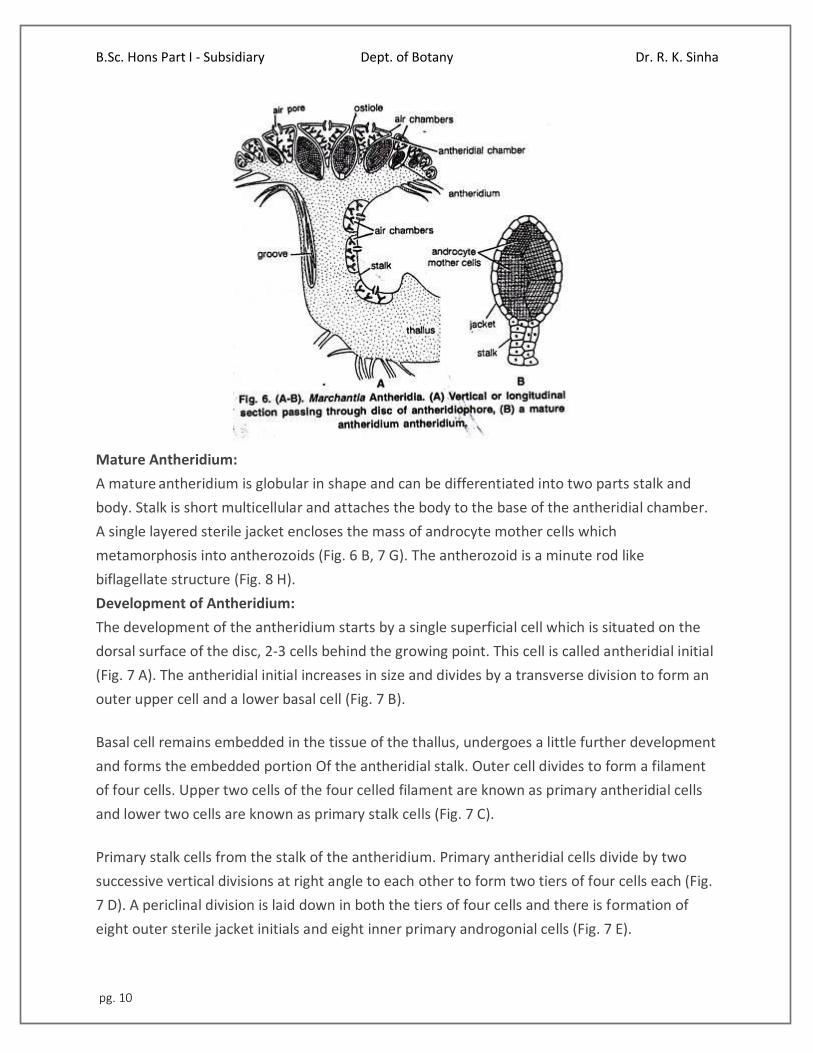
L.S. through disc of Antheridiophore:
The disc consists of air chambers alternating with heridial cavities. Air chambers are more or
less triangular and open on upper surface by n pore Called ostiole. Antheridia arise in acropetal
succession i.e., the older near the center and youngest at the margins. (fig. 6 A).
B.Sc. Hons Part I - Subsidiary Dept. of Botany Dr. R. K. Sinha
pg. 10
Mature Antheridium:
A mature antheridium is globular in shape and can be differentiated into two parts stalk and
body. Stalk is short multicellular and attaches the body to the base of the antheridial chamber.
A single layered sterile jacket encloses the mass of androcyte mother cells which
metamorphosis into antherozoids (Fig. 6 B, 7 G). The antherozoid is a minute rod like
biflagellate structure (Fig. 8 H).
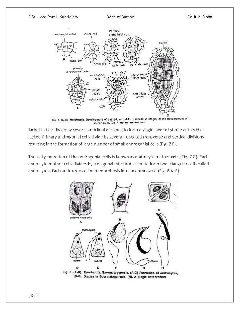
Development of Antheridium:
The development of the antheridium starts by a single superficial cell which is situated on the
dorsal surface of the disc, 2-3 cells behind the growing point. This cell is called antheridial initial
(Fig. 7 A). The antheridial initial increases in size and divides by a transverse division to form an
outer upper cell and a lower basal cell (Fig. 7 B).
Basal cell remains embedded in the tissue of the thallus, undergoes a little further development
and forms the embedded portion Of the antheridial stalk. Outer cell divides to form a filament
of four cells. Upper two cells of the four celled filament are known as primary antheridial cells
and lower two cells are known as primary stalk cells (Fig. 7 C).
Primary stalk cells from the stalk of the antheridium. Primary antheridial cells divide by two
successive vertical divisions at right angle to each other to form two tiers of four cells each (Fig.
7 D). A periclinal division is laid down in both the tiers of four cells and there is formation of
eight outer sterile jacket initials and eight inner primary androgonial cells (Fig. 7 E).
B.Sc. Hons Part I - Subsidiary Dept. of Botany Dr. R. K. Sinha
pg. 11
Jacket initials divide by several anticlinal divisions to form a single layer of sterile antheridial
jacket. Primary androgonial cells divide by several repeated transverse and vertical divisions
resulting in the formation of large number of small androgonial cells (Fig. 7 F).
The last generation of the androgonial cells is known as androcyte mother cells (Fig. 7 G). Each
androcyte mother cells divides by a diagonal mitotic division to form two triangular cells called
androcytes. Each androcyte cell metamorphosis into an antheozoid (Fig. 8 A-G).
B.Sc. Hons Part I - Subsidiary Dept. of Botany Dr. R. K. Sinha
pg. 12
Spermatogenesis:
The process of metamorphosis of androcyte mother cells into antherozoids is called
spermatogenesis.
It is completed in two phases:
(1) Development of blepharoplast.
(2) Elongation of androcyte nucleus.
1. Development of Blepharoplasty:
In the young triangular androcyte (Fig. 8 D) blepharoplast appears as a dense granule in one of
the acute angles. It elongates to some extent and puts its whole body in close contact with the
inner contour of androcyte. From the elongated blepharoplast emerge the flagella.
2 Elongation of Androcyte nucleus:
With the elongation of blepharoplast, the nucleus also elongates. The spline apparatus acts as a
cytoskeleton for the elongation of nucleus. Spline apparatus is a multilayered structure which
comprises tubules (Fig. 8 E-H).
Archegoniophore or Carpocephalum:
It arises at the apical notch and consists of a stalk and terminal disc. It is slightly longer than the
antheridiophore. It may be five to seven cm. long. The young apex of the archegoniophore
divides by three successive dichotomies to form eight lobed rosette like disc.
Each lobe of the disc contains a growing point. The archegonia begin to develop in each lobe in
acropetal succession, i.e., the oldest archegonium near the centre and the young archegonium
near the apex of the disc. (Fig. 10 A). Thus, eight groups of archegonia develop on the upper
surface of the disc. There are twelve to fourteen archegonia in a single row in each lobe of the
disc.
Development:
The development of the archegonium starts on the dorsal surface of the young receptacle in
acropetal succession. A single superficial cell which acts as archegonial initial enlarges and
divides by transverse division to form a basal cell or primary stalk cell and an outer cell or
primary archegonial cell (Fig. 9 A, B).
B.Sc. Hons Part I - Subsidiary Dept. of Botany Dr. R. K. Sinha
pg. 13
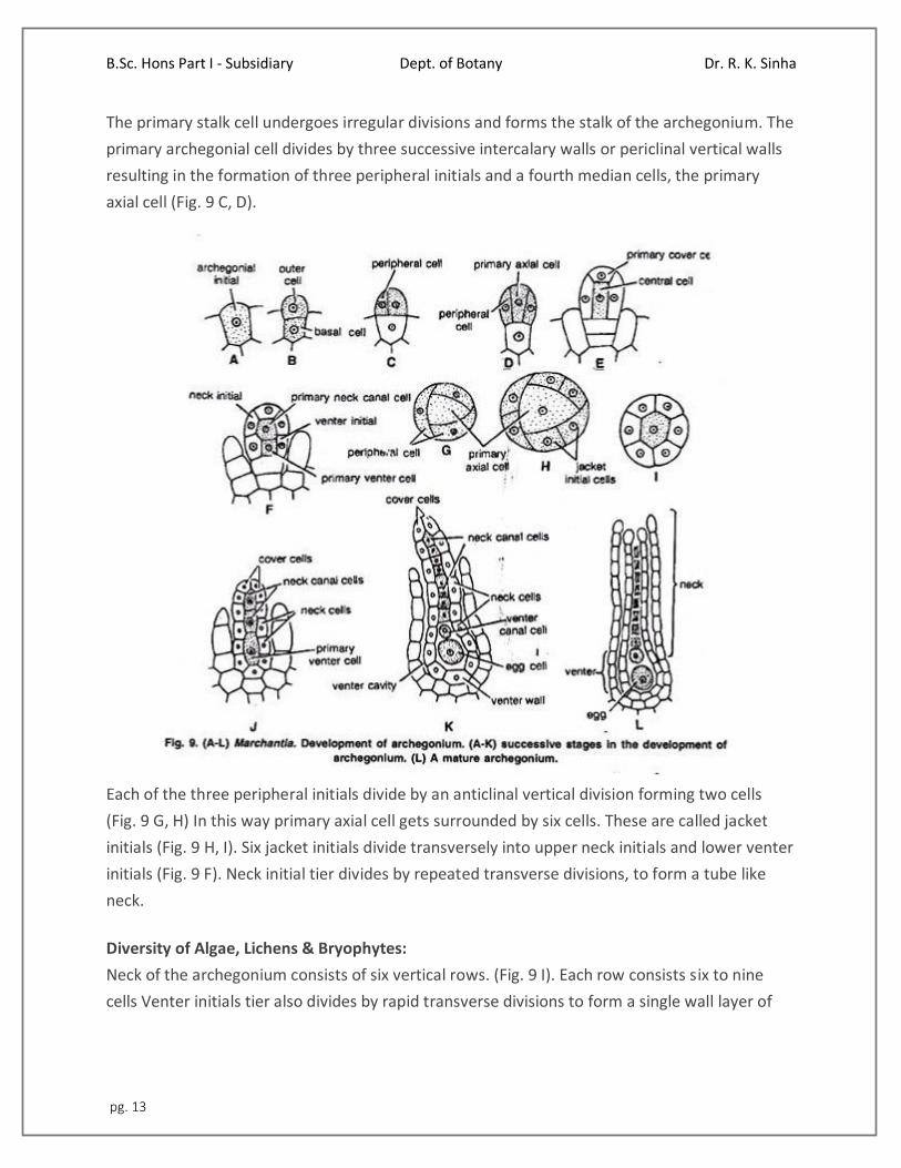
The primary stalk cell undergoes irregular divisions and forms the stalk of the archegonium. The
primary archegonial cell divides by three successive intercalary walls or periclinal vertical walls
resulting in the formation of three peripheral initials and a fourth median cells, the primary
axial cell (Fig. 9 C, D).
Each of the three peripheral initials divide by an anticlinal vertical division forming two cells
(Fig. 9 G, H) In this way primary axial cell gets surrounded by six cells. These are called jacket
initials (Fig. 9 H, I). Six jacket initials divide transversely into upper neck initials and lower venter
initials (Fig. 9 F). Neck initial tier divides by repeated transverse divisions, to form a tube like
neck.
Diversity of Algae, Lichens & Bryophytes:
Neck of the archegonium consists of six vertical rows. (Fig. 9 I). Each row consists six to nine
cells Venter initials tier also divides by rapid transverse divisions to form a single wall layer of
B.Sc. Hons Part I - Subsidiary Dept. of Botany Dr. R. K. Sinha
pg. 14
swollen venter (Fig 9 K). Simultaneously, the primary axial cell divides transversely and
unequally to form upper small primary cover cell and lower large central cell (Fig. 9 E).
The central cell divides into primary neck canal cell and a lower venter cell. Primary neck canal
cells divides by a series of transverse divisions to form a row of about eight thin walled neck
canal cells (Fig. 9 J, K).
Primary venter cell divides only once and forms a small upper venter canal cell and a lower
large egg or ovum (Fig. 9 K). The primary cover cell divide by two vertical divisions at right angle
to one another to form four cover cells which form the mouth of the archegonium.
Mature Archegonium:
A mature archegonium is a flask shaped structure. It remains attached to the archegonial disc
by a short stalk. It consists upper elongated slender neck and basal globular portion called
venter. The neck consists of six vertical rows enclosing eight neck canal cells and large egg. Four
cover cells are present at the top of the neck. (Fig. 9 L).
Fertilization in Marchantia:
Marchantia is dioecious. Fertilization takes place when male and female thalli grow near each
other. Water is essential for fertilization. The neck of the archegonium is directed upwards on
the dorsal surface of the disc of the archegoniophore (Fig. 9 A).
In the mature archegonium the venter canal cell and neck canal cells disintegrate and form a
mucilaginous mass. It absorbs water, swells up and comes out of the archegonial mouth by
pushing the cover cells apart. This mucilaginous mass consists of chemical substances.
The antherozoids are splashed by rain drops. They may fall on the nearby female receptacle or
swim the whole way by female receptacle. It is only possible if both the male and female
receptacles are surrounded by water.
Many antherozoids enter the archegonial neck by chemotactic response and reach up to egg.
This mechanism of fertilization is called splash cup mechanism. One of the antherozoids
penetrates the egg and fertilization is effected. The fusion of both male and female nuclei
results in the formation of diploid zygote or oospore. Fertilization ends the gametophytic
phase.
B.Sc. Hons Part I - Subsidiary Dept. of Botany Dr. R. K. Sinha
pg. 15
Sporophytic Phase:
Post Fertilization Changes:
After Fertilization the following changes occur simultaneously:
1. Stalk of the archegoniophore elongates.
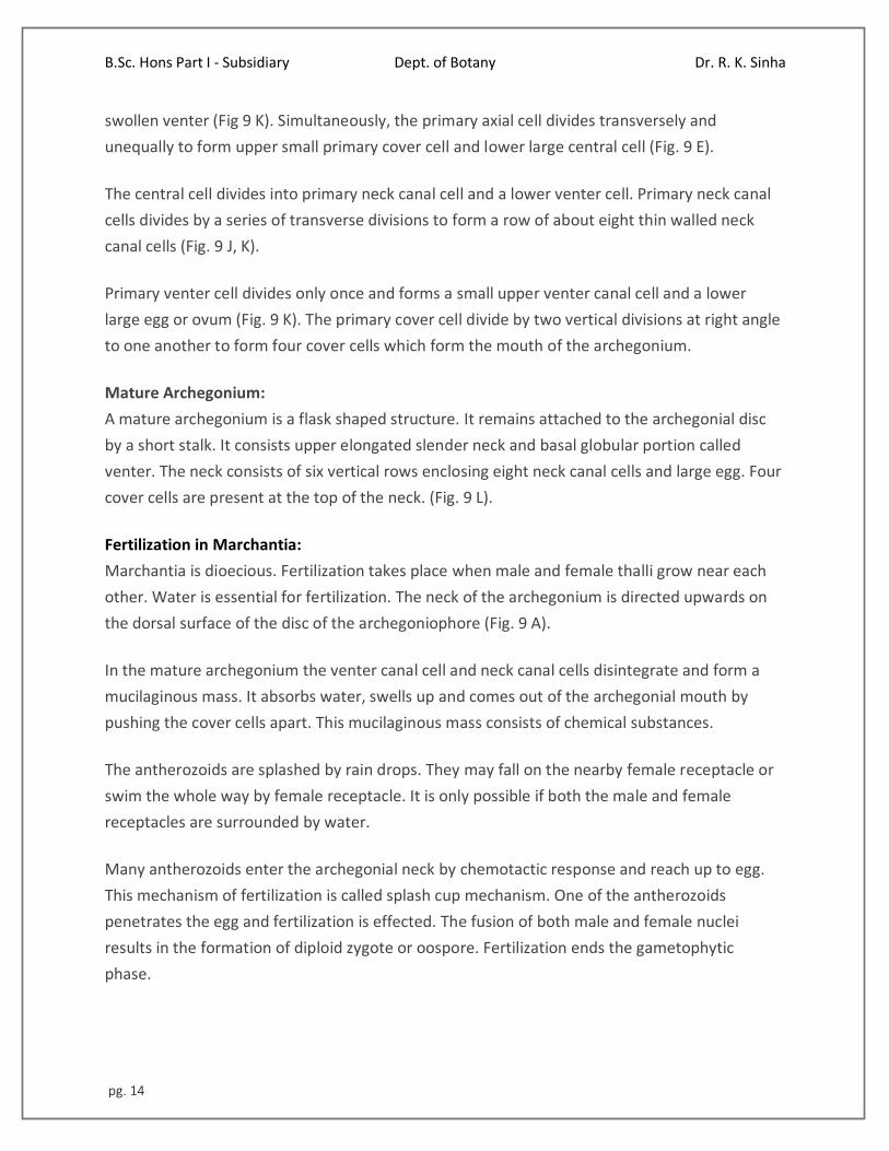
2. Remarkable over-growth takes place in the central part of the disc. As a result of this growth
the marginal region of the disc bearing archegonia is pushed downward and inward. The
archegonia are now hanging towards the lower side with their neck pointing downwards (Fig.
10 B-D).
3. Wall of the venter divides to form two to three layered calyptra.
4. A ring of cells at the base of venter divides and re-divides to form a one cell thick collar
around archegonium called perigynium (Pseudoperianth).
5. A one celled thick, fringed sheath develops on both sides of the archegonial row. It is called
perichaetium or involucre. Thus, the developing sporophyte is surrounded by three protective
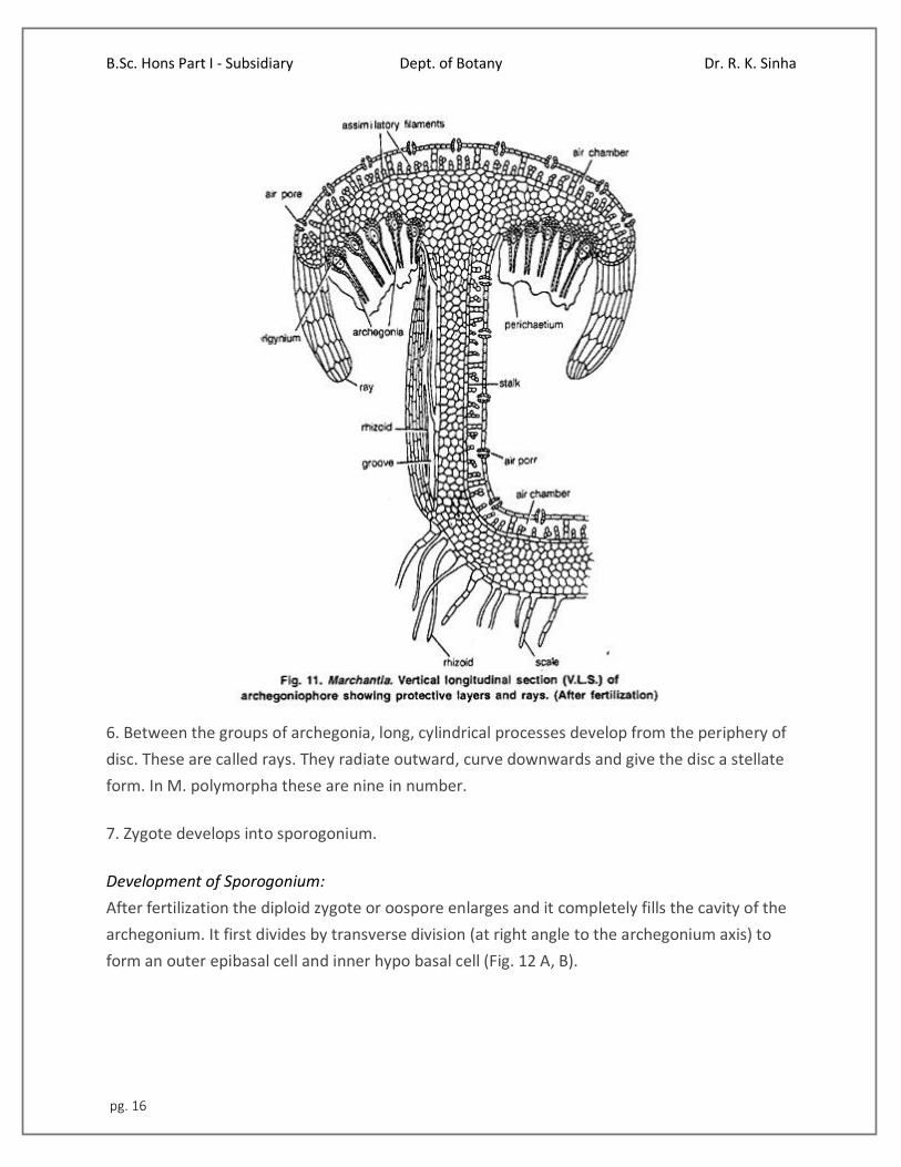
layers of gametophytic origin i.e., calyptra, perigynium and perichaetium (Fig. 11). The main
function of these layers is to provide protection, against drought, to young sporophyte.
B.Sc. Hons Part I - Subsidiary Dept. of Botany Dr. R. K. Sinha
pg. 16
6. Between the groups of archegonia, long, cylindrical processes develop from the periphery of
disc. These are called rays. They radiate outward, curve downwards and give the disc a stellate
form. In M. polymorpha these are nine in number.
7. Zygote develops into sporogonium.
Development of Sporogonium:
After fertilization the diploid zygote or oospore enlarges and it completely fills the cavity of the
archegonium. It first divides by transverse division (at right angle to the archegonium axis) to
form an outer epibasal cell and inner hypo basal cell (Fig. 12 A, B).
B.Sc. Hons Part I - Subsidiary Dept. of Botany Dr. R. K. Sinha
pg. 17
The second division is at right angle to the first and results in the formation of four cells. This
represents the quadrant stage (Fig. 12 C). The epibasal cell forms the capsule and hypo basal
cells form the foot and seta.
Since the capsule is developed from the epibasal cell and forms the apex of the sporogonium,
the type of embryogeny is known as exoscopic. The next division is also vertical and it results in
formation of eight celled stage or octant stage.
Now the divisions are irregular and globular embryo is formed (Fig. 12 D). The lower cells divide
to form a massive and bulbous foot. The cells of the seta divide in one plane to form vertical
rows of cells.
In upper region of capsule (when the young sporogonium is about a dozen or more cells in
circumference) periclinal division occurs and it differentiates it into outer single layered
amphithecium and multilayered endothecium (Fig. 12 E, F).
The cells of the endothecium divide only by anticlinal divisions to form a single layered sterile
jacket or capsule wall. The endothecium forms the archesporium. Its cells divide and re-divide
to form a mass of sporogenous cells (sporocytes). Half of the sporogenous cells become narrow
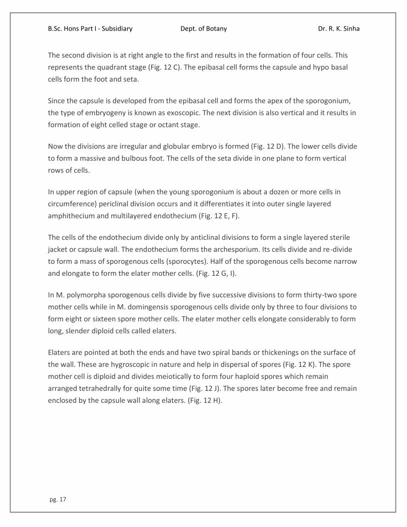
and elongate to form the elater mother cells. (Fig. 12 G, I).
In M. polymorpha sporogenous cells divide by five successive divisions to form thirty-two spore
mother cells while in M. domingensis sporogenous cells divide only by three to four divisions to
form eight or sixteen spore mother cells. The elater mother cells elongate considerably to form
long, slender diploid cells called elaters.
Elaters are pointed at both the ends and have two spiral bands or thickenings on the surface of
the wall. These are hygroscopic in nature and help in dispersal of spores (Fig. 12 K). The spore
mother cell is diploid and divides meiotically to form four haploid spores which remain
arranged tetrahedrally for quite some time (Fig. 12 J). The spores later become free and remain
enclosed by the capsule wall along elaters. (Fig. 12 H).
B.Sc. Hons Part I - Subsidiary Dept. of Botany Dr. R. K. Sinha
pg. 18
The quadrant type of development of sporogonium is quite common in many species of
Marchantia (e.g., M. polymorpha) but in a few species zygote divides by two transverse
divisions to form the 3-celled filamentous embryo. In it the hypo basal cell forms the foot, the
middle seta and the epibasal cell develops into capsule. However, it is the rare type of embryo
development in M. chenopoda.
Mature Sporogonium:
A mature sporogonium can be differentiated into three parts, viz., the foot, seta and capsule
(Fig. 13 H). Foot. It is bulbous and multicellular. It is composed of parenchymatous cells. It acts
as anchoring and absorbing organ. It absorbs the food from the adjoining gametophytic cells for
the developing sporophyte.
B.Sc. Hons Part I - Subsidiary Dept. of Botany Dr. R. K. Sinha
pg. 19
Seta:
It connects the foot and the capsule. At maturity, due to many transverse divisions it elongates
and pushes the capsule through three protective layers viz., calyptra, perigynium and
perichaetium.
Capsule:
It is oval in shape and has a single layered wall which encloses spores and elaters. It has been
estimated that as many as 3, 00,000 spores may be produced in single sporogonium and there
are 128 spores in relation to one elater.
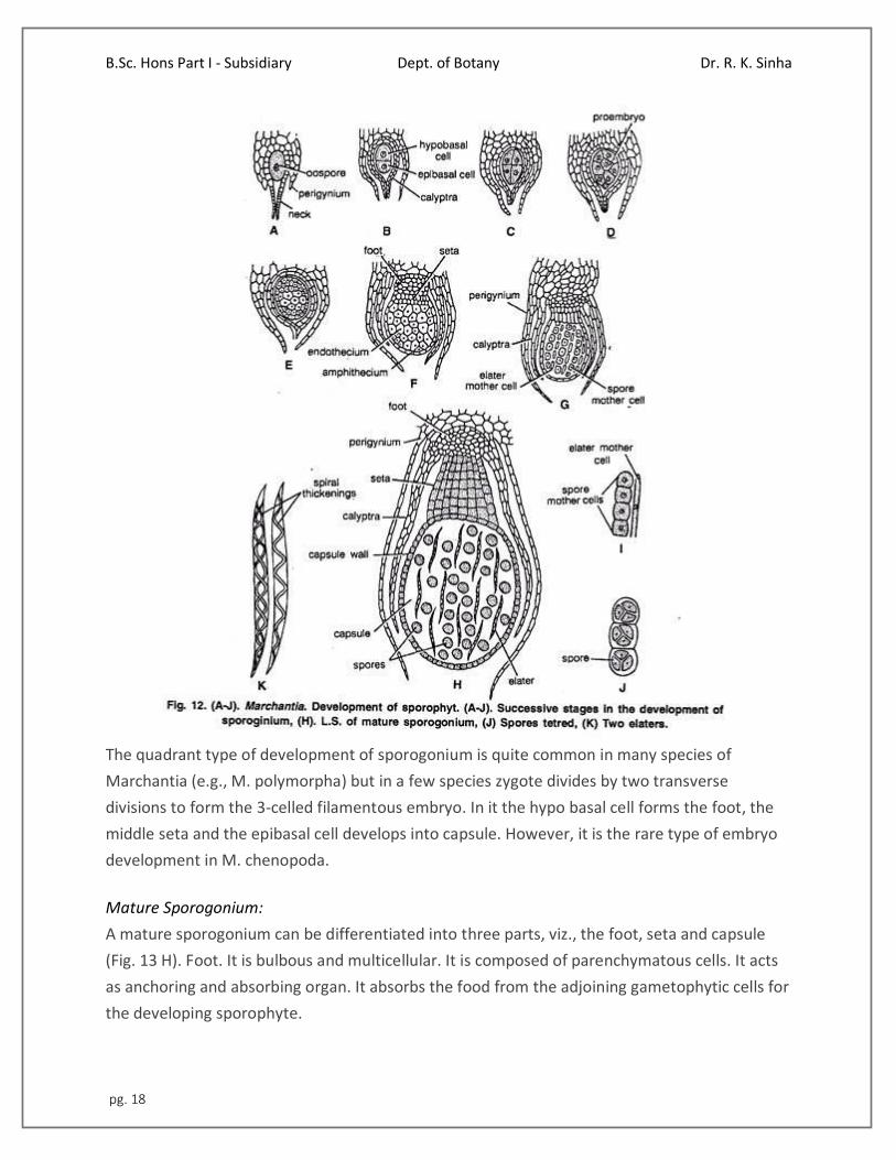
Dispersal of Spores:
As the sporogonium matures, seta elongates rapidly and pushes the capsule in the air through
the protective layers (Fig. 13 A). The ripe capsule wall dehisces from apex to middle by four to
six irregular teeth or valves. The annular thickening in the cells of the capsule wall causes the
valves to roll backward exposing the spores and elaters.
The elaters are hygroscopic in nature. In dry weather they lose water and become twisted.
When the atmosphere is wet, they become untwisted and cause the jerking action. Due to this
the spore mass loosens and spores are carried out by air currents (Fig. 13 B, C).
B.Sc. Hons Part I - Subsidiary Dept. of Botany Dr. R. K. Sinha
pg. 20
Structure of Spore:
Spores are very small (0.012 to 0.30 mm in diameter). They are haploid, uninucleate, globose
and surrounded by only two wall layers. The outer well layer is thick, smooth or reticulate and is
known as exospore or exine. The inner wall layer is thin and is called endospore or intine. In M.
torsana and M. caneiloba they are tetrahedrally arranged.
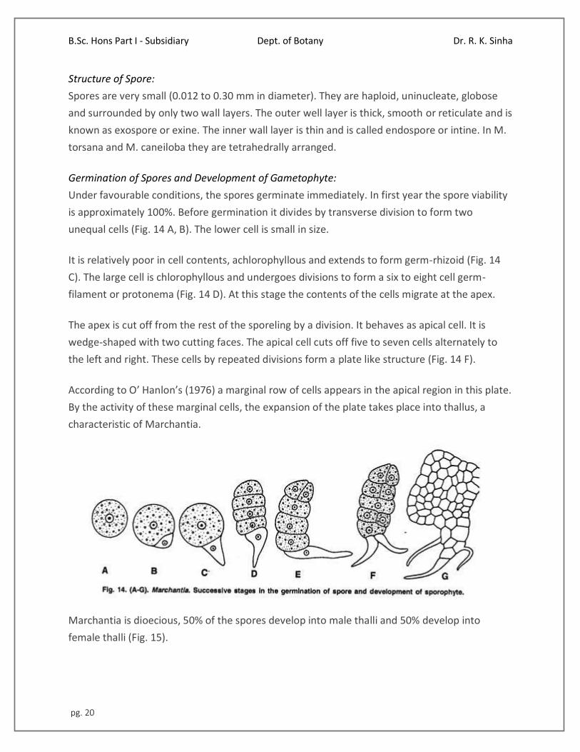
Germination of Spores and Development of Gametophyte:
Under favourable conditions, the spores germinate immediately. In first year the spore viability
is approximately 100%. Before germination it divides by transverse division to form two
unequal cells (Fig. 14 A, B). The lower cell is small in size.
It is relatively poor in cell contents, achlorophyllous and extends to form germ-rhizoid (Fig. 14
C). The large cell is chlorophyllous and undergoes divisions to form a six to eight cell germ-
filament or protonema (Fig. 14 D). At this stage the contents of the cells migrate at the apex.
The apex is cut off from the rest of the sporeling by a division. It behaves as apical cell. It is
wedge-shaped with two cutting faces. The apical cell cuts off five to seven cells alternately to
the left and right. These cells by repeated divisions form a plate like structure (Fig. 14 F).
According to O’ Hanlon’s (1976) a marginal row of cells appears in the apical region in this plate.
By the activity of these marginal cells, the expansion of the plate takes place into thallus, a
characteristic of Marchantia.
Marchantia is dioecious, 50% of the spores develop into male thalli and 50% develop into
female thalli (Fig. 15).

B.Sc. Hons Part I - Subsidiary Dept. of Botany Dr. R. K. Sinha
pg. 21
Alternation of Generation in Marchantia:
The life cycle of Marchantia shows regular alternation of two morphologically distinct phases.
One of the generations is Haplophase and the other is diplophase.
B.Sc. Hons Part I - Subsidiary Dept. of Botany Dr. R. K. Sinha
pg. 22
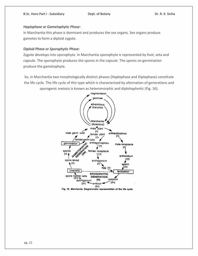
Haplophase or Gametophytic Phase:
In Marchantia this phase is dominant and produces the sex organs. Sex organs produce
gametes to form a diploid zygote.
Diploid Phase or Sporophytic Phase:
Zygote develops into sporophyte. In Marchantia sporophyte is represented by foot, seta and
capsule. The sporophyte produces the spores in the capsule. The spores on germination
produce the gametophyte.
So, in Marchantia two morphologically distinct phases (Haplophase and Diplophase) constitute
the life cycle. The life cycle of this type which is characterised by alternation of generations and
sporogenic meiosis is known as heteromorphic and diplohaplontic (Fig. 16).