Embed Size (px)
Citation preview

Syphilis
Terry Kotrla, MS, MT(ASCP)BB
Fall 2007

Introduction to Spirochetes
• Long, slender, helically tightly coiled bacteria
• Gram-negative
• Aerobic, microaerophilic or anaerobic .
• Corkscrew motility
• Can be free living or parasitic
• Best-known are those which cause disease: Syphilis and Lyme’s disease

Morphology
• Have axial filaments, which are otherwise similar to bacterial flagella
• Filaments enable movement of bacterium by rotating in place

Spirochete Diseases
• Localized skin infection disseminates to other organs.
• Latent stage, no signs/symptoms apparent.
• Cardiac and neurological involvement in untreated cases.

Serological Testing
• Important in diagnosis
• Isolation of organism very difficult
• Clinical symptoms not always apparent.

Syphilis
• Most commonly acquired spirochete disease in the U.S.
• Complex sexually transmitted disease that has a highly variable clinical course.
• Between 2005 and 2006, the number of cases of early latent syphilis reported to CDC increased 12.4% (from 8,176 to 9,186), while the number of cases of late and late latent syphilis increased 9.9% (from 16,049 to 17,644).

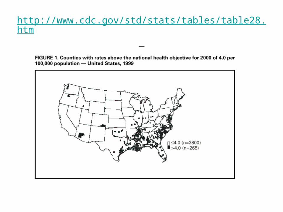
Primary and secondary syphilis — Rates by county: United States, 2006

Characteristic of the Organism
• Causative agent is Treponema pallidum
• Member of the family Spirochaetaceae.
• No natural reservoir in the environment, requires living host.
• Organism cannot be cultured from clinical specimens

Morphology
• Spiral shaped and motile due to periplasmic flagella.
• Variable length.

Scanning Electron Micrograph of T. pallidum

Other Treponemes
• Three other pathogens in the group: Treponema which are morphologically and antigenically similar to T. Pallidum
• Differences are in:– characteristics of lesions, – Amount of systemic involvement and – course of the disease.

T. pertenue• Found in tropics, causes disease Yaws.• Non-venereal transmission, transmitted by direct
contact.• Disease of bone and skin, rarely viscera• Persistent lesions, wart-like, occur primarily in
children, causes and ulcerative necrosis, scar formation, disfiguring.
• Untreated disease not as severe as syphilis, but lesions are more persistent.
• Treat with penicillin• Serologic syphilis test will be reactive.

T. pertenue
• Occurs mainly in equatorial regions and can be found in South America, Central America, the Caribbean, Africa, and Southeast Asia.
• It is associated with high humidity and rainfall.• Fifty years ago, the WHO recognized that
endemic treponematoses—yaws in particular—were a major cause of disfigurement and disability and a significant economic burden in poor countries.

T. Pertenue - Lesions

Infection with Treponema pallidum pertenue. Notice the
deformed tibiae, the so-called sabre tibiae.

T. endemicum• Causes non-venereal syphilis
known as bejel or endemic syphilis• Typically spread among children,
most commonly in the Middle East and the southern Sahara desert regions.
• Bejel is completely curable with penicillin.
• Serologic syphilis test will be reactive.

T. endemicum
• Bejel affects the skin, bones, and mucous membranes of the mouth.
• Transmission is by direct contact, with broken skin or contaminated hands, or indirectly by sharing drinking vessels and eating utensils.
• Symptoms begin with a slimy patch on the inside of the mouth followed by blisters on the trunk, arms, and legs.
• Bone infection develops later, mainly in the legs. • Also in later stages, soft, gummy lumps may appear in
the nose and on the roof of the mouth (soft palate).

Bejel

T. caroteum
• Pinta (T carateum) occurs in Central and South America and the Caribbean.
• More common in young adults.• Non-venereal, direct contact, disease of skin.• Lesion is initially a scaly patch, becomes red-
blue, later becomedepigmented and atrophy.• Treat with penicillin• Serologic syphilis test will be reactive.

Pinta
• Depigmented skin lesions.

T. cuniculi
• Not pathogenic for humans.
• Causes rabbit syphilis.

Mode of Transmission• Organism is very fragile, destroyed rapidly
by heat, cold and drying.• Sexual transmission most common,
occurs when abraded skin or mucous membranescome in contact with open lesion.
• Can be transmitted to fetus.• Rare transmission from needle stick and
blood transfusion.

Stages of Disease
• Primary
• Secondary
• Latent
• Tertiary
• Congenital Syphilis

Primary Syphilis
• Organism enters directly through skin or through mucosal tissue.
• Carried by blood throughout the body.• Organisms remaining at the site begin to
multiply.• Syphilis cannot be spread by toilet seats,
door knobs, swimming pools, hot tubs, bath tubs, shared clothing, or eating utensils.

Primary Syphilis - Chancre
• Variable incubation period of 10 days to several months, a primary lesion, chancre, forms at the entrance site.
• Chancre begins as a small, usually singular nodule; as it enlarges, the overlying epithelial tissues begins to necrose, resulting in a relatively painless ulcer.
• Unlike other bacterial infections, there is no formation of pus unless a secondary bacterial infection sets in.

Primary - Chancre• Chancre is most frequently seen on the external
genitalia– In women the lesions may form in the vagina or on
the cervix. – In men it may be inside the urethra, resulting in a
serous discharge.
• The lesion heals spontaneously after 1-5 weeks.• Swab of chancre smeared on slide, examined
under dark-field microscope, spirochetes will be present.
• Thirty percent become serologically positive one week after appearance of chancre, 90% positive after three weeks.

Primary Syphilis - Chancre

Primary Syphilis - Chancre

Darkfield Microscopy

Fluid From Chancre

Spirochetes in Blood

Intact Spirochetes in Umbilical Cord
Secondary Syphilis
• Occurs 6-8 weeks after initial chancre, becomes systemic, patient highly infectious.
• Characterized by localized or diffuse mucocutaneous lesions, often with generalized lymphadenopathy.
• Primary chancre may still be present.• Secondary lesions subside in about 2-6 weeks.• Serology tests nearly 100% positive.

Secondary Syphilis
• A widespread eruption resembling psoriasis or pityriasis rosea which prominently involves the hands should always include the differential diagnosis of secondary syphilis.

Secondary Syphilis
• Secondary syphilis lesions on back

Latent Syphilis
• Stage of infection in which organisms persist in the body of the infected person without causing symptoms or signs (asymptomatic).
• This stage may last for years.• One-third of untreated latent stage individuals
develop signs of tertiary syphilis.• After four years it is rarely communicable
sexually but can be passed from mother to fetus.

Latent Syphilis
• This stage may be further subdivided.– Early latent, initial infection occurred within
previous 12 months.– Late latent, initial infection occurred greater
than 12 months.– Latent of unknown duration, date of initial
infection cannot be established as having occurred in the previous year.

Tertiary Syphilis
• Divided into three manifestations:– Gummatous syphilis– Cardiovascular syphilis– Neurosyphilis
Tertiary Syphilis - Gummatous
• Gummas are localized areas of granulomatous inflammation found on bones, skin and subcutaneous tissue.
• Cutaneous gummas may be single or multiple, generally asymmetric and grouped together.
• Visceral lesions often cause local destruction of the affected organ.
• Contain lymphocytes, plasma cells and perivascular inflammation.
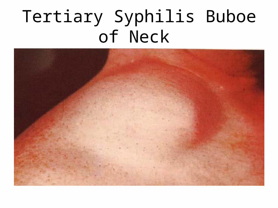
Tertiary Syphilis Buboe of Neck

Tertiary Syphilis

Tertiary Syphilis - Gumma

Tertiary - Cardiovascular
• This condition appears 20 or more years post-infection.
• Usually involves the aorta. • Invading treponemes cause scarring of the
tunica media. • Over many years, the inflammatory scarring
weakens the aortic wall, leading to aneurysm formation, which causes incompetence of the aortic valve and narrowing of the coronary ostia.

Tertiary - Cardiovascular
• Antibiotic treatment cures the syphilis infection and stops the progress of cardiovascular syphilis.
• The damage that has already occurred may not be reversed.
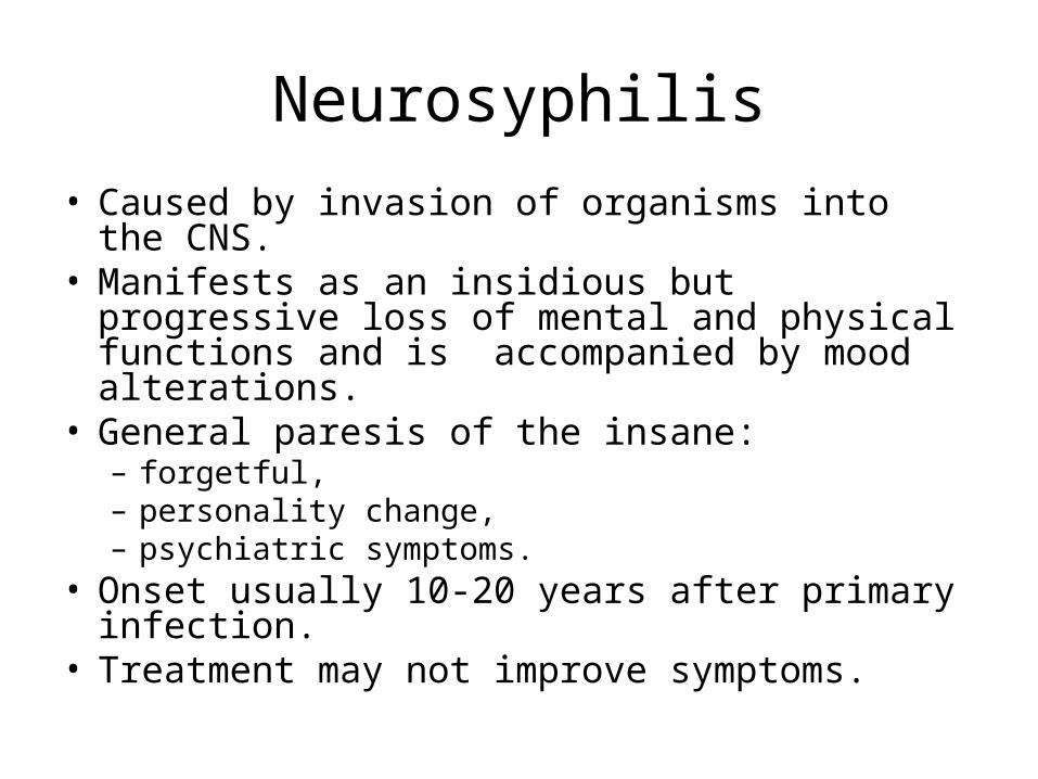
Neurosyphilis
• Caused by invasion of organisms into the CNS. • Manifests as an insidious but progressive loss of
mental and physical functions and is accompanied by mood alterations.
• General paresis of the insane: – forgetful, – personality change, – psychiatric symptoms.
• Onset usually 10-20 years after primary infection.
• Treatment may not improve symptoms.

Neurosyphilis
• Neurological complications at this stage include generalized paresis of the insane which results in personality changes, changes in emotional affect, hyperactive reflexes.
• Tabes dorsalis, degeneration of lower spinal cord, general paresis and chronic progressive dementia often results in a characteristic shuffling gait.
• Can only be diagnosed serologically by VDRL.

Neurosyphilis
• Cerebral atrophy, most prominent in frontal lobes seen in general paresis.

Congenital Syphilis
• Transmitted from mother to fetus.
• Fetus affected during second or third trimester.
• Forty percent result in syphilitic stillbirth-fetal death that occurs after a 20 week gestation and the mother had untreated or inadequately treated syphilis at delivery.

Congenital Syphilis
• According to the CDC, 40% of births to syphilitic mothers are stillborn.
• 40-70% of the survivors will be infected, and 12% of these will subsequently die prematurely
• Death from congenital syphilis is usually through pulmonary hemorrhage.

Congenital Syphilis
• After 14 years of decline in the United States, the rate of congenital syphilis increased 3.7% between 2005 and 2006 (from 8.2 to 8.5 cases per 100,000 live births)
• In 2006, 349 cases were reported, an increase from 339 in 2005.
• This small increase in the rate of congenital syphilis may relate to the increase in the rate of P&S syphilis among women that has occurred in recent years.
• Between 1996 and 2005, the average yearly percentage decrease in the congenital syphilis rate was 14.1%.
• Overall, there has been a 74.2% decrease in the rate of congenital syphilis since 1996.

Congenital Syphilis
• Bone deformities
• Blindness
• Deafness
• Deformed faces
• Dental deformities
• Skin rashes
• Neonatal death

Congenital Syphilis
• Live-born infants show no signs during first few weeks.– Sixty to 90 % develop clear or hemorrhagic
rhinitis.– skin eruptions (rash) especially around mouth,
palms of hands and soles of feet.
• Other signs: general lymphadenopathy, hepatosplenomegaly, jaundice, anemia, painful limbs, and bone abnormalities.

Congenital Syphilis
• Early onset syphilis manifests at birth or months after, exhibiting a diffuse infiltration, scabs and fissuring along the periphery of the mouth, which leave sulci in a radiated pattern or rhagades

Congenital Syphilis
• clear or hemorrhagic rhinitis

Congenital Syphilis
• Skin eruptions (rash) especially around mouth, palms of hands and soles of feet

Congenital Syphilis
• Hutchinson’s incisors.

Diagnosis of Syphilis• Evaluation based on three factors:
– Clinical findings.– Demonstration of spirochetes in clinical
specimen.– Present of antibodies in blood or
cerebrospinal fluid.
• More than one test should be performed.
• No serological test can distinguish between other treponemal infections.

Laboratory Testing• Direct examination of clinical specimen by
dark-field microscopy or fluorescent antibody testing of sample.
• Non-specific or non-treponemal serological test to detect reagin, utilized as screening test only.
• Specific Treponemal antibody tests are used as a confirmatory test for a positive reagin test.

Nontreponemal Reagin Tests• Non-specific or non-treponemal serological
test to detect reagin, utilized as screening test only.– Reagin is an antibody formed against cardiolipin.– Found in sera of patients with syphilis as well as other
diseases.– This type of reagin not to be confused with same word
originally used to describe IgE.– Non treponemal tests become positive 1 to 4 weeks
after appearance of primary chancre. – in secondary stage may have false negative due to
Prozone, in tertiary 25% are negative, after successful treatment will become nonreactive after 1 to 2 years.

Nontreponemal Reagin Tests
• VDRL
• RPR
• USR-unheated serum reagin test
• RST-reagin screen test
• ELISA

Venereal Disease Research Laboratory - VDRL
• Flocculation test, antigen consists of very fine particles that precipitate out in the presence of reagin.
• Utilizes an antigen which consists of cardiolipin, cholesterol and lecithin.– Antigen very technique dependent.– Must be made up fresh daily.
• Serum must be heated to 56 C for 30 minutes to remove anti-complementary activity which may cause false positive, if serum is not tested within 4 hours must be reheated for 10 minutes.
• Calibrated syringe utilized to dispense antigen must deliver 60 drops/mL +/- 2drops.

VDRL
• Performing the test:– 0.05 mL of serum added to circle on ceramic slide and spread.– Add one calibrated drop of antigen to each circle.– Rotate at 180 rpms for 4 minutes.– Read microscopically at 100x and grade reaction if positive.– Perform titer on positive samples, report out titer.
• Quality control:– Run three levels of control: Non-reactive, weakly reactive and
reactive.– Glass syringe with 18g delivery needle must be checked daily to
ensure delivery of 60 drops/mL.– Rotator rpms must be checked to ensure 180 rpms.– Room temperature must be 23-29 C.
• VDRL used primarily to screen cerebral spinal fluid.

VDRL
• Each preparation of antigen suspension should first be examined by testing with known positive or negative serum controls.
• The antigen particles appear as short rod forms at magnification of about 100x. Aggregation of these particles into large or small clumps is interpreted as degrees of positivity
• Reactive on left, non-reactive on right

Rapid Plasma Reagin Test - RPR
• General screening test, can be adapted to automation.
• CANNOT be performed on CSF.• Antigen
– VDRL cardiolipin antigen is modified with choline chloride to make it more stable
– attached to charcoal particles to allow macroscopic reading
– antigen comes prepared and is very stable.
• Serum or plasma may be used for testing, serum is not heated.

RPR
• Test Procedure:– Serum or plasma added to circle on card and spread.– One drop of antigen from a needle capable of delivering 60
drops/mL is added.– Rotate at 100 rpms/minute for 8 minutes.– Results are read macroscopically.
• Daily quality control:– 20 gauge needle checked for delivery of 60 drops/mL– Rotator checked for 100 rpms/minute– Room temperature must be 23-29 C.– Three levels of control must be run and give appropriate results.
• RPR appears to be more sensitive than the VDRL.

Positive RPR Serologic Test for Syphilis
• Clumping of the carbon particles indicates the person's serum contains nonspecific antilipid (reagin) antibodies.

RPR
• In the first test, the patient's serum has caused flocculation of the carbon particles; indicative of active syphilis.
• After six months, however, the test is negative. • This indicates that the antimicrobial therapy given at the time of the first test
has been successful. • Although the RPR test is a non-specific test, it is an excellent indicator of the
success of therapy. • Neg/Pos controls at top of slide, patient bottom right.

Unheated Serum Reagin Test - USR
• Modified VDRL antigen, uses choline chloride/EDTA to stabilize antigen.
• Microscopic flocculation test.• Reagent is ready-to-use and no serum heating is
required. • Chelating agents are added to neutralize the
interference due to complements. • Several types of USR tests are available.• These tests show a high incidence (8-10%) of
false negatives due to the prozone phenomenon, for this reason its preferable to run the tests at two dilutions.

Reagin Screen Test - RST• Modified VDRL antigen with Sudan Black to
make flocculation reaction macroscopically visible.
• The sensitivity and specificity of the RST are essentially the same as those of the VDRL test.
• The specimen of serum does not have to be inactivated by heat.
• The RST antigen is ready to use and it is stable for at least two years.

Specific Treponemal Tests
• Performed to confirm a positive non-specific reagin test.
• Treponema Pallidum Immobilization
• Treponema pallidum hemagglutination
• Fluorescent treponemal antibody absorption test
• ELISA

Treponema Pallidum Immobilization - TPI
• An antibody present in the serum of a syphilitic patient, in the presence of complement, causes the immobilization of actively motile Treponema pallidum obtained from testes of a rabbit infected with syphilis.
• Cumbersome and expensive, no longer used in US.

Treponema pallidum hemagglutination (TPHA)
• Adapted to microtechniques (MHA-TP)
• Tanned sheep RBCs are coated with T. pallidum antigen from Nichol’s strain.
• Agglutination of the RBCs is a positive result.

Treponema pallidum Hemagglutination (TPHA)
• Based on the agglutination of colored gelatin particle carriers sensitized with T. pallidum antigen.
• Patient sera incubated with sensitized particles in microtiter wells and unsensitized gelatin particles in control wells.
• Patient sera containing specific antibodies will react only with the antigen to form a smooth mat of agglutinated particles.
• A compact button formed by the settling of the non-agglutinated particles in the microtiter wells containing sensitized particles indicates lack of specific antibody in patient sera (-).
• If agglutination is seen with both sensitized and unsensitized particles, nonspecific agglutination is indicated.

Treponema pallidum Hemagglutination (TPHA)

Fluorescent Treponemal Antibody Absorption Test (FTA-ABS)
• Diluted, heat inactivated serum added to Reiter’s strain of T. pallidum to remove cross reactivity due to other Treponemes.
• Slides are coated with Nichol’s strain of T. pallidum and add absorbed patient serum.
• Slides are washed, and incubated with antibody bound to a fluorescent tag.
• After washing the slides are examined for fluorescence.• Requires experienced personnel to read.• Highly sensitive and specific, but time consuming to
perform.

FTA-ABS Step 1
• Teponema pallidum, the known antigen, is fixed to a microscope slide.

FTA-ABS – Step 2
• If there are antibodies against Treponema pallidum in the patient's serum, they will bind to the spirochete.
• All other antibodies are washed from the slide.

FTA-ABS- Step 3• Fluorescent anti-human gamma globulin (anti-HGG) is
added to the well. • The anti-HGG will bind with human IgG antibodies bound
to the Treponema pallidum on the slide. • All unbound anti-HGG is washed from the slide. • Viewed with a fluorescent microscope, the spirochetes
will fluoresce

Positive FTA Test for Syphilis Viewed with a Flourescent Microscope

Animation of FTA-ABS• http://www.cat.cc.md.us/courses/bio141/labmanua/lab18/ftaan.html
• Treponema pallidum, the known antigen, is fixed to a microscope slide.
• A against Treponema pallidum in the patient's serum, they will bind to the spirochete.
• All other antibodies are washed from the slide.• Fluorescent anti-human gamma globulin (anti-HGG) is
added to the well. • The anti-HGG will with any human IgG antibodies bound
to the Treponema pallidum on the slide. • All unbound anti-HGG is then washed from the slide.• When viewed with a flourescent microscope, the
spirochetes will fluoresce.

ELISA• Microtitration wells coated with T.pallidum antigens are
exposed to test specimens which may contain specific antibodies.
• After an incubation period, unbound components in the test sample are washed away.
• Specifically-bound IgG reacts with an anti-human IgG antibody bound with horseradish peroxidase during a second incubation period.
• Following a second wash cycle, specifically-bound enzyme conjugate is detected by reaction with hydrogen peroxide and the chromogen.
• The color reaction is measured spectrophotometrically to indicate the presence or absence of IgG treponemal antibodies.
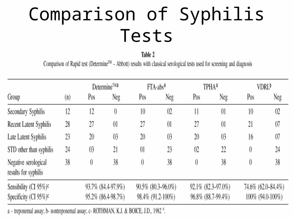
Comparison of Syphilis Tests

References
• http://www.cdc.gov/mmwr/preview/mmwrhtml/mm5007a1.htm
• http://www.cdc.gov/std/stats/tables/table22.htm• http://pathmicro.med.sc.edu/fox/spiro-neisseria.htm• http://www.dshs.state.tx.us/lab/serology_agg.shtm• http://www.cat.cc.md.us/courses/bio141/labmanua/lab18/lab18.html#fluorescent
• http://en.wikipedia.org/wiki/Syphilis • http://neuroland.com/id/neurosyph.htm• http://www.michigan.gov/documents/syphilis_flow_chart_87542_7.pdf
• CDC's Updated Plan to Eliminate Syphilis in the United States - http://tinyurl.com/yq3bn5