Embed Size (px)
Citation preview
doi.org/10.26434/chemrxiv.7680137.v1
Synthetic bPNAs as Allosteric Triggers of Hammerhead RibozymeCatalysisDennis Bong, Yufeng Liang, Jie Mao
Submitted date: 06/02/2019 • Posted date: 07/02/2019Licence: CC BY-NC-ND 4.0Citation information: Bong, Dennis; Liang, Yufeng; Mao, Jie (2019): Synthetic bPNAs as Allosteric Triggers ofHammerhead Ribozyme Catalysis. ChemRxiv. Preprint.
The biochemistry and structural biology of the hammerhead ribozyme (HHR) has been well-elucidated. Thesecondary and tertiary structural elements that enable sugar-phosphate bond scission to be be catalyzed bythis RNA are clearly understood. We have taken advantage of this knowledge base to test the extent to whichsynthetic molecules, may be used to trigger structure in secondary structure and tertiary interactions andthereby control HHR catalysis. These molecules belong to a family of molecules we call generally call“bPNAs” based on our work on bifacial peptide nucleic acid (bPNA). This family of molecules display the“bifacial” heterocycle melamine, which acts as a base-triple upon capturing two equivalents of thymine oruracil. Loosely structured internal oligouridylate bulges of 4-20 nucleotides can be restructured as triplexhybrid stems upon binding bPNAs. As such, a duplex stem element can be replaced with a bPNA triplexhybrid stem; similarly, a tertiary loop-stem interaction can be replaced with a loop-bPNA-stem complex. In thischapter, we discuss how bPNAs are prepared and applied to study structure-function turn-on in thehammerhead ribozyme system.
File list (3)
download fileview on ChemRxivMethods Enz base triple.pdf (349.84 KiB)
download fileview on ChemRxivMethods Enz base triple (2).docx (37.77 KiB)
download fileview on ChemRxivFigures.pdf (1.31 MiB)

1
Synthetic bPNAs as allosteric triggers of Hammerhead ribozyme catalysis
Yufeng Liang, Jie Mao and Dennis Bong1
Department of Chemistry and Biochemistry, The Ohio State University, Columbus, OH 43210
1Corresponding author email address: [email protected]
Contents
1. Introduction
2. Triggering RNA structure-function
3. Synthetic Protocols
3.1 Materials and instrumentation for chemical synthesis
3.2 Synthesis of bPNA amino acid derivatives
3.3 Solid phase synthesis of bPNAs
3.4 Oligoethyleneimine bPNAs from nucleophilic aromatic substitution
3.5 Oligoethyleneimine bPNAs from reductive alkylation
4. Design and preparation of U-site HHR constructs
4.1 Materials, instrumentation and general notes for transcription
4.2 Design of modified HHR transcripts with U-sites for allosteric binding
4.3 Design and use of stem/loop replacement HHR sequences
4.4 Design and use of U-loop replacement binary HHRs
5. Triggering HHR catalysis with bPNAs
5.1 General reaction protocols
5.2 Experimental design and optimizing conditions
6. Summary
Acknowledgements
References
2
Abstract
The biochemistry and structural biology of the hammerhead ribozyme (HHR) has been well-elucidated. The
secondary and tertiary structural elements that enable sugar-phosphate bond scission to be be catalyzed by
this RNA are clearly understood. We have taken advantage of this knowledge base to test the extent to which
synthetic molecules, may be used to trigger structure in secondary structure and tertiary interactions and
thereby control HHR catalysis. These molecules belong to a family of molecules we call generally call “bPNAs”
based on our work on bifacial peptide nucleic acid (bPNA). This family of molecules display the “bifacial”
heterocycle melamine, which acts as a base-triple upon capturing two equivalents of thymine or uracil. Loosely
structured internal oligouridylate bulges of 4-20 nucleotides can be restructured as triplex hybrid stems upon
binding bPNAs. As such, a duplex stem element can be replaced with a bPNA triplex hybrid stem; similarly, a
tertiary loop-stem interaction can be replaced with a loop-bPNA-stem complex. In this chapter, we discuss how
bPNAs are prepared and applied to study structure-function turn-on in the hammerhead ribozyme system.
Keywords: RNA, Hammerhead, allosteric, switch, bPNA, nucleic acid mimic, triplex, base-triple
3
1. INTRODUCTION
This chapter describes the application of bPNAs for triggering RNA structure-function in the Hammerhead
ribozyme, by virtue of the melamine base triple. Melamine hydrogen bonding has been studied extensively in
organic solvents (ten Cate, Huskens, Crego-Calama, & Reinhoudt, 2004; Whitesides et al., 1995) and the solid
state (Ranganathan, Pedireddi, & Rao, 1999). Biophysical characterization of this system (ten Cate et al.,
2004; Whitesides et al., 1995) The energetic signatures of assembly (M. Ma & Bong, 2011a) are identical to
those of nucleic acid duplexes (Kool, 2001; SantaLucia & Hicks, 2004; Yakovchuk, Protozanova, & Frank-
Kamenetskii, 2006). Accordingly, melamine recognition of native bases (Arambula, Ramisetty, Baranger, &
Zimmerman, 2009; M. Ma & Bong, 2011b, 2011c; Zhou & Bong, 2013) in a range of contexts has been
observed (Ariga & Kunitake, 1998; Kawasaki, Tokuhiro, Kimizuka, & Kunitake, 2001; Mingming Ma & Bong,
2013), presaging the possibility of triplex formation with melamine and oligo T/U domains. Unlike triplex
formation with native nucleic acids (Duca, Vekhoff, Oussedik, Halby, & Arimondo, 2008; Felsenfeld, Davies, &
Rich, 1957; Pilch, Levenson, & Shafer, 1990; Soto, Loo, & Marky, 2002) involving both Watson-Crick (WC) and
Hoogsteen base-pairing at high salt (Rougee et al., 1992; Xodo, Manzini, & Quadrifoglio, 1990) and peptide
nucleic acids (Nielsen, 1999) which invade native duplex structures, melamine triplex hybridization unites two
non-interacting oligo-T/U domains. Our group found that α-PNAs (Mittapalli et al., 2007) displaying of
melamine at alternate residue sidechains form triplex hybrids with oligo T/U DNA/RNA; we called these
peptides bifacial peptide nucleic acid (bPNA) (Figure 2) (Piao, Xia, & Bong, 2013; Zeng, Pratumyot, Piao, &
Bong, 2012). These bPNAs bind T-rich DNA to form thermally stable (Tm~58°C) triplex domains that can block
enzymatic processing of DNA and RNA (Piao et al., 2013; Xia, Piao, Fredrick, & Bong, 2013). The triplex
hybrids can functionally replace stems in RNAs and DNAs, serving as triggers of molecular recognition and
catalysis (Piao, Xia, Mao, & Bong, 2015; Xia, Piao, & Bong, 2014). The bPNA family of melamine-bearing
molecules has expanded beyond peptides to include peptoids (Mao & Bong, 2015), polyacrylates (Zhou, Xia, &
Bong, 2015) and synthetic small molecules (Mao, DeSantis, & Bong, 2017). Herein, we focus on the use of
bPNA peptides and small molecules to trigger folding and function in the Hammerhead ribozyme RNA (HHR)
scaffold.
The mini hammerhead ribozyme system (Robertson & Scott, 2007; Uhlenbeck, 1987) features a 19 nt
catalytic core supported by three structural stems. The function of the HHR is to cleave itself, and this catalytic
4
function is dependent on well-folded secondary structure in the stems and a tertiary contact between the loop
of stem 2 and stem 1. It is possible to use bPNAs to trigger both secondary and tertiary structure in the HHR
scaffold, replacing these native structures with bPNA triplex hybrids. Below we provide detailed protocols for
the synthesis of bPNAs and design of HHRs under the control of bPNA triggers.
3. SYNTHETIC PROTOCOLS
Preparation of bPNAs is relatively straightforward but requires close attention. The following synthetic
procedures have been developed to avoid chromatography where possible, but final purification of peptides
requires HPLC. Oligoethyleneimine scaffolds (tren-derived) may be purified without column chromatography.
3.1 Materials and instrumentation for chemical synthesis
All chemicals were used without further purification from commercial sources, unless otherwise noted. Solid
phase peptide synthesis was carried out under nitrogen on an AAPTEC 96 automated peptide synthesizer
equipped with a 40-well reaction block. MALDI-Mass spectra were acquired on Bruker Microflex MALDI-
TOF instrument using RP mode. Peptide samples were dissolved in ACN/H2O 1:99 v/v with 0.1% TFA while
α-cyano-4-hydroxycinnamic acid was saturated in ACN/H2O 1:1 v/v with 0.1% TFA used as the matrix.
Peptide and matrix were mixed in 1:1 ratio and then spotted on MSP 96 target ground steel (Bruker
Daltonics) for mass determination. Electrospray mass spectroscopy was acquired on a Bruker
MicroTOF equipped with an electrospray ionization source under positive mode. Mass spectrometry
instruments were provided by a grant from the Ohio BioProducts Innovation Center. NMR spectra were
acquired on a Bruker Avance DPX 400 instrument.
3.2 Synthesis of bPNA amino acid derivatives
Boc-KM-OH. This lysine derivative is prepared from Boc-Lys-OH, and must be converted to the
Fmoc derivative prior to automated solid phase peptide synthesis. It is not possible to start from the Fmoc
derivative due to basic conditions required for installation of the melamine ring. However, the derivatization
from Boc-Lys-OH is high yielding (95%), straightforward and does not require chromatography. Commercially
available Boc-Lys-OH (10 g, 40.6 mmol) was suspended in 120 mL H2O with NaOH (2 eq, 3.25 g, 81.2 mmol)
and 2,4-diamino-6-chlorotriazine (1.25 eq, 7.4 g, 50.7 mmol) and heated to 85°C for 5 hours. Thin layer
chromatography (5:1 dichloromethane-methanol) confirmed full consumption of starting material. The reaction
5
was cooled to room temperature and filtered to remove unreacted chlorotriazine. The aqueous solution was
cooled in an ice bath and the pH was adjusted to 5 using 1N HCl, resulting in a white precipitate, which was
collected via filtration and washed with cold water then dried under vacuum. This material is the derivative Boc-
Lys(M)-OH, or Boc-KM-OH, and it should be analytically pure at this point.
Fmoc-KM-OH. The Boc-KM-OH (10g, 28 mmol) deprotected by dissolution in neat TFA (80 ml)
and 1 mL of water was added as scavenger; alkylated product was never observed, and addition of water may
be unnecessary. The mixture was stirred at room temperature until complete as judged by electrospray mass
spectroscopy and TLC (~1h). The reaction was concentrated to an oil under reduced pressure, but still
contains significant quantities of TFA, which is difficult to fully remove. This residue was resuspended in 200 ml
water and the solution was neutralized to pH 7 using NaHCO3. The solution was rendered basic with another
1.5 equivalents of NaHCO3 and cooled in an ice bath. A solution of Fmoc-OSU (14 g, 42 mmol) in 200 ml
dioxane was also ice-bath cooled and added portionwise to the lysine derivative. The resulting mixture was
stirred at 0°C for 1 h and allowed to warm to room temperature overnight. The reaction was worked up removal
of dioxane under reduced pressure, addition of water (150 ml) and extraction of the aqueous layer with ethyl
acetate (2x100 ml). The aqueous layer was acidified to pH 1 with 1 N HCl and extracted again with EtOAc
(3x120 ml). The combined organic layers were concentrated under vacuum. The resulting residue was purified
CH2Cl2 to yield Fmoc-KM-OH as a white solid (8 g, 60%). Alternatively, a lower yielding purification (44%) may
be performed by trituration of the crude residue with ethanol (~200 mL).
3.3 Solid phase synthesis of bPNAs
Peptide synthesis was performed on an AAPPTEC Apex 396 Peptide Synthesizer using Rink Resin
LS (100-200 mesh) equipped with a 40-well synthesis block. The melamine base does not require protection
from SPPS or cleavage conditions and generally can be handled like any other Fmoc-amino acid derivative. To
minimize aggregation on resin due to the melamine rings a low-loading resin (0.2 mmol/g) is recommended.
Standard solid phase conditions may be used. The following solutions should be prepared:
A. 0.25M solution of Fmoc amino acid with 0.25M HOBt in NMP. (HOBt is commercially available as the
hydrate and this is acceptable).
B. 20% piperidine in NMP (by volume)
6
C. Diisopropylcarbodiimide (DIC), neat
Approximately 100-150 mg of resin is added to a reaction well, which accommodates a recommended
maximum ~2 ml liquid volume when resin is swollen. Prior to synthesis, resin is swollen in DMF for 45 min then
drained under N2 pressure and deprotected with piperidine solution B. The swelling, deprotection, washing,
coupling, shrinking and cleavage protocols are as follows:
Swelling
1 x 45 minutes. Two mL of DMF is delivered to the resin in the reaction chamber and agitated for 45 minutes at
475 rpm, then drained under N2 pressure for 2 minutes.
Deprotection cycle
2 x 5 minutes. Two mL of piperidine solution is delivered via liquid handler to reaction chamber and reaction
block is agitated at 475 rpm for 5 minutes, then drained under N2 pressure (2 minutes). This is repeated. A
wash cycle follows.
Wash cycle
3 x 2 minutes. Two mL DMF is delivered to the reaction chamber and reaction block is agitated at 475 rpm for
2 minutes, then drained under N2 pressure (2 minutes).
Coupling cycle
1 x 45-60 minutes. 2 mL (0.5 mmol) of amino acid/HOBt solution (A) is added to washed resin, followed by 75
µL (0.48 mmol) DIC (C). Reaction block is agitated at 475 rpm for 45-60 min. Standard amino acid derivatives
(eg-Fmoc-Glu(OtBu)-OH) are coupled for 45 min while Fmoc-KM-OH is coupled for 60 min. Coupling may be
extended to 90 minutes for longer peptides. After coupling is complete, reaction is drained under N2 pressure
(2 minutes).
Methanol wash
7
3 x 5 minutes. 2 mL methanol is added to washed resin and agitated for 5 minutes at 475 rpm, then drained for
2 minutes. This is repeated two additional times, then drained under N2 pressure for 15 minutes to dry the
shrunken resin.
Synthesis procedures are typically the following sequence:
1. Swell
>
2. Deprotection
3. Wash
4. Coupling
>
Repeat
5. Final wash
6. Methanol wash
Note that there is no wash after coupling as this is not necessary. After the methanol wash, the peptide resin
should then be dry, but clumped together and easy to transfer by spatula into a pre-weighed 20 mL glass
scintillation vial. This resin is then washed three times with diethyl ether, removing ether each time with a glass
pipette. The resin should be further shrunken at this time. The vial is covered with a clean kimiwipe which is
secured over the mouth of the vial with an elastic band, and then evacuated in a lyophilization bell to dry the
resin completely. The vial is then weighed to obtain the weight of the peptide-resin. It is important to obtain an
accurate weight in order to perform the peptide cleavage from resin. The following cleavage protocol is
followed:
Peptide cleavage from resin
For every 100 mg of peptide resin, 1 mL of cleavage cocktail is used. For bPNAs containing only KM and
glutamic acid or lysine, there is no need for a complex scavenger mix. Peptides were cleaved for 1 hour at RT
8
using a 95:5 volume ratio of TFA to water. The cleavage reaction including resin was filtered through cotton
into a 50 mL conical tube and the peptides were precipitated from TFA by addition of 30-50 mL chilled ether
then centrifuged to yield a white pellet. This pellet was resuspended in chilled ether and re-pelleted three times
(3x20 ml) to remove as much TFA as possible.
HPLC purification
Crude peptides were dissolved in solvent A and purified by HPLC on a semi-prep (8 mL/min) C18 reversed
phase column using a two solvent gradient. Solvent A=0.1% TFA, 1% acetonitrile, 99% water; solvent B=90%
acetonitrile, 10% water, 0.07% TFA. bPNA peptides typically elute around 40% B, depending on sequence.
Typical UV detector settings for peptides are at 230 nm while melamine itself can be observed as a distinct
peak at 238 nm under acidic conditions. Most purifications are set to observe 238 nm, with a final check for
purity at 230 nm. Purified peptide fractions were concentrated under vacuum to remove acetonitrile, then
lyophilized. Though peptides are purified on a standard gradient optimized for concentrated elution and
efficiency, peptide purity is evaluated on an analytical column with an optimized isocratic gradient and
confirmed by MALDI-TOF using ɑ-cyano-4-hydroxycinnamic acid matrix.
3.4 Oligoethyleneimine bPNAs by nucleophilic aromatic substitution
There are generally two methods employed in the preparation of oligoethyleneimine scaffolds
displaying melamine: 1) nucleophilic aromatic substitution (NAS) of chloride for amine, or 2) reductive
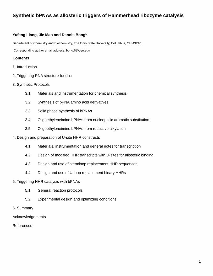
alkylation of amine using melamine acetaldehyde (Figure 1). The first method is more straightforward and can
proceed using only commercially available materials, but affords less control over substitution. The second
involves more steps but can be highly controlled. We discuss both approaches below.
t3M from NAS. This molecule can bind to a tetrauridylate bulge but only uses 2 of 3 rings in the
binding. This binding event can serve as a folding trigger for RNA. If the presence of a 3rd non-participating
melamine ring is acceptable, then it is recommended to prepare this molecule, as it is the easiest to
synthesize. Dissolve commercially available tris(2-aminoethyl)amine (0.75 mL, 5 mmol) was dissolved in 30
mL H2O, followed by addition of 2,4-diamino-6-chlorotriazine (4.36 g, 30 mmol) and NaHCO3 (1.5 g, 18 mmol).
Diaminochlorotriazine may be purchased but the synthesis from cyanuric chloride and ammonium hydroxide in
water is straightforward. The reaction was heated to 85°C and left to stir overnight. After cooling to room
9
temperature, the solution was filtered, the solid was washed with water twice, and dried under vacuum (1.63 g,
73%). The crude solid was further purified by prep HPLC using gradient of 0% to 15% acetonitrile over 40
minutes.
Boc-tren. In order to prepare a tren scaffold with only two melamines (t2M), a protected
derivative, mono-Boc tren, is required. Though this starting material is only available in modest yield, the
preparation is convenient. Tris(2-aminoethyl)amine (6 mL, 40 mmol) was dissolved in 50 mL DCM and cooled
in an ice bath. A solution of Boc anhydride (1.09 g, 5 mmol) in 20 mL DCM was added dropwise, followed by
dropwise addition of triethylamine (0.7 mL, 5 mmol) in 20 mL DCM. The reaction was warmed up to room
temperature slowly and stirred overnight. After removal of the solvent under reduced pressure, the crude oil
was resuspended in 50 mL H2O and extracted with DCM (50 mL x 5). The organic phase was dried over
Na2SO4 and solvent removed under reduced pressure. Chromatographic purification of the crude product
(DCM:MeOH=5:1, 2% concentrated ammonium hydroxide) yielded 700 mg (57%) of a light yellow oil.
t2M from NAS. 6-chloro-2,4-diamino-1,3,5-triazine(550 mg, 3.78 mmol) and NaHCO3 (500 mg,
5.95 mmol) were added into a 40 mL H2O solution of mono-Boc protected tren (220 mg, 0.89 mmol). The slurry
was heated to 85 °C and reacted overnight. The reaction was then cooled to room temperature and filtered.
The solid was washed twice with water and dried under vacuum. The crude product was then purified by silica
gel chromatography using DCM:MeOH:conc NH4OH=80:20:2 to yield Boc-t2M as a white solid (90 mg, 22%).
Boc t2M was dissolved in trifluoroacetic acid (TFA) and reacted at room temperature for 10 min. The TFA was
evaporated under N2 flow, the residue was dissolved in H2O, and solution lyophilized to obtain the TFA salt of
t2M.
t2MMe from NAS. An appropriate control molecule to evaluate selective binding of melamine to
T/U domains is the N-methylated melamine derivative. These molecules have the same overall electrostatics
and similar shape, but with hydrogen bond donors replaced with methyl groups. Though this control changes
steric interactions somewhat, this is unavoidable. The tetramethylated chloro triazine can be prepared in a
manner similar to the diamino chlorotriazine by reaction of cyanuric chloride with dimethyamine instead of
ammonium hydroxide. Thus, 6-chloro-N2,N2,N4,N4-tetramethyl-2,4-diamino-1,3,5-triazine (80 mg, 0.4 mmol)
and NaHCO3 (84 mg, 1 mmol) were added into 10 mL 1,4-dioxane:H2O=1:1 solution of mono Boc-protected
tren (40 mg, 0.162 mmol). The slurry was heated to 85°C and reacted overnight. The reaction was then cooled
10
to RT and extracted with DCM twice. The organic layer was then dried and condensed to obtain a crude
mixture which was purified by silica gel chromatography using 2% MeOH in DCM with 2% concentrated
ammonia in MeOH. The product was obtained as a white solid (45 mg, 48.4%). Boc t2MMe was dissolved in
TFA and reacted at room temperature for 10 min. The TFA was evaporated under N2 flow, the residue was
dissolved in H2O and lyophilized to obtain the TFA salt of t2MMe.
[insert Figure 1 here]
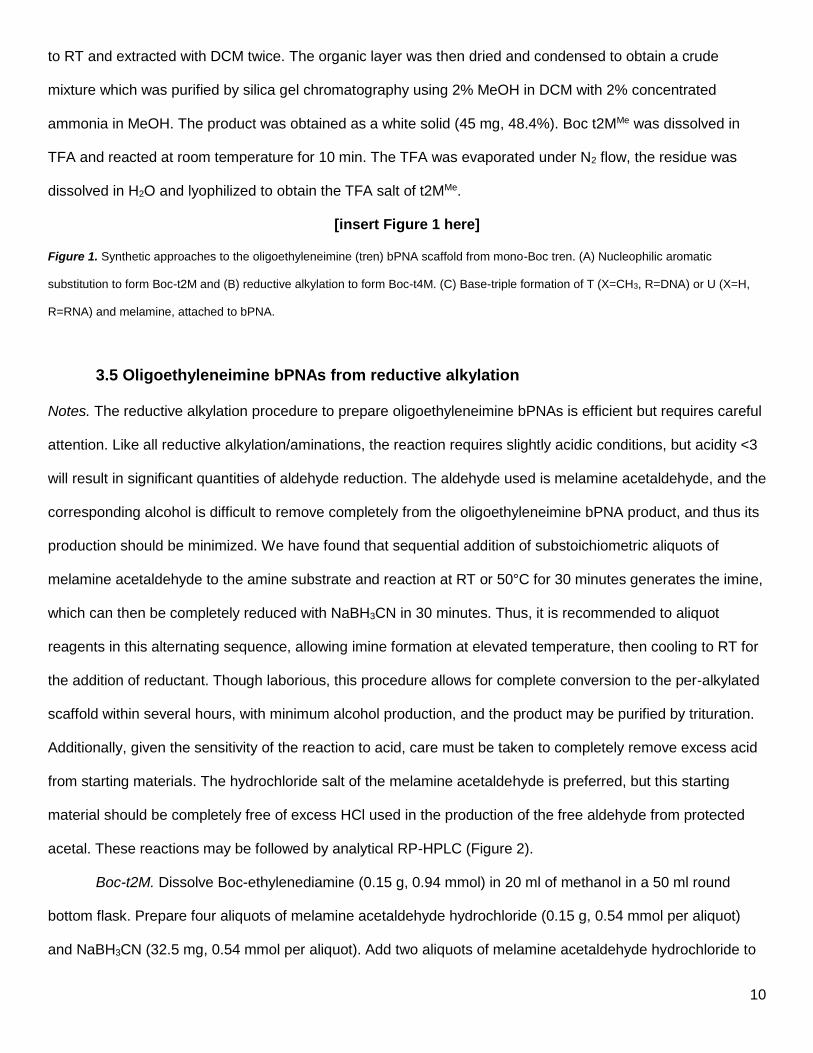
Figure 1. Synthetic approaches to the oligoethyleneimine (tren) bPNA scaffold from mono-Boc tren. (A) Nucleophilic aromatic
substitution to form Boc-t2M and (B) reductive alkylation to form Boc-t4M. (C) Base-triple formation of T (X=CH3, R=DNA) or U (X=H,
R=RNA) and melamine, attached to bPNA.
3.5 Oligoethyleneimine bPNAs from reductive alkylation
Notes. The reductive alkylation procedure to prepare oligoethyleneimine bPNAs is efficient but requires careful
attention. Like all reductive alkylation/aminations, the reaction requires slightly acidic conditions, but acidity <3
will result in significant quantities of aldehyde reduction. The aldehyde used is melamine acetaldehyde, and the
corresponding alcohol is difficult to remove completely from the oligoethyleneimine bPNA product, and thus its
production should be minimized. We have found that sequential addition of substoichiometric aliquots of
melamine acetaldehyde to the amine substrate and reaction at RT or 50°C for 30 minutes generates the imine,
which can then be completely reduced with NaBH3CN in 30 minutes. Thus, it is recommended to aliquot
reagents in this alternating sequence, allowing imine formation at elevated temperature, then cooling to RT for
the addition of reductant. Though laborious, this procedure allows for complete conversion to the per-alkylated
scaffold within several hours, with minimum alcohol production, and the product may be purified by trituration.
Additionally, given the sensitivity of the reaction to acid, care must be taken to completely remove excess acid
from starting materials. The hydrochloride salt of the melamine acetaldehyde is preferred, but this starting
material should be completely free of excess HCl used in the production of the free aldehyde from protected
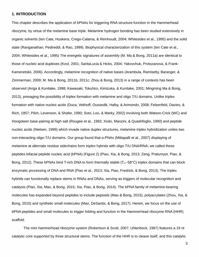
acetal. These reactions may be followed by analytical RP-HPLC (Figure 2).
Boc-t2M. Dissolve Boc-ethylenediamine (0.15 g, 0.94 mmol) in 20 ml of methanol in a 50 ml round
bottom flask. Prepare four aliquots of melamine acetaldehyde hydrochloride (0.15 g, 0.54 mmol per aliquot)
and NaBH3CN (32.5 mg, 0.54 mmol per aliquot). Add two aliquots of melamine acetaldehyde hydrochloride to
11
the methanol solution and stir for 30 minutes at room temperature to allow imine formation to proceed. The
hydrochloride salt is preferred here to generate the slightly acidic conditions favorable for reductive alkylation.
Add aliquots of aldehyde and borohydride sequentially every 30 minutes over 3-4 hours. A major side product
of the reaction is the alcohol from reduction of melamine acetaldehyde, which can be difficult to separate from
the desired product. If the pH drops to <3, then significant aldehyde reduction is observed. It was empirically
determined that addition of aldehyde and reductant in aliquots over time minimizes formation of this side
product as compared to addition of larger portions at the start of the reaction. A typical treatment is shown
below:
Reaction start:
1) 2 aliquots (1.15 eq) melamine acetaldehyde with Boc-ethylenediamine, stir 30 min
2) 1 aliquot NaBH3CN added, stirred 30 min
3) 1 aliquot melamine acetaldehyde added, react 30 min
4) 1 aliquot NaBH3CN added, react 30 min
5) 1 aliquot melamine acetaldehyde and 1 aliquot NaCNBH3 added, react 30 min
6) Final aliquot of NaCNBH3 added
Monitor the reaction by analytical HPLC to follow the progression of the alkylation (Figure 2). The imine
intermediate may be detected by HPLC/MS (ESI). To work up the reaction, reduce the amount of methanol to
~4ml and transfer to two 2-ml-centrifuge tubes. Centrifuge to obtain the solid and discard the methanol
solution. The residue will be sticky, but can be triturated and sonicated in acetone 3 times, using 3 mL acetone
for each wash. Each time after trituration, centrifuge to obtain the solid and discard the acetone. Combine the
solid into one centrifuge tube and repeat the trituration procedure with ~0.5 ml of neutral water to wash away
inorganic salts. A final trituration with 0.5 ml of ethanol yields a solid that can be air-dried to yield 210 mg white
powder (0.45 mmol, yield=48%).
[insert Figure 2 here]
Figure 2. Analytical C18RP-HPLC traces of reaction progress for reductive alkylation with melamine acetaldehyde of (A-D) Boc-t2M
12
from Boc-ethylenediamine and (E-H) Boc-t4M from Boc-tren. t2M gradient: 0%B (5 min); 0-20%B (5 min); 20%B (10 min); 20-100%B (5
min); 5 min B-wash. t4M gradient: 10%B (5 min); 10-30%B (10 min) 30%B (5 min); 30-100%B (2 min); 5 min B-wash. Early products
observed and identified by ESI-MS in (A) are: monoalkylated (15.9 min), dialkylated (16.5 min) and monoalkylated + imine (18.2 min).
Early products in Boc-t4M synthesis identified in (B) are: monoalkylated (6.2 min), dialkylated (8.8, 10.3 min) trialkylated (13.1, 14.1
min) tetraalkylated (14.7 min) and trialkylated + imine (15.5 min).
Boc-t4M. The procedure to produce Boc-t4M is similar to Boc-t2M, using portionwise addition of
melamine acetaldehyde and NaBH3CN to an amine scaffold to minimize production of the reduced
acetaldehyde side product. To prepare Boc-t4M, dissolve Boc-tren (0.2 g, 0.81 mmol) in 40 ml methanol.
Prepare 5 aliquots of melamine acetaldehyde (0.83 g, 1 equivalent, 0.81 mmol) and NaBH3CN (51 mg, 1
equivalent, 0.81 mmol). Reactants are aliquoted into the reaction every 30 minutes until addition is complete.
The reaction rate slows considerably as it progresses, resulting in small quantities of trialkylated material in
addition to the desired t4M. This may be separated, or further reacted addition of further aldehyde and
borohydride with extended reaction times. Work up the reaction as described for t2M, by concentration,
sedimentation and washing with acetone and water. Isolate the final product and wash with 5 ml of ethanol,
then air-dry to obtain 480 mg of white powder (0.56 mmol, yield=69%). The reaction can be followed by RP-
HPLC (Figure 2), though HPLC is not needed for purification.
4. DESIGN AND PREPARATION OF U-site HHR CONSTRUCTS
4.1 Materials, instrumentation and general notes for transcription
DNA templates were purchased from Integrated DNA Technologies (IDT) purified by gel prior to use in
transcription. SYBR gold was purchased from Thermo Fisher Scientific. DNA and RNA stock solutions were
prepared in deionized water and concentrations were determined by measuring solution absorbance at 260 nm
by Thermo Fisher Nanodrop 2000. The promoter sequence in each DNA template is underlined and does not
appear in the final RNA transcripts. Buffers were prepared in house with the following composition: 1X PBS
(137 mM NaCl, 2.7 mM KCl and 10 mM PO43-, pH 7.4), Tris-Cl (50 mM tris(hydroxymethyl)aminomethane, pH
13
7.6, adjusted with HCl). RNA transcription buffer (10X) was 1M HEPES-KOH pH 7.5, 100 mM MgCl2, 20 mM
Spermidine-HCl, 400 mM DTT.
4.1.1 General protocol for transcription. RNAs were prepared via runoff transcription using wild-
type T7 RNA polymerase (Milligan & Uhlenbeck, 1989). A general starting point for transcriptions used the
following conditions: 1x transcription buffer with an additional 10 mM DTT, 10 mM MgCl2, 20 mM rNTP, 7.5%
glycerol, 350 nM DNA template, and 2 μL of T7 polymerase stock per 100 μl of transcription reaction solution.
These conditions require optimization for new template sequences. The transcriptions were run for 2 hours at
37°C, then quenched with 1.5 equivalents of EDTA and an equal volume of 2x TBE/urea loading buffer.
Samples were heated at 95°C for 5 minutes, cooled to room temperature, and loaded onto acrylamide (5%
bisacrylamide) denaturing (8M urea) gel (8.3cm X 7.3cm X 1.5mm). The RNA product bands were visualized
on the gel by UV shadowing and cut from the gel. Desired RNA can be isolated from the cut band by standard
“crush and soak” (gel is forced through a syringe and RNA extracted into water by overnight agitation at RT) or
electroelution, which generally gives higher recovery. Extracted or electroeluted RNA was precipitated by
addition of 10% volume of 5M NH4OAc pH 5.2 and 2.5 volume equivalents of 200 proof ethanol, and
incubation at -20 °C for at least 2 hrs. RNA was centrifuged down at high speed for 10 minutes at 4 °C and the
pellet was washed with 500 μl of ice-cold 70% ethanol in water and centrifuged again at high speed for 10
minutes at 4 °C. RNA pellets were then dried in a speed-vac at room temperature for 20 minutes and
suspended in water and stored at -20 °C. After confirming RNA purity by denaturing gel, this pelleted material
can be used to prepare stock solutions.
4.1.2. General design considerations and challenges in transcription. Transcription targets
containing extended oligo-U domains can be challenging as these domains are considered “slippery” for the
polymerase and can also be read as termination signals (Martin & Tinoco, 1980; von Hippel, 1998; Yarnell &
Roberts, 1999), thus leading to less pure transcription reactions with truncations and closely eluting products.
This problem is exacerbated when the starting point for transcription is the U-domain. Yield and purity of
transcription is greatly improved by the addition of several non-U nucleotides (preferably a GC rich sequence)
prior to the U-site. While it is difficult to predict which transcripts will be the most problematic, it is generally true
that shorter, internal oligo-U sites give better results than longer sites near start of transcription. If it is
unavoidable to have the U-site near the start of the transcript, then addition of a few nts prior can be very
14
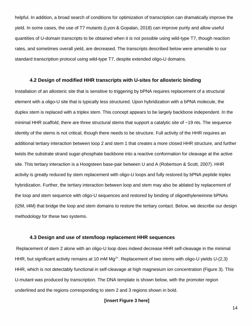
helpful. In addition, a broad search of conditions for optimization of transcription can dramatically improve the
yield. In some cases, the use of T7 mutants (Lyon & Gopalan, 2018) can improve purity and allow useful
quantities of U-domain transcripts to be obtained when it is not possible using wild-type T7, though reaction
rates, and sometimes overall yield, are decreased. The transcripts described below were amenable to our
standard transcription protocol using wild-type T7, despite extended oligo-U domains.
4.2 Design of modified HHR transcripts with U-sites for allosteric binding
Installation of an allosteric site that is sensitive to triggering by bPNA requires replacement of a structural
element with a oligo-U site that is typically less structured. Upon hybridization with a bPNA molecule, the
duplex stem is replaced with a triplex stem. This concept appears to be largely backbone independent. In the
minimal HHR scaffold, there are three structural stems that support a catalytic site of ~19 nts. The sequence
identity of the stems is not critical, though there needs to be structure. Full activity of the HHR requires an
additional tertiary interaction between loop 2 and stem 1 that creates a more closed HHR structure, and further
twists the substrate strand sugar-phosphate backbone into a reactive conformation for cleavage at the active
site. This tertiary interaction is a Hoogsteen base-pair between U and A (Robertson & Scott, 2007). HHR
activity is greatly reduced by stem replacement with oligo-U loops and fully restored by bPNA peptide triplex
hybridization. Further, the tertiary interaction between loop and stem may also be ablated by replacement of
the loop and stem sequence with oligo-U sequences and restored by binding of oligoethyleneimine bPNAs
(t2M, t4M) that bridge the loop and stem domains to restore the tertiary contact. Below, we describe our design
methodology for these two systems.
4.3 Design and use of stem/loop replacement HHR sequences
Replacement of stem 2 alone with an oligo-U loop does indeed decrease HHR self-cleavage in the minimal
HHR, but significant activity remains at 10 mM Mg2+. Replacement of two stems with oligo-U yields U-(2,3)
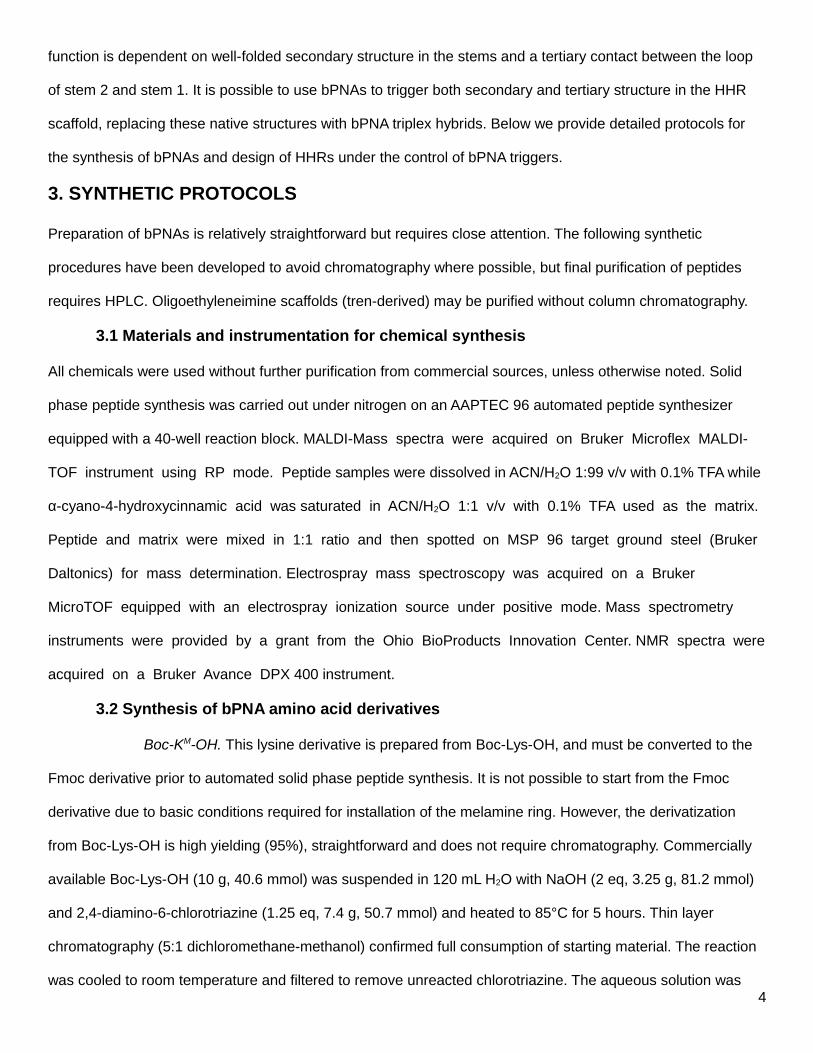
HHR, which is not detectably functional in self-cleavage at high magnesium ion concentration (Figure 3). This
U-mutant was produced by transcription. The DNA template is shown below, with the promoter region
underlined and the regions corresponding to stem 2 and 3 regions shown in bold.
[insert Figure 3 here]
15
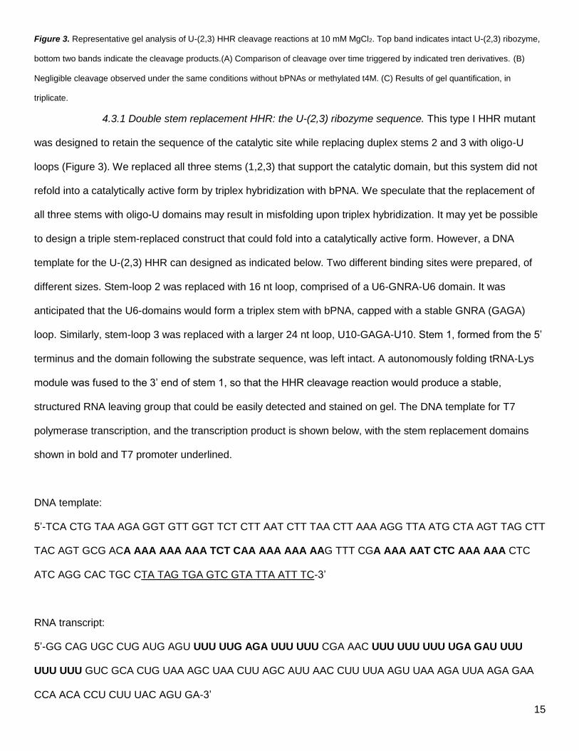
Figure 3. Representative gel analysis of U-(2,3) HHR cleavage reactions at 10 mM MgCl2. Top band indicates intact U-(2,3) ribozyme,
bottom two bands indicate the cleavage products.(A) Comparison of cleavage over time triggered by indicated tren derivatives. (B)
Negligible cleavage observed under the same conditions without bPNAs or methylated t4M. (C) Results of gel quantification, in
triplicate.
4.3.1 Double stem replacement HHR: the U-(2,3) ribozyme sequence. This type I HHR mutant
was designed to retain the sequence of the catalytic site while replacing duplex stems 2 and 3 with oligo-U
loops (Figure 3). We replaced all three stems (1,2,3) that support the catalytic domain, but this system did not
refold into a catalytically active form by triplex hybridization with bPNA. We speculate that the replacement of
all three stems with oligo-U domains may result in misfolding upon triplex hybridization. It may yet be possible
to design a triple stem-replaced construct that could fold into a catalytically active form. However, a DNA
template for the U-(2,3) HHR can designed as indicated below. Two different binding sites were prepared, of
different sizes. Stem-loop 2 was replaced with 16 nt loop, comprised of a U6-GNRA-U6 domain. It was
anticipated that the U6-domains would form a triplex stem with bPNA, capped with a stable GNRA (GAGA)
loop. Similarly, stem-loop 3 was replaced with a larger 24 nt loop, U10-GAGA-U10. Stem 1, formed from the 5’
terminus and the domain following the substrate sequence, was left intact. A autonomously folding tRNA-Lys
module was fused to the 3’ end of stem 1, so that the HHR cleavage reaction would produce a stable,
structured RNA leaving group that could be easily detected and stained on gel. The DNA template for T7
polymerase transcription, and the transcription product is shown below, with the stem replacement domains
shown in bold and T7 promoter underlined.
DNA template:
5’-TCA CTG TAA AGA GGT GTT GGT TCT CTT AAT CTT TAA CTT AAA AGG TTA ATG CTA AGT TAG CTT
TAC AGT GCG ACA AAA AAA AAA TCT CAA AAA AAA AAG TTT CGA AAA AAT CTC AAA AAA CTC
ATC AGG CAC TGC CTA TAG TGA GTC GTA TTA ATT TC-3’
RNA transcript:
5ʼ-GG CAG UGC CUG AUG AGU UUU UUG AGA UUU UUU CGA AAC UUU UUU UUU UGA GAU UUU
UUU UUU GUC GCA CUG UAA AGC UAA CUU AGC AUU AAC CUU UUA AGU UAA AGA UUA AGA GAA
CCA ACA CCU CUU UAC AGU GA-3ʼ
16
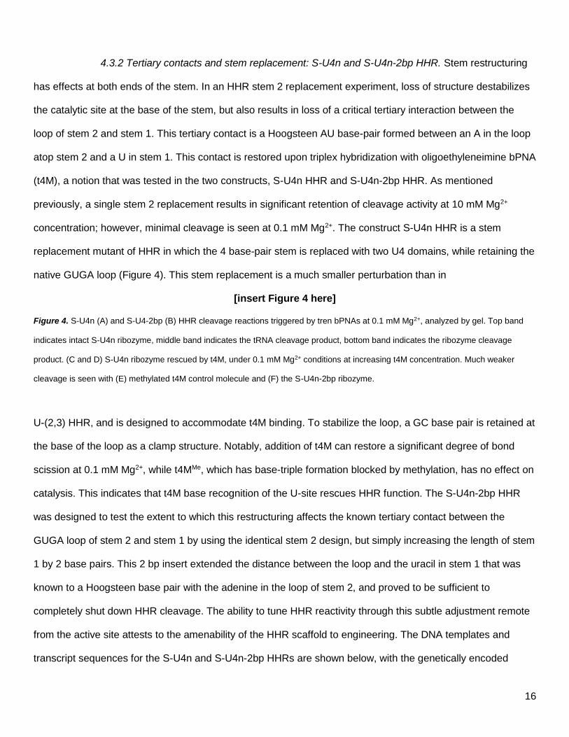
4.3.2 Tertiary contacts and stem replacement: S-U4n and S-U4n-2bp HHR. Stem restructuring
has effects at both ends of the stem. In an HHR stem 2 replacement experiment, loss of structure destabilizes
the catalytic site at the base of the stem, but also results in loss of a critical tertiary interaction between the
loop of stem 2 and stem 1. This tertiary contact is a Hoogsteen AU base-pair formed between an A in the loop
atop stem 2 and a U in stem 1. This contact is restored upon triplex hybridization with oligoethyleneimine bPNA
(t4M), a notion that was tested in the two constructs, S-U4n HHR and S-U4n-2bp HHR. As mentioned
previously, a single stem 2 replacement results in significant retention of cleavage activity at 10 mM Mg2+
concentration; however, minimal cleavage is seen at 0.1 mM Mg2+. The construct S-U4n HHR is a stem
replacement mutant of HHR in which the 4 base-pair stem is replaced with two U4 domains, while retaining the
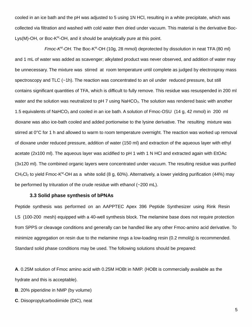
native GUGA loop (Figure 4). This stem replacement is a much smaller perturbation than in
[insert Figure 4 here]
Figure 4. S-U4n (A) and S-U4-2bp (B) HHR cleavage reactions triggered by tren bPNAs at 0.1 mM Mg2+, analyzed by gel. Top band
indicates intact S-U4n ribozyme, middle band indicates the tRNA cleavage product, bottom band indicates the ribozyme cleavage
product. (C and D) S-U4n ribozyme rescued by t4M, under 0.1 mM Mg2+ conditions at increasing t4M concentration. Much weaker
cleavage is seen with (E) methylated t4M control molecule and (F) the S-U4n-2bp ribozyme.
U-(2,3) HHR, and is designed to accommodate t4M binding. To stabilize the loop, a GC base pair is retained at
the base of the loop as a clamp structure. Notably, addition of t4M can restore a significant degree of bond
scission at 0.1 mM Mg2+, while t4MMe, which has base-triple formation blocked by methylation, has no effect on
catalysis. This indicates that t4M base recognition of the U-site rescues HHR function. The S-U4n-2bp HHR
was designed to test the extent to which this restructuring affects the known tertiary contact between the
GUGA loop of stem 2 and stem 1 by using the identical stem 2 design, but simply increasing the length of stem
1 by 2 base pairs. This 2 bp insert extended the distance between the loop and the uracil in stem 1 that was
known to a Hoogsteen base pair with the adenine in the loop of stem 2, and proved to be sufficient to
completely shut down HHR cleavage. The ability to tune HHR reactivity through this subtle adjustment remote
from the active site attests to the amenability of the HHR scaffold to engineering. The DNA templates and
transcript sequences for the S-U4n and S-U4n-2bp HHRs are shown below, with the genetically encoded
17
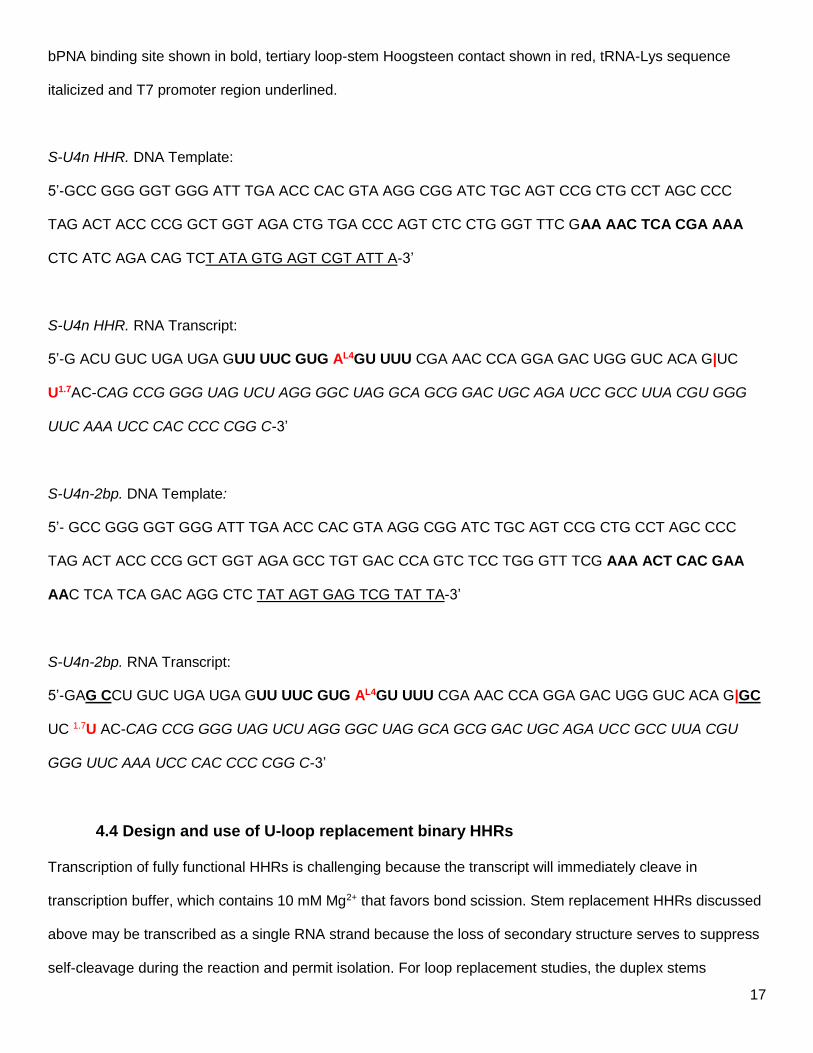
bPNA binding site shown in bold, tertiary loop-stem Hoogsteen contact shown in red, tRNA-Lys sequence
italicized and T7 promoter region underlined.
S-U4n HHR. DNA Template:
5’-GCC GGG GGT GGG ATT TGA ACC CAC GTA AGG CGG ATC TGC AGT CCG CTG CCT AGC CCC
TAG ACT ACC CCG GCT GGT AGA CTG TGA CCC AGT CTC CTG GGT TTC GAA AAC TCA CGA AAA
CTC ATC AGA CAG TCT ATA GTG AGT CGT ATT A-3’
S-U4n HHR. RNA Transcript:
5’-G ACU GUC UGA UGA GUU UUC GUG AL4GU UUU CGA AAC CCA GGA GAC UGG GUC ACA G|UC
U1.7AC-CAG CCG GGG UAG UCU AGG GGC UAG GCA GCG GAC UGC AGA UCC GCC UUA CGU GGG
UUC AAA UCC CAC CCC CGG C-3’
S-U4n-2bp. DNA Template:
5’- GCC GGG GGT GGG ATT TGA ACC CAC GTA AGG CGG ATC TGC AGT CCG CTG CCT AGC CCC
TAG ACT ACC CCG GCT GGT AGA GCC TGT GAC CCA GTC TCC TGG GTT TCG AAA ACT CAC GAA
AAC TCA TCA GAC AGG CTC TAT AGT GAG TCG TAT TA-3’
S-U4n-2bp. RNA Transcript:
5’-GAG CCU GUC UGA UGA GUU UUC GUG AL4GU UUU CGA AAC CCA GGA GAC UGG GUC ACA G|GC
UC 1.7U AC-CAG CCG GGG UAG UCU AGG GGC UAG GCA GCG GAC UGC AGA UCC GCC UUA CGU
GGG UUC AAA UCC CAC CCC CGG C-3’
4.4 Design and use of U-loop replacement binary HHRs
Transcription of fully functional HHRs is challenging because the transcript will immediately cleave in
transcription buffer, which contains 10 mM Mg2+ that favors bond scission. Stem replacement HHRs discussed
above may be transcribed as a single RNA strand because the loss of secondary structure serves to suppress
self-cleavage during the reaction and permit isolation. For loop replacement studies, the duplex stems
18
supporting the catalytic site are intact, and thus a full-length HHR will completely self-cleave prior to isolation.
For this reason, loop replacement studies must be carried out on binary HHRs, wherein one strand is an
enzyme strand, and the other is a substrate strand. These binary HHRs exhibit significant rate dependence on
the sequence identity of the stem 2 loop. However, though a GUGA loop is found in the wild type HHR, with
the adenine base of the loop interacting with uracil base in stem 1, a GUUU loop also functions well in
cleavage. This suggests that the HHR loop-stem interaction may be quite plastic, and may accept alternative
non-canonical interactions such as a GU wobble pair in addition to the AU Hoogsteen pair. To study the turn
on of tertiary interactions with t4M, we transcribed the enzyme strand for a binary HHR in which the GUGA
loop of stem 2 was replaced with UUUU. To provide a baseline of reactivity, a the HHR enzyme strand with the
native GUGA loop was also transcribed. It is convenient to follow this reaction by fluorescently tagged RNA
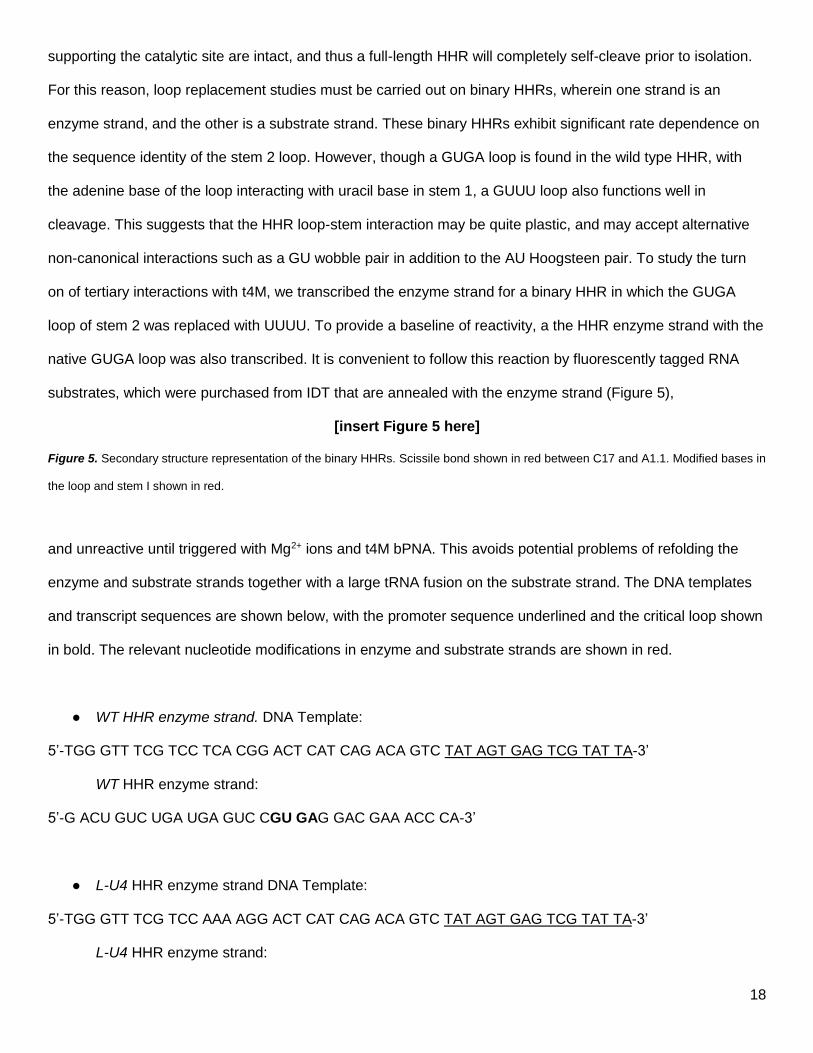
substrates, which were purchased from IDT that are annealed with the enzyme strand (Figure 5),
[insert Figure 5 here]
Figure 5. Secondary structure representation of the binary HHRs. Scissile bond shown in red between C17 and A1.1. Modified bases in
the loop and stem I shown in red.
and unreactive until triggered with Mg2+ ions and t4M bPNA. This avoids potential problems of refolding the
enzyme and substrate strands together with a large tRNA fusion on the substrate strand. The DNA templates
and transcript sequences are shown below, with the promoter sequence underlined and the critical loop shown
in bold. The relevant nucleotide modifications in enzyme and substrate strands are shown in red.
WT HHR enzyme strand. DNA Template:
5’-TGG GTT TCG TCC TCA CGG ACT CAT CAG ACA GTC TAT AGT GAG TCG TAT TA-3’
WT HHR enzyme strand:
5’-G ACU GUC UGA UGA GUC CGU GAG GAC GAA ACC CA-3’
L-U4 HHR enzyme strand DNA Template:
5’-TGG GTT TCG TCC AAA AGG ACT CAT CAG ACA GTC TAT AGT GAG TCG TAT TA-3’
L-U4 HHR enzyme strand:
19
5’-G ACU GUC UGA UGA GUC CUU UUG GAC GAA ACC CA-3’
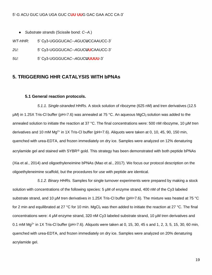
Substrate strands (Scissile bond: C--A.)
WT-HHR: 5’ Cy3-UGGGUCAC--AGUCUCCAAUCC-3’
2U: 5’ Cy3-UGGGUCAC--AGUCUUCAAUCC-3’
5U: 5’ Cy3-UGGGUCAC--AGUCUUUUU-3’
5. TRIGGERING HHR CATALYSIS WITH bPNAs
5.1 General reaction protocols.
5.1.1. Single-stranded HHRs. A stock solution of ribozyme (625 nM) and tren derivatives (12.5
µM) in 1.25X Tris-Cl buffer (pH=7.6) was annealed at 75 °C. An aqueous MgCl2 solution was added to the
annealed solution to initiate the reaction at 37 °C. The final concentrations were: 500 nM ribozyme, 10 µM tren
derivatives and 10 mM Mg2+ in 1X Tris-Cl buffer (pH=7.6). Aliquots were taken at 0, 10, 45, 90, 150 min,
quenched with urea-EDTA, and frozen immediately on dry ice. Samples were analyzed on 12% denaturing
acrylamide gel and stained with SYBR gold. This strategy has been demonstrated with both peptide bPNAs
(Xia et al., 2014) and oligoethyleneimine bPNAs (Mao et al., 2017). We focus our protocol description on the
oligoethyleneimine scaffold, but the procedures for use with peptide are identical.
5.1.2. Binary HHRs. Samples for single turnover experiments were prepared by making a stock
solution with concentrations of the following species: 5 µM of enzyme strand, 400 nM of the Cy3 labeled
substrate strand, and 10 µM tren derivatives in 1.25X Tris-Cl buffer (pH=7.6). The mixture was heated at 75 °C
for 2 min and equilibrated at 27 °C for 10 min. MgCl2 was then added to initiate the reaction at 27 °C. The final
concentrations were: 4 µM enzyme strand, 320 nM Cy3 labeled substrate strand, 10 µM tren derivatives and
0.1 mM Mg2+ in 1X Tris-Cl buffer (pH=7.6). Aliquots were taken at 0, 15, 30, 45 s and 1, 2, 3, 5, 15, 30, 60 min,
quenched with urea-EDTA, and frozen immediately on dry ice. Samples were analyzed on 20% denaturing
acrylamide gel.
20
5.2 Experimental design and optimizing conditions
5.2.1. Cleavage reactions of single-stranded HHRs. The single stranded HHR U-mutants
such as U-(2,3), S-U4n and S-U4n-2bp HHRs can be transcribed under normal conditions and do not self-
cleave appreciably, despite 10 mM MgCl2 in the reaction. However, their reactivity remains sensitive to Mg2+
concentration, and the combination of both t4M and Mg2+ is required to trigger RNA cleavage. The presence of
the tRNA fusions in these constructs yields two RNAs of comparable size upon cleavage, the HHR itself and
the tRNA. These two RNAs may be easily observed by gel (Figure 4). Addition of the methylated t4M molecule
generally does not cause HHR cleavage. This control is essential to rule out the effects of the oligoamine
backbone itself; molecules of this type can affect RNA function, but typically at much higher concentrations
(Pingoud et al., 1984; Quigley, Teeter, & Rich, 1978). Reactions should be run in triplicate and gels quantified
using standard software (Imagequant) to obtain HHR cleavage rates (Figure 4). Reactivity should be very
reproducible for each construct, resulting in relatively tight error bars. If the reaction rate does not allow
monitoring by reasonable data time point sampling, the rate may be tuned by decreasing or increasing
Mg2+concentration to decrease or increase reaction rate, respectively. Decreasing Mg2+ to 0.1 mM enabled
convenient monitoring of HHR cleavage in the sU4n HHR as a function of t4M concentration. Typically,
cleavage reactions were monitored at a single t4M concentration over time. In addition, rates were measured
as a function of increasing t4M concentrations. In this way, it can be clearly established that the t4M molecules
can directly influence HHR cleavage.
5.2.2. Cleavage reactions of binary HHRs. Binary HHRs were annealed together prior to use in
cleavage reactions as described above. Interestingly, it is possible to mix different substrate strands with each
HHR enzyme strand, yielding cleavage rates reflective of their dependence on t4M as well as substrate-
enzyme matching. While both loop modified L-GU3 and LU4 HHRs do not effectively cleave the native
substrate, L-GU3 can cleave a substrate strand that features U’s at both position 1.7 (native) and 1.8 in stem 1,
which suggests that the L1 guanine may be forming a tertiary contact with the U 1.8. The labeled substrate
strand provides a convenient way to quantitatively follow the cleavage reaction, signaling unreacted and
cleaved substrate with the same dye (Figure 6).
[insert Figure 6 here]

21
Figure 6. Representative gel image for binary ribozyme cleavage. Top band is the intact Cy3 labelled substrate strand RNA, bottom
band indicates the cleavage product. (Left) L-U4/5U; (right) L-U4/5U with 10 µM t4M, under 0.1 mM Mg2+ conditions.
6. Summary
The synthetic bPNAs described herein can bridge native and artificial architectures, uniting nucleic acid
biotechnology (Burnett & Rossi, 2012) with new materials (Roh, Ruiz, Peng, Lee, & Luo, 2011). This protocol
provides a general guide on the synthesis of bPNA peptides and oligoethyleneimine derivatives that have been
used to trigger cleavage in engineered hammerhead ribozymes. This strategy enables additional control over
RNA chemistry.
Acknowledgments
This work was supported in part by funding from the National Institutes of Health (GM111995-01A1 to D.B.), NASA
(GRT00044810 to D.B.) and NSF (DMR 1802432 to D.B).
22
References
Arambula, J. F., Ramisetty, S. R., Baranger, A. M., & Zimmerman, S. C. (2009). A simple ligand that selectively
targets CUG trinucleotide repeats and inhibits MBNL protein binding. Proceedings of the National
Academy of Sciences of the United States of America, 106, 16068–16073.
https://doi.org/10.1073/pnas.0901824106
Ariga, K., & Kunitake, T. (1998). Molecular Recognition at Air-Water and Related Interfaces: Complementary
Hydrogen Bonding and Multisite Interaction. Accounts of Chemical Research, 31, 371–378. Retrieved
from http://pubs3.acs.org/acs/journals/doilookup?in_doi=10.1021/ar970014i
Burnett, J. C., & Rossi, J. J. (2012). RNA-Based Therapeutics: Current Progress and Future Prospects.
Chemistry & Biology, 19, 60–71. https://doi.org/10.1016/j.chembiol.2011.12.008
Duca, M., Vekhoff, P., Oussedik, K., Halby, L., & Arimondo, P. B. (2008). The triple helix: 50 years later, the
outcome. Nucleic Acids Research, 36(16), 5123–5138. https://doi.org/10.1093/nar/gkn493
Felsenfeld, G., Davies, D. R., & Rich, A. (1957). Formation of a three-stranded polynucleotide molecule.
Journal of the American Chemical Society, 79, 2023–2024. https://doi.org/10.1021/ja01565a074
Kawasaki, T., Tokuhiro, M., Kimizuka, N., & Kunitake, T. (2001). Hierarchical self-assembly of chiral
complementary hydrogen-bond networks in water: reconstitution of supramolecular membranes. Journal
of the American Chemical Society, 123, 6792–6800. Retrieved from
http://www.ncbi.nlm.nih.gov/entrez/query.fcgi?cmd=Retrieve&db=PubMed&dopt=Citation&list_uids=11448
183
Kool, E. T. (2001). Hydrogen Bonding, Base Stacking, and Steric Effects in DNA Replication. Annual Review of
Biophysics and Biomolecular Structure, 30(1), 1–22. https://doi.org/10.1146/annurev.biophys.30.1.1
Lyon, S., & Gopalan, V. (2018). A T7 RNA Polymerase Mutant Enhances the Yield of 5’-Thienoguanosine-
Initiated RNAs. Chembiochem: A European Journal of Chemical Biology, 19(2), 142–146.
https://doi.org/10.1002/cbic.201700538
Ma, M., & Bong, D. (2011a). Determinants of cyanuric acid and melamine assembly in water. Langmuir: The
ACS Journal of Surfaces and Colloids, 27, 8841–8853. https://doi.org/10.1021/la201415d
Ma, M., & Bong, D. (2011b). Directed peptide assembly at the lipid-water interface cooperatively enhances
23
membrane binding and activity. Langmuir: The ACS Journal of Surfaces and Colloids, 27, 1480–1486.
https://doi.org/10.1021/la104405r
Ma, M., & Bong, D. (2011c). Protein assembly directed by synthetic molecular recognition motifs. Organic &
Biomolecular Chemistry, 9, 7296–7299. https://doi.org/10.1039/c1ob05998j
Ma, M., & Bong, D. (2013). Controlled Fusion of Synthetic Lipid Membrane Vesicles. Accounts of Chemical
Research, 46, 2988–2997.
Mao, J., & Bong, D. (2015). Synthesis of DNA-Binding Peptoids. Synlett: Accounts and Rapid Communications
in Synthetic Organic Chemistry, 26(11), 1581–1585. https://doi.org/10.1055/s-0034-1380698
Mao, J., DeSantis, C., & Bong, D. (2017). Small Molecule Recognition Triggers Secondary and Tertiary
Interactions in DNA Folding and Hammerhead Ribozyme Catalysis. Journal of the American Chemical
Society, 139(29), 9815–9818. https://doi.org/10.1021/jacs.7b05448
Martin, F. H., & Tinoco, I., Jr. (1980). DNA-RNA hybrid duplexes containing oligo(dA:rU) sequences are
exceptionally unstable and may facilitate termination of transcription. Nucleic Acids Research, 8(10),
2295–2299. Retrieved from https://www.ncbi.nlm.nih.gov/pubmed/6159577
Milligan, J. F., & Uhlenbeck, O. C. (1989). [5] Synthesis of small RNAs using T7 RNA polymerase. In Methods
in Enzymology (Vol. 180, pp. 51–62). Academic Press. https://doi.org/10.1016/0076-6879(89)80091-6
Mittapalli, G. K., Reddy, K. R., Xiong, H., Munoz, O., Han, B., De Riccardis, F., … Eschenmoser, A. (2007).
Mapping the landscape of potentially primordial informational oligomers: oligodipeptides and
oligodipeptoids tagged with triazines as recognition elements. Angewandte Chemie, International Edition,
46, 2470–2477. https://doi.org/10.1002/anie.200603207
Nielsen, P. E. (1999). Peptide Nucleic Acid. A Molecule with Two Identities. Accounts of Chemical Research,
32(7), 624–630. https://doi.org/10.1021/ar980010t
Piao, X., Xia, X., & Bong, D. (2013). Bifacial Peptide Nucleic Acid Directs Cooperative Folding and Assembly of
Binary, Ternary, and Quaternary DNA Complexes. Biochemistry, 52, 6313–6323.
https://doi.org/10.1021/bi4008963
Piao, X., Xia, X., Mao, J., & Bong, D. (2015). Peptide ligation and RNA cleavage via an abiotic template
interface. Journal of the American Chemical Society, 137(11), 3751–3754.
https://doi.org/10.1021/jacs.5b00236
24
Pilch, D. S., Levenson, C., & Shafer, R. H. (1990). Structural analysis of the (dA)10.2(dT)10 triple helix.
Proceedings of the National Academy of Sciences of the United States of America, 87, 1942–1946.
Retrieved from
http://www.ncbi.nlm.nih.gov/entrez/query.fcgi?cmd=Retrieve&db=PubMed&dopt=Citation&list_uids=23089
55
Pingoud, A., Urbanke, C., Alves, J., Ehbrecht, H. J., Zabeau, M., & Gualerzi, C. (1984). Effect of polyamines
and basic proteins on cleavage of DNA by restriction endonucleases. Biochemistry, 23(24), 5697–5703.
Retrieved from https://www.ncbi.nlm.nih.gov/pubmed/6098296
Quigley, G. J., Teeter, M. M., & Rich, A. (1978). Structural analysis of spermine and magnesium ion binding to
yeast phenylalanine transfer RNA. Proceedings of the National Academy of Sciences of the United States
of America, 75(1), 64–68. Retrieved from https://www.ncbi.nlm.nih.gov/pubmed/343112
Ranganathan, A., Pedireddi, V. R., & Rao, C. N. R. (1999). Hydrothermal Synthesis of Organic Channel
Structures: 1:1 Hydrogen-Bonded Adducts of Melamine with Cyanuric and Trithiocyanuric Acids. Journal
of the American Chemical Society, 121, 1752–1753. Retrieved from
http://pubs3.acs.org/acs/journals/doilookup?in_doi=10.1021/ja983928o
Robertson, M. P., & Scott, W. G. (2007). The structural basis of ribozyme-catalyzed RNA assembly. Science,
315(5818), 1549–1553. https://doi.org/10.1126/science.1136231
Roh, Y. H., Ruiz, R. C. H., Peng, S., Lee, J. B., & Luo, D. (2011). Engineering DNA-based functional materials.
Chemical Society Reviews, 40(12), 5730–5744. https://doi.org/10.1039/c1cs15162b
Rougee, M., Faucon, B., Mergny, J. L., Barcelo, F., Giovannangeli, C., Garestier, T., & Hélène, C. (1992).
Kinetics and thermodynamics of triple-helix formation: effects of ionic strength and mismatches.
Biochemistry, 31, 9269–9278. Retrieved from
http://www.ncbi.nlm.nih.gov/entrez/query.fcgi?cmd=Retrieve&db=PubMed&dopt=Citation&list_uids=13907
13
SantaLucia, J., Jr., & Hicks, D. (2004). The thermodynamics of DNA structural motifs. Annual Review of
Biophysics and Biomolecular Structure, 33, 415–440.
https://doi.org/10.1146/annurev.biophys.32.110601.141800
Soto, A. M., Loo, J., & Marky, L. A. (2002). Energetic contributions for the formation of TAT/TAT, TAT/CGC(+),
25
and CGC(+)/CGC(+) base triplet stacks. Journal of the American Chemical Society, 124, 14355–14363.
Retrieved from
http://www.ncbi.nlm.nih.gov/entrez/query.fcgi?cmd=Retrieve&db=PubMed&dopt=Citation&list_uids=12452
709
ten Cate, M. G. J., Huskens, J., Crego-Calama, M., & Reinhoudt, D. N. (2004). Thermodynamic stability of
hydrogen-bonded nanostructures: a calorimetric study. Chemistry , 10(15), 3632–3639.
https://doi.org/10.1002/chem.200400085
Uhlenbeck, O. C. (1987). A small catalytic oligoribonucleotide. Nature, 328(6131), 596–600.
https://doi.org/10.1038/328596a0
von Hippel, P. H. (1998). An integrated model of the transcription complex in elongation, termination, and
editing. Science, 281(5377), 660–665. Retrieved from https://www.ncbi.nlm.nih.gov/pubmed/9685251
Whitesides, G. M., Simanek, E. E., Mathias, J. P., Seto, C. T., Chin, D., Mammen, M., & Gordon, D. M. (1995).
Noncovalent Synthesis: Using Physical-Organic Chemistry To Make Aggregates. Accounts of Chemical
Research, 28, 37–44. Retrieved from
http://pubs3.acs.org/acs/journals/doilookup?in_doi=10.1021/ar00049a006
Xia, X., Piao, X., & Bong, D. (2014). Bifacial peptide nucleic acid as an allosteric switch for aptamer and
ribozyme function. Journal of the American Chemical Society, 136(20), 7265–7268.
https://doi.org/10.1021/ja5032584
Xia, X., Piao, X., Fredrick, K., & Bong, D. (2013). Bifacial PNA complexation inhibits enzymatic access to DNA
and RNA. Chembiochem: A European Journal of Chemical Biology, 15, 31–36.
Xodo, L. E., Manzini, G., & Quadrifoglio, F. (1990). Spectroscopic and calorimetric investigation on the DNA
triplex formed by d(CTCTTCTTTCTTTTCTTTCTTCTC) and d(GAGAAGAAAGA) at acidic pH. Nucleic
Acids Research, 18, 3557–3564. https://doi.org/10.1093/nar/18.12.3557
Yakovchuk, P., Protozanova, E., & Frank-Kamenetskii, M. D. (2006). Base-stacking and base-pairing
contributions into thermal stability of the DNA double helix. Nucleic Acids Research, 34, 564–574.
https://doi.org/10.1093/nar/gkj454
Yarnell, W. S., & Roberts, J. W. (1999). Mechanism of intrinsic transcription termination and antitermination.
Science, 284(5414), 611–615. Retrieved from https://www.ncbi.nlm.nih.gov/pubmed/10213678
26
Zeng, Y., Pratumyot, Y., Piao, X., & Bong, D. (2012). Discrete assembly of synthetic peptide-DNA triplex
structures from polyvalent melamine-thymine bifacial recognition. Journal of the American Chemical
Society, 134(2), 832–835. https://doi.org/10.1021/ja2099326
Zhou, Z., & Bong, D. (2013). Small-Molecule/Polymer Recognition Triggers Aqueous-Phase Assembly and
Encapsulation. Langmuir: The ACS Journal of Surfaces and Colloids, 29, 144–150.
https://doi.org/10.1021/la304457y
Zhou, Z., Xia, X., & Bong, D. (2015). Synthetic Polymer Hybridization with DNA and RNA Directs Nanoparticle
Loading, Silencing Delivery, and Aptamer Function. Journal of the American Chemical Society, 137(28),
8920–8923. https://doi.org/10.1021/jacs.5b05481

download fileview on ChemRxivMethods Enz base triple.pdf (349.84 KiB)
Synthetic bPNAs as allosteric triggers of Hammerhead ribozyme catalysis
Yufeng Liang, Jie Mao and Dennis Bong1
Department of Chemistry and Biochemistry, The Ohio State University, Columbus, OH 43210
1Corresponding author email address: [email protected]
Contents
1. Introduction
2. Triggering RNA structure-function
3. Synthetic Protocols
3.1 Materials and instrumentation for chemical synthesis
3.2 Synthesis of bPNA amino acid derivatives
3.3 Solid phase synthesis of bPNAs
3.4 Oligoethyleneimine bPNAs from nucleophilic aromatic substitution
3.5 Oligoethyleneimine bPNAs from reductive alkylation
4. Design and preparation of U-site HHR constructs
4.1 Materials, instrumentation and general notes for transcription
4.2 Design of modified HHR transcripts with U-sites for allosteric binding
4.3 Design and use of stem/loop replacement HHR sequences
4.4 Design and use of U-loop replacement binary HHRs
5. Triggering HHR catalysis with bPNAs
5.1 General reaction protocols
5.2 Experimental design and optimizing conditions
6. Summary
Acknowledgements
References
1

Abstract
The biochemistry and structural biology of the hammerhead ribozyme (HHR) has been well-elucidated. The
secondary and tertiary structural elements that enable sugar-phosphate bond scission to be be catalyzed by
this RNA are clearly understood. We have taken advantage of this knowledge base to test the extent to which
synthetic molecules, may be used to trigger structure in secondary structure and tertiary interactions and
thereby control HHR catalysis. These molecules belong to a family of molecules we call generally call “bPNAs”
based on our work on bifacial peptide nucleic acid (bPNA). This family of molecules display the “bifacial”
heterocycle melamine, which acts as a base-triple upon capturing two equivalents of thymine or uracil. Loosely
structured internal oligouridylate bulges of 4-20 nucleotides can be restructured as triplex hybrid stems upon
binding bPNAs. As such, a duplex stem element can be replaced with a bPNA triplex hybrid stem; similarly, a
tertiary loop-stem interaction can be replaced with a loop-bPNA-stem complex. In this chapter, we discuss how
bPNAs are prepared and applied to study structure-function turn-on in the hammerhead ribozyme system.
Keywords: RNA, Hammerhead, allosteric, switch, bPNA, nucleic acid mimic, triplex, base-triple
2
1. INTRODUCTION
This chapter describes the application of bPNAs for triggering RNA structure-function in the Hammerhead
ribozyme, by virtue of the melamine base triple. Melamine hydrogen bonding has been studied extensively in
organic solvents (ten Cate, Huskens, Crego-Calama, & Reinhoudt, 2004; Whitesides et al., 1995) and the solid
state (Ranganathan, Pedireddi, & Rao, 1999). Biophysical characterization of this system (ten Cate et al.,
2004; Whitesides et al., 1995) The energetic signatures of assembly (M. Ma & Bong, 2011a) are identical to
those of nucleic acid duplexes (Kool, 2001; SantaLucia & Hicks, 2004; Yakovchuk, Protozanova, & Frank-
Kamenetskii, 2006). Accordingly, melamine recognition of native bases (Arambula, Ramisetty, Baranger, &
Zimmerman, 2009; M. Ma & Bong, 2011b, 2011c; Zhou & Bong, 2013) in a range of contexts has been
observed (Ariga & Kunitake, 1998; Kawasaki, Tokuhiro, Kimizuka, & Kunitake, 2001; Mingming Ma & Bong,
2013), presaging the possibility of triplex formation with melamine and oligo T/U domains. Unlike triplex
formation with native nucleic acids (Duca, Vekhoff, Oussedik, Halby, & Arimondo, 2008; Felsenfeld, Davies, &
Rich, 1957; Pilch, Levenson, & Shafer, 1990; Soto, Loo, & Marky, 2002) involving both Watson-Crick (WC) and
Hoogsteen base-pairing at high salt (Rougee et al., 1992; Xodo, Manzini, & Quadrifoglio, 1990) and peptide
nucleic acids (Nielsen, 1999) which invade native duplex structures, melamine triplex hybridization unites two
non-interacting oligo-T/U domains. Our group found that α-PNAs (Mittapalli et al., 2007) displaying of
melamine at alternate residue sidechains form triplex hybrids with oligo T/U DNA/RNA; we called these
peptides bifacial peptide nucleic acid (bPNA) (Figure 2) (Piao, Xia, & Bong, 2013; Zeng, Pratumyot, Piao, &
Bong, 2012). These bPNAs bind T-rich DNA to form thermally stable (Tm~58°C) triplex domains that can block
enzymatic processing of DNA and RNA (Piao et al., 2013; Xia, Piao, Fredrick, & Bong, 2013). The triplex
hybrids can functionally replace stems in RNAs and DNAs, serving as triggers of molecular recognition and
catalysis (Piao, Xia, Mao, & Bong, 2015; Xia, Piao, & Bong, 2014). The bPNA family of melamine-bearing
molecules has expanded beyond peptides to include peptoids (Mao & Bong, 2015), polyacrylates (Zhou, Xia, &
Bong, 2015) and synthetic small molecules (Mao, DeSantis, & Bong, 2017). Herein, we focus on the use of
bPNA peptides and small molecules to trigger folding and function in the Hammerhead ribozyme RNA (HHR)
scaffold.
The mini hammerhead ribozyme system (Robertson & Scott, 2007; Uhlenbeck, 1987) features a 19 nt
catalytic core supported by three structural stems. The function of the HHR is to cleave itself, and this catalytic
3
function is dependent on well-folded secondary structure in the stems and a tertiary contact between the loop
of stem 2 and stem 1. It is possible to use bPNAs to trigger both secondary and tertiary structure in the HHR
scaffold, replacing these native structures with bPNA triplex hybrids. Below we provide detailed protocols for
the synthesis of bPNAs and design of HHRs under the control of bPNA triggers.
3. SYNTHETIC PROTOCOLS
Preparation of bPNAs is relatively straightforward but requires close attention. The following synthetic
procedures have been developed to avoid chromatography where possible, but final purification of peptides
requires HPLC. Oligoethyleneimine scaffolds (tren-derived) may be purified without column chromatography.
3.1 Materials and instrumentation for chemical synthesis
All chemicals were used without further purification from commercial sources, unless otherwise noted. Solid
phase peptide synthesis was carried out under nitrogen on an AAPTEC 96 automated peptide synthesizer
equipped with a 40-well reaction block. MALDI-Mass spectra were acquired on Bruker Microflex MALDI-
TOF instrument using RP mode. Peptide samples were dissolved in ACN/H2O 1:99 v/v with 0.1% TFA while
α-cyano-4-hydroxycinnamic acid was saturated in ACN/H2O 1:1 v/v with 0.1% TFA used as the matrix.
Peptide and matrix were mixed in 1:1 ratio and then spotted on MSP 96 target ground steel (Bruker
Daltonics) for mass determination. Electrospray mass spectroscopy was acquired on a Bruker
MicroTOF equipped with an electrospray ionization source under positive mode. Mass spectrometry
instruments were provided by a grant from the Ohio BioProducts Innovation Center. NMR spectra were
acquired on a Bruker Avance DPX 400 instrument.
3.2 Synthesis of bPNA amino acid derivatives
Boc-KM-OH. This lysine derivative is prepared from Boc-Lys-OH, and must be converted to the
Fmoc derivative prior to automated solid phase peptide synthesis. It is not possible to start from the Fmoc
derivative due to basic conditions required for installation of the melamine ring. However, the derivatization
from Boc-Lys-OH is high yielding (95%), straightforward and does not require chromatography. Commercially
available Boc-Lys-OH (10 g, 40.6 mmol) was suspended in 120 mL H2O with NaOH (2 eq, 3.25 g, 81.2 mmol)
and 2,4-diamino-6-chlorotriazine (1.25 eq, 7.4 g, 50.7 mmol) and heated to 85°C for 5 hours. Thin layer
chromatography (5:1 dichloromethane-methanol) confirmed full consumption of starting material. The reaction
was cooled to room temperature and filtered to remove unreacted chlorotriazine. The aqueous solution was 4
cooled in an ice bath and the pH was adjusted to 5 using 1N HCl, resulting in a white precipitate, which was
collected via filtration and washed with cold water then dried under vacuum. This material is the derivative Boc-
Lys(M)-OH, or Boc-KM-OH, and it should be analytically pure at this point.
Fmoc-KM-OH. The Boc-KM-OH (10g, 28 mmol) deprotected by dissolution in neat TFA (80 ml)
and 1 mL of water was added as scavenger; alkylated product was never observed, and addition of water may
be unnecessary. The mixture was stirred at room temperature until complete as judged by electrospray mass
spectroscopy and TLC (~1h). The reaction was concentrated to an oil under reduced pressure, but still
contains significant quantities of TFA, which is difficult to fully remove. This residue was resuspended in 200 ml
water and the solution was neutralized to pH 7 using NaHCO3. The solution was rendered basic with another
1.5 equivalents of NaHCO3 and cooled in an ice bath. A solution of Fmoc-OSU (14 g, 42 mmol) in 200 ml
dioxane was also ice-bath cooled and added portionwise to the lysine derivative. The resulting mixture was
stirred at 0°C for 1 h and allowed to warm to room temperature overnight. The reaction was worked up removal
of dioxane under reduced pressure, addition of water (150 ml) and extraction of the aqueous layer with ethyl
acetate (2x100 ml). The aqueous layer was acidified to pH 1 with 1 N HCl and extracted again with EtOAc
(3x120 ml). The combined organic layers were concentrated under vacuum. The resulting residue was purified
CH2Cl2 to yield Fmoc-KM-OH as a white solid (8 g, 60%). Alternatively, a lower yielding purification (44%) may
be performed by trituration of the crude residue with ethanol (~200 mL).
3.3 Solid phase synthesis of bPNAs
Peptide synthesis was performed on an AAPPTEC Apex 396 Peptide Synthesizer using Rink Resin
LS (100-200 mesh) equipped with a 40-well synthesis block. The melamine base does not require protection
from SPPS or cleavage conditions and generally can be handled like any other Fmoc-amino acid derivative. To
minimize aggregation on resin due to the melamine rings a low-loading resin (0.2 mmol/g) is recommended.
Standard solid phase conditions may be used. The following solutions should be prepared:
A. 0.25M solution of Fmoc amino acid with 0.25M HOBt in NMP. (HOBt is commercially available as the
hydrate and this is acceptable).
B. 20% piperidine in NMP (by volume)
C. Diisopropylcarbodiimide (DIC), neat
5
Approximately 100-150 mg of resin is added to a reaction well, which accommodates a recommended
maximum ~2 ml liquid volume when resin is swollen. Prior to synthesis, resin is swollen in DMF for 45 min then
drained under N2 pressure and deprotected with piperidine solution B. The swelling, deprotection, washing,
coupling, shrinking and cleavage protocols are as follows:
Swelling
1 x 45 minutes. Two mL of DMF is delivered to the resin in the reaction chamber and agitated for 45 minutes at
475 rpm, then drained under N2 pressure for 2 minutes.
Deprotection cycle
2 x 5 minutes. Two mL of piperidine solution is delivered via liquid handler to reaction chamber and reaction
block is agitated at 475 rpm for 5 minutes, then drained under N2 pressure (2 minutes). This is repeated. A
wash cycle follows.
Wash cycle
3 x 2 minutes. Two mL DMF is delivered to the reaction chamber and reaction block is agitated at 475 rpm for 2
minutes, then drained under N2 pressure (2 minutes).
Coupling cycle
1 x 45-60 minutes. 2 mL (0.5 mmol) of amino acid/HOBt solution (A) is added to washed resin, followed by 75
µL (0.48 mmol) DIC (C). Reaction block is agitated at 475 rpm for 45-60 min. Standard amino acid derivatives
(eg-Fmoc-Glu(OtBu)-OH) are coupled for 45 min while Fmoc-KM-OH is coupled for 60 min. Coupling may be
extended to 90 minutes for longer peptides. After coupling is complete, reaction is drained under N2 pressure
(2 minutes).
Methanol wash
3 x 5 minutes. 2 mL methanol is added to washed resin and agitated for 5 minutes at 475 rpm, then drained for
6
2 minutes. This is repeated two additional times, then drained under N2 pressure for 15 minutes to dry the
shrunken resin.
Synthesis procedures are typically the following sequence:
1. Swell
>
2. Deprotection
3. Wash
4. Coupling
>
Repeat
5. Final wash
6. Methanol wash
Note that there is no wash after coupling as this is not necessary. After the methanol wash, the peptide resin
should then be dry, but clumped together and easy to transfer by spatula into a pre-weighed 20 mL glass
scintillation vial. This resin is then washed three times with diethyl ether, removing ether each time with a glass
pipette. The resin should be further shrunken at this time. The vial is covered with a clean kimiwipe which is
secured over the mouth of the vial with an elastic band, and then evacuated in a lyophilization bell to dry the
resin completely. The vial is then weighed to obtain the weight of the peptide-resin. It is important to obtain an
accurate weight in order to perform the peptide cleavage from resin. The following cleavage protocol is
followed:
Peptide cleavage from resin
For every 100 mg of peptide resin, 1 mL of cleavage cocktail is used. For bPNAs containing only KM and
glutamic acid or lysine, there is no need for a complex scavenger mix. Peptides were cleaved for 1 hour at RT
using a 95:5 volume ratio of TFA to water. The cleavage reaction including resin was filtered through cotton into
7
a 50 mL conical tube and the peptides were precipitated from TFA by addition of 30-50 mL chilled ether then
centrifuged to yield a white pellet. This pellet was resuspended in chilled ether and re-pelleted three times
(3x20 ml) to remove as much TFA as possible.
HPLC purification
Crude peptides were dissolved in solvent A and purified by HPLC on a semi-prep (8 mL/min) C18 reversed
phase column using a two solvent gradient. Solvent A=0.1% TFA, 1% acetonitrile, 99% water; solvent B=90%
acetonitrile, 10% water, 0.07% TFA. bPNA peptides typically elute around 40% B, depending on sequence.
Typical UV detector settings for peptides are at 230 nm while melamine itself can be observed as a distinct
peak at 238 nm under acidic conditions. Most purifications are set to observe 238 nm, with a final check for
purity at 230 nm. Purified peptide fractions were concentrated under vacuum to remove acetonitrile, then
lyophilized. Though peptides are purified on a standard gradient optimized for concentrated elution and
efficiency, peptide purity is evaluated on an analytical column with an optimized isocratic gradient and
confirmed by MALDI-TOF using -cyano-4-hydroxycinnamic acid matrix.ɑ
3.4 Oligoethyleneimine bPNAs by nucleophilic aromatic substitution
There are generally two methods employed in the preparation of oligoethyleneimine scaffolds
displaying melamine: 1) nucleophilic aromatic substitution (NAS) of chloride for amine, or 2) reductive
alkylation of amine using melamine acetaldehyde (Figure 1). The first method is more straightforward and can
proceed using only commercially available materials, but affords less control over substitution. The second
involves more steps but can be highly controlled. We discuss both approaches below.
t3M from NAS. This molecule can bind to a tetrauridylate bulge but only uses 2 of 3 rings in the
binding. This binding event can serve as a folding trigger for RNA. If the presence of a 3rd non-participating
melamine ring is acceptable, then it is recommended to prepare this molecule, as it is the easiest to
synthesize. Dissolve commercially available tris(2-aminoethyl)amine (0.75 mL, 5 mmol) was dissolved in 30
mL H2O, followed by addition of 2,4-diamino-6-chlorotriazine (4.36 g, 30 mmol) and NaHCO3 (1.5 g, 18 mmol).
Diaminochlorotriazine may be purchased but the synthesis from cyanuric chloride and ammonium hydroxide in
water is straightforward. The reaction was heated to 85°C and left to stir overnight. After cooling to room
temperature, the solution was filtered, the solid was washed with water twice, and dried under vacuum (1.63 g,
8
73%). The crude solid was further purified by prep HPLC using gradient of 0% to 15% acetonitrile over 40
minutes.
Boc-tren. In order to prepare a tren scaffold with only two melamines (t2M), a protected
derivative, mono-Boc tren, is required. Though this starting material is only available in modest yield, the
preparation is convenient. Tris(2-aminoethyl)amine (6 mL, 40 mmol) was dissolved in 50 mL DCM and cooled
in an ice bath. A solution of Boc anhydride (1.09 g, 5 mmol) in 20 mL DCM was added dropwise, followed by
dropwise addition of triethylamine (0.7 mL, 5 mmol) in 20 mL DCM. The reaction was warmed up to room
temperature slowly and stirred overnight. After removal of the solvent under reduced pressure, the crude oil
was resuspended in 50 mL H2O and extracted with DCM (50 mL x 5). The organic phase was dried over
Na2SO4 and solvent removed under reduced pressure. Chromatographic purification of the crude product
(DCM:MeOH=5:1, 2% concentrated ammonium hydroxide) yielded 700 mg (57%) of a light yellow oil.
t2M from NAS. 6-chloro-2,4-diamino-1,3,5-triazine(550 mg, 3.78 mmol) and NaHCO3 (500 mg,
5.95 mmol) were added into a 40 mL H2O solution of mono-Boc protected tren (220 mg, 0.89 mmol). The slurry
was heated to 85 °C and reacted overnight. The reaction was then cooled to room temperature and filtered.
The solid was washed twice with water and dried under vacuum. The crude product was then purified by silica
gel chromatography using DCM:MeOH:conc NH4OH=80:20:2 to yield Boc-t2M as a white solid (90 mg, 22%).
Boc t2M was dissolved in trifluoroacetic acid (TFA) and reacted at room temperature for 10 min. The TFA was
evaporated under N2 flow, the residue was dissolved in H2O, and solution lyophilized to obtain the TFA salt of
t2M.
t2MMe from NAS. An appropriate control molecule to evaluate selective binding of melamine to
T/U domains is the N-methylated melamine derivative. These molecules have the same overall electrostatics
and similar shape, but with hydrogen bond donors replaced with methyl groups. Though this control changes
steric interactions somewhat, this is unavoidable. The tetramethylated chloro triazine can be prepared in a
manner similar to the diamino chlorotriazine by reaction of cyanuric chloride with dimethyamine instead of
ammonium hydroxide. Thus, 6-chloro-N2,N2,N4,N4-tetramethyl-2,4-diamino-1,3,5-triazine (80 mg, 0.4 mmol)
and NaHCO3 (84 mg, 1 mmol) were added into 10 mL 1,4-dioxane:H2O=1:1 solution of mono Boc-protected
tren (40 mg, 0.162 mmol). The slurry was heated to 85°C and reacted overnight. The reaction was then cooled
to RT and extracted with DCM twice. The organic layer was then dried and condensed to obtain a crude
9
mixture which was purified by silica gel chromatography using 2% MeOH in DCM with 2% concentrated
ammonia in MeOH. The product was obtained as a white solid (45 mg, 48.4%). Boc t2MMe was dissolved in
TFA and reacted at room temperature for 10 min. The TFA was evaporated under N2 flow, the residue was
dissolved in H2O and lyophilized to obtain the TFA salt of t2MMe.
[insert Figure 1 here]
Figure 1. Synthetic approaches to the oligoethyleneimine (tren) bPNA scaffold from mono-Boc tren. (A) Nucleophilic aromatic
substitution to form Boc-t2M and (B) reductive alkylation to form Boc-t4M. (C) Base-triple formation of T (X=CH3, R=DNA) or U (X=H,
R=RNA) and melamine, attached to bPNA.
3.5 Oligoethyleneimine bPNAs from reductive alkylation
Notes. The reductive alkylation procedure to prepare oligoethyleneimine bPNAs is efficient but requires careful
attention. Like all reductive alkylation/aminations, the reaction requires slightly acidic conditions, but acidity <3
will result in significant quantities of aldehyde reduction. The aldehyde used is melamine acetaldehyde, and the
corresponding alcohol is difficult to remove completely from the oligoethyleneimine bPNA product, and thus its
production should be minimized. We have found that sequential addition of substoichiometric aliquots of
melamine acetaldehyde to the amine substrate and reaction at RT or 50°C for 30 minutes generates the imine,
which can then be completely reduced with NaBH3CN in 30 minutes. Thus, it is recommended to aliquot
reagents in this alternating sequence, allowing imine formation at elevated temperature, then cooling to RT for
the addition of reductant. Though laborious, this procedure allows for complete conversion to the per-alkylated
scaffold within several hours, with minimum alcohol production, and the product may be purified by trituration.
Additionally, given the sensitivity of the reaction to acid, care must be taken to completely remove excess acid
from starting materials. The hydrochloride salt of the melamine acetaldehyde is preferred, but this starting
material should be completely free of excess HCl used in the production of the free aldehyde from protected
acetal. These reactions may be followed by analytical RP-HPLC (Figure 2).
Boc-t2M. Dissolve Boc-ethylenediamine (0.15 g, 0.94 mmol) in 20 ml of methanol in a 50 ml round
bottom flask. Prepare four aliquots of melamine acetaldehyde hydrochloride (0.15 g, 0.54 mmol per aliquot)
and NaBH3CN (32.5 mg, 0.54 mmol per aliquot). Add two aliquots of melamine acetaldehyde hydrochloride to
the methanol solution and stir for 30 minutes at room temperature to allow imine formation to proceed. The
10
hydrochloride salt is preferred here to generate the slightly acidic conditions favorable for reductive alkylation.
Add aliquots of aldehyde and borohydride sequentially every 30 minutes over 3-4 hours. A major side product
of the reaction is the alcohol from reduction of melamine acetaldehyde, which can be difficult to separate from
the desired product. If the pH drops to <3, then significant aldehyde reduction is observed. It was empirically
determined that addition of aldehyde and reductant in aliquots over time minimizes formation of this side
product as compared to addition of larger portions at the start of the reaction. A typical treatment is shown
below:
Reaction start:
1) 2 aliquots (1.15 eq) melamine acetaldehyde with Boc-ethylenediamine, stir 30 min
2) 1 aliquot NaBH3CN added, stirred 30 min
3) 1 aliquot melamine acetaldehyde added, react 30 min
4) 1 aliquot NaBH3CN added, react 30 min
5) 1 aliquot melamine acetaldehyde and 1 aliquot NaCNBH3 added, react 30 min
6) Final aliquot of NaCNBH3 added
Monitor the reaction by analytical HPLC to follow the progression of the alkylation (Figure 2). The imine
intermediate may be detected by HPLC/MS (ESI). To work up the reaction, reduce the amount of methanol to
~4ml and transfer to two 2-ml-centrifuge tubes. Centrifuge to obtain the solid and discard the methanol
solution. The residue will be sticky, but can be triturated and sonicated in acetone 3 times, using 3 mL acetone
for each wash. Each time after trituration, centrifuge to obtain the solid and discard the acetone. Combine the
solid into one centrifuge tube and repeat the trituration procedure with ~0.5 ml of neutral water to wash away
inorganic salts. A final trituration with 0.5 ml of ethanol yields a solid that can be air-dried to yield 210 mg white
powder (0.45 mmol, yield=48%).
[insert Figure 2 here]
Figure 2. Analytical C18RP-HPLC traces of reaction progress for reductive alkylation with melamine acetaldehyde of (A-D) Boc-t2M
from Boc-ethylenediamine and (E-H) Boc-t4M from Boc-tren. t2M gradient: 0%B (5 min); 0-20%B (5 min); 20%B (10 min); 20-100%B (5
11
min); 5 min B-wash. t4M gradient: 10%B (5 min); 10-30%B (10 min) 30%B (5 min); 30-100%B (2 min); 5 min B-wash. Early products
observed and identified by ESI-MS in (A) are: monoalkylated (15.9 min), dialkylated (16.5 min) and monoalkylated + imine (18.2 min).
Early products in Boc-t4M synthesis identified in (B) are: monoalkylated (6.2 min), dialkylated (8.8, 10.3 min) trialkylated (13.1, 14.1
min) tetraalkylated (14.7 min) and trialkylated + imine (15.5 min).
Boc-t4M. The procedure to produce Boc-t4M is similar to Boc-t2M, using portionwise addition of
melamine acetaldehyde and NaBH3CN to an amine scaffold to minimize production of the reduced
acetaldehyde side product. To prepare Boc-t4M, dissolve Boc-tren (0.2 g, 0.81 mmol) in 40 ml methanol.
Prepare 5 aliquots of melamine acetaldehyde (0.83 g, 1 equivalent, 0.81 mmol) and NaBH3CN (51 mg, 1
equivalent, 0.81 mmol). Reactants are aliquoted into the reaction every 30 minutes until addition is complete.
The reaction rate slows considerably as it progresses, resulting in small quantities of trialkylated material in
addition to the desired t4M. This may be separated, or further reacted addition of further aldehyde and
borohydride with extended reaction times. Work up the reaction as described for t2M, by concentration,
sedimentation and washing with acetone and water. Isolate the final product and wash with 5 ml of ethanol,
then air-dry to obtain 480 mg of white powder (0.56 mmol, yield=69%). The reaction can be followed by RP-
HPLC (Figure 2), though HPLC is not needed for purification.
4. DESIGN AND PREPARATION OF U-site HHR CONSTRUCTS
4.1 Materials, instrumentation and general notes for transcription
DNA templates were purchased from Integrated DNA Technologies (IDT) purified by gel prior to use in
transcription. SYBR gold was purchased from Thermo Fisher Scientific. DNA and RNA stock solutions were
prepared in deionized water and concentrations were determined by measuring solution absorbance at 260 nm
by Thermo Fisher Nanodrop 2000. The promoter sequence in each DNA template is underlined and does not
appear in the final RNA transcripts. Buffers were prepared in house with the following composition: 1X PBS
(137 mM NaCl, 2.7 mM KCl and 10 mM PO43-, pH 7.4), Tris-Cl (50 mM tris(hydroxymethyl)aminomethane, pH
7.6, adjusted with HCl). RNA transcription buffer (10X) was 1M HEPES-KOH pH 7.5, 100 mM MgCl2, 20 mM
Spermidine-HCl, 400 mM DTT.
12
4.1.1 General protocol for transcription. RNAs were prepared via runoff transcription using wild-
type T7 RNA polymerase (Milligan & Uhlenbeck, 1989). A general starting point for transcriptions used the
following conditions: 1x transcription buffer with an additional 10 mM DTT, 10 mM MgCl2, 20 mM rNTP, 7.5%
glycerol, 350 nM DNA template, and 2 μL of T7 polymerase stock per 100 μl of transcription reaction solution.
These conditions require optimization for new template sequences. The transcriptions were run for 2 hours at
37°C, then quenched with 1.5 equivalents of EDTA and an equal volume of 2x TBE/urea loading buffer.
Samples were heated at 95°C for 5 minutes, cooled to room temperature, and loaded onto acrylamide (5%
bisacrylamide) denaturing (8M urea) gel (8.3cm X 7.3cm X 1.5mm). The RNA product bands were visualized
on the gel by UV shadowing and cut from the gel. Desired RNA can be isolated from the cut band by standard
“crush and soak” (gel is forced through a syringe and RNA extracted into water by overnight agitation at RT) or
electroelution, which generally gives higher recovery. Extracted or electroeluted RNA was precipitated by
addition of 10% volume of 5M NH4OAc pH 5.2 and 2.5 volume equivalents of 200 proof ethanol, and incubation
at -20 °C for at least 2 hrs. RNA was centrifuged down at high speed for 10 minutes at 4 °C and the pellet was
washed with 500 μl of ice-cold 70% ethanol in water and centrifuged again at high speed for 10 minutes at 4
°C. RNA pellets were then dried in a speed-vac at room temperature for 20 minutes and suspended in water
and stored at -20 °C. After confirming RNA purity by denaturing gel, this pelleted material can be used to
prepare stock solutions.
4.1.2. General design considerations and challenges in transcription. Transcription targets
containing extended oligo-U domains can be challenging as these domains are considered “slippery” for the
polymerase and can also be read as termination signals (Martin & Tinoco, 1980; von Hippel, 1998; Yarnell &
Roberts, 1999), thus leading to less pure transcription reactions with truncations and closely eluting products.
This problem is exacerbated when the starting point for transcription is the U-domain. Yield and purity of
transcription is greatly improved by the addition of several non-U nucleotides (preferably a GC rich sequence)
prior to the U-site. While it is difficult to predict which transcripts will be the most problematic, it is generally true
that shorter, internal oligo-U sites give better results than longer sites near start of transcription. If it is
unavoidable to have the U-site near the start of the transcript, then addition of a few nts prior can be very
helpful. In addition, a broad search of conditions for optimization of transcription can dramatically improve the
yield. In some cases, the use of T7 mutants (Lyon & Gopalan, 2018) can improve purity and allow useful
13
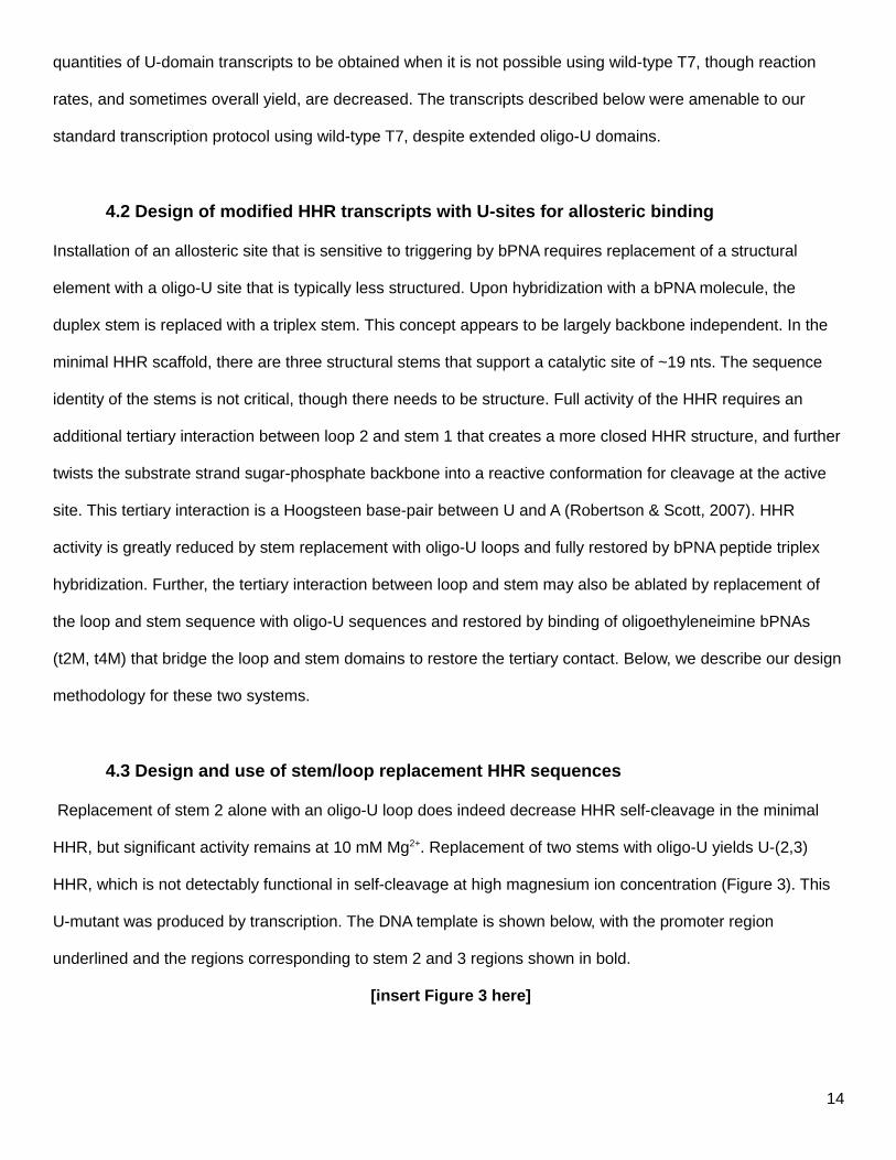
quantities of U-domain transcripts to be obtained when it is not possible using wild-type T7, though reaction
rates, and sometimes overall yield, are decreased. The transcripts described below were amenable to our
standard transcription protocol using wild-type T7, despite extended oligo-U domains.
4.2 Design of modified HHR transcripts with U-sites for allosteric binding
Installation of an allosteric site that is sensitive to triggering by bPNA requires replacement of a structural
element with a oligo-U site that is typically less structured. Upon hybridization with a bPNA molecule, the
duplex stem is replaced with a triplex stem. This concept appears to be largely backbone independent. In the
minimal HHR scaffold, there are three structural stems that support a catalytic site of ~19 nts. The sequence
identity of the stems is not critical, though there needs to be structure. Full activity of the HHR requires an
additional tertiary interaction between loop 2 and stem 1 that creates a more closed HHR structure, and further
twists the substrate strand sugar-phosphate backbone into a reactive conformation for cleavage at the active
site. This tertiary interaction is a Hoogsteen base-pair between U and A (Robertson & Scott, 2007). HHR
activity is greatly reduced by stem replacement with oligo-U loops and fully restored by bPNA peptide triplex
hybridization. Further, the tertiary interaction between loop and stem may also be ablated by replacement of
the loop and stem sequence with oligo-U sequences and restored by binding of oligoethyleneimine bPNAs
(t2M, t4M) that bridge the loop and stem domains to restore the tertiary contact. Below, we describe our design
methodology for these two systems.
4.3 Design and use of stem/loop replacement HHR sequences
Replacement of stem 2 alone with an oligo-U loop does indeed decrease HHR self-cleavage in the minimal
HHR, but significant activity remains at 10 mM Mg2+. Replacement of two stems with oligo-U yields U-(2,3)
HHR, which is not detectably functional in self-cleavage at high magnesium ion concentration (Figure 3). This
U-mutant was produced by transcription. The DNA template is shown below, with the promoter region
underlined and the regions corresponding to stem 2 and 3 regions shown in bold.
[insert Figure 3 here]
14
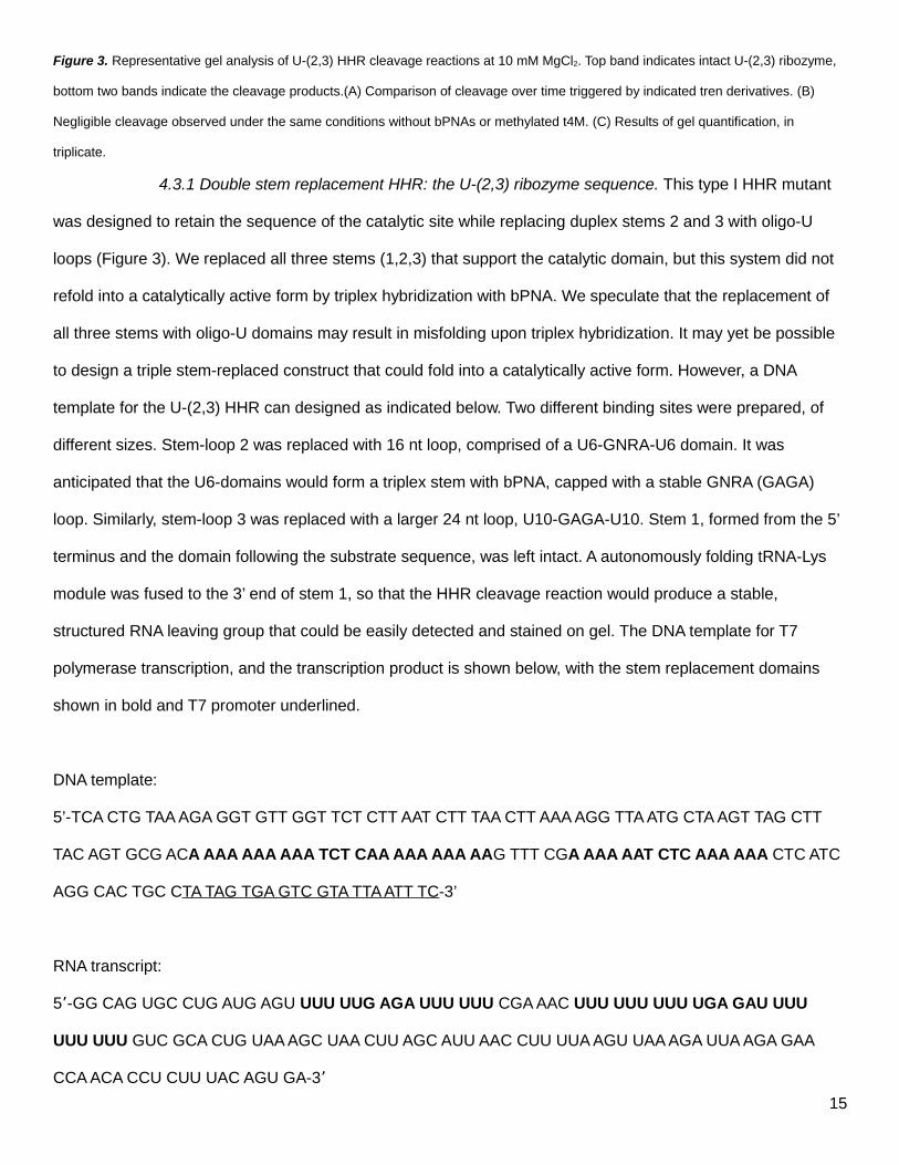
Figure 3. Representative gel analysis of U-(2,3) HHR cleavage reactions at 10 mM MgCl2. Top band indicates intact U-(2,3) ribozyme,
bottom two bands indicate the cleavage products.(A) Comparison of cleavage over time triggered by indicated tren derivatives. (B)
Negligible cleavage observed under the same conditions without bPNAs or methylated t4M. (C) Results of gel quantification, in
triplicate.
4.3.1 Double stem replacement HHR: the U-(2,3) ribozyme sequence. This type I HHR mutant
was designed to retain the sequence of the catalytic site while replacing duplex stems 2 and 3 with oligo-U
loops (Figure 3). We replaced all three stems (1,2,3) that support the catalytic domain, but this system did not
refold into a catalytically active form by triplex hybridization with bPNA. We speculate that the replacement of
all three stems with oligo-U domains may result in misfolding upon triplex hybridization. It may yet be possible
to design a triple stem-replaced construct that could fold into a catalytically active form. However, a DNA
template for the U-(2,3) HHR can designed as indicated below. Two different binding sites were prepared, of
different sizes. Stem-loop 2 was replaced with 16 nt loop, comprised of a U6-GNRA-U6 domain. It was
anticipated that the U6-domains would form a triplex stem with bPNA, capped with a stable GNRA (GAGA)
loop. Similarly, stem-loop 3 was replaced with a larger 24 nt loop, U10-GAGA-U10. Stem 1, formed from the 5’
terminus and the domain following the substrate sequence, was left intact. A autonomously folding tRNA-Lys
module was fused to the 3’ end of stem 1, so that the HHR cleavage reaction would produce a stable,
structured RNA leaving group that could be easily detected and stained on gel. The DNA template for T7
polymerase transcription, and the transcription product is shown below, with the stem replacement domains
shown in bold and T7 promoter underlined.
DNA template:
5’-TCA CTG TAA AGA GGT GTT GGT TCT CTT AAT CTT TAA CTT AAA AGG TTA ATG CTA AGT TAG CTT
TAC AGT GCG ACA AAA AAA AAA TCT CAA AAA AAA AAG TTT CGA AAA AAT CTC AAA AAA CTC ATC
AGG CAC TGC CTA TAG TGA GTC GTA TTA ATT TC-3’
RNA transcript:
5 -GG CAG UGC CUG AUG AGU ʼ UUU UUG AGA UUU UUU CGA AAC UUU UUU UUU UGA GAU UUU
UUU UUU GUC GCA CUG UAA AGC UAA CUU AGC AUU AAC CUU UUA AGU UAA AGA UUA AGA GAA
CCA ACA CCU CUU UAC AGU GA-3 ʼ
15
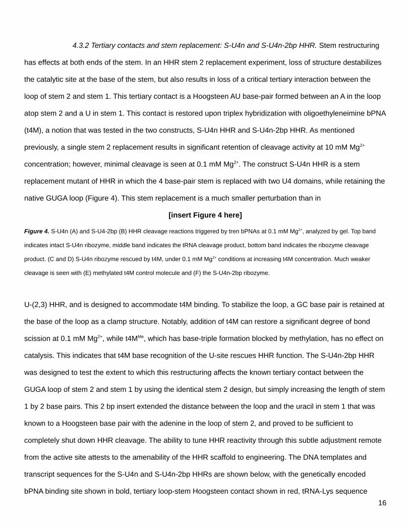
4.3.2 Tertiary contacts and stem replacement: S-U4n and S-U4n-2bp HHR. Stem restructuring
has effects at both ends of the stem. In an HHR stem 2 replacement experiment, loss of structure destabilizes
the catalytic site at the base of the stem, but also results in loss of a critical tertiary interaction between the
loop of stem 2 and stem 1. This tertiary contact is a Hoogsteen AU base-pair formed between an A in the loop
atop stem 2 and a U in stem 1. This contact is restored upon triplex hybridization with oligoethyleneimine bPNA
(t4M), a notion that was tested in the two constructs, S-U4n HHR and S-U4n-2bp HHR. As mentioned
previously, a single stem 2 replacement results in significant retention of cleavage activity at 10 mM Mg2+
concentration; however, minimal cleavage is seen at 0.1 mM Mg2+. The construct S-U4n HHR is a stem
replacement mutant of HHR in which the 4 base-pair stem is replaced with two U4 domains, while retaining the
native GUGA loop (Figure 4). This stem replacement is a much smaller perturbation than in
[insert Figure 4 here]
Figure 4. S-U4n (A) and S-U4-2bp (B) HHR cleavage reactions triggered by tren bPNAs at 0.1 mM Mg2+, analyzed by gel. Top band
indicates intact S-U4n ribozyme, middle band indicates the tRNA cleavage product, bottom band indicates the ribozyme cleavage
product. (C and D) S-U4n ribozyme rescued by t4M, under 0.1 mM Mg2+ conditions at increasing t4M concentration. Much weaker
cleavage is seen with (E) methylated t4M control molecule and (F) the S-U4n-2bp ribozyme.
U-(2,3) HHR, and is designed to accommodate t4M binding. To stabilize the loop, a GC base pair is retained at
the base of the loop as a clamp structure. Notably, addition of t4M can restore a significant degree of bond
scission at 0.1 mM Mg2+, while t4MMe, which has base-triple formation blocked by methylation, has no effect on
catalysis. This indicates that t4M base recognition of the U-site rescues HHR function. The S-U4n-2bp HHR
was designed to test the extent to which this restructuring affects the known tertiary contact between the
GUGA loop of stem 2 and stem 1 by using the identical stem 2 design, but simply increasing the length of stem
1 by 2 base pairs. This 2 bp insert extended the distance between the loop and the uracil in stem 1 that was
known to a Hoogsteen base pair with the adenine in the loop of stem 2, and proved to be sufficient to
completely shut down HHR cleavage. The ability to tune HHR reactivity through this subtle adjustment remote
from the active site attests to the amenability of the HHR scaffold to engineering. The DNA templates and
transcript sequences for the S-U4n and S-U4n-2bp HHRs are shown below, with the genetically encoded
bPNA binding site shown in bold, tertiary loop-stem Hoogsteen contact shown in red, tRNA-Lys sequence
16
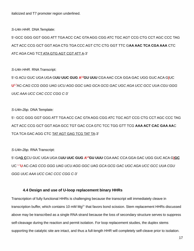
italicized and T7 promoter region underlined.
S-U4n HHR. DNA Template:
5’-GCC GGG GGT GGG ATT TGA ACC CAC GTA AGG CGG ATC TGC AGT CCG CTG CCT AGC CCC TAG
ACT ACC CCG GCT GGT AGA CTG TGA CCC AGT CTC CTG GGT TTC GAA AAC TCA CGA AAA CTC
ATC AGA CAG TCT ATA GTG AGT CGT ATT A-3’
S-U4n HHR. RNA Transcript:
5’-G ACU GUC UGA UGA GUU UUC GUG AL4GU UUU CGA AAC CCA GGA GAC UGG GUC ACA G|UC
U1.7AC-CAG CCG GGG UAG UCU AGG GGC UAG GCA GCG GAC UGC AGA UCC GCC UUA CGU GGG
UUC AAA UCC CAC CCC CGG C-3’
S-U4n-2bp. DNA Template:
5’- GCC GGG GGT GGG ATT TGA ACC CAC GTA AGG CGG ATC TGC AGT CCG CTG CCT AGC CCC TAG
ACT ACC CCG GCT GGT AGA GCC TGT GAC CCA GTC TCC TGG GTT TCG AAA ACT CAC GAA AAC
TCA TCA GAC AGG CTC TAT AGT GAG TCG TAT TA-3’
S-U4n-2bp. RNA Transcript:
5’-GAG CCU GUC UGA UGA GUU UUC GUG AL4GU UUU CGA AAC CCA GGA GAC UGG GUC ACA G|GC
UC 1.7U AC-CAG CCG GGG UAG UCU AGG GGC UAG GCA GCG GAC UGC AGA UCC GCC UUA CGU
GGG UUC AAA UCC CAC CCC CGG C-3’
4.4 Design and use of U-loop replacement binary HHRs
Transcription of fully functional HHRs is challenging because the transcript will immediately cleave in
transcription buffer, which contains 10 mM Mg2+ that favors bond scission. Stem replacement HHRs discussed
above may be transcribed as a single RNA strand because the loss of secondary structure serves to suppress
self-cleavage during the reaction and permit isolation. For loop replacement studies, the duplex stems
supporting the catalytic site are intact, and thus a full-length HHR will completely self-cleave prior to isolation.
17
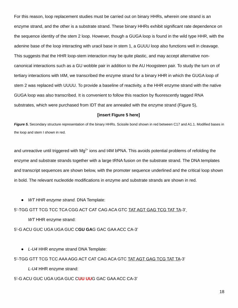
For this reason, loop replacement studies must be carried out on binary HHRs, wherein one strand is an
enzyme strand, and the other is a substrate strand. These binary HHRs exhibit significant rate dependence on
the sequence identity of the stem 2 loop. However, though a GUGA loop is found in the wild type HHR, with the
adenine base of the loop interacting with uracil base in stem 1, a GUUU loop also functions well in cleavage.
This suggests that the HHR loop-stem interaction may be quite plastic, and may accept alternative non-
canonical interactions such as a GU wobble pair in addition to the AU Hoogsteen pair. To study the turn on of
tertiary interactions with t4M, we transcribed the enzyme strand for a binary HHR in which the GUGA loop of
stem 2 was replaced with UUUU. To provide a baseline of reactivity, a the HHR enzyme strand with the native
GUGA loop was also transcribed. It is convenient to follow this reaction by fluorescently tagged RNA
substrates, which were purchased from IDT that are annealed with the enzyme strand (Figure 5),
[insert Figure 5 here]
Figure 5. Secondary structure representation of the binary HHRs. Scissile bond shown in red between C17 and A1.1. Modified bases in
the loop and stem I shown in red.
and unreactive until triggered with Mg2+ ions and t4M bPNA. This avoids potential problems of refolding the
enzyme and substrate strands together with a large tRNA fusion on the substrate strand. The DNA templates
and transcript sequences are shown below, with the promoter sequence underlined and the critical loop shown
in bold. The relevant nucleotide modifications in enzyme and substrate strands are shown in red.
WT HHR enzyme strand. DNA Template:
5’-TGG GTT TCG TCC TCA CGG ACT CAT CAG ACA GTC TAT AGT GAG TCG TAT TA-3’
WT HHR enzyme strand:
5’-G ACU GUC UGA UGA GUC CGU GAG GAC GAA ACC CA-3’
L-U4 HHR enzyme strand DNA Template:
5’-TGG GTT TCG TCC AAA AGG ACT CAT CAG ACA GTC TAT AGT GAG TCG TAT TA-3’
L-U4 HHR enzyme strand:
5’-G ACU GUC UGA UGA GUC CUU UUG GAC GAA ACC CA-3’
18
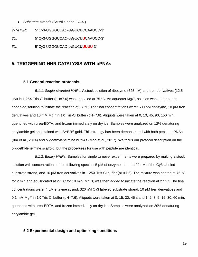
Substrate strands (Scissile bond: C--A.)
WT-HHR: 5’ Cy3-UGGGUCAC--AGUCUCCAAUCC-3’
2U: 5’ Cy3-UGGGUCAC--AGUCUUCAAUCC-3’
5U: 5’ Cy3-UGGGUCAC--AGUCUUUUU-3’
5. TRIGGERING HHR CATALYSIS WITH bPNAs
5.1 General reaction protocols.
5.1.1. Single-stranded HHRs. A stock solution of ribozyme (625 nM) and tren derivatives (12.5
µM) in 1.25X Tris-Cl buffer (pH=7.6) was annealed at 75 °C. An aqueous MgCl2 solution was added to the
annealed solution to initiate the reaction at 37 °C. The final concentrations were: 500 nM ribozyme, 10 µM tren
derivatives and 10 mM Mg2+ in 1X Tris-Cl buffer (pH=7.6). Aliquots were taken at 0, 10, 45, 90, 150 min,
quenched with urea-EDTA, and frozen immediately on dry ice. Samples were analyzed on 12% denaturing
acrylamide gel and stained with SYBR gold. This strategy has been demonstrated with both peptide bPNAs
(Xia et al., 2014) and oligoethyleneimine bPNAs (Mao et al., 2017). We focus our protocol description on the
oligoethyleneimine scaffold, but the procedures for use with peptide are identical.
5.1.2. Binary HHRs. Samples for single turnover experiments were prepared by making a stock
solution with concentrations of the following species: 5 µM of enzyme strand, 400 nM of the Cy3 labeled
substrate strand, and 10 µM tren derivatives in 1.25X Tris-Cl buffer (pH=7.6). The mixture was heated at 75 °C
for 2 min and equilibrated at 27 °C for 10 min. MgCl2 was then added to initiate the reaction at 27 °C. The final
concentrations were: 4 µM enzyme strand, 320 nM Cy3 labeled substrate strand, 10 µM tren derivatives and
0.1 mM Mg2+ in 1X Tris-Cl buffer (pH=7.6). Aliquots were taken at 0, 15, 30, 45 s and 1, 2, 3, 5, 15, 30, 60 min,
quenched with urea-EDTA, and frozen immediately on dry ice. Samples were analyzed on 20% denaturing
acrylamide gel.
5.2 Experimental design and optimizing conditions
19
5.2.1. Cleavage reactions of single-stranded HHRs. The single stranded HHR U-mutants
such as U-(2,3), S-U4n and S-U4n-2bp HHRs can be transcribed under normal conditions and do not self-
cleave appreciably, despite 10 mM MgCl2 in the reaction. However, their reactivity remains sensitive to Mg2+
concentration, and the combination of both t4M and Mg2+ is required to trigger RNA cleavage. The presence of
the tRNA fusions in these constructs yields two RNAs of comparable size upon cleavage, the HHR itself and
the tRNA. These two RNAs may be easily observed by gel (Figure 4). Addition of the methylated t4M molecule
generally does not cause HHR cleavage. This control is essential to rule out the effects of the oligoamine
backbone itself; molecules of this type can affect RNA function, but typically at much higher concentrations
(Pingoud et al., 1984; Quigley, Teeter, & Rich, 1978). Reactions should be run in triplicate and gels quantified
using standard software (Imagequant) to obtain HHR cleavage rates (Figure 4). Reactivity should be very
reproducible for each construct, resulting in relatively tight error bars. If the reaction rate does not allow
monitoring by reasonable data time point sampling, the rate may be tuned by decreasing or increasing
Mg2+concentration to decrease or increase reaction rate, respectively. Decreasing Mg2+ to 0.1 mM enabled
convenient monitoring of HHR cleavage in the sU4n HHR as a function of t4M concentration. Typically,
cleavage reactions were monitored at a single t4M concentration over time. In addition, rates were measured
as a function of increasing t4M concentrations. In this way, it can be clearly established that the t4M molecules
can directly influence HHR cleavage.
5.2.2. Cleavage reactions of binary HHRs. Binary HHRs were annealed together prior to use in
cleavage reactions as described above. Interestingly, it is possible to mix different substrate strands with each
HHR enzyme strand, yielding cleavage rates reflective of their dependence on t4M as well as substrate-
enzyme matching. While both loop modified L-GU3 and LU4 HHRs do not effectively cleave the native
substrate, L-GU3 can cleave a substrate strand that features U’s at both position 1.7 (native) and 1.8 in stem 1,
which suggests that the L1 guanine may be forming a tertiary contact with the U 1.8. The labeled substrate
strand provides a convenient way to quantitatively follow the cleavage reaction, signaling unreacted and
cleaved substrate with the same dye (Figure 6).
[insert Figure 6 here]
20
Figure 6. Representative gel image for binary ribozyme cleavage. Top band is the intact Cy3 labelled substrate strand RNA, bottom
band indicates the cleavage product. (Left) L-U4/5U; (right) L-U4/5U with 10 µM t4M, under 0.1 mM Mg2+ conditions.
6. Summary
The synthetic bPNAs described herein can bridge native and artificial architectures, uniting nucleic acid
biotechnology (Burnett & Rossi, 2012) with new materials (Roh, Ruiz, Peng, Lee, & Luo, 2011). This protocol
provides a general guide on the synthesis of bPNA peptides and oligoethyleneimine derivatives that have been
used to trigger cleavage in engineered hammerhead ribozymes. This strategy enables additional control over
RNA chemistry.
Acknowledgments
This work was supported in part by funding from the National Institutes of Health (GM111995-01A1 to D.B.), NASA
(GRT00044810 to D.B.) and NSF (DMR 1802432 to D.B).
21
References
Arambula, J. F., Ramisetty, S. R., Baranger, A. M., & Zimmerman, S. C. (2009). A simple ligand that selectively
targets CUG trinucleotide repeats and inhibits MBNL protein binding. Proceedings of the National
Academy of Sciences of the United States of America, 106, 16068–16073.
https://doi.org/ 10.1073/pnas.0901824106
Ariga, K., & Kunitake, T. (1998). Molecular Recognition at Air-Water and Related Interfaces: Complementary
Hydrogen Bonding and Multisite Interaction. Accounts of Chemical Research, 31, 371–378. Retrieved
from http://pubs3.acs.org/acs/journals/doilookup?in_doi=10.1021/ar970014i
Burnett, J. C., & Rossi, J. J. (2012). RNA-Based Therapeutics: Current Progress and Future Prospects.
Chemistry & Biology, 19, 60–71. https://doi.org/ 10.1016/j.chembiol.2011.12.008
Duca, M., Vekhoff, P., Oussedik, K., Halby, L., & Arimondo, P. B. (2008). The triple helix: 50 years later, the
outcome. Nucleic Acids Research, 36(16), 5123–5138. https://doi.org/ 10.1093/nar/gkn493
Felsenfeld, G., Davies, D. R., & Rich, A. (1957). Formation of a three-stranded polynucleotide molecule.
Journal of the American Chemical Society, 79, 2023–2024. https://doi.org/ 10.1021/ja01565a074
Kawasaki, T., Tokuhiro, M., Kimizuka, N., & Kunitake, T. (2001). Hierarchical self-assembly of chiral
complementary hydrogen-bond networks in water: reconstitution of supramolecular membranes. Journal
of the American Chemical Society, 123, 6792–6800. Retrieved from
http://www.ncbi.nlm.nih.gov/entrez/query.fcgi?
cmd=Retrieve&db=PubMed&dopt=Citation&list_uids=11448183
Kool, E. T. (2001). Hydrogen Bonding, Base Stacking, and Steric Effects in DNA Replication. Annual Review of
Biophysics and Biomolecular Structure, 30(1), 1–22. https://doi.org/ 10.1146/annurev.biophys.30.1.1
Lyon, S., & Gopalan, V. (2018). A T7 RNA Polymerase Mutant Enhances the Yield of 5’-Thienoguanosine-
Initiated RNAs. Chembiochem: A European Journal of Chemical Biology, 19(2), 142–146.
https://doi.org/ 10.1002/cbic.201700538
Ma, M., & Bong, D. (2011a). Determinants of cyanuric acid and melamine assembly in water. Langmuir: The
ACS Journal of Surfaces and Colloids, 27, 8841–8853. https://doi.org/ 10.1021/la201415d
Ma, M., & Bong, D. (2011b). Directed peptide assembly at the lipid-water interface cooperatively enhances
22

membrane binding and activity. Langmuir: The ACS Journal of Surfaces and Colloids, 27, 1480–1486.
https://doi.org/ 10.1021/la104405r
Ma, M., & Bong, D. (2011c). Protein assembly directed by synthetic molecular recognition motifs. Organic &
Biomolecular Chemistry, 9, 7296–7299. https://doi.org/ 10.1039/c1ob05998j
Ma, M., & Bong, D. (2013). Controlled Fusion of Synthetic Lipid Membrane Vesicles. Accounts of Chemical
Research, 46, 2988–2997.
Mao, J., & Bong, D. (2015). Synthesis of DNA-Binding Peptoids. Synlett: Accounts and Rapid Communications
in Synthetic Organic Chemistry, 26(11), 1581–1585. https://doi.org/ 10.1055/s-0034-1380698
Mao, J., DeSantis, C., & Bong, D. (2017). Small Molecule Recognition Triggers Secondary and Tertiary
Interactions in DNA Folding and Hammerhead Ribozyme Catalysis. Journal of the American Chemical
Society, 139(29), 9815–9818. https://doi.org/ 10.1021/jacs.7b05448
Martin, F. H., & Tinoco, I., Jr. (1980). DNA-RNA hybrid duplexes containing oligo(dA:rU) sequences are
exceptionally unstable and may facilitate termination of transcription. Nucleic Acids Research, 8(10),
2295–2299. Retrieved from https://www.ncbi.nlm.nih.gov/pubmed/6159577
Milligan, J. F., & Uhlenbeck, O. C. (1989). [5] Synthesis of small RNAs using T7 RNA polymerase. In Methods
in Enzymology (Vol. 180, pp. 51–62). Academic Press. https://doi.org/ 10.1016/0076-6879(89)80091-6
Mittapalli, G. K., Reddy, K. R., Xiong, H., Munoz, O., Han, B., De Riccardis, F., … Eschenmoser, A. (2007).
Mapping the landscape of potentially primordial informational oligomers: oligodipeptides and
oligodipeptoids tagged with triazines as recognition elements. Angewandte Chemie, International Edition,
46, 2470–2477. https://doi.org/ 10.1002/anie.200603207
Nielsen, P. E. (1999). Peptide Nucleic Acid. A Molecule with Two Identities. Accounts of Chemical Research,
32(7), 624–630. https://doi.org/ 10.1021/ar980010t
Piao, X., Xia, X., & Bong, D. (2013). Bifacial Peptide Nucleic Acid Directs Cooperative Folding and Assembly of
Binary, Ternary, and Quaternary DNA Complexes. Biochemistry, 52, 6313–6323.
https://doi.org/ 10.1021/bi4008963
Piao, X., Xia, X., Mao, J., & Bong, D. (2015). Peptide ligation and RNA cleavage via an abiotic template
interface. Journal of the American Chemical Society, 137(11), 3751–3754.
https://doi.org/ 10.1021/jacs.5b00236
23
Pilch, D. S., Levenson, C., & Shafer, R. H. (1990). Structural analysis of the (dA)10.2(dT)10 triple helix.
Proceedings of the National Academy of Sciences of the United States of America, 87, 1942–1946.
Retrieved from http://www.ncbi.nlm.nih.gov/entrez/query.fcgi?
cmd=Retrieve&db=PubMed&dopt=Citation&list_uids=2308955
Pingoud, A., Urbanke, C., Alves, J., Ehbrecht, H. J., Zabeau, M., & Gualerzi, C. (1984). Effect of polyamines
and basic proteins on cleavage of DNA by restriction endonucleases. Biochemistry, 23(24), 5697–5703.
Retrieved from https://www.ncbi.nlm.nih.gov/pubmed/6098296
Quigley, G. J., Teeter, M. M., & Rich, A. (1978). Structural analysis of spermine and magnesium ion binding to
yeast phenylalanine transfer RNA. Proceedings of the National Academy of Sciences of the United States
of America, 75(1), 64–68. Retrieved from https://www.ncbi.nlm.nih.gov/pubmed/343112
Ranganathan, A., Pedireddi, V. R., & Rao, C. N. R. (1999). Hydrothermal Synthesis of Organic Channel
Structures: 1:1 Hydrogen-Bonded Adducts of Melamine with Cyanuric and Trithiocyanuric Acids. Journal
of the American Chemical Society, 121, 1752–1753. Retrieved from
http://pubs3.acs.org/acs/journals/doilookup?in_doi=10.1021/ja983928o
Robertson, M. P., & Scott, W. G. (2007). The structural basis of ribozyme-catalyzed RNA assembly. Science,
315(5818), 1549–1553. https://doi.org/ 10.1126/science.1136231
Roh, Y. H., Ruiz, R. C. H., Peng, S., Lee, J. B., & Luo, D. (2011). Engineering DNA-based functional materials.
Chemical Society Reviews, 40(12), 5730–5744. https://doi.org/ 10.1039/c1cs15162b
Rougee, M., Faucon, B., Mergny, J. L., Barcelo, F., Giovannangeli, C., Garestier, T., & Hélène, C. (1992).
Kinetics and thermodynamics of triple-helix formation: effects of ionic strength and mismatches.
Biochemistry, 31, 9269–9278. Retrieved from http://www.ncbi.nlm.nih.gov/entrez/query.fcgi?
cmd=Retrieve&db=PubMed&dopt=Citation&list_uids=1390713
SantaLucia, J., Jr., & Hicks, D. (2004). The thermodynamics of DNA structural motifs. Annual Review of
Biophysics and Biomolecular Structure, 33, 415–440.
https://doi.org/ 10.1146/annurev.biophys.32.110601.141800
Soto, A. M., Loo, J., & Marky, L. A. (2002). Energetic contributions for the formation of TAT/TAT, TAT/CGC(+),
and CGC(+)/CGC(+) base triplet stacks. Journal of the American Chemical Society, 124, 14355–14363.
Retrieved from http://www.ncbi.nlm.nih.gov/entrez/query.fcgi?
24
cmd=Retrieve&db=PubMed&dopt=Citation&list_uids=12452709
ten Cate, M. G. J., Huskens, J., Crego-Calama, M., & Reinhoudt, D. N. (2004). Thermodynamic stability of
hydrogen-bonded nanostructures: a calorimetric study. Chemistry , 10(15), 3632–3639.
https://doi.org/ 10.1002/chem.200400085
Uhlenbeck, O. C. (1987). A small catalytic oligoribonucleotide. Nature, 328(6131), 596–600.
https://doi.org/ 10.1038/328596a0
von Hippel, P. H. (1998). An integrated model of the transcription complex in elongation, termination, and
editing. Science, 281(5377), 660–665. Retrieved from https://www.ncbi.nlm.nih.gov/pubmed/9685251
Whitesides, G. M., Simanek, E. E., Mathias, J. P., Seto, C. T., Chin, D., Mammen, M., & Gordon, D. M. (1995).
Noncovalent Synthesis: Using Physical-Organic Chemistry To Make Aggregates. Accounts of Chemical
Research, 28, 37–44. Retrieved from http://pubs3.acs.org/acs/journals/doilookup?
in_doi=10.1021/ar00049a006
Xia, X., Piao, X., & Bong, D. (2014). Bifacial peptide nucleic acid as an allosteric switch for aptamer and
ribozyme function. Journal of the American Chemical Society, 136(20), 7265–7268.
https://doi.org/ 10.1021/ja5032584
Xia, X., Piao, X., Fredrick, K., & Bong, D. (2013). Bifacial PNA complexation inhibits enzymatic access to DNA
and RNA. Chembiochem: A European Journal of Chemical Biology, 15, 31–36.
Xodo, L. E., Manzini, G., & Quadrifoglio, F. (1990). Spectroscopic and calorimetric investigation on the DNA
triplex formed by d(CTCTTCTTTCTTTTCTTTCTTCTC) and d(GAGAAGAAAGA) at acidic pH. Nucleic
Acids Research, 18, 3557–3564. https://doi.org/ 10.1093/nar/18.12.3557
Yakovchuk, P., Protozanova, E., & Frank-Kamenetskii, M. D. (2006). Base-stacking and base-pairing
contributions into thermal stability of the DNA double helix. Nucleic Acids Research, 34, 564–574.
https://doi.org/ 10.1093/nar/gkj454
Yarnell, W. S., & Roberts, J. W. (1999). Mechanism of intrinsic transcription termination and antitermination.
Science, 284(5414), 611–615. Retrieved from https://www.ncbi.nlm.nih.gov/pubmed/10213678
Zeng, Y., Pratumyot, Y., Piao, X., & Bong, D. (2012). Discrete assembly of synthetic peptide-DNA triplex
structures from polyvalent melamine-thymine bifacial recognition. Journal of the American Chemical
Society, 134(2), 832–835. https://doi.org/ 10.1021/ja2099326
25
Zhou, Z., & Bong, D. (2013). Small-Molecule/Polymer Recognition Triggers Aqueous-Phase Assembly and
Encapsulation. Langmuir: The ACS Journal of Surfaces and Colloids, 29, 144–150.
https://doi.org/ 10.1021/la304457y
Zhou, Z., Xia, X., & Bong, D. (2015). Synthetic Polymer Hybridization with DNA and RNA Directs Nanoparticle
Loading, Silencing Delivery, and Aptamer Function. Journal of the American Chemical Society, 137(28),
8920–8923. https://doi.org/ 10.1021/jacs.5b05481
26

download fileview on ChemRxivMethods Enz base triple (2).docx (37.77 KiB)
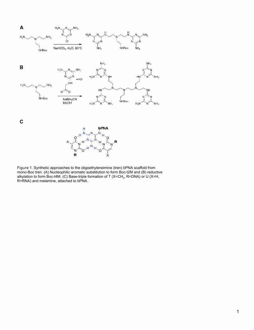
2/6/2019 Methods Enz figures - Google Docs
https://docs.google.com/document/d/1gyCiCUU4iCHpf4-feXlr7vcavhzuzJTgK01S0dsfXiA/edit# 1/6
C
Figure 1. Synthetic approaches to the oligoethyleneimine (tren) bPNA scaffold from monoBoc tren. (A) Nucleophilic aromatic substitution to form Boct2M and (B) reductive alkylation to form Boct4M. (C) Basetriple formation of T (X=CH 3 , R=DNA) or U (X=H, R=RNA) and melamine, attached to bPNA.
1
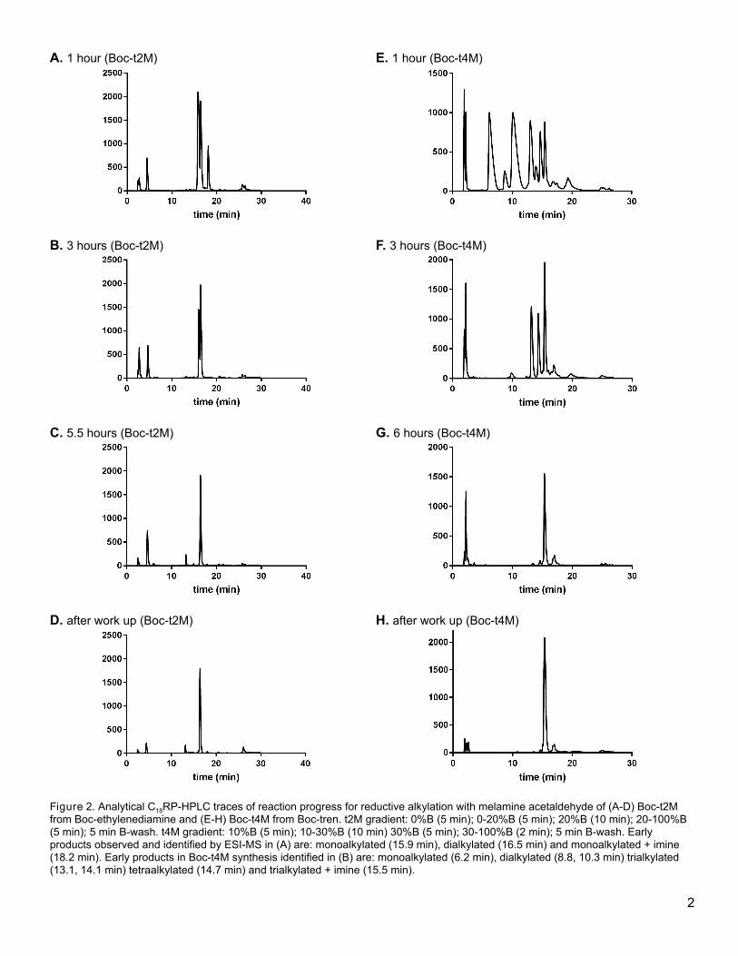
2/6/2019 Methods Enz figures - Google Docs
https://docs.google.com/document/d/1gyCiCUU4iCHpf4-feXlr7vcavhzuzJTgK01S0dsfXiA/edit# 2/6
A. 1 hour (Boct2M)
E. 1 hour (Boct4M)
B. 3 hours (Boct2M)
F. 3 hours (Boct4M)
C. 5.5 hours (Boct2M)
G. 6 hours (Boct4M)
D. after work up (Boct2M)
H. after work up (Boct4M)
Figure 2. Analytical C 18 RPHPLC traces of reaction progress for reductive alkylation with melamine acetaldehyde of (AD) Boct2M from Bocethylenediamine and (EH) Boct4M from Boctren. t2M gradient: 0%B (5 min); 020%B (5 min); 20%B (10 min); 20100%B (5 min); 5 min Bwash. t4M gradient: 10%B (5 min); 1030%B (10 min) 30%B (5 min); 30100%B (2 min); 5 min Bwash. Early products observed and identified by ESIMS in (A) are: monoalkylated (15.9 min), dialkylated (16.5 min) and monoalkylated + imine (18.2 min). Early products in Boct4M synthesis identified in (B) are: monoalkylated (6.2 min), dialkylated (8.8, 10.3 min) trialkylated (13.1, 14.1 min) tetraalkylated (14.7 min) and trialkylated + imine (15.5 min).
2
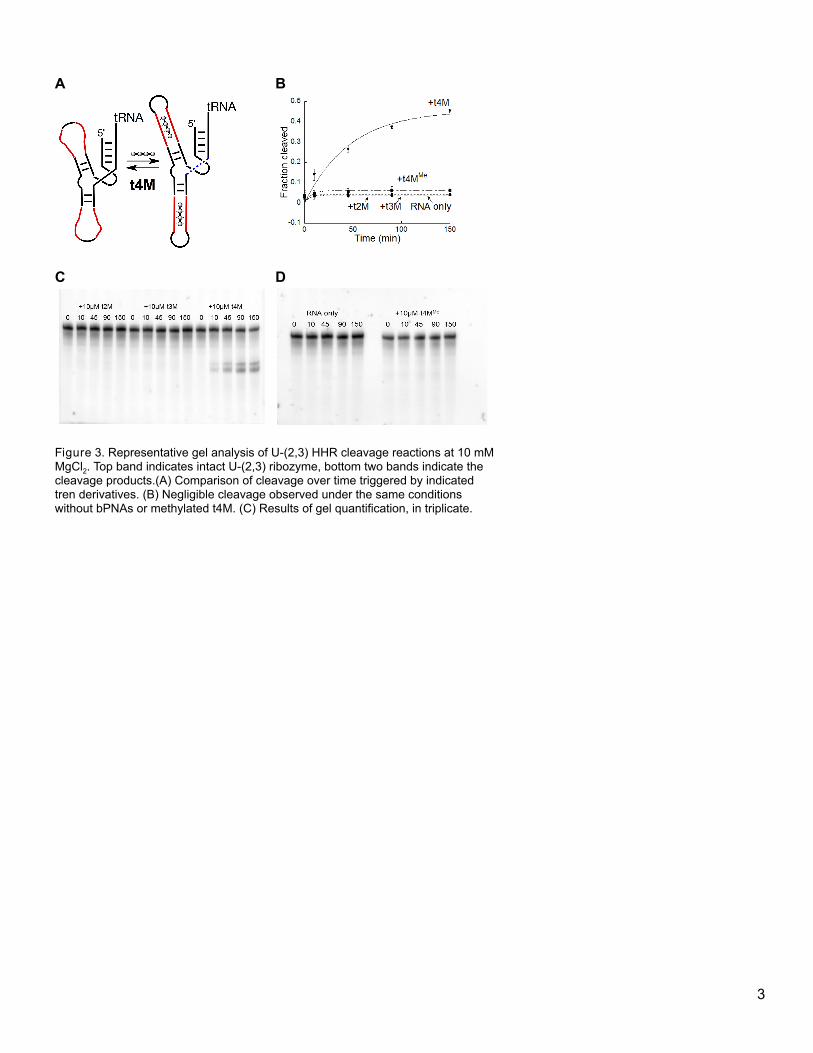
2/6/2019 Methods Enz figures - Google Docs
https://docs.google.com/document/d/1gyCiCUU4iCHpf4-feXlr7vcavhzuzJTgK01S0dsfXiA/edit# 3/6
A
B
C
D
Figure 3. Representative gel analysis of U(2,3) HHR cleavage reactions at 10 mM MgCl 2 . Top band indicates intact U(2,3) ribozyme, bottom two bands indicate the cleavage products.(A) Comparison of cleavage over time triggered by indicated tren derivatives. (B) Negligible cleavage observed under the same conditions without bPNAs or methylated t4M. (C) Results of gel quantification, in triplicate.
3
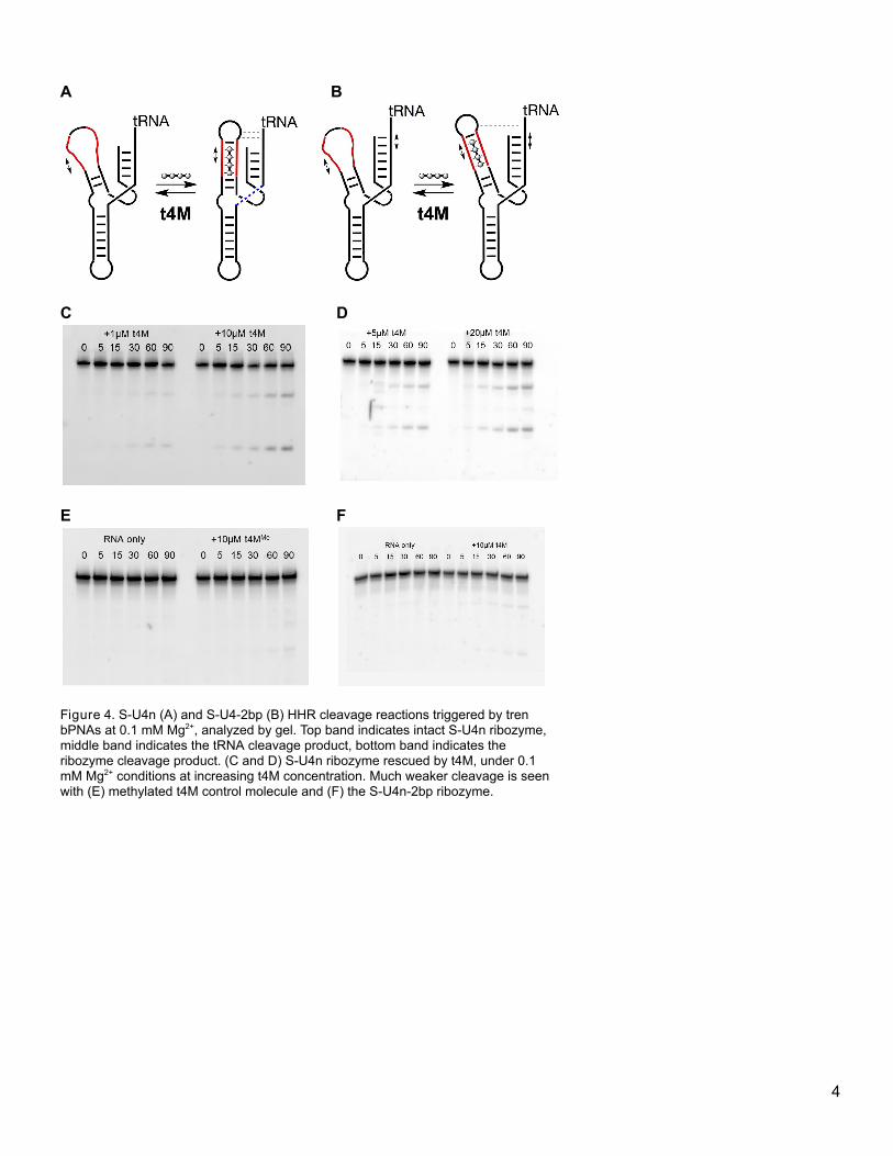
2/6/2019 Methods Enz figures - Google Docs
https://docs.google.com/document/d/1gyCiCUU4iCHpf4-feXlr7vcavhzuzJTgK01S0dsfXiA/edit# 4/6
A B
C
D
E
F
Figure 4. SU4n (A) and SU42bp (B) HHR cleavage reactions triggered by tren bPNAs at 0.1 mM Mg 2+ , analyzed by gel. Top band indicates intact SU4n ribozyme, middle band indicates the tRNA cleavage product, bottom band indicates the ribozyme cleavage product. (C and D) SU4n ribozyme rescued by t4M, under 0.1 mM Mg 2+ conditions at increasing t4M concentration. Much weaker cleavage is seen with (E) methylated t4M control molecule and (F) the SU4n2bp ribozyme.
4
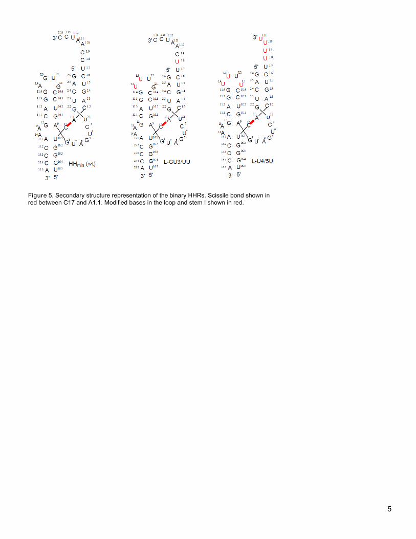
2/6/2019 Methods Enz figures - Google Docs
https://docs.google.com/document/d/1gyCiCUU4iCHpf4-feXlr7vcavhzuzJTgK01S0dsfXiA/edit# 5/6
Figure 5. Secondary structure representation of the binary HHRs. Scissile bond shown in red between C17 and A1.1. Modified bases in the loop and stem I shown in red.
5
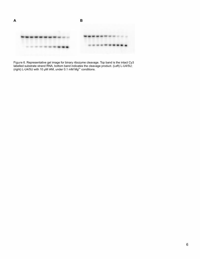
2/6/2019 Methods Enz figures - Google Docs
https://docs.google.com/document/d/1gyCiCUU4iCHpf4-feXlr7vcavhzuzJTgK01S0dsfXiA/edit# 6/6
A B
Figure 6. Representative gel image for binary ribozyme cleavage. Top band is the intact Cy3 labelled substrate strand RNA, bottom band indicates the cleavage product. (Left) LU4/5U; (right) LU4/5U with 10 µM t4M, under 0.1 mM Mg 2+ conditions.
6

download fileview on ChemRxivFigures.pdf (1.31 MiB)