Embed Size (px)
Citation preview
Hindawi Publishing CorporationResearch Letters in NanotechnologyVolume 2008, Article ID 389512, 4 pagesdoi:10.1155/2008/389512
Research LetterSynthesis of PET-PLA/Drug Nanoparticles andTheir Effect with Gold Nanoparticles for ControlledDrug Release in Cancer Chemotherapy
K. Sathish Kumar,1 V. Selvaraj,2 and M. Alagar2
1 Department of Chemical Engineering, SSN College of Engineering, Kalavakkam 603110, India2 Department of Chemical Engineering, Alagappa College of Technology, Anna University, Chennai 600025, India
Correspondence should be addressed to M. Alagar, [email protected]
Received 31 December 2007; Accepted 4 April 2008
Recommended by Lyudmila Bronstein
Polyethylene terephthalate-polylactic acid copolymer (PET-PLA) was synthesized from bis (2-hydroxyethyl terephthalate) and L-lactic acid oligomer in the presence of manganese antimony glycoxide as a catalyst. The synthesized PET-PLA copolymer was usedfor controlled drug release systems with gold nanoparticles. Fluorouracil containing PET-PLA nanocapsules was prepared in thepresence of gold nanoparticles by solvent evaporation method. The morphologies of the nanocapsules were characterized usingscanning electron microscopy and transmission electron microscopy. Controlled release of Fu and Fu@Au was carried out in 0.1 Mphosphate buffer (pH 7.4) and 0.1 M HCl solution. The results indicated that the drug release for gold nanoparticles/fluorouracil(Au@Fu) incorporated PET-PLA nanocapsules was controlled and slow compared to Fu incorporated PET-PLA nanocapsules.This may be due to the interaction between the gold nanoparticles and fluorouracil in PET-PLA nanocapsules.
Copyright © 2008 K. Sathish Kumar et al. This is an open access article distributed under the Creative Commons AttributionLicense, which permits unrestricted use, distribution, and reproduction in any medium, provided the original work is properlycited.
1. INTRODUCTION
Nanotechnology provides a novel route for many biomedicalapplications especially in the case of incurable diseases suchas cancer, diabetes, and so forth. Cancer can affect just everyorgan in human body. The various treatments of cancer arechemotherapy, radiation therapy, surgery, biological therapy,hormone, and gene therapies. Chemotherapy uses chemicalagents (anticancer or cytotoxic drugs) to interact withcancer cells to eradicate or control the growth of cancer.Depending on the type of cancer and kind of drug used,chemotherapy drugs may be administered differently. 5-Fluorouracil (5-Fu) is one of the oldest chemotherapy drugsand has been used for decades. It is an active medicineagainst many cancers. Over the past 20 years, increasedunderstanding of the mechanism of action of 5-Fu has ledto the development of strategies that increase its anticanceractivity. 5-Fu is given for treatment of cancers like bowel,breast, stomach, and gullet cancer [1]. However, anticancerdrugs normally attack both normal cells and cancerous cellswhen the drug was given as an injection or tablet formfor a long time. In order to over come this side effect,
targeting the drug delivery and sustained release of drugsare required. Many research investigations are focused in thepreparation of drug encapsulated polymer nanoparticles forthe controlled release applications. Biodegradable polymershave become increasingly important in the developmentof drug delivery systems [2–5]. There are several meth-ods that can be used to make microcapsules [6] frombiodegradable polymers. Polyethylene terephthalate (PET)is a semicrystalline polymer with high mechanical strengthand an excellent thermal stability. Copolyesters such as PET-PLA are biodegradable materials used for tissue engineering,bone reconstruction, and controlled drug delivery systems[7–9]. Further gold nanoparticles play an important rolein cancer therapy to detect or to deliver the drug to thecancerous cell without affecting the normal cells and havegood ability to form complex with many drugs throughchemical bonding. Nanoparticles have uniform shape andsize and are soluble in an aqueous medium. With this viewin mind, the present work is undertaken to synthesize PET-PLA copolyester and the preparation of PET-PLA/Fu andPET-PLA/Fu-Au nanocapsules to study their drug releasebehavior. A comparative study of sustained release of 5-Fu
2 Research Letters in Nanotechnology
2
1.8
1.6
1.4
1.2
1
0.8
0.6
0.4
0.2
0200 300 400 500 600 700 800 900
Abs
orba
nce
(a.u
.)
Wavelength (nm)
(a)
2
1.5
1
0.5
0200 300 400 500 600 700 800 900
Abs
orba
nce
(a.u
.)
Wavelength (nm)
(b)
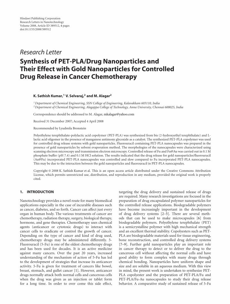
Figure 1: (a) and (b) show the UV-spectrum of Au nanoparticles and Fu@Au nanoparticles.
under different pH conditions was also carried out. Thepresent work is the continuation of our previous work withgold nanoparticles [10, 11].
2. EXPERIMENTAL
2.1. Synthesis of PET-PLA copolyester
Bis (2-hydroxy ethyl terephthalate) and L-lactic acidoligomers (30/70) wt% were reacted in the presence of 10 mgof manganese antimony glycoxide catalyst [12]. The reactionmixture was placed [13] in a 500 mL flask connected to avacuum line (0.4 kPa) and immersed in an oil bath at 180◦Cfor 6 hours. As the reactants were stirred, glycol and waterwere slowly distilled out. The copolyesters were dissolved inchloroform, precipitated in methanol, filtered and dried at70◦C. This was used without further purification.
2.2. Preparation of citrate-capped gold nanoparticles
Trisodium citrate (38.8 mM, 50 cm3) was added to a boilingHAuCl4 solution (1 mM, 500 cm3). As a result, the previouslyyellow solution of gold chloride turned to wine red colorand gave a characteristic absorbance at 518 nm in the UV-visspectrum.
2.3. Preparation of nanocapsules
Nanocapsules were prepared by the solvent evaporationmethod similar to that reported previously by Beck et al.[14]. For PET/PLA/5Fu-Au experiment, 5-Fluorouracil(50 mg) was added in 0.5 mL nanogold aqueous suspension(with a 30-minute shaking under ultrasound to help theadsorption of 5-Fluorouracil on gold nanoparticles) andthis solution was added to an organic polymer solution(300 mg PET-PLA + 5 mL CH2Cl2) under stirring condition.This was continued until the organic solvent was com-pletely evaporated. The suspension became clear after allthe nanocapsules precipitated out of the solution. Thesenanocapsules were collected by filtration and washed withdeionized water to remove any undesirable residuals. Finally,
the clean nanocapsules were dried in a vacuum oven at 40◦Cfor 24 hours to ensure a complete removal of the organicsolvent and deionized water. All the nanocapsules werestored in a desiccator at 25◦C. PET-PLA/Fu nanocapsuleswere also prepared under similar conditions.
Encapsulation efficiency (%) = [(Mass of drug addedduring Nanoparticle (NP) preparation − Mass of free drugin supernatant)/Mass of Drug added during NP preparation]× 100.
Drug loading (%) = [Mass of 5-Fluouracil in NP/Mass ofNP recovered] × 100.
3. RESULTS AND DISCUSSION
3.1. UV-vis characterization
Figure 1 shows the UV-visible spectrum of citrate stabilizedgold nanoparticles. The plasmon band observed for the winered colloidal gold at 518 nm is characteristic of the goldnanoparticles. The pure drug shows a maximum at 273 nm,but with the addition of 5-Fu to colloidal gold, both thebands at 273 and 518 nm pertaining to pure drug and Aucolloids decrease in intensity steadily with time.
This decrease is accompanied by the emergence of anadditional peak at 650 nm (Figure 1(b)), that is, a changefrom wine red to blue with the addition of drug tocolloidal gold. The appearance of the new peak is due tothe aggregation of gold nanoparticles and the replacementof citrate by 5-Fu, leading to the formation of gold-drugcomplex. Citrate ions are readily replaced by the –NH ligandon the surface of gold nanoparticles. This ligand exchangereaction provides an important means for the chemicalfunctionalization of the nanoparticles and greatly extends theversatility of these systems.
3.2. Morphological characterization
PET-PLA/5-Fu-Au and PET-PLA/5-Fu nanocapsules wereeasily distinguished from PET-PLA/5-Fu nanocapsules bytheir color. While the color of PET-PLA/5-Fu nanocapsules
K. Sathish Kumar et al. 3
0.1 μm
(a)
0.2 μm
(b)
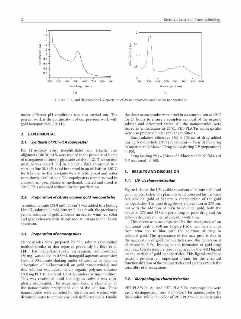
Figure 2: The TEM images of (a) Au@Fu, and (b) Au-Fu/PET-PLAnanoparticles.
800 nm
(a)
800 nm
(b)
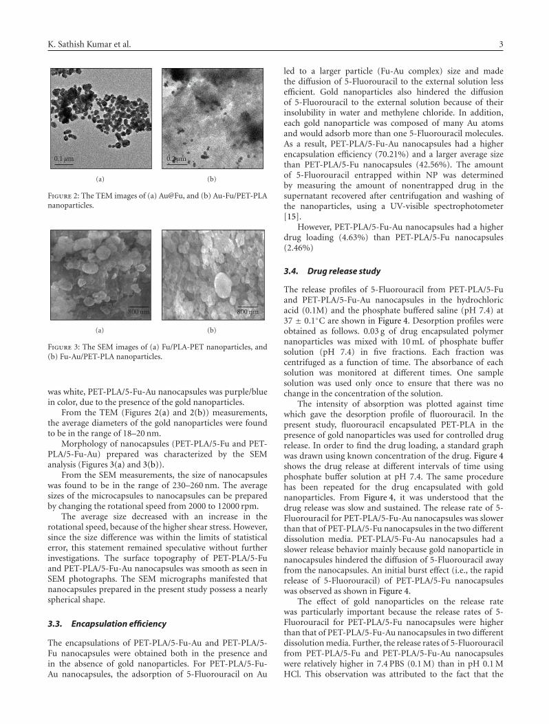
Figure 3: The SEM images of (a) Fu/PLA-PET nanoparticles, and(b) Fu-Au/PET-PLA nanoparticles.
was white, PET-PLA/5-Fu-Au nanocapsules was purple/bluein color, due to the presence of the gold nanoparticles.
From the TEM (Figures 2(a) and 2(b)) measurements,the average diameters of the gold nanoparticles were foundto be in the range of 18–20 nm.
Morphology of nanocapsules (PET-PLA/5-Fu and PET-PLA/5-Fu-Au) prepared was characterized by the SEManalysis (Figures 3(a) and 3(b)).
From the SEM measurements, the size of nanocapsuleswas found to be in the range of 230–260 nm. The averagesizes of the microcapsules to nanocapsules can be preparedby changing the rotational speed from 2000 to 12000 rpm.
The average size decreased with an increase in therotational speed, because of the higher shear stress. However,since the size difference was within the limits of statisticalerror, this statement remained speculative without furtherinvestigations. The surface topography of PET-PLA/5-Fuand PET-PLA/5-Fu-Au nanocapsules was smooth as seen inSEM photographs. The SEM micrographs manifested thatnanocapsules prepared in the present study possess a nearlyspherical shape.
3.3. Encapsulation efficiency
The encapsulations of PET-PLA/5-Fu-Au and PET-PLA/5-Fu nanocapsules were obtained both in the presence andin the absence of gold nanoparticles. For PET-PLA/5-Fu-Au nanocapsules, the adsorption of 5-Fluorouracil on Au
led to a larger particle (Fu-Au complex) size and madethe diffusion of 5-Fluorouracil to the external solution lessefficient. Gold nanoparticles also hindered the diffusionof 5-Fluorouracil to the external solution because of theirinsolubility in water and methylene chloride. In addition,each gold nanoparticle was composed of many Au atomsand would adsorb more than one 5-Fluorouracil molecules.As a result, PET-PLA/5-Fu-Au nanocapsules had a higherencapsulation efficiency (70.21%) and a larger average sizethan PET-PLA/5-Fu nanocapsules (42.56%). The amountof 5-Fluorouracil entrapped within NP was determinedby measuring the amount of nonentrapped drug in thesupernatant recovered after centrifugation and washing ofthe nanoparticles, using a UV-visible spectrophotometer[15].
However, PET-PLA/5-Fu-Au nanocapsules had a higherdrug loading (4.63%) than PET-PLA/5-Fu nanocapsules(2.46%)
3.4. Drug release study
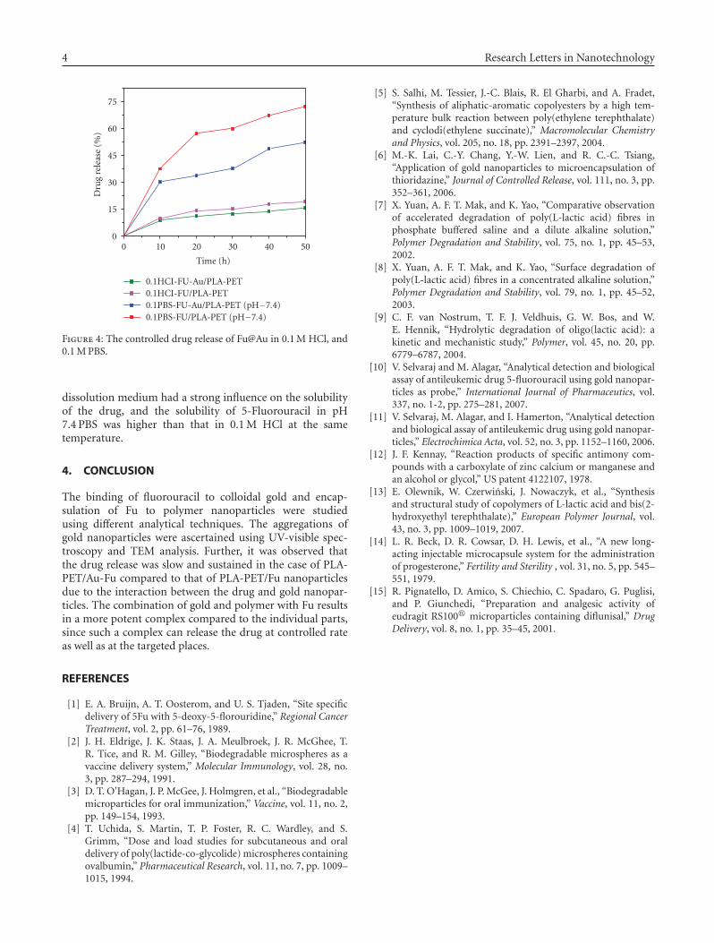
The release profiles of 5-Fluorouracil from PET-PLA/5-Fuand PET-PLA/5-Fu-Au nanocapsules in the hydrochloricacid (0.1M) and the phosphate buffered saline (pH 7.4) at37 ± 0.1◦C are shown in Figure 4. Desorption profiles wereobtained as follows. 0.03 g of drug encapsulated polymernanoparticles was mixed with 10 mL of phosphate buffersolution (pH 7.4) in five fractions. Each fraction wascentrifuged as a function of time. The absorbance of eachsolution was monitored at different times. One samplesolution was used only once to ensure that there was nochange in the concentration of the solution.
The intensity of absorption was plotted against timewhich gave the desorption profile of fluorouracil. In thepresent study, fluorouracil encapsulated PET-PLA in thepresence of gold nanoparticles was used for controlled drugrelease. In order to find the drug loading, a standard graphwas drawn using known concentration of the drug. Figure 4shows the drug release at different intervals of time usingphosphate buffer solution at pH 7.4. The same procedurehas been repeated for the drug encapsulated with goldnanoparticles. From Figure 4, it was understood that thedrug release was slow and sustained. The release rate of 5-Fluorouracil for PET-PLA/5-Fu-Au nanocapsules was slowerthan that of PET-PLA/5-Fu nanocapsules in the two differentdissolution media. PET-PLA/5-Fu-Au nanocapsules had aslower release behavior mainly because gold nanoparticle innanocapsules hindered the diffusion of 5-Fluorouracil awayfrom the nanocapsules. An initial burst effect (i.e., the rapidrelease of 5-Fluorouracil) of PET-PLA/5-Fu nanocapsuleswas observed as shown in Figure 4.
The effect of gold nanoparticles on the release ratewas particularly important because the release rates of 5-Fluorouracil for PET-PLA/5-Fu nanocapsules were higherthan that of PET-PLA/5-Fu-Au nanocapsules in two differentdissolution media. Further, the release rates of 5-Fluorouracilfrom PET-PLA/5-Fu and PET-PLA/5-Fu-Au nanocapsuleswere relatively higher in 7.4 PBS (0.1 M) than in pH 0.1 MHCl. This observation was attributed to the fact that the
4 Research Letters in Nanotechnology
75
60
45
30
15
0
0.1HCI-FU-Au/PLA-PET0.1HCI-FU/PLA-PET0.1PBS-FU-Au/PLA-PET (pH−7.4)0.1PBS-FU/PLA-PET (pH−7.4)
0 10 20 30 40 50
Dru
gre
leas
e(%
)
Time (h)
Figure 4: The controlled drug release of Fu@Au in 0.1 M HCl, and0.1 M PBS.
dissolution medium had a strong influence on the solubilityof the drug, and the solubility of 5-Fluorouracil in pH7.4 PBS was higher than that in 0.1 M HCl at the sametemperature.
4. CONCLUSION
The binding of fluorouracil to colloidal gold and encap-sulation of Fu to polymer nanoparticles were studiedusing different analytical techniques. The aggregations ofgold nanoparticles were ascertained using UV-visible spec-troscopy and TEM analysis. Further, it was observed thatthe drug release was slow and sustained in the case of PLA-PET/Au-Fu compared to that of PLA-PET/Fu nanoparticlesdue to the interaction between the drug and gold nanopar-ticles. The combination of gold and polymer with Fu resultsin a more potent complex compared to the individual parts,since such a complex can release the drug at controlled rateas well as at the targeted places.
REFERENCES
[1] E. A. Bruijn, A. T. Oosterom, and U. S. Tjaden, “Site specificdelivery of 5Fu with 5-deoxy-5-florouridine,” Regional CancerTreatment, vol. 2, pp. 61–76, 1989.
[2] J. H. Eldrige, J. K. Staas, J. A. Meulbroek, J. R. McGhee, T.R. Tice, and R. M. Gilley, “Biodegradable microspheres as avaccine delivery system,” Molecular Immunology, vol. 28, no.3, pp. 287–294, 1991.
[3] D. T. O’Hagan, J. P. McGee, J. Holmgren, et al., “Biodegradablemicroparticles for oral immunization,” Vaccine, vol. 11, no. 2,pp. 149–154, 1993.
[4] T. Uchida, S. Martin, T. P. Foster, R. C. Wardley, and S.Grimm, “Dose and load studies for subcutaneous and oraldelivery of poly(lactide-co-glycolide) microspheres containingovalbumin,” Pharmaceutical Research, vol. 11, no. 7, pp. 1009–1015, 1994.
[5] S. Salhi, M. Tessier, J.-C. Blais, R. El Gharbi, and A. Fradet,“Synthesis of aliphatic-aromatic copolyesters by a high tem-perature bulk reaction between poly(ethylene terephthalate)and cyclodi(ethylene succinate),” Macromolecular Chemistryand Physics, vol. 205, no. 18, pp. 2391–2397, 2004.
[6] M.-K. Lai, C.-Y. Chang, Y.-W. Lien, and R. C.-C. Tsiang,“Application of gold nanoparticles to microencapsulation ofthioridazine,” Journal of Controlled Release, vol. 111, no. 3, pp.352–361, 2006.
[7] X. Yuan, A. F. T. Mak, and K. Yao, “Comparative observationof accelerated degradation of poly(L-lactic acid) fibres inphosphate buffered saline and a dilute alkaline solution,”Polymer Degradation and Stability, vol. 75, no. 1, pp. 45–53,2002.
[8] X. Yuan, A. F. T. Mak, and K. Yao, “Surface degradation ofpoly(L-lactic acid) fibres in a concentrated alkaline solution,”Polymer Degradation and Stability, vol. 79, no. 1, pp. 45–52,2003.
[9] C. F. van Nostrum, T. F. J. Veldhuis, G. W. Bos, and W.E. Hennik, “Hydrolytic degradation of oligo(lactic acid): akinetic and mechanistic study,” Polymer, vol. 45, no. 20, pp.6779–6787, 2004.
[10] V. Selvaraj and M. Alagar, “Analytical detection and biologicalassay of antileukemic drug 5-fluorouracil using gold nanopar-ticles as probe,” International Journal of Pharmaceutics, vol.337, no. 1-2, pp. 275–281, 2007.
[11] V. Selvaraj, M. Alagar, and I. Hamerton, “Analytical detectionand biological assay of antileukemic drug using gold nanopar-ticles,” Electrochimica Acta, vol. 52, no. 3, pp. 1152–1160, 2006.
[12] J. F. Kennay, “Reaction products of specific antimony com-pounds with a carboxylate of zinc calcium or manganese andan alcohol or glycol,” US patent 4122107, 1978.
[13] E. Olewnik, W. Czerwinski, J. Nowaczyk, et al., “Synthesisand structural study of copolymers of L-lactic acid and bis(2-hydroxyethyl terephthalate),” European Polymer Journal, vol.43, no. 3, pp. 1009–1019, 2007.
[14] L. R. Beck, D. R. Cowsar, D. H. Lewis, et al., “A new long-acting injectable microcapsule system for the administrationof progesterone,” Fertility and Sterility , vol. 31, no. 5, pp. 545–551, 1979.
[15] R. Pignatello, D. Amico, S. Chiechio, C. Spadaro, G. Puglisi,and P. Giunchedi, “Preparation and analgesic activity ofeudragit RS100� microparticles containing diflunisal,” DrugDelivery, vol. 8, no. 1, pp. 35–45, 2001.

Submit your manuscripts athttp://www.hindawi.com
ScientificaHindawi Publishing Corporationhttp://www.hindawi.com Volume 2014
CorrosionInternational Journal of
Hindawi Publishing Corporationhttp://www.hindawi.com Volume 2014
Polymer ScienceInternational Journal of
Hindawi Publishing Corporationhttp://www.hindawi.com Volume 2014
Hindawi Publishing Corporationhttp://www.hindawi.com Volume 2014
CeramicsJournal of
Hindawi Publishing Corporationhttp://www.hindawi.com Volume 2014
CompositesJournal of
NanoparticlesJournal of
Hindawi Publishing Corporationhttp://www.hindawi.com Volume 2014
Hindawi Publishing Corporationhttp://www.hindawi.com Volume 2014
International Journal of
Biomaterials
Hindawi Publishing Corporationhttp://www.hindawi.com Volume 2014
NanoscienceJournal of
TextilesHindawi Publishing Corporation http://www.hindawi.com Volume 2014
Journal of
NanotechnologyHindawi Publishing Corporationhttp://www.hindawi.com Volume 2014
Journal of
CrystallographyJournal of
Hindawi Publishing Corporationhttp://www.hindawi.com Volume 2014
The Scientific World JournalHindawi Publishing Corporation http://www.hindawi.com Volume 2014
Hindawi Publishing Corporationhttp://www.hindawi.com Volume 2014
CoatingsJournal of
Advances in
Materials Science and EngineeringHindawi Publishing Corporationhttp://www.hindawi.com Volume 2014
Smart Materials Research
Hindawi Publishing Corporationhttp://www.hindawi.com Volume 2014
Hindawi Publishing Corporationhttp://www.hindawi.com Volume 2014
MetallurgyJournal of
Hindawi Publishing Corporationhttp://www.hindawi.com Volume 2014
BioMed Research International
MaterialsJournal of
Hindawi Publishing Corporationhttp://www.hindawi.com Volume 2014
Nano
materials
Hindawi Publishing Corporationhttp://www.hindawi.com Volume 2014
Journal ofNanomaterials


![s 1Lanter C!&ffsbore, ]Jnt](https://img.pdfslide.us/doc/110x75/61ee5f460ac9e13f1e2fa5da/s-1lanter-campffsbore-jnt.jpg)