Embed Size (px)
Citation preview

1
Synthesis, preclinical evaluation, and a pilot clinical PET imaging
study of 68Ga-labeled FAPI dimer
Authors: Liang Zhao1,2*, Bo Niu3*, Jianyang Fang4*, Yizhen Pang1, Siyang Li3, Chengrong
Xie5, Long Sun1, Xianzhong Zhang4, Zhide Guo4†, Qin Lin2†, and Haojun Chen1†
1 Department of Nuclear Medicine & Minnan PET Center, The First Affiliated Hospital of
Xiamen University, Xiamen, China
2 Department of Radiation Oncology, The First Affiliated Hospital of Xiamen University,
Xiamen, China;
3 School of medicine, Xiamen University, Xiamen, China;
4 State Key Laboratory of Molecular Vaccinology and Molecular Diagnostics & Center for
Molecular Imaging and Translational Medicine, School of Public Health, Xiamen University,
Xiamen, China;
5 Fujian Provincial Key Laboratory of Chronic Liver Disease and Hepatocellular Carcinoma,
Xiamen, China;
* First authors, contributed equally to this work
Liang Zhao, Email: [email protected]
Bo Niu, Email: [email protected]
Jianyang Fang, Email: [email protected]
Journal of Nuclear Medicine, published on September 23, 2021 as doi:10.2967/jnumed.121.263016

2
† Corresponding authors:
Zhide Guo, PhD
School of Public Health, Xiamen University, China
Email: [email protected]
Qin Lin, MD, PhD
Department of Radiation Oncology, The First Affiliated Hospital of Xiamen University, Xiamen,
China
Email: [email protected]
Haojun Chen, MD, PhD
Department of Nuclear Medicine & Minnan PET Center, The First Affiliated Hospital of
Xiamen University, Xiamen, China
Email: [email protected]
Tel: +86-592-2137077
ORCID ID: 0000-0002-9101-8884
Funding: This work was funded by the National Natural Science Foundation of China (Grant
number 82071961, 81901805, and 81772893) and the key medical and health projects in
Xiamen (Grant number 3502Z20191104).
Running title: Pilot study of 68Ga-labeled FAPI dimer
Immediate Open Access: Creative Commons Attribution 4.0 International License (CC BY)
allows users to share and adapt with attribution, excluding materials credited to previous
publications.
License: https://creativecommons.org/licenses/by/4.0/.
Details: https://jnm.snmjournals.org/page/permissions.

3
ABSTRACT
Cancer-associated fibroblasts (CAFs) are crucial components of the tumor
microenvironment. Fibroblast activation protein (FAP) is overexpressed in CAFs.
FAP-targeted molecular imaging agents, including FAP inhibitor (FAPI)-04 and
FAPI-46, have shown promising results in tumor diagnosis. However, these
molecules have relatively short tumor-retention time for peptide-targeted
radionuclide therapy applications. We aimed to design a 68Ga-labeled FAPI dimer
(denoted as 68Ga-DOTA-2P(FAPI)2) to optimize the pharmacokinetics and
evaluate whether this form is more effective than its monomeric analogs.
Methods: 68Ga-DOTA-2P(FAPI)2 was synthesized based on the quinoline-based
FAPI variants (FAPI-46), and its binding properties were assayed in CAFs.
Preclinical pharmacokinetics was determined in FAP-positive patient-derived
xenografts (PDXs) using small-animal PET and biodistribution experiments. The
effective dosimetry of 68Ga-DOTA-2P(FAPI)2 was evaluated in three healthy
volunteers, and PET/ CT imaging of 68Ga-FAPI-46 and 68Ga-DOTA-2P(FAPI)2
was performed in three cancer patients. Results: 68Ga-DOTA-2P(FAPI)2 was
stable in phosphate-buffered saline and fetal bovine serum for 4 h. The FAPI
dimer showed high affinity and specificity for FAP in-vitro and in-vivo. The tumor
uptake of 68Ga-DOTA-2P(FAPI)2 was approximately two-fold stronger than that of

4
68Ga-FAPI-46 in PDXs, while the healthy organs showed low tracer uptake and
fast body clearance. The effective dose of 68Ga-DOTA-2P(FAPI)2 was 1.19E-02
mSv/MBq, calculated using OLINDA. Finally, PET/CT scans in three cancer
patients revealed higher intratumoral uptake of 68Ga-DOTA-2P(FAPI)2 than that of
68Ga-FAPI-46 in all tumor lesions (maximum standardized uptake value: 8.1-39.0
vs. 1.7-24.0, respectively; P < 0.001). Conclusion: 68Ga-DOTA-2P(FAPI)2 has
increased tumor uptake and retention properties compared to 68Ga-FAPI-46, and
it could be a promising tracer for both diagnostic imaging and targeted therapy of
malignant tumors with positive expression of FAP.
Keywords: fibroblast activation protein; cancer-associated fibroblasts; FAPI
dimer; patient-derived xenografts; PET imaging

5
INTRODUCTION
Cancer-associated fibroblasts (CAFs) are crucial components of the tumor
microenvironment and can constitute over half of the mass in various tumors.
According to previous reports, CAFs play important roles in tumor growth,
immune suppression, and cancer invasion (1,2). Thus, CAFs may be promising
targets for tumor diagnosis and therapy. Fibroblast activation protein (FAP) is
overexpressed in the CAFs in numerous epithelial carcinomas and weakly
expressed in healthy tissues; therefore, an attractive target for cancer research.
The past few years have witnessed the expansion of research on FAP-targeted
molecular imaging in tumor diagnosis (3-5).
Recently, there has been a growing application of FAP targeting, from
diagnostic imaging to peptide-targeted radionuclide therapy (PTRT) (6-10).
However, the reports on PTRT are mainly based on the FAP-inhibitor (FAPI)-04
and FAPI-46 peptides, which showed a relatively short tumor-retention time in
preclinical models and human subjects (10-12). Another FAPI variant, FAP-2286,
has been studied in PTRT to improve tumor-retention time (13). Moreover, mouse
models used to evaluate the pharmacokinetics of FAPI variants were cancer
cell-derived xenografts (CDXs) in previous research (7,10,11). When
xenotransplanted, CDXs adequately recruit mouse fibroblasts during tumor
6
growth; thus, they are highly suitable for direct tracer comparisons (14). However,
patient-derived xenografts (PDXs), established by direct implantation of fresh
surgical tissue fragments into immunodeficient mice, could be more attractive
because they retain the tumor environment and molecular signatures of the
corresponding parental tumor compared to that of CDXs (15). However, the
potential of PDXs for PET imaging of CAFs has been rarely investigated.
The polyvalency effect has been applied to develop multimeric peptides to
enhance the tumor-targeting efficacy of the tracers and improve the quality of
in-vivo imaging (16,17). Moreover, adding an amphiphilic polyethylene glycol
linker (PEGylation) has been widely used to improve the in-vivo kinetics of
various pharmaceuticals (16,18). In the present study, we designed and
synthesized a novel FAPI dimer with two mini-PEG spacers
(11-amino-3,6,9-trioxaundecanoic acid, with three ethylene oxide units) between
the two FAPI motifs in the homodimeric peptides, denoted as DOTA-2P(FAPI)2.
The novel dimeric FAP-targeted molecule was labeled with positron-emitting
radionuclide 68Ga (68Ga-DOTA-2P(FAPI)2) for PET imaging. We present the
results of 68Ga-DOTA-2P(FAPI)2 testing in PDX models, healthy volunteers, and
cancer patients. We hypothesized that the dimeric FAPI is more effective than
monomeric analogs in terms of tumor uptake and tumor-retention time.

7
MATERIALS AND METHODS
Chemistry and Radiochemistry
The vender information of chemicals, cells, reagents, synthesis procedure, HPLC,
liquid chromatography-mass spectrometry, and flow diagram of DOTA-2P(FAPI)2
are provided in the Supplemental material and Supplemental Fig. 1.
Radiolabeling of FAPI variants was performed by adjusting a mixture of 50 μg
(56.4 nmol) FAPI-46 or 50 μg (25.3 nmol) DOTA-2P(FAPI)2, and 4 mL 68Ga
solution (1.3 GBq in 0.6 M HCl) to pH 3.3-3.6 with 1 mL sodium acetate (2.5 M in
water; total volume of reaction: 5 mL). After heating to 100 °C for 15 min, the
product was isolated by a C18 Sep-Pak cartridge (WAT020515, Waters) using
ethanol (0.5 mL) as the eluent. Quality control of radiosynthesis was performed
using ultraviolet and radio-HPLC (details presented in Supplemental material).
The radiolabeled compound was incubated in phosphate-buffered saline
(PBS) and fetal bovine serum (FBS) at 37 °C for 1, 2, and 4 h to measure the
in-vitro stability. Then, 0.5 mL of acetonitrile was added to remove plasma
proteins from the serum after the sample was centrifuged at 1, 2, and 4 h. Finally,
the radiochemical purities were analyzed using radio-HPLC.

8
PDX Models Establishment
Written informed consent was obtained from all patients, and the research
protocol was approved by the Clinical Research Ethics Committee of the First
Affiliated Hospital of Xiamen University (ID KYZ-2017-001). All animal care and
experimental procedure were reviewed and approved by the Animal Care and
Use Committee of the Xiamen University Laboratory Animal Center (ID
XMULAC20170063). The establishment of PDX models was based on our
previous protocols detailed in the Supplemental material (19).
Western Blot and Histopathological Staining
Western blot analysis was performed in CAFs and Huh7 cell line to select cells
expressing FAP. CAFs or Huh7 cells were cultured in the RPMI 1640 or DMEM
medium containing 10% FBS at 37 °C in 5% CO2. Western blotting and
histopathological staining were performed as described in the Supplemental
Material according to our previous protocol (20).
Radioligand Binding Studies
Radioligand binding studies included cell uptake, cell uptake blocking, and FAP
binding assay. CAFs expressing FAP were seeded in 24-well plates with 1640

9
medium containing 10% FBS and cultivated for 48 h to a density of approximately
80% before the experiments. The medium was replaced with 1640 medium
without FBS. 68Ga-DOTA-2P(FAPI)2 or 68Ga-FAPI-46 or 68Ga-DOTA-2P(FAPI)2
with 11.3 nmol unlabeled FAPI-46 (for the blocking experiment) was added to the
24-well plates and incubated for the scheduled times (10, 30, 60, 90, and 120
min). The FAP-binding assays were performed by simultaneous exposure to
unlabeled FAPI variants (1.27 × 10⁻⁴ to 10-13 M for 68Ga-DOTA-2P(FAPI)2; 2.83 ×
10-4 to 10-13 M for 68Ga-FAPI-46) and radiolabeled compounds for 60 min. The
inhibitory concentration of 50% (IC50s) was calculated by fitting the data by
nonlinear regression using GraphPad Prism. In each step of the experiments, the
cells were washed twice with 1 mL of PBS. Finally, CAFs were lysed with 0.5 mL
of 1 M NaOH for radioactivity counting (counts per minute, cpm), examined in a
γ-counter. Each independent experiment was repeated three times.
PET Imaging and Biodistribution Study in HCC-PDXs Models
The products of 68Ga-DOTA-2P(FAPI)2 and 68Ga-FAPI-46 were diluted to the
concentration of 74 MBq/mL. 7.4 MBq (bout 0.1 mL) of 68Ga-DOTA-2P(FAPI)2 or
68Ga-FAPI-46 were intravenously injected into HCC-PDXs (n = 3 for each group).
All PET scans were conducted using an Inveon small-animal PET scanner
10
(Siemens Preclinical Solution). Dynamic and static PET imaging procedures are
provided in the Supplemental material.
In the biodistribution study, the products of 68Ga-DOTA-2P(FAPI)2 and
68Ga-FAPI-46 were diluted to the concentration of 14.8 MBq/mL. HCC-PDX mice
were injected with the same batch of 1.48 MBq 68Ga-DOTA-2P(FAPI)2 and
sacrificed at different times (1 and 4 h p.i.; n = 3 for each time point). The main
organs and tumors were isolated, weighed, and analyzed. The biodistribution in
the 68Ga-FAPI-46 group (1.48 MBq) and the blocking group
(68Ga-DOTA-2P(FAPI)2 [1.48 MBq] with 30 nmol of unlabeled FAPI-46) was also
evaluated for comparison. The radioactivity (counts per minute, cpm) was
measured with a γ-counter.
PET Imaging in Healthy Volunteers and Cancer Patients
The clinical study was registered at ClinicalTrials.gov (NCT04941872). The
Clinical Research Ethics Committee of the First Affiliated Hospital of Xiamen
University approved this study and all subjects signed a written informed consent.
Safety data were collected before and 4 h after injection of 68Ga-DOTA-2P(FAPI)2,
including vital signs (blood pressure, heart rate, respiratory frequency, and
temperature) and adverse events. The scan and reconstruction procedures are
presented in the Supplemental material according to a previously described

11
protocol (21). Time-activity curve fitting and subsequent dose calculations were
performed using OLINDA/EXM v.1.1 (22).
Statistical Analysis
All quantitative data are expressed as mean ± SD. All statistical analyses were
conducted using SPSS 22.0, a statistical analysis software (IBM). One-way
analysis of variance, Student’s t-test, and Wilcoxon matched-pairs signed-rank
test were used to compare means. Statistical significance was set at P < 0.05.
RESULTS
Synthesis and Radiolabeling
The dimer of FAPI-46 with two PEG3 groups and the chelator DOTA was
synthesized (Fig. 1 and Supplemental Fig. 1). Subsequently, two radioligands
were prepared as controls and tests: 68Ga-FAPI-46, first reported by Loktev A et
al. (3), and 68Ga-DOTA-2P(FAPI)2, the dimer of FAPI-46. 68Ga-DOTA-2P(FAPI)2
and 68Ga-FAPI-46 were radiolabeled at an average specific activity of 37 and 16.5
GBq/μmol, respectively, with > 95% radiochemical purity after purification
(Supplemental Fig. 2A-B).
The stability of 68Ga-DOTA-2P(FAPI)2 was evaluated in PBS and FBS 1, 2,
and 4 h after incubation. HPLC analysis results showed that

12
68Ga-DOTA-2P(FAPI)2 had high stability for up to 4 h with no significant
demetalation observed in either PBS (92.78%) or FBS (97.80%) (Supplemental
Fig. 2C-D).
Cell-based Experiments
The binding properties of FAPI variants were first verified and evaluated in a
FAP-expressing cell line. Western blotting results revealed that FAP was highly
expressed in CAFs, and negatively expressed in Huh7 cells (Fig. 2A). Since FAP
is expressed on the CAFs surface, 68Ga-DOTA-2P(FAPI)2 and 68Ga-FAPI-46
could bind to FAP rapidly. The uptake of 68Ga-FAPI-46 reached approximately 1.5%
after 10 min of incubation and then slightly increased until 120 min. The uptake
pattern of 68Ga-DOTA-2P(FAPI)2 was similar; however, the uptake value was
approximately double. Regarding the cell uptake-blocking experiment, the
FAPI-46 precursor could significantly block the binding between
68Ga-DOTA-2P(FAPI)2 and FAP (Fig. 2B). The mean value ± SD and error of the
IC50 values for the monomer were 2.06 ± 1.84 nM and 1.06 nM, respectively, and
for the dimer were 3.68 ± 1.82 nM and 1.05 nM (Fig. 2C-D). The comparable IC50
values of DOTA-2P(FAPI)2 and FAPI-46 suggest that the dimerization of the FAPI
structure has a minimal effect on the receptor-binding avidity.
13
HCC-PDXs Establishment and Validation
Two different groups of HCC-PDXs were successfully established, denoted as
HCC-PDX-1 and HCC-PDX-2. The two groups had different clinical data (Patient
1: male, T1aN0M0, poorly differentiated HCC; Patient 2: male, T1bN0M0,
moderately differentiated HCC), both with high levels of FAP expression
(Supplemental Fig. 3). Both groups showed histopathological characteristics (FAP,
Ki67, and H&E; Supplemental Fig. 3) consistent with their corresponding primary
HCC and were chosen as the experimental models to evaluate the in-vivo
behavior of 68Ga-DOTA-2P(FAPI)2.
Small-animal PET Studies
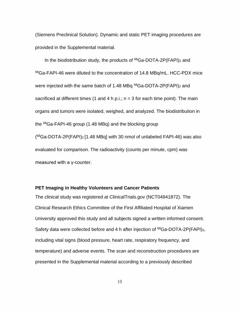
In HCC-PDX-1, both radiotracers were absorbed highly by the tumor at 0.5 h after
injection, and the uptake decreased relatively slowly until 4 h (Figure 3). However,
the tumor uptake of 68Ga-DOTA-2P(FAPI)2 was significantly higher than that of
68Ga-FAPI-46. The detailed %ID/g tumor values for both tracers from
small-animal PET are shown in Supplemental Fig. 4A. Other organs
demonstrated low non-specific binding that quickly decreased (Supplemental Fig.
4B-E), resulting in low background signal and favorable tumor-to-background

14
ratios. For a comprehensive investigation of the early pharmacokinetics of
68Ga-DOTA-2P(FAPI)2, 60-min dynamic PET was performed in HCC-PDX-1. The
tumor accumulation of 68Ga-DOTA-2P(FAPI)2 was rapid, and the time
dependency of the FAPI-dimer uptake was similar to other FAPI tracers. In
contrast, the heart, kidney, and liver uptake showed sharp elimination (Fig. 3).
Regarding 68Ga-DOTA-2P(FAPI)2 PET in HCC-PDX-2, the tumor accumulation
was rapid. Slightly decreased tumor uptake was observed from 30 min to 1h, then
it remained constant between 1 and 4 h (Fig. 4A and Supplemental Fig. 5),
similarly to that in HCC-PDX-1. The 60-min dynamic PET was also performed in
HCC-PDX-2 (Supplemental Fig. 6).
Target specificity was evaluated by simultaneous administration of unlabeled
FAPI-46 as a competitor with 68Ga-DOTA-2P(FAPI)2. The tumor uptake 1 h after
injection was suppressed greatly by blocking in HCC-PDX-1 and HCC-PDX-2,
and the radiotracer clearance in most organs was faster than that without blocking
(Fig. 4B). The uptake values of the tumor and key organ with or without
competitor are presented in Supplemental Figures 4F and 5F.
Organ Distribution in HCC-PDX-1

15
The biodistribution of 68Ga-FAPI-46 in HCC-PDX-1 was determined by ex-vivo
counting in tissues collected 1 and 4 h after injection (Fig. 5A). At 1 h p.i.,
68Ga-FAPI-46 accumulated mainly in the tumor (4.60 ± 1.12 %ID/g) and kidney
(4.42 ± 0.97 %ID/g), and the tumor-to-kidney (T/K) ratio was 1.05 ± 0.18. Four
hours p.i., 68Ga-FAPI-46 in the blood, heart, liver, lung, and spleen decreased
sharply, whereas the tumor uptake was steady (3.81 ± 0.18 %ID/g).
The biodistribution of 68Ga-DOTA-2P(FAPI)2 was also assessed in the same PDX
model by comparison (Figure 5B). Consistently with the PET findings,
68Ga-DOTA-2P(FAPI)2 demonstrated higher uptake in the tumor than
68Ga-FAPI-46, 1 h p.i. (8.97 ± 0.32 vs. 4.60 ± 1.12 %ID/g, P = 0.003) and 4 h p.i.
(7.61 ± 0.64 vs. 3.81 ± 0.18 %ID/g, P = 0.001). The organ uptake of
68Ga-DOTA-2P(FAPI)2 was slightly greater than that of 68Ga-FAPI-46 both 1 and
4 h p.i. As a result, 68Ga-DOTA-2P(FAPI)2 had a higher T/K ratio than
68Ga-FAPI-46 (T/K: 1.60 ± 0.26 vs 1.05 ± 0.18, P = 0.039, 1 h p.i.), although the
difference was not significant 4 h p.i. (1.33 ± 0.29 vs 0.95 ± 0.09, P = 0.093).
Regarding the blocking group, a dramatic decrease of radioactivity was
detected in most organs (Fig. 5B), and the tumor uptake decreased most
significant (8.97 ± 0.32 vs. 1.07 ± 0.19 % ID/g 1 h p.i., P < 0.001, Student’s t-test;
7.61 ± 0.64 vs.1.14 ± 0.15 % ID/g 4 h p.i., P = 0.002, Student’s t-test).

16
Additional biodistribution and PET studies were performed to rule out the
effect of molar activity on comparative experiments between FAPI dimer and
FAPI-46. The amount of precursor administered was 23 μg (25.9 nmol) for
FAPI-46 and 50 μg (25.3 nmol) for DOTA-2P(FAPI)2, resulting in the same
specific activity for 68Ga-FAPI-46 and 68Ga-FAPI-dimer. Under these
circumstances, the results from the biodistribution study demonstrated that
68Ga-FAPI-dimer had higher tumor uptake than 68Ga-FAPI-46 (8.45 ± 2.19 vs.
4.03 ± 0.69 %ID/g; P = 0.029, Student’s t-test, Supplemental Fig. 7). Similar
results were observed from the PET imaging study (Supplemental Fig. 8).
Adverse Events
All observed vital signs (including blood pressure, heart rate, and body
temperature) remained normal during the injection and at 4-h follow-up.
No individuals reported any adverse events.
Dosimetry Estimate
The dosimetry reports and a representative figure for three healthy volunteers are
shown in Table 1 and Fig. 6. There was no time dependency of tracer uptake,
showing that the tracer distribution was not obviously changing after 10 min. The

17
effective dose of 68Ga-DOTA-2P(FAPI)2 was 1.19E-02 mSv/MBq, calculated
using OLINDA. The organ with the highest effective dose was the thyroid
(3.11E-03 mSv/MBq), followed by the liver (1.65E-03 mSv/MBq) and lungs
(1.36E-03 mSv/MBq). Overall, the effective dose of 68Ga-DOTA-2P(FAPI)2 was
comparable with those of 68Ga-FAPI-02 (1.80E−02 mSv/MBq) and 68Ga-FAPI-04
(1.64E−02 mSv/MBq) (4), and higher than 68Ga-FAPI-46 (7.80E−03 mSv/MBq)
(23).
68Ga-DOTA-2P(FAPI)2 PET Imaging in Cancer Patients
68Ga-FAPI-46 and 68Ga-DOTA-2P(FAPI)2 PET/CT scans were performed after 60
min of intravenous administration in three patients: one with nasopharyngeal
non-keratinized undifferentiated carcinoma, wild diffuse bone metastases after
chemoradiotherapy and immunotherapy; one with papillary thyroid carcinoma,
wild diffuse lymph node metastases after total thyroidectomy and multiple cycles
of radioiodine treatment; and one with HCC, treatment-naïve. Representative PET
images of these three patients after administration of 68Ga-FAPI-46 and
68Ga-DOTA-2P(FAPI)2 are shown in Fig. 7 and Supplemental Fig. 9 and 10. In the
patient with metastatic thyroid cancer, 68Ga-DOTA-2P(FAPI)2 was mainly
accumulated in the tumor, pancreas, submandibular glands, and blood pool.
Interestingly, the activity of FAPI dimer in the blood pool remained at a high level

18
(maximum standardized uptake value, SUVmax 8.3) 4 h p.i. All tumor lesions
were clearly visible owing to the favorable tumor-to-background ratios. In the
lesion-to-lesion comparison, the dimer uptake in 21 lesions (from 3 patients) was
higher than monomer uptake (SUVmax 1 h p.i.: 8.1-39.0 vs. 1.7-24.0, respectively;
P < 0.001 by Wilcoxon matched-pairs signed-rank test, mean SUVmax: 15.3 vs.
23.9; Supplemental Table 1). In addition, the 68Ga-2P(FAPI)2 uptake in tumors
was slightly decreased from 1 to 4 h (SUVmax 1 h p.i.: 8.1-39.0; 4 h p.i.: 6.6-35.0).
DISCUSSION
With a burst of preclinical and clinical research on quinoline-based FAPI variants,
two main hurdles remain: improving the tumor retention time and finding the
appropriate preclinical models. Since FAP is mainly overexpressed in CAFs and
not in tumor cells, the tumor cell-line transfected with human or murine FAP could
not reflect the tumor microenvironment (3,7). In contrast, PDXs can reliably
reproduce a patient’s parental tumor for histopathology and genetics (15).
Therefore, PDXs are suitable models for studying tumor biology, including the
microenvironment and patient sensitivity to target agents. Despite an increasing
number of case studies (6-9) and two clinical trials with a small patient population
(13,24) for FAP-based PTRT, basic research on this topic is rare (10). In the
present study, PDXs derived from HCC could maintain the principal

19
histopathological characterization of the human tumor, confirming the robustness
of this model for testing the properties of the new FAPI variant.
As a pan-cancer target, labelling FAPI monomers with different imaging
isotopes has shown impressive results in several tumor diagnoses (3-5,25);
however, the pharmacokinetics with fast clearance from blood and short retention
in tumors are problematic issues for PTRT application. Thus, structural
modification of FAPI for optimizing tumor uptake and tumor retention time for
PTRT is another key research direction.
Based on the polyvalent effect, multimeric peptides can help improve
tumor-targeting efficacy and generate higher quality in-vivo imaging. This strategy
has been widely used in the development of multimeric Arg-Gly-Asp (RGD)
peptides (16,17). Indeed, given the distance between two FAPI motifs in
DOTA-2P(FAPI)2 may not be long enough, it is unlikely that they would bind to two
adjacent FAP sites simultaneously. However, the binding of one FAPI motif to
FAP will significantly increase the local concentration of a second FAP motif in the
vicinity of FAP sites. The locally enhanced FAPI concentration may explain the
higher tumor uptake of radiolabeled FAPI dimers compared to their monomeric
analogs. Similar findings were observed in the studies of radiolabeled
RGD-dimers (26). Nonetheless, it should be noted that although tetrameric and

20
octameric peptides possess higher receptor-binding affinity and higher tumor
uptake than their dimeric and monomeric counterparts, they also have
substantially higher background activity, especially in the kidney (27). Therefore,
dimeric peptides seem to be an optimal choice owing to their increased tumor
uptake and favorable pharmacokinetics (27). PEGylation is another widely used
strategy to improve the in-vivo pharmacokinetics of radiotracers. According to
previous reports where PEGylated RGD peptides were labeled with different
isotopes, PEGylation improved the labeling yield and in-vivo pharmacokinetics
(16,18). However, PEGylation also induces hydrophilicity and increases kidney
uptake, partially explaining the high initial kidney uptake compared to other FAPI
derivatives. In this study, we designed and synthesized a novel FAPI dimer with
two mini-PEG spacers between the FAPI motifs in homodimeric peptides. The
in-vitro binding assays demonstrated that DOTA-2P(FAPI)2 had specific and high
binding affinity to FAP expressed on CAFs, revealing that the polyvalent strategy
did not compromise its FAP-binding affinity.
After radiolabeling with 68Ga, the FAPI dimers exhibited improved in-vivo
pharmacokinetics and enhanced tumor uptake compared to the FAPI monomer.
Dynamic PET scans in the two HCC-PDX groups showed prominent tumor uptake
and predominant organ clearance. After applying the tracers to static PET scans,

21
68Ga-DOTA-2P(FAPI)2 demonstrated higher tumor uptake than 68Ga-FAPI-46 at
all time points examined in both PDXs. In the small-animal PET imaging study,
higher initial (30 min p.i.) kidney and liver uptake for 68Ga-DOTA-2P(FAPI)2 was
observed compared to FAPI-monomers, including 68Ga-FAPI-04, 68Ga-FAPI-46,
and 18F-FGlc-FAPI (14). However, the kidney and liver uptake were quickly
eliminated at 60 min p.i. in PET imaging and the biodistribution study. The high
initial kidney uptake and rapid renal clearance may be attributed to the insertion of
two PEG groups, which improved the hydrophilic properties (16,18). Nevertheless,
the main organs uptake should be carefully estimated for safety dose limitation
when FAPI dimer is labeled with 177Lu for targeted radionuclide therapy. The FAP
specificity of 68Ga-DOTA-2P(FAPI)2 was strongly confirmed by effective uptake
inhibition in the presence of unlabeled FAPI-46 in cell-uptake, PET scan, and
biodistribution experiments.
It was reported that the FAP-blocking dose (cold mass of FAPI) in one mouse
was 30 nmol (7,11). Since the specific activity of FAPI dimer and FAPI-46 was 37
GBq/μmol and 16.5 GBq/μmol, respectively, in this study, a dose of 7.4 MBq
68Ga-FAPI-46 (0.45 nmol, hot and cold mass) or 68Ga-FAPI-dimer (0.2 nmol, hot
and cold mass) per mouse for PET imaging may have minimal impact on the
tumor uptake. Therefore, there is no effect of the different injected cold mass of

22
the radiotracers that might have caused the significant differences in tumor uptake
values. Moreover, additional PET imaging and biodistribution experiments have
been performed to rule out the effect of molar activity on all comparative
experiments of dimeric and monomeric inhibitors.
The encouraging results of the in-vitro and mouse studies led to the clinical
translation of FAPI dimer into human subjects. The radiation dose deposition of
68Ga-DOTA-2P(FAPI)2 in healthy organs was estimated using the PET data of
three healthy volunteers at four time points. The average effective whole-body
dose was 1.19E-02 mSv/MBq. This estimate is comparable with the previously
reported effective doses of those of 68Ga-FAPI-02 and 68Ga-FAPI-04 (1.80E-02
and 1.64E-02 mSv/MBq), and higher than 68Ga-FAPI-46 (7.80E−03 mSv/MBq)
(4,23).
Regarding the clinical diagnosis, 68Ga-DOTA-2P(FAPI)2 PET/CT imaging in
the patients examined showed a rapid and stable accumulation of the dimer in
tumorous lesions, consistently with the results of animal experiments. Tumor
uptake in most lesions was significantly higher with 68Ga-DOTA-2P(FAPI)2 than
with 68Ga-FAPI-46, leading to visualization of primary lesions and metastases
more clearly. Interestingly, the retention of the tracer in the patient blood pool
remained high 4 h p.i., in contrast with mice findings. The prolonged retention in
23
the blood pool may make DOTA-2P(FAPI)2 an attractive tracer for PTRT
applications. Although the PET/CT results were encouraging in patients, high
physiological uptake in the thyroid and pancreas should be noted. Nevertheless,
DOTA-2P(FAPI)2 labeling with 68Ga demonstrated favorable data in cells, mice,
and patients. Future development, especially for anti-tumor therapeutic
applications, labeling the ligand with therapeutic radionuclides such as 177Lu and
90Y should be considered to compare the FAPI dimer with the monomer.
CONCLUSION
68Ga-DOTA-2P(FAPI)2 provides an improved tumor uptake and longer
tumor-retention time compared with 68Ga-FAPI-46, and it could be a promising
tracer for both diagnostic imaging and targeted radionuclide therapy in malignant
tumors with positive FAP expression. Further work to optimize the
pharmacokinetics of DOTA-2P(FAPI)2 and evaluate its anti-tumor efficacy after
labeling with therapeutic isotopes should be envisaged.
Disclosures:

24
Funding: This work was funded by the National Natural Science Foundation of
China (Grant number 82071961, 81901805,and 81772893), and the key medical
and health projects in Xiamen (Grant number 3502Z20191104).
Conflicts of interest: None.
Acknowledgments: The authors gratefully acknowledge Dr. Dongyan Shen (The
First Affiliated Hospital of Xiamen University, Xiamen University, China) for
providing the CAFs.

25
KEY POINTS
QUESTION:
How is it possible to optimize tumor uptake and retention of the fibroblast
activation protein (FAP)-targeting molecular agents?
PERTINENT FINDINGS:
In a pilot clinical cancer imaging study, 68Ga-DOTA-2P(FAPI)2 was synthesized as
a FAP-inhibitor dimer and tested for its pharmacokinetic properties. Its tumor
uptake was higher than that of monomeric FAPIs in-vitro, in-vivo, and in cancer
patients.
IMPLICATIONS FOR PATIENT CARE:
68Ga-DOTA-2P(FAPI)2 shows improved tumor uptake and retention properties. It
could be a promising candidate tracer for both diagnostic imaging and targeted
therapy of malignant tumors with positive FAP expression.

26
REFERENCES
1. Gaggioli C, Hooper S, Hidalgo-Carcedo C, et al. Fibroblast-led collective invasion of carcinoma
cells with differing roles for RhoGTPases in leading and following cells. Nat Cell Biol.
2007;9:1392-1400.
2. Lakins MA, Ghorani E, Munir H, Martins CP, Shields JD. Cancer-associated fibroblasts induce
antigen-specific deletion of CD8 (+) T Cells to protect tumour cells. Nat Commun. 2018;9:948.
3. Loktev A, Lindner T, Burger EM, et al. Development of fibroblast activation protein-targeted
radiotracers with improved tumor retention. J Nucl Med. 2019;60:1421-1429.
4. Giesel FL, Kratochwil C, Lindner T, et al. (68)Ga-FAPI PET/CT: biodistribution and preliminary
dosimetry estimate of 2 DOTA-containing FAP-targeting agents in patients with various cancers. J
Nucl Med. 2019;60:386-392.
5. Chen H, Pang Y, Wu J, et al. Comparison of [(68)Ga]Ga-DOTA-FAPI-04 and [(18)F] FDG PET/CT
for the diagnosis of primary and metastatic lesions in patients with various types of cancer. Eur J Nucl
Med Mol Imaging. 2020;47:1820-1832.
6. Ballal S, Yadav MP, Kramer V, et al. A theranostic approach of [(68)Ga]Ga-DOTA.SA.FAPi
PET/CT-guided [(177)Lu]Lu-DOTA.SA.FAPi radionuclide therapy in an end-stage breast cancer
patient: new frontier in targeted radionuclide therapy. Eur J Nucl Med Mol Imaging. 2021;48:942-944.
7. Lindner T, Loktev A, Altmann A, et al. Development of quinoline-based theranostic ligands for
the targeting of fibroblast activation protein. J Nucl Med. 2018;59:1415-1422.
8. Lindner T, Altmann A, Kramer S, et al. Design and development of (99m)Tc-labeled FAPI tracers
for SPECT imaging and (188)Re therapy. J Nucl Med. 2020;61:1507-1513.
9. Kratochwil C, Giesel FL, Rathke H, et al. [(153)Sm]Samarium-labeled FAPI-46 radioligand therapy
in a patient with lung metastases of a sarcoma. Eur J Nucl Med Mol Imaging. 2021;48:3011-3013.
10. Watabe T, Liu Y, Kaneda-Nakashima K, et al. Theranostics targeting fibroblast activation protein
in the tumor stroma: (64)Cu- and (225)Ac-labeled FAPI-04 in pancreatic cancer xenograft mouse
models. J Nucl Med. 2020;61:563-569.
11. Loktev A, Lindner T, Mier W, et al. A tumor-imaging method targeting cancer-associated
fibroblasts. J Nucl Med. 2018;59:1423-1429.

27
12. Assadi M, Rekabpour SJ, Jafari E, et al. Feasibility and therapeutic potential of 177Lu-fibroblast
activation protein inhibitor-46 for patients with relapsed or refractory cancers: a preliminary study.
Clin Nucl Med. 2021. Epub ahead of print.
13. Baum RP, Schuchardt C, Singh A, et al. Feasibility, biodistribution and preliminary dosimetry in
peptide-targeted radionuclide therapy (PTRT) of diverse adenocarcinomas using (177)Lu-FAP-2286:
first-in-human results. J Nucl Med. 2021. Epub ahead of print.
14. Toms J, Kogler J, Maschauer S, et al. Targeting fibroblast activation protein: radiosynthesis and
preclinical evaluation of an (18)F-labeled FAP inhibitor. J Nucl Med. 2020;61:1806-1813.
15. Hidalgo M, Amant F, Biankin AV, et al. Patient-derived xenograft models: an emerging platform
for translational cancer research. Cancer Discov. 2014;4:998-1013.
16. Lang L, Li W, Guo N, et al. Comparison study of [18F]FAl-NOTA-PRGD2, [18F]FPPRGD2, and
[68Ga]Ga-NOTA-PRGD2 for PET imaging of U87MG tumors in mice. Bioconjug Chem.
2011;22:2415-2422.
17. Li ZB, Cai W, Cao Q, et al. (64)Cu-labeled tetrameric and octameric RGD peptides for
small-animal PET of tumor alpha(v)beta(3) integrin expression. J Nucl Med. 2007;48:1162-1171.
18. Wu Z, Li ZB, Chen K, et al. MicroPET of tumor integrin alphavbeta3 expression using 18F-labeled
PEGylated tetrameric RGD peptide (18F-FPRGD4). J Nucl Med. 2007;48:1536-1544.
19. Zhao L, Chen H, Guo Z, et al. Targeted radionuclide therapy in patient-derived xenografts using
(177)Lu-EB-RGD. Mol Cancer Ther. 2020;19:2034-2043.
20. Chen H, Zhao L, Fu K, et al. Integrin alphavbeta3-targeted radionuclide therapy combined with
immune checkpoint blockade immunotherapy synergistically enhances anti-tumor efficacy.
Theranostics. 2019;9:7948-7960.
21. Chen H, Pang Y, Wu J, et al. Comparison of [(68)Ga]Ga-DOTA-FAPI-04 and [(18)F] FDG PET/CT
for the diagnosis of primary and metastatic lesions in patients with various types of cancer. Eur J Nucl
Med Mol Imaging. 2020. Epub ahead of print.
22. Stabin MG, Sparks RB, Crowe E. OLINDA/EXM: the second-generation personal computer
software for internal dose assessment in nuclear medicine. J Nucl Med. 2005;46:1023-1027.

28
23. Meyer C, Dahlbom M, Lindner T, et al. Radiation dosimetry and biodistribution of
(68)Ga-FAPI-46 PET imaging in cancer patients. J Nucl Med. 2020;61:1171-1177.
24. Jokar N, Velikyan I, Ahmadzadehfar H, et al. Theranostic approach in breast cancer: a treasured
tailor for future oncology. Clin Nucl Med. 2021;46:e410-e420.
25. Wang S, Zhou X, Xu X, et al. Clinical translational evaluation of Al(18)F-NOTA-FAPI for fibroblast
activation protein-targeted tumour imaging. Eur J Nucl Med Mol Imaging. 2021. Epub ahead of print.
26. Liu S. Radiolabeled cyclic RGD peptides as integrin alpha(v)beta(3)-targeted radiotracers:
maximizing binding affinity via bivalency. Bioconjug Chem. 2009;20:2199-2213.
27. Li ZB, Chen K, Chen X. (68)Ga-labeled multimeric RGD peptides for microPET imaging of integrin
alpha(v)beta (3) expression. Eur J Nucl Med Mol Imaging. 2008;35:1100-1108.

29
Figures
FIGURE 1. Chemical structure of DOTA-2P(FAPI)2.

30
FIGURE 2. (A) Fibroblast activation protein (FAP) expression in Huh7 cells and
cancer-associated fibroblasts assayed using western blotting. (B) Cell uptake
assay of 68Ga-DOTA-2P(FAPI)2, 68Ga-FAPI-46, and blocking experiment on CAFs
(n=3). (C) Inhibition of 68Ga-FAPI-46 binding to FAP on CAFs by unlabeled
FAPI-46 (2.83 × 10⁻⁴ to 10-13 M; n=3). (D) Inhibition of 68Ga-DOTA-2P(FAPI)2
binding to FAP on CAFs by unlabeled FAPI-46 (1.27 × 10⁻⁴ to 10-13 M; n=3). CAFs:
cancer-associated fibroblasts.
31
FIGURE 3. Representative static PET imaging of 68Ga-DOTA-2P(FAPI)2 and
68Ga-FAPI-46 in HCC-PDX-1, and dynamic time-activity curves of
68Ga-DOTA-2P(FAPI)2 in the heart, kidney, liver, muscle, and tumor tissues.

32
FIGURE 4. (A) Representative static PET imaging of 68Ga-DOTA-2P(FAPI)2 and
68Ga-FAPI-46 in HCC-PDX-2. (B) Representative static PET imaging of
68Ga-DOTA-2P(FAPI)2 in HCC-PDX-1 and HCC-PDX-2 with and without
simultaneous injection of unlabeled FAPI-46 as competitor 1 h after
administration.

33
FIGURE 5. (A) Ex-vivo biodistribution of 68Ga-FAPI-46 in HCC-PDX-1, 1 and 4 h
post-injection (n = 3/group); (B) Ex-vivo biodistribution of 68Ga-DOTA-2P(FAPI)2
in HCC-PDX-1, 1 and 4 h post-injection, with and without co-administration of
unlabeled FAPI-46 as a blocking agent (n = 3/group).

34
FIGURE 6. 68Ga-DOTA-2P(FAPI)2 10, 30, 60, and 180 min after injection in
healthy volunteers, and the SUVmean values of healthy organs at different time
points.

35
FIGURE 7. 68Ga-FAPI-46, 1 h after injection, and 68Ga-DOTA-2P(FAPI)2, 1 and 4
h after injection, in a patient with metastatic thyroid cancer. Hematoxylin and
eosin (H&E) staining and FAP immunohistochemistry staining showed high FAP
expression in the tumor stroma (original magnification, ×100).

36
Tables
Table 1 68Ga-DOTA-2P(FAPI)2 dosimetry summary of effective doses using
OLINDA/EXM v.1.1.
Target organ Mean
(mSv/MBq)
SD
(mSv/MBq)
Adrenal glands 7.98E-05 3.04E-05
Brain 3.16E-05 1.96E-05
Breasts 6.36E-05 1.09E-05
Gallbladder Wall - -
LLI Wall 9.22E-04 2.33E-04
Small Intestine 5.18E-05 1.84E-05
Stomach Wall 7.12E-04 3.16E-05
ULI Wall 2.79E-05 1.13E-05
Heart Wall - -
Kidneys 1.01E-04 4.96E-05
Liver 1.65E-03 4.12E-04
Lungs 1.36E-03 4.59E-04
Muscle 4.19E-05 2.07E-05
Ovaries 5.94E-04 1.06E-04
Pancreas 7.61E-04 8.05E-04
Red Marrow 1.12E-03 1.33E-04
Osteogenic Cells 6.47E-05 6.87E-06
Skin 1.22E-05 2.32E-06
Spleen 1.58E-04 9.12E-05
Thymus 9.64E-06 4.47E-06
Thyroid 3.11E-03 4.68E-04
Urinary Bladder Wall 1.04E-03 5.22E-04
Uterus 1.27E-05 6.00E-06
Effective Dose Equivalent 1.69E-02 1.92E-03
Effective Dose 1.19E-02 9.45E-04
LLI: lower large intestine; SD: standard deviation; ULI: upper large intestine.

37
Graphical Abstract

Supplemental materials
Chemicals and Reagents
All chemicals were purchased from Energy Chemical Co. and Nanchang
Tanzhen Biological Technology Co., Ltd.; FAPI-46 was purchased from C.S. Bio.
Anti-FAP mAb was purchased from Abcam (Cat. No. ab207178) and Abclonal
(Cat. No. A6349). The human hepatocellular carcinoma (HCC) cell line Huh7 was
purchased from the China National Infrastructure of Cell Line Resource. The
CAFs from surgical specimens of HCC patients were kindly provided by Dr.
Dongyan Shen (The First Affiliated Hospital of Xiamen University, Xiamen
University, China). The labeling efficiency and radiochemical purity were tested
using a radio-TLC scanner (MSFC1-00220, Eckert & Ziegler) Dionex Ulti-Mate
3000 high-performance liquid chromatography (HPLC; Thermo Scientific), an
SPD-20A UV detector (λ = 254 nm), and an Elysia Raytest Gabi Star γ-radiation
detector. Radioactivity (counts per minute) was measured using a γ-counter
(WIZARD 2480; Perkin-Elmer) and CRC-25R dose calibrators (CAPIN-TEC Inc.).

Procedure of DOTA-2P(FAPI)2 synthesis
Compound 2 (500 mg, 1 mmol) and Boc-PEG3-OH (338 mg, 1.1 mmol) in
DMF (3 mL) were treated with HATU (570 mg, 1.5 mmol) and DIPEA (710 mg,
5.5 mmol). The reaction mixture was stirred for 16 h at 80°C. After completion of
the reaction, the mixture was transferred into an ice bath and TFA (1.15 mL, 15
mmol) was added dropwise. After another 3 h of stirring at room temperature, the
mixture was adjusted to pH=5.5 by NaOH solution and purified by prep-HPLC to
afford 200 mg of compound 3. LC-MS (ESI+): m/z 689.6 [M+H]+, 345.4
[M+2H]+/2.
Compound 3 (68.9 mg, 0.1 mmol) and compound 4 (56 mg, 1.1 mmol) in
DMF (1 mL) were treated with HATU (57 mg, 0.15 mmol) and DIPEA (71 mg,
0.55 mmol). The reaction mixture was stirred for 8 h at 80°C. After completion of
the reaction, the mixture was transferred into an ice bath and TFA (0.2 mL, 1.8
mmol) was added dropwise. After another 1 h of stirring at room temperature, the
mixture was adjusted to pH=5.5 by NaOH solution and purified by prep-HPLC to
afford compound 5 (50 mg). LC-MS (ESI+): m/z 1039.9 [M+H]+, 520.5
[M+2H]+/2.
Compound 5 (30 mg, 0.028 mmol) and compound 6 (20 mg, 0.035 mmol)
were dissolved in DMF (1.5 mL) and treated with HATU (16 mg, 0.042 mmol) and

DIPEA (20 mg, 0.154 mmol). The reaction mixture was stirred for 6 h at room
temperature. After completion of the reaction, DMF was removed under reduced
pressure, and 25% DEA/THF (1 mL) was added dropwise. After another 1 h of
stirring at room temperature, the mixture was purified by prep-HPLC to afford
compound 7 (10 mg). LC-MS (ESI+): m/z 686.8 [M+2H]+/2
Compound 7 (16 mg, 0.01 mmol) and Compound 8 (9.5 mg, 0.011 mmol) in
DMF (1 mL) were treated with HATU (5.7 mg, 0.015 mmol) followed by DIPEA
(7.1 mg, 0.055 mmol). The reaction mixture was stirred for 4 h at 80°C. After
completion of the reaction, the mixture was transferred into an ice bath and TFA
(0.02 mL, 0.18 mmol) dropwise. After another 1 h of stirring at room temperature,
the mixture was adjusted to pH5.5 by NaOH solution and purified by prep-HPLC
to afford compound 9 (5 mg). LC-MS (ESI+): m/z 659.3 [M+3H]+/3.

The high-performance liquid chromatography (HPLC) of DOTA-2P(FAPI)2

The liquid chromatography–mass spectrometry of DOTA-2P(FAPI)2.

Radio-HPLC
HPLC chromatogram analysis was performed using the analytic C-18 reversed-
phase column (4.6 × 250 mm, 5 μm, 120Å, Thermo).
HPLC conditions were as follows: trifluoroacetic acid (TFA) (0.1%) and CH3CN
(0.1%TFA) flow rate = 1 mL/min; λ = 254 nm; A = 0.1% trifluoroacetic acid
(TFA)/H2O; B = 0.1% TFA/acetonitrile. B gradient: 0–16 min, from 5% to 75%;
17–20 min, from 75% to 95%; 21-25 min, 95%; 26–30 min, 5%. The retention
time was 10.66 min for 68Ga-DOTA-2P(FAPI)2 and 9.41 min for 68Ga-FAPI-46.
PDX-models production
The tumor specimens were obtained from surgical resection, and the
hepatocellular carcinoma (HCC) specimens were immediately placed in DMEM
(Cat. #C11995599BT, Gibco, USA) supplemented with 2% antibiotics (penicillin
and streptomycin) and stored in an ice box. Immunodeficient BALB/c nude mice
were bred under specific pathogen free (SPF) conditions at Xiamen University
Laboratory Animal Center from founders originally obtained from Shanghai SLAC
Laboratory Animal Co., Ltd (China). In brief, the necrotic tissue was removed
from the fresh tumor specimen (less than 2 h after the surgery). Then the
specimen were cut into approximately 30 mm3 pieces with scissors and washed

three times by DMEM supplemented with 2% antibiotics before subcutaneous
implantation into the mice right side of the trunk. All procedures were performed
in super-clean benches. Tumor growth was monitored until it reached 1500 mm3,
then the mice were sacrificed, and the tumor was minced for passaging to the
next generation BALB/c nude mice. The remaining fragments were used for the
histological verification, including western blot of FAP, immunohistochemistry
staining of FAP and Ki67, and Hematoxylin and eosin (H&E) staining.
Western blot and histopathological staining
Proteins were extracted with lysis buffer (150 mM NaCl, 50 mM Tris-HCl [pH 8.0],
1mM EDTA and 1% Triton X-100). Approximately 20 μg of total protein per
sample was separated by SDS-PAGE and transferred to a PVDF membrane
(Millipore). The membranes were pre-incubated with 5% skimmed milk in TBST
for 1 h, followed by incubation with human FAP antibody (ab207178; Abcam).
Membranes were washed with TBST three times and incubated with horseradish
peroxidase-labelled secondary antibody, which was detected using an enhanced
chemiluminescence detection system (C280, Azure).
Anti-FAP mAb (ab207178, Abcam and A6349, Abclonal) was used to label
surgical specimens of HCC patients and mice tumors during

immunohistochemistry (IHC). FAP expression IHC staining was conducted
according to our previous protocol using the above mAb (1).
Dynamic PET imaging and static PET imaging for mice
For dynamic PET imaging, the duration of the scan was 60 min, and the
reconstruction frames were 10×30 s, 10×60 s, 10×120 s, and 9×160 s. For 10-
min static PET imaging, the acquisition times were 0.5, 1, 2, and 4 h post
injection (p.i.). For the blocking experiment, 30 nmol of unlabelled FAPI-46 was
added to the solution of 68Ga-DOTA-2P(FAPI)2 before injection. Images were
reconstructed iteratively using a 3D OPMAP 256.pPetRcn (Siemens), and
converted to % ID/g images. Regions of interest (ROIs) in the tumor, liver, heart,
kidney, and muscle were counted on the PET images to quantify the radioactive
signals.
The dose of intravenously injected 68Ga-FAPI-46 and 68Ga-DOTA-2P(FAPI)2 was
calculated according to the patient’s weight (1.8-2.2 MBq [0.05-0.06 mCi]/kg for
FAPI). The time interval in cancer patients between 68Ga-DOTA-2P(FAPI)2 and
68Ga-FAPI-46 was three days. For healthy volunteers, data were acquired using
a hybrid PET/CT scanner (Discovery MI, GE Healthcare, Milwaukee, WI, USA)

after 10 min, 30 min, 60 min, and 180 min of intravenous injection. For cancer
patients, PET/CT images were acquired after 60 min of intravenous injection
(one patient was scanned twice at 60 min and 240 min p.i.). All scans and
reconstruction were performed according to a previously described protocol (2).
The maximum standard uptake values (SUVmax) were automatically calculated
using a region of interest (ROI) drawn on the transaxial images.

Supplemental Figures
Supplemental Figure 1. Flow diagram of DOTA-2P(FAPI)2 synthesis

Supplemental Figure 2. (A, B) HPLC profiles of pure 68Ga-DOTA-2P(FAPI)2 and 68Ga-FAPI-46, respectively; (C, D)
Stability of 68Ga-DOTA-2P(FAPI)2 in phosphate buffer saline (PBS) and fetal bovine serum (FBS), respectively, 1, 2, and 4
h after incubation.

Supplemental Figure 3. (A) Immunohistochemistry staining of FAP and Ki67, and Hematoxylin and eosin (H&E) staining
in human hepatocellular carcinoma and corresponding PDXs (positive FAP in CAFs are indicated by arrows). (B) Western
blot of FAP in PDXs

Supplemental Figure 4 (A-E) Comparison of tumor and organ uptake of 68Ga-FAPI-46 and 68Ga-DOTA-2P(FAPI)2 in
HCC-PDX-1 0.5, 1, 2, and 4 h post-injection (n = 3/group). (F) Tumor and organ uptake of 68Ga-DOTA-2P(FAPI)2 in HCC-
PDX-1 1 h post-injection with and without co-administration of unlabeled FAPI-46 as a blocking agent (n = 3/group).

Supplemental Figure 5 (A-E) Comparison of tumor and organ uptake of 68Ga-FAPI-46 and 68Ga-DOTA-2P(FAPI)2 in
HCC-PDX-2 0.5, 1, 2, and 4 h post-injection (n = 3/group); (F) Tumor and organ uptake of 68Ga-DOTA-2P(FAPI)2 in HCC-
PDX-2 1 h post-injection with and without co-administration of unlabeled FAPI-46 as a blocking agent (n = 3/group).

Supplemental Figure 6 Dynamic time-activity curves of 68Ga-DOTA-2P(FAPI)2 in the heart, kidney, liver, muscle, and
tumor in HCC-PDX-2.

Supplemental Figure 7. Ex-vivo biodistribution of 68Ga-FAPI-46 and 68Ga-DOTA-2P(FAPI)2 (with the same specific
activity) in HCC-PDX-1 at 1 h post-injection (n = 3/group).
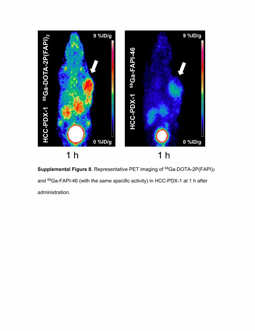
Supplemental Figure 8. Representative PET imaging of 68Ga-DOTA-2P(FAPI)2
and 68Ga-FAPI-46 (with the same specific activity) in HCC-PDX-1 at 1 h after
administration.
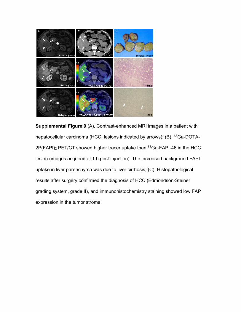
Supplemental Figure 9 (A). Contrast-enhanced MRI images in a patient with
hepatocellular carcinoma (HCC, lesions indicated by arrows); (B). 68Ga-DOTA-
2P(FAPI)2 PET/CT showed higher tracer uptake than 68Ga-FAPI-46 in the HCC
lesion (images acquired at 1 h post-injection). The increased background FAPI
uptake in liver parenchyma was due to liver cirrhosis; (C). Histopathological
results after surgery confirmed the diagnosis of HCC (Edmondson-Steiner
grading system, grade II), and immunohistochemistry staining showed low FAP
expression in the tumor stroma.

Supplemental Figure 10. 68Ga-FAPI-46 and 68Ga-DOTA-2P(FAPI)2 PET/CT
showed multiple bone metastases in a patient with metastatic nasopharyngeal
carcinoma (non-keratinized undifferentiated carcinoma, who presented disease
progression after chemoradiotherapy and immunotherapy). The uptake of 68Ga-
DOTA-2P(FAPI)2 was higher than 68Ga-FAPI-46 in most of the bone metastases
(images acquired at 1 h post-injection). The bone metastases were further
observed in the radionuclide bone scan.

Supplemental Table 1: Lesion-by-lesion comparison of 68Ga-FAPI-46 and 68Ga-
DOTA-2P(FAPI)2 uptake in three cancer patients.
Patient No. Age Sex Status No. of
lesions Site of lesions 68Ga-FAPI-46 SUVmax
68Ga-DOTA-2P(FAPI)2 SUVmax
P
Patient1 (NPC) 71 Male Recurrence >10 Bone 1 16.3 17.8 0.005
Bone 2 24.7 27.5 Bone 3 23.4 27.4 Bone 4 14.4 25.1 Bone 5 22.0 29.8 Bone 6 9.8 20.8 Bone 7 8.1 17.2 Bone 8 13.3 22.1 Bone 9 17.4 27.6 Bone 10 11.6 23.3 Patient 2 (Thyroid cancer)
34 Male Recurrence >10 Cervical node 1 20.0 32.8 0.005
Cervical node 2 1.7* 8.1 Supraclavicular node 1 24.0 39.0 Supraclavicular node 2 12.8 24.1 Axillary node 1 17.8 23.2 Axillary node 2 17.7 33.2 Axillary node 3 16.7 28.9 Bone 1 11.4 26.4 Mediastinal node 1 23.4 24.6 Hilar node 1 11.5 18.1 Patient 3 (HCC) 46 Male Initial stage 1 Primary tumor 2.7 9.8 NA
Note: HCC, hepatocellular carcinoma; NPC, nasopharyngeal carcinoma; No.,
number; SUVmax, maximum standardized uptake value; *, negative in 68Ga-
FAPI-46 PET/CT

References 1. Chen H, Zhao L, Fu K, et al. Integrin alphavbeta3-targeted
radionuclide therapy combined with immune checkpoint blockade
immunotherapy synergistically enhances anti-tumor efficacy.
Theranostics. 2019;9:7948-7960.
2. Chen H, Pang Y, Wu J, et al. Comparison of [(68)Ga]Ga-DOTA-FAPI-04
and [(18)F] FDG PET/CT for the diagnosis of primary and metastatic
lesions in patients with various types of cancer. Eur J Nucl Med Mol Imaging. 2020.