Embed Size (px)
Citation preview
Synthesis of Silver Nanoparticles Using Klebsiella pneumonia and its Bio-Medical Applications
Amar Ratan1#, Ekta Gupta1 and R.Ragunathan*
1Amity Institute of Nanotechnology, Amity University, Noida, Uttar Pradesh, India.*Centre for Bioscience and Nano science Research, Coimbatore, Tamil Nadu, India.
Abstract
The antimicrobial effects of sliver ions have been known for long but their activity against microorganisms has not yet been conclusively studied. In the present study, biological synthesis of silver nanoparticles using Klebsiella pneumonia has been investigated. The bacterial strain was grown the appropriate production media. The cell free filtrate obtained on filtration was bought in contact with different concentrations of AgNO 3. . The change in the pH with respect to the different concentrations was studied .The nanoparticles were further characterized by SEM studies. The antimicrobial activity of silver nanoparticles was investigated against E. coli, Bacillus and ESBL .Also the antimicrobial activity of encapsulated beads of silver nanoparticles and 2mM silver nanoparticles was compared. The anti-microbial fabrics activity was studied by coating the silver nanoparticles on gauge cotton fabric and then compared with standard disc Oxacillin. UV-VIS studies showed an increased in absorbance at 363 nm. The antimicrobial tests against E.coli showed increased inhibition at 1mM while for Bacillus, inhibition was more prominent at 2mM concentration and for ESBL-Pseudomonas inhibitory concentration was 2mM .The zone of inhibition for the encapsulated beads was less than that of the 2mM silver nanoparticles.
Key Words- Silver nanoparticles, Klebsiella pneumonia, UV-VIS studies, antimicrobial, Nano-fabrics, device
applications.
INTRODUCTION
The field of nanotechnology is one of the most emerging area of research in modern medical applications .Nanoparticles are the particles reduced at molecular level which are the building pillar of nanoscience and nanotechnology . Biological synthesis is most preferred method for synthesizing silver Nano-particles because in other methods like in chemical reduction, the use of toxic chemicals for the reduction and stabilization of silver nanoparticles is needed, which are eco-friendly .Biological methods are regarded as safe, cost-effective, sustainable and environment friendly processes for the synthesis of silver nanoparticles [1].Silver Nanoparticles have been successfully synthesized using various bacteria [2-8] ,fungi[9-13] ,actinomycetes [14] and plant extracts[14-18]. Silver nanoparticles have been successfully employed in catalysis, pharmaceutical nanoengineering, drug delivery, sensor development, electronics-DSSC and allied sectors. [19-22].Recent advancement in silver nanotechnology, the application of silver nanoparticles has also been introduced into the medical field. The success of silver nanoparticles against bacterial growth is due to the damage of the plasma membrane or bacterial enzymes. These results to a morphological distortion of the bacterial cells which in turn results in leading to impairment of bacterial metabolism and escape of cytoplasmic substance to the surroundings [23].
Here we report synthesis of silver nanoparticles, reducing Ag+ ions present in the aqueous solution of Silver Nitrate by the supernatant of Klebsiella pneumonia. Through elaborate screening process involving number of bacteria we observed that Klebsiella pneumonia were potential candidate for synthesis of silver nanoparticles. We also study the antibacterial property of silver nanoparticles toward E.coli,Bacillus and ESBL Although, several previous reports have been study the antibacterial activity, DNA toxicity assay and silver coated Nano-fabrics of chemically synthesized silver nanoparticles [23-29] but here we study the biologically (using Klebsiella pneumonia ) synthesized silver nanoparticles.
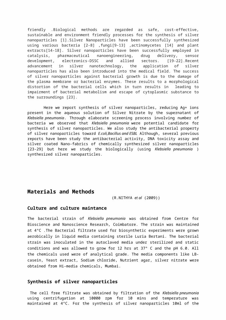
Materials and Methods (R.NITHYA et al (2009))
Culture and culture maintance
The bacterial strain of Klebsiella pneumonia was obtained from Centre for Bioscience and Nanoscience Research, Coimbatore. The strain was maintained at 4°C .The Bacterial filtrate used for biosynthetic experiments were grown aerobically in liquid media containing sterile Luria Bertani. The bacterial strain was inoculated in the autoclaved media under sterilized and static conditions and was allowed to grow for 12 hrs at 37° C and the pH 6.0. All the chemicals used were of analytical grade. The media components like LB-casein, Yeast extract, Sodium chloride, Nutrient agar, silver nitrate were obtained from Hi-media chemicals, Mumbai.
Synthesis of silver nanoparticles The cell free filtrate was obtained by filtration of the Klebsiella pneumonia using centrifugation at 10000 rpm for 10 mins and temperature was maintained at 4°C. For the synthesis of silver nanoparticles 10ml of the cell free filtrate was brought in contact with 1mM and 2mM of silver nitrate final concentration in 100 ml Erlen Meyer flask and agitated at 37°C in dark conditions under normal pH. Simultaneously, control without AgNO3 solution was incubated under same conditions.
UV-VIS studies
The reduction of silver ions was monitored by measuring the UV-VIS spectrum of the reactionMedium at 24 hrs time interval by drawing 1cm3 of the samples and their absorbance was recorded at a resolution of 0.5m at 350-800nm using UV-VIS spectrophotometer – (Elico, UV-VIS SL 159).
FTIR Analysis
The chemical bonds present in the analyzed chemicals can be interpreted by FTIR spectrum, by using the KBr pellets with prominent resonance spectra. The filtrate containing the extra cellular proteins secreted by the bacteria in the presence of Ag was salted out overnight at 40 ̊ C using ammonium sulphate precipitate followed by centrifugation at 5000 rpm for 10 mins. The protein obtained thereafter was dissolved in the minimal volume of deionized water and dialysed using a 12 kDa cut off dialysis membrane.
SEM studies
Scanning Electron Microscopic (SEM) analysis was done in Cochin University, Kerala. Thin films of the sample was prepared on a carbon coated copper grid by just dropping a very small amount of the sample on the grid, extra solution was removed using a blotting paper and then the film on the SEM grid was allowed to dry by putting it under a mercury lamp for 5 mins for emitting characteristic X-rays. These characteristic X-rays are used to identify the composition and measure the abundance of elements in the sample.,
Bacterial susceptibility
Susceptibility of Klebsiella pneumonia silver nanoparticles to different Gram positive and negative organisms like E. coli, Bacillus and ESBL using well diffusion method on nutrient agar plates was examined. The action of encapsulated nanoparticles (Sodium alginate and test sample –drop wise in CaCl2 solution) was studied against Nutrient ESBL-Pseudomonas. The zone of inhibition was calculated for its antimicrobial studies.
Preparation of silver Nano-coated Fabrics
To study the anti-microbial activity of silver nanoparticles when coated on fabrics like gauge-cotton, the fabric was sterilized and few drops of synthesized silver nanoparticles were mixed to it and plated on agar. After few hours, the zone of inhibition was calculated.
Results
Synthesis of Silver Nanoparticles
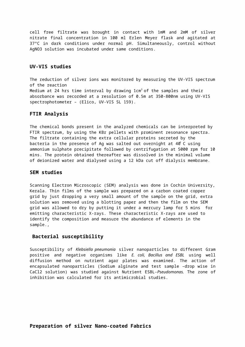
The colour change occurred in the cell free extract when challenged with 1mM and 2mM AgNo3 changed colour from pale yellow (Fig 1) to dark brown colour (Fig 2) in 12 hrs and attained maximum intensity after 24 hrs with intensity increasing during the period of incubation indicative of the formation of silver nanoparticles. Control without silver ions showed no change in colour of the cell filtrates when incubated under same conditions.
Fig.1 At the time inoculation Fig2.After 24 hrs of inoculation
UV – VIS spectral studies
Graph1. Control (AgNO3)
2mM 1mM controlcontrol 1mM 2mM
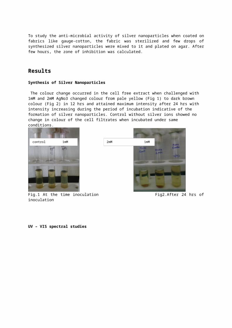
Graph.2. after 12 hrs of inoculation
Graph 1 (Control) and Graph 2 (Experimental) depicts a series of typical UV- VIS spectra of theReaction solution recorded at an interval between 0 – 24 hrs. Under normal pH 6.0 the change in light absorption profile of the medium and change in intensity of the brown colour during long term incubation (24hrs). In the filtrate obtained from the bacterial extract, a new path was observed in the visible region of (363nm) and suggested that the organism reduced the silver nitrate to silver oxide as the growth of the organisms preceded in the medium.
FTIR studies.
Graph 3.without bacterial extract (AgNO3 solution)Source- G. Pandiarajan et. al. Journal of Ecobiotechnology 2/11: 13-18, 2010
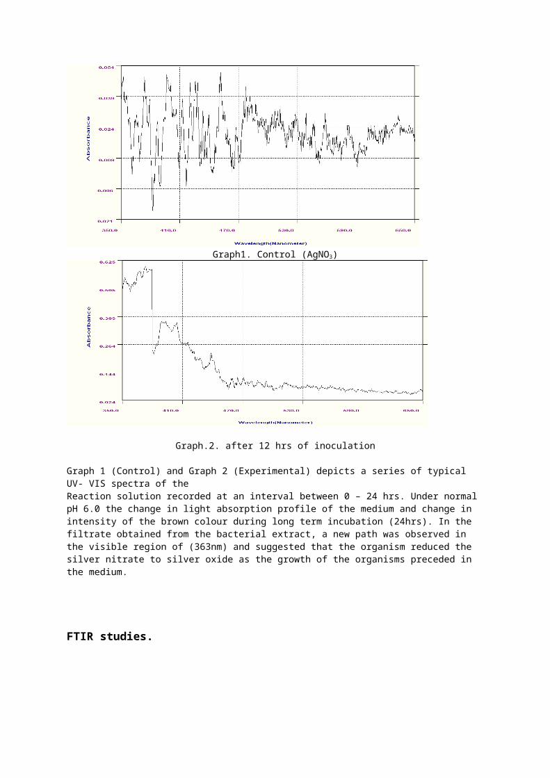
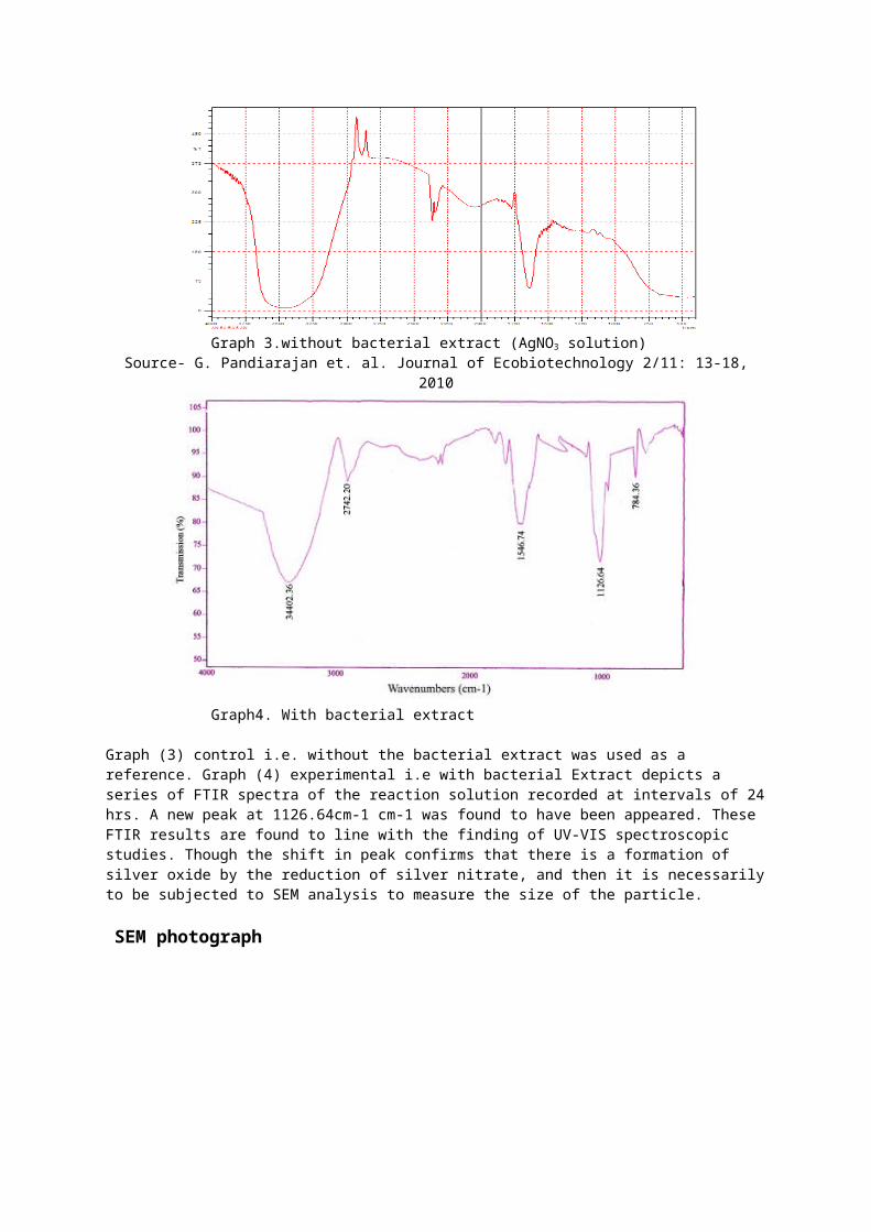
Graph4. With bacterial extract
Graph (3) control i.e. without the bacterial extract was used as a reference. Graph (4) experimental i.e with bacterial Extract depicts a series of FTIR spectra of the reaction solution recorded at intervals of 24 hrs. A new peak at 1126.64cm-1 cm-1 was found to have been appeared. These FTIR results are found to line with the finding of UV-VIS spectroscopic studies. Though the shift in peak confirms that there is a formation of silver oxide by the reduction of silver nitrate, and then it is necessarily to be subjected to SEM analysis to measure the size of the particle.
SEM photograph
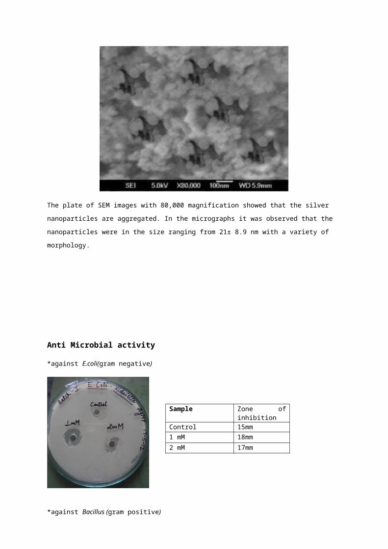
The plate of SEM images with 80,000 magnification showed that the silver nanoparticles are aggregated. In the
micrographs it was observed that the nanoparticles were in the size ranging from 21± 8.9 nm with a variety of
morphology.
Anti Microbial activity
*against E.coli(gram negative)
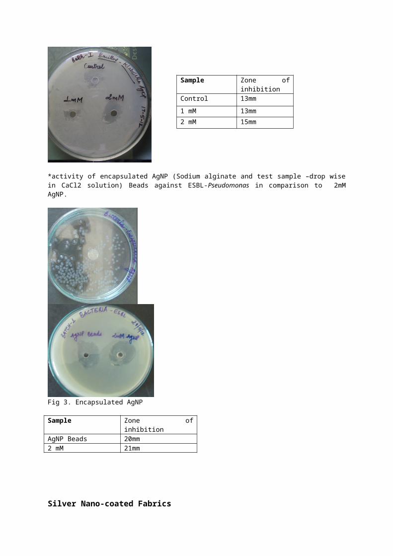
*against Bacillus (gram positive)
*activity of encapsulated AgNP (Sodium alginate and test sample –drop wise in CaCl2 solution) Beads against ESBL-Pseudomonas in comparison to 2mM AgNP.
Fig 3. Encapsulated AgNP
Sample Zone of inhibitionAgNP Beads 20mm2 mM 21mm
Sample Zone of inhibition
Control 15mm1 mM 18mm
2 mM 17mm
Sample Zone of inhibitionControl 13mm
1 mM 13mm
2 mM 15mm
Silver Nano-coated Fabrics
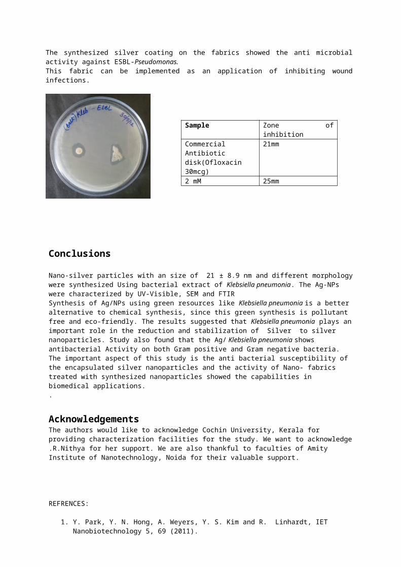
The synthesized silver coating on the fabrics showed the anti microbial activity against ESBL-Pseudomonas.This fabric can be implemented as an application of inhibiting wound infections.
Conclusions
Nano-silver particles with an size of 21 ± 8.9 nm and different morphology were synthesized Using bacterial extract of Klebsiella pneumonia. The Ag-NPs were characterized by UV-Visible, SEM and FTIRSynthesis of Ag/NPs using green resources like Klebsiella pneumonia is a better alternative to chemical synthesis, since this green synthesis is pollutant free and eco-friendly. The results suggested that Klebsiella pneumonia plays an important role in the reduction and stabilization of Silver to silver nanoparticles. Study also found that the Ag/ Klebsiella pneumonia shows antibacterial Activity on both Gram positive and Gram negative bacteria. The important aspect of this study is the anti bacterial susceptibility of the encapsulated silver nanoparticles and the activity of Nano- fabrics treated with synthesized nanoparticles showed the capabilities in biomedical applications..
AcknowledgementsThe authors would like to acknowledge Cochin University, Kerala for providing characterization facilities for the study. We want to acknowledge .R.Nithya for her support. We are also thankful to faculties of Amity Institute of Nanotechnology, Noida for their valuable support.
REFRENCES:
1. Y. Park, Y. N. Hong, A. Weyers, Y. S. Kim and R. Linhardt, IET Nanobiotechnology 5, 69 (2011).
2. Ratan Das,Sneha Gang,Siddhartha Sankar Nath, Journal of Biomaterials and Nanobiotechnology,2011,2,472-475
3. P. R. Chaudhari, S. A. Masurkar, V. B.Shidore and S. P. Kamble, Int. J. Pharma and Bio Sciences 3, 222 (2012).
4. N. Saifuddin, C. W. Wong and A. N. Yasumira, E-J. Chem 6, 61 (2009 5. K. Kalimuthu, R. S. Babu, D. Venkataraman, B. Mohd and S. Gurunathan- Colloids Surf. B 65, 150 (2008).
5. Lengke Maggy, Southam Gordon, Acta 70 (14), (2006).
Sample Zone of inhibitionCommercial Antibiotic disk(Ofloxacin 30mcg)
21mm
2 mM 25mm
6. Binoj Nair and T. Pradeep, Crystal Growth & Design, 2, (2000).
7. Shiying He, Zhirui Guo, Yu Zhang, Song Zhang, Jing Wang and Ning Gu, Materials Letters 61, (2007)
8. K. Kathiresan., S. Manivannan, M. A. Nabeel and B. Dhivya- Colloids Surf. B 71, 133 (2009).
9. Ahmad, P. Mukherjee, S. Senapati, D. Man- dal, M. I. Khan and R. Kumar- Colloids Surf. B 28, 313 (2003).
10. Holmes J.D., Smith P.R, Evans-Gowing R., Richardson D.J., Russel D.A., Sodeau J.R, Arch.Microbiol.163, (1995).
11. Mukherjee P, Senapati S, Mandal D, Ahmad A, Khan M.I, Kumar R, Sastry M, J. Nanotechnology 5, (2005).
12. Ahmad A, Mukherjee P, Senapati S, Mandal D, Khan M. I,Kumar R and Sastry M, Colloids and Surfaces B: Biointerfaces 28, (2003).
13. Kuber C. Bhainsa and S.F. D'Souza, Colloids and Surfaces B: Biointerfaces 47, (2006).
14. Prakasam, Reddy Shetty, Buddana Sudheer kumar,Yannam Sudheer Kumar, and Guntuku Girija Shankar- J.Microbiol.Biotechnol(2012)
15. R. Raut, J. R. Lakkakula, N. Kolekar, V. D.Mendhulkar and S. B. Kashid, Nano-Micro Lett. 2,106 (2010). p106-113.
16. S. A. Masurkar, P. R. Chaudhari, V. B. Shidore and S. P. Kamble, Nano-Micro Lett. 3, 189 (2011).
17. Shankar S. Shiv, Rai Akhilesh, Ahmad Absar and Sastry Murali, Colloid and interface science 275, (2004).
18. Gardea-Torresdey J.L, Parsons J.G, Gomez E, Peralta-Videa J, Troiani H.E, Santiago P and Jose Yacaman M, Nano Letters 2, (2002).
19. Catauro M, Raucci MG, De Gaaetano FD, Marotta A, J.Mater Sci Mater Med. 15(7), (2004).
20. Crabtree JH, Brruchette RJ, Siddiqi Ra,Huen IT, Handott LL, Fishman A, Perit Dial Int.23(4), (2003).
21. Krolikowska A, Kudelski A, Michota A, Bukowska, J.Surf Sci 532, (2003).
22. Zhao G, Stevens, J. Se. Biometals. 11, (1998).
23. Rani Catherine, C.R.Ragunathan and K.PrasannaKumar –International journal of Nanotechnology and Applications(2010)pp.217-222
24. Raffi, M. Hussain, F. Bhatti, T.M. Akhter, J .I.,Hameed, A. Hasan, Journal of Material Sciences and Technology 24 (2), (2000).
25. Monica Heger, A Silver Coating in the Fight Against Microbes, Silver nanoparticles could be the next step forward in antibacterial products, New York University's Science, Health andEnvironmental Reporting Program, (2008).
26. S. Pal, Y. T. Tak, J. M. Song,Does, Applied and environmental Microbiology, 73 (6), (2007).
27. M. Singh, S. Singh, S. Prasad, I. S. Gambhir, Digest Journal of Nanomaterials and Biostructures, 3(3), (2008).
28. R. E. K.León, Study of Silver Nanoparticles Biocidal Impact on Escherichia coli Using Optical and Atomic Force Microscopy, Research Accomplishments, (2007).
29. R. G. Cutler, E. J. Evans, Journal of Bacteriology, 9(2), (1996).